1
PP-319 Cheloid. Is it a Marker of Response to Endothelial Injury? Sinan S ¸ahin 1 , Murat Ziyrek 1 , Zeydin Acar 2 , Muammer Bilici 3 , Mustafa TarıkA gaç 2 1 Trabzon Kanuni Training and Research Hospital, Trabzon, 2 Ahi Evren Thorasic and Cardiovascular Surgery Training and Research Hospital, Trabzon, 3 Orhangazi Government Hospital, Bursa Background: Cheloid is an excessive brous growth as a result of abnormal wound healing in response to skin injury. As postulated in response to injury hypothesis, atherosclerosis could also be an abnormal wound healing response to an endothelial injury. It is well known that endothelial dysfunction is an early nding of athero- sclerosis. As a result, it could be hypothesized that endothelial dysfunction would be more prominent in patients having cheloid. The aim of this study was to assess the relationship between ow mediated diatation and cheloid formation. Method: Consecutive patients, who were admitted to the cardiology outpatient clinic with a history of coronary artery bypass grefting operation were evaluated. After application of exclusion criteria 69 patients enrolled into the study. 33 patients having cheloid formed cheloid group and 36 patients not having cheloid formed normal group. Endothelial function was measured with the help of ow mediated vasodila- tation (FMD) of the brachial artery. Results: There is no signicant difference according to the demographical data, biochemical parameters, clinical parameters and number of grefts between cheloid and normal groups. Only fasting blood glucose was signicantly higher in normal group (p¼0.02) (Table 1). In cheloid group nomber of revascularization was s higher than normal group (p¼0.025). Mean baseline brachial artery diameter was signicantly lower in normal group than cheloid group (35.04.59, 37.74.03 respectively; p¼0.012). No signicant difference was found in mean hyperemia diameter of brachial artery between cheloid and normal groups. FMD was lower in cheloid group than normal group. (9.303.5, 18.688.2 respectively p¼0.001) (Table 2). Conclusıon: This study showed that; endothalial function is signicantly worsened in patients having cheloid after coronary artery bypass grefting operation than patients who did not. If we assume that endothelial function is an early nding of athero- sclerosis we might say that atherosclerotic process would grow more agressive in patients havig cheloid than who do not. Hence, patients having cheloid might need more redo coronary artery bypass grefting or percutaneous coronary interventions. PP-320 Evaluation of Galectin-3 Levels in Acute Coronary Syndrome and its Relation with the Burden of Atherosclerosis Esra Gucuk Ipek 1 , Senay Akin Suljevic 2 , Habibe Kafes 5 , Funda Basyigit 3 , Neslihan Karalok 4 , Yesim Guray 5 , Lale Dinc Asarcikli 5 , Haydar Demirel 2 1 Polatli Hospital, Department of Cardiology, Ankara, 2 School of Sports Sciences and Technology, Hacettepe University, Ankara, 3 Cinnah Heart Center, Cardiology Department, Ankara, 4 Polatli Hospital, Biochemistry Department, Ankara, 5 Yuksek Ihtisas Hospital Cardiology Department, Ankara Background: Galectin-3 is a new biomarker assumed to reect brogenesis and inammation. In this study, we aimed to evaluate the levels of galectin-3 in patients with acute coronary syndrome and the relation of galectin-3 with the severity of the coronary artery disease. Methods: Nineteen patients with acute coronary syndrome who underwent coronary angiography and 17 age-matched controls without a history of coronary artery disease or heart failure were enrolled to the study. The burden of atherosclerosis was assessed by the number of involved vessels and the Gensini score. Serum samples were ob- tained to evaluate galectin-3 levels and ELISA test was used to measure galectin-3 levels. Continuous variables were expressed as mean standard deviation or median (interquartile range). Results: The mean age of the patients was 62.810.6 and 56% of the patients were male. Of all patients, 14% had diabetes, 31% had hypertension, 42% had hyperlip- idemia, and 33% were active smokers. In the acute coronary syndrome group, 63% of the patients had non ST elevation myocardial infarction and 37% had ST elevation myocardial infarction. Compared to control group, the median galectin-3 levels were signicantly higher in acute coronary syndrome patients [0.77 ng/ml (0.50-1.19) vs. 0.51 ng/ml (0.41-0.78), p¼0.01]. When patients were classied into three groups according to their number of involved vessels, median galectin-3 levels did not differ signicantly among groups [One vessel: 0.68 ng/ml (0.55-0.74), two vessels: 0.67 ng/ ml (0.46-1.84), three vessels 0.90 ng/ml (0.53-1.38), p¼0.62]. There was a strong correlation between galectin-3 levels and Gensini score (r¼0.625, p¼0.004). Galectin- 3 levels were also correlated with other parameters such as glomerular ltration rate (r¼ -0.409, p¼0.01), left ventricular ejection fraction (r¼ -0.479, p¼0.03), and white blood cell count (r¼0.381, p¼0.02). Conclusıons: In our study, levels of galectin-3 were elevated in patients with acute coronary syndrome and correlated with Gensini score. PP-321 Smoking May Induce Inammation, Platelet Activation and Prothrombotic State in Isolated Coronary Artery Ectasia Abdullah Do gan 1 , Fatih Aksoy 1 , Akif Arslan 1 , Atilla _ Içli 2 , Habil Yücel 3 , Ercan Varol 1 , Hatem Arı 1 , Mehmet Özaydın 1 , Do gan Erdo gan 1 1 Suleyman Demirel University Medical Faculty Cardiology Department, Isparta, 2 Kırsehir State Hospital, Kırsehir, 3 Isparta State Hospital, Isparta Background: It is unclear whether smoking may induce platelet activation, inam- mation and prothrombotic state in patients with coronary artery ectasia (CAE). We thus investigated its effect on platelet indices, high sensitivity C-reactive protein (hs- CRP) and D-dimer levels in isolated CAE patients. Methods: This study included 64 smokers with CAE, 103 non-smokers with CAE and 64 non-smoking controls with normal coronary arteries whose age and gender were comparable to smokers with CAE. We evaluated mean platelet volume (MPV), platelet and white blood cell (WBC) counts, serum levels of D-dimer, brinogen and hs-CRP. Table-1. Demographical, biochemical and clinical characteristics of cheloid and normal groups. Table-2. Comparison of endothelial function parameters of cheloid and normal groups. Table 1 Total (n¼36) AKS (n¼19) Control (n¼17) p Age (years) 62.810.6 64.57.6 60.97.6 0.3 Gender (%F/%M) 44/56 42/58 47/53 1 DM (%) 14 21 16 0.34 HT (%) 31 37 23 0.48 HL (%) 42 50 37 0.67 Smoking (%) 33 32 35 1 BMI (kg/m 2 ) 25.44.3 24.33.6 26.64.7 0.1 GFR (ml/min) 73.422.4 65.823.1 8218.8 0.02 Glc (mg/dl) 114.5 (101.7-131.5) 123 (112.4-177) 111 (96-116.5) 0.01 Galectin (ng/ml) 0.67 (0.46-0.84) 0.77 (0.50-1.19 0.51 (0.41-0.78) 0.01 AKS (acute coronary syndrome), BMI (body mass index), DM (diabetes mellitus), GFR (glomerular ltration rate), GLC (glucose), HL(hyperlipidemia), HT (hypertension) JACC Vol 62/18/Suppl C j October 2629, 2013 j TSC Abstracts/POSTERS C209 POSTERS

Evaluation of Galectin-3 Levels in Acute Coronary Syndrome and its Relation with the Burden of Atherosclerosis

  • Upload
    haydar

  • View
    215

  • Download
    2

Embed Size (px)

Citation preview

PP-319

Cheloid. Is it a Marker of Response to Endothelial Injury?

Sinan Sahin1, Murat Ziyrek1, Zeydin Acar2, Muammer Bilici3, Mustafa Tarık A�gaç21Trabzon Kanuni Training and Research Hospital, Trabzon, 2Ahi Evren Thorasic andCardiovascular Surgery Training and Research Hospital, Trabzon, 3OrhangaziGovernment Hospital, Bursa

Background: Cheloid is an excessive fibrous growth as a result of abnormal woundhealing in response to skin injury. As postulated in “response to injury hypothesis”,atherosclerosis could also be an abnormal wound healing response to an endothelialinjury. It is well known that endothelial dysfunction is an early finding of athero-sclerosis. As a result, it could be hypothesized that endothelial dysfunction would bemore prominent in patients having cheloid. The aim of this study was to assess therelationship between flow mediated diatation and cheloid formation.Method: Consecutive patients, who were admitted to the cardiology outpatient clinicwith a history of coronary artery bypass grefting operation were evaluated. Afterapplication of exclusion criteria 69 patients enrolled into the study. 33 patients havingcheloid formed cheloid group and 36 patients not having cheloid formed normalgroup. Endothelial function was measured with the help of flow mediated vasodila-tation (FMD) of the brachial artery.Results: There is no signicant difference according to the demographical data,biochemical parameters, clinical parameters and number of grefts between cheloid andnormal groups. Only fasting blood glucose was significantly higher in normal group(p¼0.02) (Table 1). In cheloid group nomber of revascularization was s higher thannormal group (p¼0.025). Mean baseline brachial artery diameter was significantlylower in normal group than cheloid group (35.0�4.59, 37.7�4.03 respectively;p¼0.012). No significant difference was found in mean hyperemia diameter ofbrachial artery between cheloid and normal groups. FMD was lower in cheloid groupthan normal group. (9.30�3.5, 18.68�8.2 respectively p¼0.001) (Table 2).Conclusıon: This study showed that; endothalial function is significantly worsened inpatients having cheloid after coronary artery bypass grefting operation than patientswho did not. If we assume that endothelial function is an early finding of athero-sclerosis we might say that atherosclerotic process would grow more agressive inpatients havig cheloid than who do not. Hence, patients having cheloid might needmore redo coronary artery bypass grefting or percutaneous coronary interventions.

Table-1. Demographical, biochemical and clinical characteristics of cheloidand normal groups.

Table-2. Comparison of endothelial function parameters of cheloid and normalgroups.

JACC Vol 62/18/Suppl C j October 26–29, 2013 j TSC Abstracts/POST

PP-320

Evaluation of Galectin-3 Levels in Acute Coronary Syndrome and its Relationwith the Burden of Atherosclerosis

Esra Gucuk Ipek1, Senay Akin Suljevic2, Habibe Kafes5, Funda Basyigit3,Neslihan Karalok4, Yesim Guray5, Lale Dinc Asarcikli5, Haydar Demirel21Polatli Hospital, Department of Cardiology, Ankara, 2School of Sports Sciences andTechnology, Hacettepe University, Ankara, 3Cinnah Heart Center, CardiologyDepartment, Ankara, 4Polatli Hospital, Biochemistry Department, Ankara, 5YuksekIhtisas Hospital Cardiology Department, Ankara

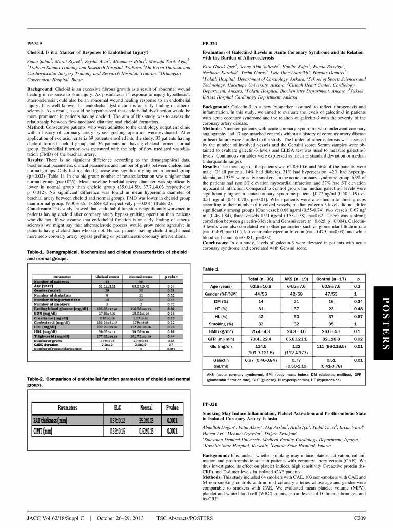
Background: Galectin-3 is a new biomarker assumed to reflect fibrogenesis andinflammation. In this study, we aimed to evaluate the levels of galectin-3 in patientswith acute coronary syndrome and the relation of galectin-3 with the severity of thecoronary artery disease.Methods: Nineteen patients with acute coronary syndrome who underwent coronaryangiography and 17 age-matched controls without a history of coronary artery diseaseor heart failure were enrolled to the study. The burden of atherosclerosis was assessedby the number of involved vessels and the Gensini score. Serum samples were ob-tained to evaluate galectin-3 levels and ELISA test was used to measure galectin-3levels. Continuous variables were expressed as mean � standard deviation or median(interquartile range).Results: The mean age of the patients was 62.8�10.6 and 56% of the patients weremale. Of all patients, 14% had diabetes, 31% had hypertension, 42% had hyperlip-idemia, and 33% were active smokers. In the acute coronary syndrome group, 63% ofthe patients had non ST elevation myocardial infarction and 37% had ST elevationmyocardial infarction. Compared to control group, the median galectin-3 levels weresignificantly higher in acute coronary syndrome patients [0.77 ng/ml (0.50-1.19) vs.0.51 ng/ml (0.41-0.78), p¼0.01]. When patients were classified into three groupsaccording to their number of involved vessels, median galectin-3 levels did not differsignificantly among groups [One vessel: 0.68 ng/ml (0.55-0.74), two vessels: 0.67 ng/ml (0.46-1.84), three vessels 0.90 ng/ml (0.53-1.38), p¼0.62]. There was a strongcorrelation between galectin-3 levels and Gensini score (r¼0.625, p¼0.004). Galectin-3 levels were also correlated with other parameters such as glomerular filtration rate(r¼ -0.409, p¼0.01), left ventricular ejection fraction (r¼ -0.479, p¼0.03), and whiteblood cell count (r¼0.381, p¼0.02).Conclusıons: In our study, levels of galectin-3 were elevated in patients with acutecoronary syndrome and correlated with Gensini score.

Table 1

Total (n¼36) AKS (n¼19) Control (n¼17) p

Age (years) 62.8�10.6 64.5�7.6 60.9�7.6 0.3

Gender (%F/%M) 44/56 42/58 47/53 1

DM (%) 14 21 16 0.34

HT (%) 31 37 23 0.48

HL (%) 42 50 37 0.67

Smoking (%) 33 32 35 1

BMI (kg/m2) 25.4�4.3 24.3�3.6 26.6�4.7 0.1

GFR (ml/min) 73.4�22.4 65.8�23.1 82�18.8 0.02

Glc (mg/dl) 114.5(101.7-131.5)

123(112.4-177)

111 (96-116.5) 0.01

Galectin(ng/ml)

0.67 (0.46-0.84) 0.77(0.50-1.19

0.51(0.41-0.78)

0.01

AKS (acute coronary syndrome), BMI (body mass index), DM (diabetes mellitus), GFR(glomerular filtration rate), GLC (glucose), HL(hyperlipidemia), HT (hypertension)

POSTERS

PP-321

Smoking May Induce Inflammation, Platelet Activation and Prothrombotic Statein Isolated Coronary Artery Ectasia

Abdullah Do�gan1, Fatih Aksoy1, Akif Arslan1, Atilla _Içli2, Habil Yücel3, Ercan Varol1,Hatem Arı1, Mehmet Özaydın1, Do�gan Erdo�gan11Suleyman Demirel University Medical Faculty Cardiology Department, Isparta,2Kırsehir State Hospital, Kırsehir, 3Isparta State Hospital, Isparta

Background: It is unclear whether smoking may induce platelet activation, inflam-mation and prothrombotic state in patients with coronary artery ectasia (CAE). Wethus investigated its effect on platelet indices, high sensitivity C-reactive protein (hs-CRP) and D-dimer levels in isolated CAE patients.Methods: This study included 64 smokers with CAE, 103 non-smokers with CAE and64 non-smoking controls with normal coronary arteries whose age and gender werecomparable to smokers with CAE. We evaluated mean platelet volume (MPV),platelet and white blood cell (WBC) counts, serum levels of D-dimer, fibrinogen andhs-CRP.

ERS C209