Embed Size (px)
Citation preview
EVALUATION OF ELECTRICAL STI ULATI
ASA TREATMENT FOR THE REDUCTION OF SPASTICITY 0
Ross Davis, M .D.
John W. Gesink, Ph .D.
Spinal Cord Injury Service, Veterans Administration Hospita1201 Northwest 16th Street, Miami, Florida 33125
The purpose of this study is to examine ways in which electricalstimulation, both transcutaneously and via implanted stimulators, maybe used to treat spasticity . Spasticity is one of the complications whichfrequently follows CNS (central nervous system) lesion, and it is com-monly identified in the following ways : The involved muscles develop apathological resistance to passive stretch . This resistance increases withthe rate of stretch and also exhibits the "knife-clasp" release phenome-non. The involved muscle groups also exhibit exag-gerated flexor and extensor reflexes . Another neuromuscular compli-cation, the muscle spasm (sometimes included as a characteristic ofspasticity), frequently accompanies the spasticity of CNS lesion and isoften observed in the spinal-cord-injured patient.
Spasticity is generally considered to be detrimental to the patient (1).Gross spasm in the paralyzed limbs of a spinal-cord-injured patient canobviously interfere with activities of daily living . Also, spasm can lead tocontractures, dislocation of joints, and can mask volitional control in caseof paresis.
Currently, spasticity is managed in a variety of ways (2) . Thesemethods include drug therapy, chemical and surgical denervation,physical therapy, and sometimes surgical alteration of tendons . Thephysical therapy treatment involves passive exercise of the spastic mus-cles . This treatment, in addition to preventing contracture, has a lastingeffect in quieting the spasticity . Apparently, when the paralyzed musclesare exercised, muscle stretch receptors and tendon organs are activated.This sensory information flow into the spinal cord and/or the passiveactivation of the reflex loop appears to quiet the spasticity . Unfortu-
nately, the therapeutic effect that exercise has on the spasticity lasts onlyfor a period of hours . With electrical stimulation we hope to utilize thesame exercise-triggered mechanisms that reduce spasticity in passiveexercise. The scheme includes equipping the patient with a portableelectrical muscle stimulator . Thus equipped, the patient is free to exer-cise his paralyzed muscles at his convenience . The potential secondary
a Paper submitted for publication but not presented at the conference.
302
Davis and Gesink : State of Effort—Elec. Stim. for Spasticity
benefits of this treatment include discontinued use of drugs, with theirundesirable side effects, and elimination of the need for regular physicaltherapy. Also, the electrically exercised muscle acts as a pump returningblood, which otherwise might pool in the legs, to the heart . Because 70percent of spinal-cord-injured patients suffer from deep vein throm-bosis (thrornbophlebitis), a condition arising from pooling of venousblood, this secondary effect could be very beneficial.
Our program begins with electrical exercise of recently injuredspinal-cord patients with transcutaneous stimulation of motor pointsand motor nerves . If this phase of the treatment shows promise, thepatient is considered for implantation of a permanent radio-frequencycoupled transmitter-receiver-type electrical stimulator . The electrodesof the implanted receiver are placed around the motor nerves, and thetransmitter is worn on the patient's belt.
Thus far, effectiveness of the treatment has been evaluated subjec-tively with the aid of a standard neurological examination . Here, judg-ments are made regarding changes in the indicators of spasticity, such asresistance to passive stretch, strength of the stretch and flexor reflexes,and duration of clonus. We are also in the process of developing twoadditional measures to evaluate the effectiveness of the treatment . Bothof these measures are attempts to define changes in spasticity in asquantitative a manner as possible . The first of these two measures is thereflexogram . Our use of this measure is based on the knowledge that thestretch reflex is a primary indicator of level of spasticity . Calibrated tapsare delivered to the Achilles tendon with a motor driven hammer . Theresulting isometric torque produced by the stretch reflex is measuredwith a torque transducer attached to the foot . Both the tendon tap andthe isometric torque are recorded on FM tape for later processing bycomputer . The second measure we are employing to quantify level ofspasticity is the standard H-reflex (3) . This reflex, except for bypassingthe muscle spindle, follows the same pathway as the Achilles tendonstretch reflex. Use of this reflex as a diagnostic tool is standard practice(4) . Also, its use as a measure of motor neuron excitability, a prime in-dicator of level of spasticity, is common and is well documented (5).
Our progress to date on this project includes a paraplegic patient whohas been implanted with bilateral peroneal nerve stimulators (Oct.1972) . The equipment used in this case was a portion of MedtronicNeuromuscular Assist Device (Fig . 1) which has been developed tocorrect the footdrop condition that frequently accompanies hemi-plegia. This device is supplied with a cycling module which may be usedto exercise paralyzed muscle . In this case the anterior tibial groups ofboth legs were exercised . Because our objective measure of spasticity(reflexogram and H-reflex) had not yet been developed when this
303

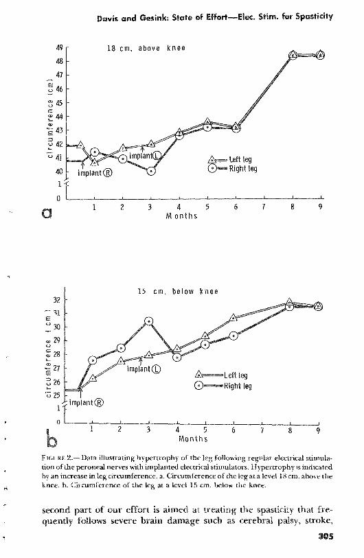
patient received his implant, only the subjective evaluation of thechanges in his spasticity was made . This evaluation indicated a signific-ant reduction in the clinical indicators of spasticity . In addition to theseprimary effects, an additional beneficial side effect was noted . Theelectrical exercise initially arrested atrophy of the stimulated muscle,and as time passed, resulted in an increase of muscle mass not only of thestimulated muscle but also of the adjacent musculature above the knee(Fig . 2) . Further, the patient was able to discontinue the use of musclerelaxant drugs as well as regular physical therapy sessions.
f ibWa
T
FIGURE 1 .-scheme illustrating method used to electrically exercise the paralyzed muscu-lature . a. Receivers, lead, and peroneal electrodes are implanted in the patient . b.Battery-powered transmitter is worn on a belt and an antenna is taped over an implantedreceiver . Copyright 1972 by Medtronic, Inc . Reprinted with permission.
Our immediate plans for the future in this part of our researchinclude providing a second paraplegic patient with an implanted bilat-eral peroneal stimulator system similar to the one shown in Figure 1.Evaluation of the effectiveness of the treatment in this patient willinclude the use of both our subjective and quantitative measures.
The above study constitutes only part of our effort in evaluating theeffectiveness of electrical stimulation for the treatment of spasticity . The
Davis and Gesink: State of Effort—Elec . Stan . for Spasticity
A ., ;
0 Implan ~. . Left leg
nolant
0
Right leg
2
3
4
5Monthsa
6
7 8
9
04
5
7Months
FIGURE 2 .--Data illustrating hypertrophy of the leg following regular electrical stimula-tion of the peroneal nerves with implanted electrical stimulators . Hypertrophy is indicatedby an increase in leg circumference . a . Circumference of the leg at a level 18 cm . above theknee. b . Circumference of the leg at a level 15 cm. below the knee.
second part of our effort is aimed at treating the spasticity that fre-quently follows severe brain damage such as cerebral palsy, stroke,
305

and head injury . While this type of spasticity (cerebral spasticity b) hasmany characteristics that are similar to spinal spasticity, there are severaldifferences . Spasticity of cerebral origin is frequently less violent andmore regular than that arising from spinal lesions (2) . Also, cerebralspasticity frequently masks voluntary muscular control, while in spinalspasticity, because the neural pathways for volitional control are fre-quently transected, there is no control left to mask.
Thus, successful treatment of cerebral spasticity potentially can bedoubly beneficial in that it both reduces the spasticity and unmasksvoluntary control . Many of the current treatments for cerebral syxm6-ci/yare the same as those already discussed . Medication such as Valium(Roche), Dantralene (Eaton Laboratories), and other muscle relaxantshave been partially successful with mild and moderate spasticity . Physi-cal therapy and stereotaxically placed deep thalamic and cerebellarlesions have also been used to manage the spasticity . Very recently atreatment using c!ecurical stimulation has been described (6) . Thistreatment involves electrical stimulation of the cerebellum through elec-trodes placed directly on its cortex . The rationale for this treatment isbased on a broad spectrum of neurophysiological studies which indicatethat the cerebellar cortex acts as a significant inhibitor of the rest of thebrain (7) . This powerful inhibitory influence of the cerebellum wasdemonstrated before the turn of the century by Sherrington (8) whoshowed that decerebrate rigidity in cats could be inhibited by stimulationof the anterior cerebellar cortex . More recently, Moruzzi (9) observedthat the stimulation of the anterior cerebellar cortex could increase orJecreayc6eccre6rate rigidity depending upon the frequency of stimula-tion . He found that a frequency of 100 to 300 Hz decreased rigiditywhile a frequency of 10 Hz resulted in an increase.
These and other observations led to the conclusion that the cerebellarcortex acts primarily in an inhibitory capacity, and that this inhibitoryfunction can be increased and modulated with chronic electrical stimula-tion. The pioneering work in implanting cerebellar electrodes forchronic stimulation of the cortex was done by Cooper (6) . Be has
b it should be noted that all spas,icityoriginates in the spinal cord and is the result of ahyperexcitable reflex loop. The hyperexcitability, however, can result from lesions at anypoint in the CNS. The terms "cerebral spasticity" and 'spinal spasticity' thus refer to thelocation of the lesions which resulted in the hyperexcitability at the reflex loop.
~
a ^
306
Davis and Gesink: State of Effort—Elec. Stim. for Spasticity
reported at least 32 cases where the treatment was beneficial in reducingchronic cerebral spasticity.
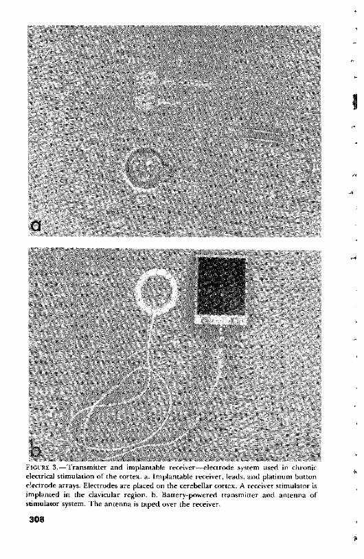
The aim of our effort in this project is to continue and extend the workof Cooper. Thus far we have moved toward this objective by developinga new technique which significantly reduces the complexity of implant-ing the cerebellar stimulators (10) . Using the technique, we have im-planted two patients suffering from cerebellar spasticity . In both cases,two arrays of silicon rubber-backed platinum button electrodes wereused (Fig . 3) . In the first case, the electrodes were placed on the anteriorand posterior lobes of the right cerebellar cortex (Fig . 4) . In the secondcase, the electrodes were placed on the anterior cerebellar lobes bilater-ally . Electrical stimuli were delivered to the electrodes by means of atranscutaneous RF-coupled transmitter-receiver system supplied byAvery Laboratories (Fig . 3) . Eight minute bursts of stimulus at a rate of200 Hz were delivered alternately to the two electrode arrays . Stimula-tion was continuous . At this time we are still evaluating the effectivenessof the treatment in reducing the patient's spasticity . Preliminary subjec-tive observations in the first patient indicate an improvement in speechand in gait and, in both patients, a somewhat reduced level of spasticity.H-reflex measurements on the first patient were made both pre- andpost-implantation . These measurements indicate that the stimulationproduced a decrease in spasticity, in that they showed a significantreduction in H-reflex detectable motorneuron excitability.
While our study of these two patients is not yet complete, our observa-tions and measurements have led us to a number of conclusions regard-ing the cerebellar stimulation treatment . First, the treatment appears tobe most effective when the electrodes are placed on the anterior cerebel-lar cortex . Second, placement of the electrodes on the posterior cortexhas no detectable effect on reducing spasticity . Our third finding is thatthe beneficial effects of the treatment can be achieved even when thestimulation frequency is reduced from 200 Hz to 100 Hz . This reductionis beneficial in that it significantly reduces the rate of discharge of thebatteries of the patient-worn transmitter.
Because this procedure appears to provide some relief from apathological condition for which there are not other proven treatments,we plan to extend the study . Additional patients with cerebral palsy anddystonia are being considered for implantation and study . Also, fol-lowup studies are continuing on those patients who have already had animplant .
307
,4
FIGURE 3 .—Transmitter and implantable receiver—electrode system used in chronicelectrical stimulation of the cortex . a . Implantable receiver, leads, and platinum buttonelectrode arrays. Electrodes are placed on the cerebellar cortex . A receiver stimulator isimplanted in the clavicular region. b. Battery-powered transmitter and antenna ofstimulator system . The antenna is taped over the receiver.
308

i
Davis and Gesink: State of Effort—Elec . Stirn . for Spasticity
FIGURE 4 .-X-ray illustrating placement of chronic cerebellar stimulating electrodes ar-rays on the anterior and posterior cerebellar cortex.
REFERENCES
1. Khalili, A .A . : Pathophysiology, Clinical Picture and Management of Spasticity . Clini-cal Anesthesia, F .A . Davis Co ., 1967.
2. Pedersen, E . : Spasticity . Charles C . Thomas, Springfield, Ill ., 1969.3. Hoffman, P. : Llber die Beziehungen der Sehnenreflexe zur wilkurlichen Bewegung
and zum Tonus . Z . Biol ., 68 :351-370, 1918.4. Braddom, R .I . and E .W. Johnson: Standardization of H-reflex and Diagnostic Use in
SI Radiculopathy . Arch . Phys . Med . & Rehab ., 55 :161-166, 1974.5. Angel, R .W . and W .W. Hoffman: The H-reflex in Normal, Spastic and Rigid Subjects.
Arch . Neurol ., 8 :591-596, 1963.6. Cooper, I . : Cerebellar Stimulation Aids Victims of Intractable Hypertonia, Epilepsy.
Interview by Gail McBride, Associate Editor, J . Amer . Med . Assn ., 225(12), Sept. 17,1973.
7. Cooper, I ., M. Riklan, and R .S . Snider : The Cerebellum, Epilepsy, and Behavior.Plenum Press, New York—London, 1974.
8 Sherrington, C .S. : Double (Antidrone) Conduction in the Central Nervous System.Proc. Roy . Soc . London, 618 :243-246, 1897.
9. Moruzzi, G. : Problems in Cerebellar Physiology . Charles C . Thomas, Springfield, Ill .,1969.
10. Davis, R . and M . Dimanceseu: Technique for Implanting Cerebellar Stimulator.Exhibit, Congress of Neurological Surgeons, Vancouver, Canada, 1974 .
309