Embed Size (px)
Citation preview
1516
Evaluation of Efficacy of the Smear Layer Removaland Micro Hardness of Root Dentin after using
Different Irrigants - An In Vitro StudySangeetha Vallikanthan1, Sainath Dinapadu2, Swathi Aravelli3,
Aliveni Manga4, Sujayeendranath Reddy5, Sindhura Reddy6
ABSTRACT:
Aim: To evaluate the efficacy of the smear layer removal and microhardness of root dentin after using different irrigants. Materialsand methods: Sixty freshly extracted single rooted premolars wereselected. Root canal preparation is done using ProTaper Rotaryinstruments. The teeth were randomly divided into six groups with10 specimens each and final irrigation of 5 ml each was done asper the groups. The teeth were sectioned longitudinally into twoequal halves along the long axis using a hard tissue microtomemachine. The one half of the specimen is used for the SEMevaluation and the other one half with the acrylic block is used forthe micro hardness study. The data was analysed using Kruskel -Wallis One - way ANOVA followed by Mann - Whitney U - test, p< 0.05 was considered as the level of significance. Results: Thebest result for the smear layer removal was observed in Group IIMTAD (0.2 ± 0.4), followed by Group VI 5.2% NaOCl + 17% EDTA(0.3 ± 0.7), Group IV SAEW (0.5 ±0.7), Group III 17% EDTA (0.8 ±0.5) and Group I 2.6% NaOCl (1.8 ± 0.6). Dentin Microhardness inGroup I (67.42 ± 10.9), Group II (67.0 ± 7.8 ) and in Group V (67.4± 3.4) showed significantly higher when compared to the meanvalues in Group III (59.7 ± 4.3) and in Group VI (58.3 ± 5.0) with p< 0.05. Conclusion: Strong Acid Electrolytic Water had removedthe smear layer, but has reduced the microhardness. But MTADsolution effectively removed the smear layer without significantlyaltering the microhardness of the root dentin.
Key words: chelation, dentine, irrigation, smear layer, sodiumhypochlorite.
O R I G I N A L R E S E A R C H
doi: 10.5866/2014.621516
1,3,4&6Senior Lecturer,2&5Reader,Department of Conservative Dentistry and Endodontics,SVS Institute of Dental Sciences,Mahabubnagar,Telangana, INDIA.
Article Info:
Received: January 13, 2014Review Completed: February 10, 2014Accepted: March 11, 2014Available Online: July, 2014 (www.nacd.in)© NAD, 2014 - All rights reserved
Email for correspondence:[email protected]
Quick Response Code
INDIAN JOURNAL OF DENTAL ADVANCEMENTS
Jour nal homepage: www. nacd. in
Introduction
An important objective in root canal treatmentis the removal of pulp tissue and dentinal debrisfrom the root canal system. In addition tobiomechanical preparation, it is essential to use anirrigant or combination of irrigants. Since, it helpsto clean the areas of the root canal system that could
not be directly reached by root canalinstrumentation.1
Subsequent to biomechanical preparation, anamorphous irregular layer known as the “smearlayer” is formed on the root canal walls. Smear layercontains inorganic and organic substances thatinclude fragments of odontoblastic process, micro
Indian J Dent Adv 2014; 6(2): 1516-1522
1517
organisms and necrotic debris.2 The smear layerconsists of a superficial layer on the surface of thecanal wall approximately 1 to 2 µm in thickness anda deeper layer packed into the dentinal tubules to adepth of 40 µm.3 The presence of smear layerprevents the penetration of the intra canalmedication into the irregularities of the complex rootcanal system and also prevents the completeadaptation of obturation material to the preparedroot canal surface.4
Ethylene diamine tetra acetic acid (EDTA) isoften suggested as an irrigating solution because itcan chelate and remove the mineralized portion ofsmear layer. It also can decalcify up to a 50µm layerof the root canal wall if used liberally. 17% EDTA isnormally used to remove smear layer in less than 1min if the fluid is able to reach the surface of theroot canal wall.
The most common organism, which is beingisolated particularly in reinfected root canals, isEnterococcus faecalis. The irrigating solutions likeNaOCl and EDTA helps in eradicating the microorganisms commonly found in the infected rootcanals effectively.
Various investigators have demonstrated thatMTAD also has an ability to remove the smear layer,disinfect contaminated root canals, and found to beeffective in eradicating Enterococcus faecalis.MTAD, a new irrigating solution, comprised of aMixture of Tetracycline isomer, Acid and Detergenthas shown promising results. It was reported thatthe cleanest canals were obtained when NaOCl wasused before a final irrigation and flushing withMTAD.5
Recently Strong acid electrolytic water (SAEW),also called as “Electrochemically activated water”or “Oxidative potential water” was used extensivelyin Japan for disinfecting purpose for its safety andantibacterial effect. Since 1996, it has been tried asa root canal irrigant and was reported that, Strongacid electrolytic water could reduce bacteria andremove the smear layer.6
The use of the irrigating agents influences thedentin structure and change in the dentinproperties. Calcium ions present in hydroxyapatitecrystals are one of the main inorganic elements ofdentin. Any change in the calcium ion ratio may
change the original proportion of the organic andinorganic components. This in turn affects the microhardness, permeability and solubility characteristicsof dentin, sealing ability and adhesion of dentalmaterials such as resin-based cements and rootcanal sealers to dentin.7 It has been furtherestablished that removal of the smear layer willenhance the adhesion of root canal sealers to thesurface of dentin walls.
Therefore, the present study has beenundertaken to evaluate the effectiveness of removalof smear layer and its effect on the micro-hardnessof the root dentin after using different irrigantsduring endodontic procedure.
Materials and Methods
Sixty single rooted freshly extracted premolarsfor the orthodontic and periodontic purpose wereused for the study. It was stored in normalphysiological saline at room temperature. The softtissue covering the root surface was removed usinggauze piece and a fine brush. Later, the teeth weredecoronated at the cemento-enamel junction usinga diamond disc. Barbed broach (size 20) was used toremove the pulp tissue. A size 10 no K file wasintroduced into the canal until its tip appeared atthe apical foramen. The working length wasestablished by subtracting 1mm from thismeasurement.
Root canal enlargement and preparation wasperformed by crown down technique using ProTaperRotary instruments to a size of file F2. 1 ml of salinewas used between each file size. The teeth were thenrandomly divided into six groups with ten specimensin each, a final irrigation of 5 ml each was done asper the groups.
The treatment groups namely:
Group 1 - 2.6%Naocl
Group 2 - MTAD
Group 3 - 17% EDTA
Group 4 - Strong Acid Electrolytic Water(SAEW)
Group 5 - Saline (Negative Control)
Group 6 - 5.25% Naocl + 17% EDTA (Positivecontrol)
Evaluation of Efficacy of the Smear Layer Removal and Micro Hardness Sangeetha, et, al.
Indian J Dent Adv 2014; 6(2): 1516-1522
1518
Microtome Sectioning
After complete irrigation, the teeth weremounted on an acrylic block and sectionedlongitudinally into two equal halves along the longaxis using a hard tissue microtome machine. Theone half of the specimen is used for the SEMevaluation and the other one half with the acrylicblock is used for the micro hardness study.
Surface of Root Canal Dentin - SEMObservation
The one half of the specimens was dried,mounted on metallic stubs and gold sputtered andstudied under a scanning electron microscope atmagnification of 2000 × at the middle third of theroot canal. The amount of debris and the smear layerpresent was scored (Table 1) and frequencies of thescores were subjected to statistical evaluation.
Root Canal Dentin Micro-Hardness
The other half of the section with acrylic blockwas taken and 3 mm sectioning was done. Thesespecimens were divided into 6 groups, immersed in20 ml of the respective irrigating solution forduration of 5 minutes. Later the section wassubjected to Vicker’s micro hardness testing.Hardness was measured under the condition of load50 grams and a retentive duration of 15 seconds infive dentin areas on the middle third of the root canalof each specimen and the mean average of the fiveindentations was taken and the results werestatistically evaluated.
Statistical Analysis
Mean and standard deviation were estimatedfrom the samples from each study group. Meanvalues were compared among different study groupsby using Kruskel-Wallis, One-way ANOVA followedby Mann - Whitney U test. Kolmogrov-Smirnov testwas used to test the normality of the data in eachstudy group. Pearson’s chi-square test was used toassess the relationship between SEM scores anddifferent study groups. In the present study, p < 0.05was considered as the level of significance.
Results
Among the Groups observed under the scanningelectron microscope, Group II, IV and VI smear layerwas removed successfully. The best result for the
smear layer removal was observed in Group IIMTAD (0.2 ± 0.4), followed by Group VI 5.2% NaOCl+ 17% EDTA (0.3 ± 0.7), Group IV SAEW (0.5 ±0.7),Group III 17% EDTA (0.8 ± 0.5) and Group I 2.6%NaOCl (1.8 ± 0.6). For the smear scores thesignificance level was p < 0.001. (Table 2)
The mean debris removal was best observed inGroup II MTAD (0.2 ± 0.4), then Group VI 5.2%NaOCl + 17% EDTA (0.4 ± 0.5), Group IV SAEW(0.6 ±0.8) followed by Group III 17% EDTA (0.9 ±0.4) and Group I 2.6% NaOCl (1.8 ± 0.6). (Table 3)
Dentin Microhardness in Group I (67.42 ± 10.9),Group II (67.0 ± 7.8) and in Group V (67.4 ± 3.4)showed significantly higher when compared to themean values in Group III (59.7 ± 4.3) and Group VI(58.3 ± 5.0) with p < 0.05. Further the mean of DentinMicrohardness (VHN) in Group V (67.4 ± 3.4) issignificantly lower than the Group IV (60.6 ± 9.9)with p < 0.05.(Table 4)
Discussion
Cleaning and shaping of the root canal systemare considered the key requirements for success inroot canal treatment.8 Irrigation of root canal systemprovides gross debridement, lubrication anddestruction of microbes and dissolution of tissues.9
Further they help in cleaning those areas that areinaccessible for mechanical cleansing.3 During canalpreparation dentinal debris created by the action ofendodontic instrumentation and remnants of organicmatter, forming a smear layer, may adheres to thecanal walls.10
This layer can form two zones; the first zone issuperficial zone which is made up of 1-2 µm oforganic matter and dentin particles; the second layeris deeper zone which extends into the dentinaltubules to a depth of 40µm.10 Removal of the smearlayer requires the use of irrigants that can dissolveboth the organic and inorganic components.7 Acidsolutions have been recommended for removing thesmear layer.
The decalcifying efficacy of these acids andchelating agents depends on the root length,duration of application time, diffusion in the dentinand the pH of the solution.11
In the present study, Group I - 2.6% sodiumhypochlorite was used as the irrigant. It was notcapable of removing the smear layer that remained
Evaluation of Efficacy of the Smear Layer Removal and Micro Hardness Sangeetha, et, al.
Indian J Dent Adv 2014; 6(2): 1516-1522
1519
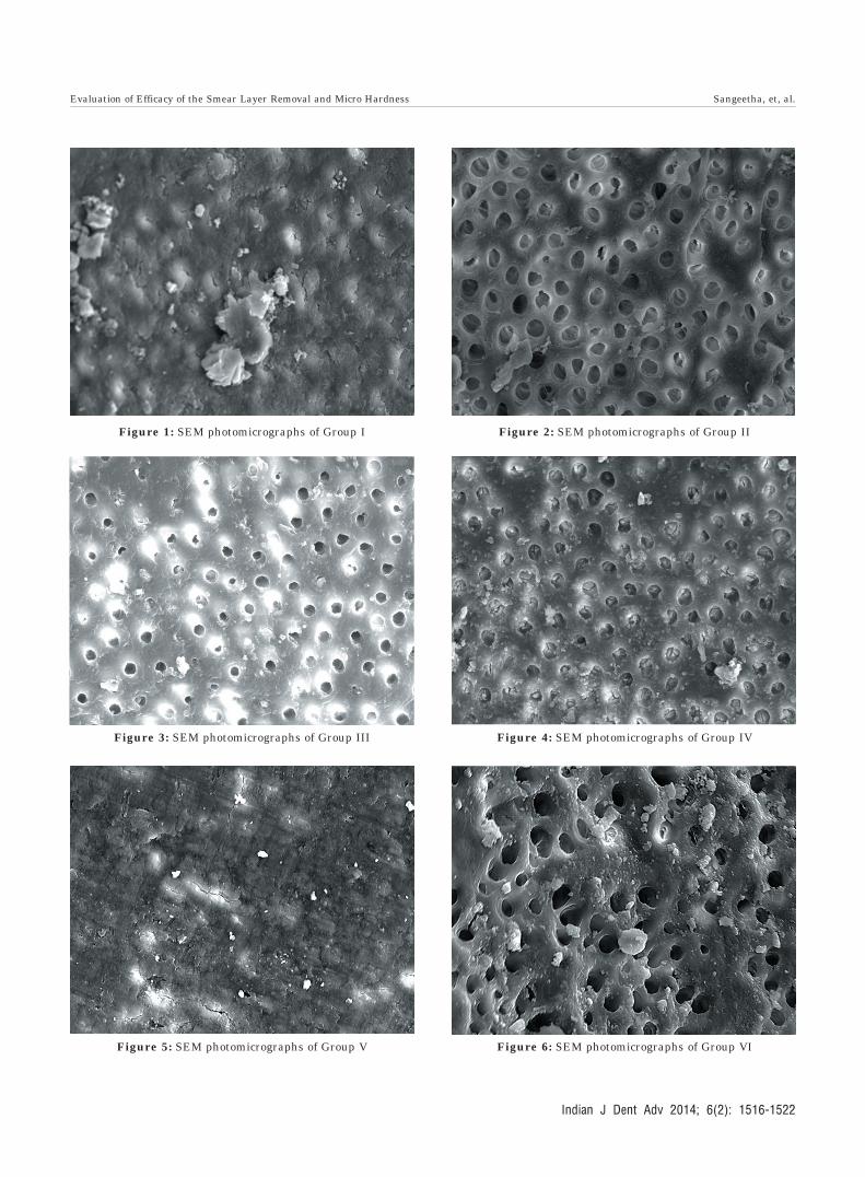
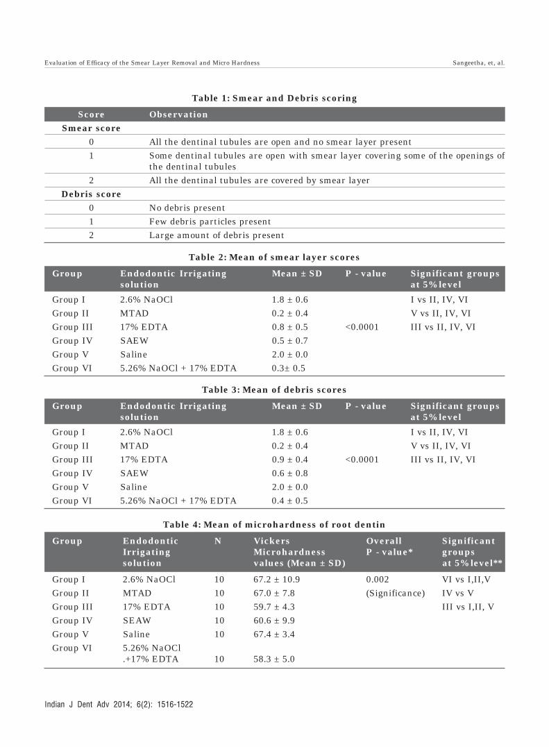
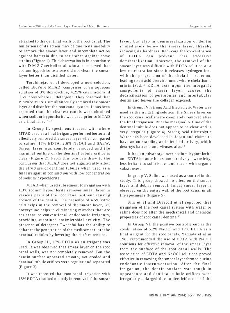
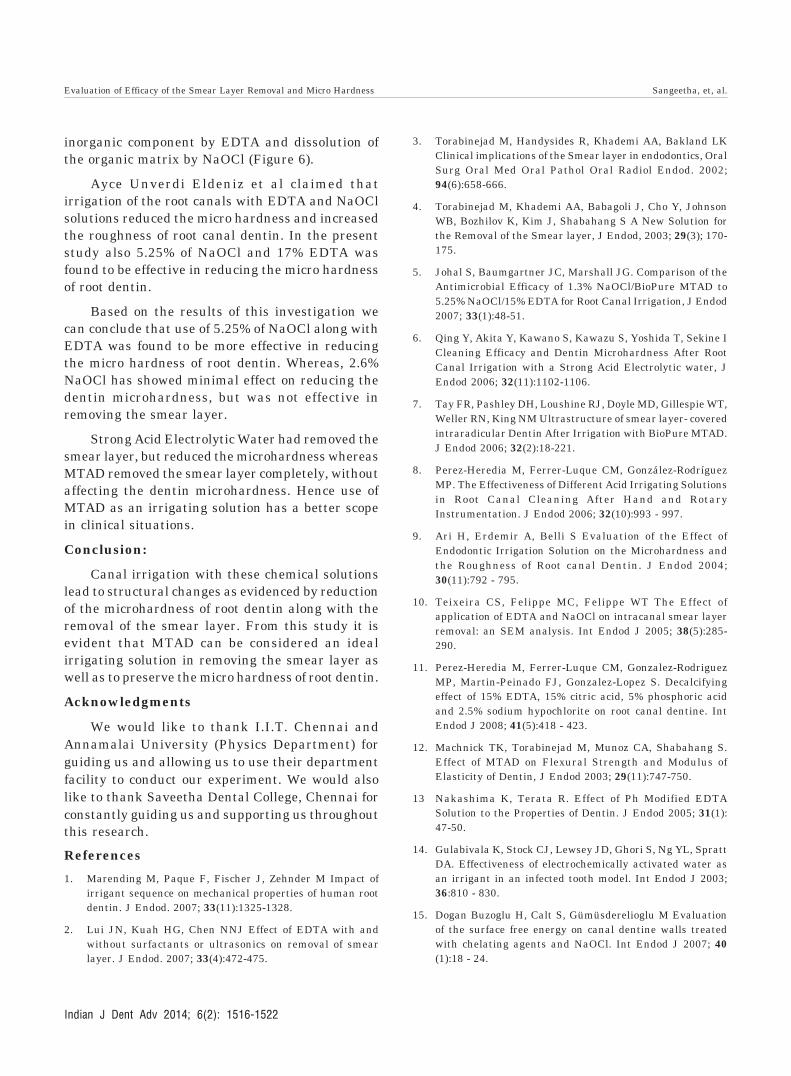
Figure 1: SEM photomicrographs of Group I Figure 2: SEM photomicrographs of Group II
Figure 3: SEM photomicrographs of Group III Figure 4: SEM photomicrographs of Group IV
Figure 5: SEM photomicrographs of Group V Figure 6: SEM photomicrographs of Group VI
Evaluation of Efficacy of the Smear Layer Removal and Micro Hardness Sangeetha, et, al.
Indian J Dent Adv 2014; 6(2): 1516-1522
1520
Table 1: Smear and Debris scoring
Score ObservationSmear score
0 All the dentinal tubules are open and no smear layer present1 Some dentinal tubules are open with smear layer covering some of the openings of
the dentinal tubules2 All the dentinal tubules are covered by smear layer
Debris score0 No debris present1 Few debris particles present2 Large amount of debris present
Table 2: Mean of smear layer scores
Group Endodontic Irrigating Mean ± SD P - value Significant groupssolution at 5% level
Group I 2.6% NaOCl 1.8 ± 0.6 I vs II, IV, VIGroup II MTAD 0.2 ± 0.4 V vs II, IV, VIGroup III 17% EDTA 0.8 ± 0.5 <0.0001 III vs II, IV, VIGroup IV SAEW 0.5 ± 0.7Group V Saline 2.0 ± 0.0Group VI 5.26% NaOCl + 17% EDTA 0.3± 0.5
Table 3: Mean of debris scores
Group Endodontic Irrigating Mean ± SD P - value Significant groupssolution at 5% level
Group I 2.6% NaOCl 1.8 ± 0.6 I vs II, IV, VIGroup II MTAD 0.2 ± 0.4 V vs II, IV, VIGroup III 17% EDTA 0.9 ± 0.4 <0.0001 III vs II, IV, VIGroup IV SAEW 0.6 ± 0.8Group V Saline 2.0 ± 0.0Group VI 5.26% NaOCl + 17% EDTA 0.4 ± 0.5
Table 4: Mean of microhardness of root dentin
Group Endodontic N Vickers Overall SignificantIrrigating Microhardness P - value* groupssolution values (Mean ± SD) at 5% level**
Group I 2.6% NaOCl 10 67.2 ± 10.9 0.002 VI vs I,II,VGroup II MTAD 10 67.0 ± 7.8 (Significance) IV vs VGroup III 17% EDTA 10 59.7 ± 4.3 III vs I,II, VGroup IV SEAW 10 60.6 ± 9.9Group V Saline 10 67.4 ± 3.4Group VI 5.26% NaOCl
.+17% EDTA 10 58.3 ± 5.0
Evaluation of Efficacy of the Smear Layer Removal and Micro Hardness Sangeetha, et, al.
Indian J Dent Adv 2014; 6(2): 1516-1522
1521
attached to the dentinal walls of the root canal. Thelimitations of its action may be due to its in-abilityto remove the smear layer and incomplete actionagainst bacteria due to resistance against somestrains (Figure 1). This observation is in accordancewith D M Z Guerisoli et al, who also observed thatsodium hypochlorite alone did not clean the smearlayer better than distilled water.
Torabinejad et al developed a new solution,called BioPure MTAD, comprises of an aqueoussolution of 3% doxycycline, 4.25% citric acid and0.5% polysorbate 80 detergent. They observed thatBioPure MTAD simultaneously removed the smearlayer and disinfect the root canal system. It has beenreported that the cleanest canals were obtainedwhen sodium hypochlorite was used prior to MTADas a final rinse.7, 12
In Group II, specimens treated with whereMTAD used as a final irrigant, performed better andeffectively removed the smear layer when comparedto saline, 17% EDTA, 2.6% NaOCl and SAEW.Smear layer was completely removed and themarginal outline of the dentinal tubule orifice isclear (Figure 2). From this one can draw to theconclusion that MTAD does not significantly affectthe structure of dentinal tubules when used as afinal irrigant in conjunction with low concentrationof sodium hypochlorite.
MTAD when used subsequent to irrigation with1.3% sodium hypochlorite removes smear layer invarious parts of the root canal without causingerosion of the dentin. The presence of 4.5% citricacid helps in the removal of the smear layer, 3%doxycycline helps in eliminating microbes that areresistant to conventional endodontic irrigants,providing sustained antimicrobial activity. Thepresence of detergent Tween80 has the ability toenhance the penetration of the medicament into thedentinal tubules by lowering the surface tension.
In Group III, 17% EDTA as an irrigant wasused. It was observed that smear layer on the rootcanal walls, was not completely removed. But thedentin surface appeared smooth, not eroded anddentinal tubule orifices were regular and separated(Figure 3).
It was reported that root canal irrigation with15% EDTA resulted not only in removal of the smear
layer, but also in demineralization of dentinimmediately below the smear layer, therebyreducing its hardness. Reducing the concentrationof EDTA can prevent this excessivedemineralization. However, the removal of thesmear layer was difficult with EDTA solution at alow concentration since it releases hydrogen ionswith the progression of the chelation reaction,leading to an acidic environment where chelation isminimized.13 EDTA acts upon the inorganiccomponents of smear layer, causes thedecalcification of peritubular and intertubulardentin and leaves the collagen exposed.
In Group IV, Strong Acid Electrolytic Water wasused as the irrigating solution, the Smear layer onthe root canal walls were completely removed afterthe final irrigation. But the marginal outline of thedentinal tubule does not appear to be clear and isvery irregular (Figure 4). Strong Acid ElectrolyticWater has been developed in Japan and claims tohave an outstanding antimicrobial activity, whichdestroys bacteria and viruses also.14
It has an advantage over sodium hypochloriteand EDTA because it has comparatively low toxicity,less irritant to soft tissues and reacts with organicsubstances.
In group V, Saline was used as a control in thestudy. This group showed no effect on the smearlayer and debris removal. Infact smear layer isobserved on the entire wall of the root canal in allthe specimens (Figure 5).
Sim et al and Driscoll et al reported thatirrigation of the root canal system with water orsaline does not alter the mechanical and chemicalproperties of root canal dentine.15
In Group VI, the positive control group is thecombination of 5.2% NaOCl and 17% EDTA as afinal irrigant for the root canals. Yamada et al in1983 recommended the use of EDTA with NaOClsolutions for effective removal of the smear layerfrom the surface of the root canal walls. Theassociation of EDTA and NaOCl solutions provedeffective in removing the smear layer formed duringendodontic instrumentation. After the finalirrigation, the dentin surface was rough inappearance and dentinal tubule orifices wereirregularly enlarged due to decalcification of the
Evaluation of Efficacy of the Smear Layer Removal and Micro Hardness Sangeetha, et, al.
Indian J Dent Adv 2014; 6(2): 1516-1522
1522
inorganic component by EDTA and dissolution ofthe organic matrix by NaOCl (Figure 6).
Ayce Unverdi Eldeniz et al claimed thatirrigation of the root canals with EDTA and NaOClsolutions reduced the micro hardness and increasedthe roughness of root canal dentin. In the presentstudy also 5.25% of NaOCl and 17% EDTA wasfound to be effective in reducing the micro hardnessof root dentin.
Based on the results of this investigation wecan conclude that use of 5.25% of NaOCl along withEDTA was found to be more effective in reducingthe micro hardness of root dentin. Whereas, 2.6%NaOCl has showed minimal effect on reducing thedentin microhardness, but was not effective inremoving the smear layer.
Strong Acid Electrolytic Water had removed thesmear layer, but reduced the microhardness whereasMTAD removed the smear layer completely, withoutaffecting the dentin microhardness. Hence use ofMTAD as an irrigating solution has a better scopein clinical situations.
Conclusion:
Canal irrigation with these chemical solutionslead to structural changes as evidenced by reductionof the microhardness of root dentin along with theremoval of the smear layer. From this study it isevident that MTAD can be considered an idealirrigating solution in removing the smear layer aswell as to preserve the micro hardness of root dentin.
Acknowledgments
We would like to thank I.I.T. Chennai andAnnamalai University (Physics Department) forguiding us and allowing us to use their departmentfacility to conduct our experiment. We would alsolike to thank Saveetha Dental College, Chennai forconstantly guiding us and supporting us throughoutthis research.
References
1. Marending M, Paque F, Fischer J, Zehnder M Impact ofirrigant sequence on mechanical properties of human rootdentin. J Endod. 2007; 33(11):1325-1328.
2. Lui JN, Kuah HG, Chen NNJ Effect of EDTA with andwithout surfactants or ultrasonics on removal of smearlayer. J Endod. 2007; 33(4):472-475.
3. Torabinejad M, Handysides R, Khademi AA, Bakland LKClinical implications of the Smear layer in endodontics, OralSurg Oral Med Oral Pathol Oral Radiol Endod. 2002;94(6):658-666.
4. Torabinejad M, Khademi AA, Babagoli J, Cho Y, JohnsonWB, Bozhilov K, Kim J, Shabahang S A New Solution forthe Removal of the Smear layer, J Endod, 2003; 29(3); 170-175.
5. Johal S, Baumgartner JC, Marshall JG. Comparison of theAntimicrobial Efficacy of 1.3% NaOCl/BioPure MTAD to5.25% NaOCl/15% EDTA for Root Canal Irrigation, J Endod2007; 33(1):48-51.
6. Qing Y, Akita Y, Kawano S, Kawazu S, Yoshida T, Sekine ICleaning Efficacy and Dentin Microhardness After RootCanal Irrigation with a Strong Acid Electrolytic water, JEndod 2006; 32(11):1102-1106.
7. Tay FR, Pashley DH, Loushine RJ, Doyle MD, Gillespie WT,Weller RN, King NM Ultrastructure of smear layer- coveredintraradicular Dentin After Irrigation with BioPure MTAD.J Endod 2006; 32(2):18-221.
8. Perez-Heredia M, Ferrer-Luque CM, González-RodríguezMP. The Effectiveness of Different Acid Irrigating Solutionsin Root Canal Cleaning After Hand and RotaryInstrumentation. J Endod 2006; 32(10):993 - 997.
9. Ari H, Erdemir A, Belli S Evaluation of the Effect ofEndodontic Irrigation Solution on the Microhardness andthe Roughness of Root canal Dentin. J Endod 2004;30(11):792 - 795.
10. Teixeira CS, Felippe MC, Felippe WT The Effect ofapplication of EDTA and NaOCl on intracanal smear layerremoval: an SEM analysis. Int Endod J 2005; 38(5):285-290.
11. Perez-Heredia M, Ferrer-Luque CM, Gonzalez-RodriguezMP, Martin-Peinado FJ, Gonzalez-Lopez S. Decalcifyingeffect of 15% EDTA, 15% citric acid, 5% phosphoric acidand 2.5% sodium hypochlorite on root canal dentine. IntEndod J 2008; 41(5):418 - 423.
12. Machnick TK, Torabinejad M, Munoz CA, Shabahang S.Effect of MTAD on Flexural Strength and Modulus ofElasticity of Dentin, J Endod 2003; 29(11):747-750.
13 Nakashima K, Terata R. Effect of Ph Modified EDTASolution to the Properties of Dentin. J Endod 2005; 31(1):47-50.
14. Gulabivala K, Stock CJ, Lewsey JD, Ghori S, Ng YL, SprattDA. Effectiveness of electrochemically activated water asan irrigant in an infected tooth model. Int Endod J 2003;36:810 - 830.
15. Dogan Buzoglu H, Calt S, Gümüsderelioglu M Evaluationof the surface free energy on canal dentine walls treatedwith chelating agents and NaOCl. Int Endod J 2007; 40(1):18 - 24.
Evaluation of Efficacy of the Smear Layer Removal and Micro Hardness Sangeetha, et, al.
Indian J Dent Adv 2014; 6(2): 1516-1522