Embed Size (px)
Citation preview

Evaluation of Conventional Chest X-Ray in the Trauma Bay
Degree Project in Medicine
Christoffer Örtenwall
Programme in Medicine
Gothenburg, Sweden 2021
Supervisors: Dr. Ragnar Ang
Doc. Jenny Skytte
University of Gothenburg, Sahlgrenska Academy
SAHLGRENSKA ACADEMY

1
Table of Contents ABSTRACT ............................................................................................................................................ 2
1. Introduction ......................................................................................................................................... 3
1.1 Background ................................................................................................................................... 3
1.2 Evolution ....................................................................................................................................... 4
1.3 Chest Radiography ........................................................................................................................ 6
1.4 Computer Tomography ................................................................................................................. 6
1.5 Extended Focused Assessment Sonography in Trauma (eFAST) ................................................. 7
1.6 Lodox/Statscan .............................................................................................................................. 8
1.7 Aim ................................................................................................................................................ 8
2. Material and methods .......................................................................................................................... 9
2.1 Study design .................................................................................................................................. 9
2.2 Population and data collection....................................................................................................... 9
2.3 Variables ........................................................................................................................................ 9
2.4 Statistical methods ....................................................................................................................... 10
3. Ethics ................................................................................................................................................. 11
4. Results ............................................................................................................................................... 11
4.1.1 Quality of CXR ........................................................................................................................ 11
4.1.2 Reliability of CXR .................................................................................................................... 13
4.1.3 Clinical findings ....................................................................................................................... 15
4.2 Tube thoracostomy ...................................................................................................................... 16
4.3 Survey .......................................................................................................................................... 16
5. Discussion ......................................................................................................................................... 17
5.1 Findings 1 .................................................................................................................................... 17
5.2 Findings 2 .................................................................................................................................... 18
5.3 Limitations................................................................................................................................... 19
6. Conclusions and Implications............................................................................................................ 19
Populärvetenskaplig sammanfattning .................................................................................................... 20
Acknowledgements ............................................................................................................................... 21
References ............................................................................................................................................. 22
Appendices ............................................................................................................................................ 23

2
ABSTRACT Title: Evaluation of Conventional Chest X-Ray in the Trauma Bay
Christoffer Örtenwall
Degree Project, Programme in Medicine, Sahlgrenska Academy, 2021, Gothenburg, Sweden
Introduction: The Advanced Trauma Life Support (ATLS™) provides guidelines for the
initial management of major trauma patients, which have become the golden standard at
Sahlgrenska University Hospital (SU/S). These guidelines recommend conventional chest x-
ray (CXR) to be used in the trauma bay. Extended Focused Assessment Sonography in
Trauma (eFAST) has in recent years become more frequently used as an alternative to CXR.
Since almost all trauma patients are subject to a trauma computed tomography (CT) scan, the
value of CXR in the trauma bay has been questioned.
Aim: The aim of this study was to evaluate the usage of CXR to diagnose pneumo- and
hemothoraces in the trauma bay at SU/S.
Methods: The study was partly retrospective, where an expert radiologist examined the
quality of all CXRs taken at SU/S between 2016 and 2018. Also, findings on CXR as
compared to CT were compared. Additionally, all tube thoracostomies placed during the same
period were investigated. Finally, a survey, where surgeons and radiologists assessed CXR-
images to assess their ability to diagnose pneumo- and/or hemothoraces, was performed.
Results: A total of 77 chest x-ray (CXR) images were obtained in the trauma bay at SU/S
between 2016 to 2018. Of these, 74% had one or more remarks regarding quality. Comparison
between findings on CXR and following trauma CT showed a sensitivity of 50% regarding
pneumo- and/or hemothoraces of clinical relevance. Based on CXR findings a chest drain was
placed in 10 patients. The survey showed that surgeons correctly assessed the presence of
pneumothorax and/or pleural fluid in 66% of the cases.
Conclusions: The findings in this study suggest that the value of current routines as well as
radiography equipment in the trauma bay is limited.

3
Key words: chest x-ray, trauma, pneumothorax, hemothorax.
1. Introduction
1.1 Background
Worldwide, more than nine individuals perish from injuries per minute, and
approximately five million die every year due to trauma (1). Measuring in DALY (disability
in life years), trauma accounts for 12% of the total loss, thus causing a significant burden of
disease. Rapid diagnosis and treatment are usually the key elements in dealing with major
trauma patients. When these patients arrive to the hospital, the initial management is a critical
and demanding period, including assessment, resuscitation and diagnostic studies. This
systematic approach is outlined in the Advanced Trauma Life Support (ATLS™) guidelines.
The ATLS course was introduced in 1978 by The American College of
Surgeons (ACS). ATLS presented a model of structured care of persons injured by external
violence. The concept of the course was spread across the world, and in 1996 it was
introduced in Sweden. It is now mandatory for all Swedish residents in surgery, orthopaedics
and anaesthesiology. Therefore, the principles that are given in ATLS have become a standard
in the primary assessment and resuscitation of trauma patients. The content of the course is
revised every four years.
An adjunct to the primary evaluation of trauma patients according to ATLS is
the use of AP (anterior-posterior) plain-film radiography of the chest (CXR) and pelvis, as
well as AP and lateral x-ray of the cervical spine in the trauma bay. The purpose is to early
diagnose any major chest injuries, fractures of the pelvis and dislocations or fractures of the
cervical spine. In case of penetrating injuries, it is also recommended to mark the entrance
(and exit) wounds, with x-ray dense markers. This in order to obtain a perception of the
trajectory (projectile path) in the body (1).
When the emergency department (ED) at Sahlgrenska University Hospital
(SU/S) was renovated 20 years ago, radiology equipment was installed. It consists of an x-ray

4
tube hung on a rail attached to ceiling, able to slide over the three available beds. For
technical reasons only AP exposures can be obtained on digital plates placed under the
patient. The image is not immediately available since the plate must be removed and put in a
scanner. The delay between exposure and presentation of the image on a monitor in the
trauma bay is a few minutes. At this point in time, it can also be reviewed thru the internal
network by the radiologist on call at the department of radiology.
Since the installation of x-ray equipment every member of the trauma team at
SU/S is required to wear radiation protection gear. When a level 1 trauma alert is activated,
two nurses from the department of radiology are summoned to the trauma bay. These are part
of the trauma team and responsible for conducting any of the radiological examinations
mentioned above requested by the trauma team leader.
1.2 Evolution
ATLS is revised every fourth year and the recommendations regarding x-ray
examinations have changed over the years. Previously, the ATLS course recommended plain-
film radiography of the chest, pelvis and cervical spine in all patients with blunt trauma (1).
Nowadays, conventional radiography of the pelvis and cervical spine are only recommended
to be performed in hemodynamically unstable patients or those with abnormal findings on
physical examination. The reason for this is based on new evidence. Duane et al. (2) showed
that clinical examination of the awake patient readily identified the need of pelvic x-ray. CCR
(Canadian C-spine Rule) as well as NEXUS (National Emergency X-radiography Utilization
Study) are two decision making rules that have been shown to have a high sensitivity and are
recommended to be used in clinical practice by international guidelines (3). Nevertheless, in
the latest edition (10th) of the ATLS course, CXR is still considered to be a valid adjunct to
the primary survey, alongside and parallel to the introduction of physician operated
ultrasound.
5
Besides the implementation of clinical protocols, much progress has been made
in the field of radiology as well as other imaging modalities since the introduction of ATLS.
Computed tomography (CT scanning) has replaced many of the radiological examinations
previously used in various acute situations, including trauma. CT scanners are today readily
available in all Swedish emergency hospitals and when emergency departments are renovated
efforts are made to locate scanners within or near the ED.
An example of when conventional x-ray has been replaced by CT is in the care
of haemodynamically stable patients injured through blunt trauma. These cases make up for
most trauma patients at SU/S, and so called “trauma-CT” has become the golden standard for
further diagnostic workup. Trauma-CT provides a detailed view of the head, (face), cervical
spine, thorax, abdomen, pelvis and upper parts of the femur, and has a higher diagnostic
accuracy than plain-film radiography (4). However, at SU/S, the examination requires
transportation of the patient to the CT-scanner located approximately 200 meters from the
ED. This, as well as the transfer of the patient from the stretcher to the scanner and reverse
takes time and should only be done in patients assessed as circulatory stable (5).
In recent years, another type of examination that has become more frequently
used in trauma patients is the Extended Focused Assessment Sonography in Trauma (eFAST)
(6). eFAST can be used bedside to diagnose pneumothorax, haemothorax, cardiac tamponade
as well as fluid (blood) in the abdomen.
Overall, the diagnostic accuracy and increased availability of CT scanning have
made this examination the golden standard for trauma patients, and the delay between
exposure and access to the x-ray image in combination with the introduction of eFAST in the
trauma bay has made the use of plain x-ray examinations more and more rare at SU/S.
Nevertheless, personnel from the department of radiology are summoned on every level 1
trauma alert. The present radiography equipment needs regular service as well as replacement

6
of this 20 years old equipment. This has led to questioning the value of radiography in the
trauma bay at SU/S, and if investing in new hardware seems reasonable.
1.3 Chest Radiography
Plain-film chest radiography (CXR) is useful for detecting serious life-
threatening conditions such as tension pneumothorax, hemothorax, gross pulmonary
contusions, mediastinal hematoma, flail chest or malpositioned tubes (4). The availability of
the CXR in the trauma bay gives the physician possibility to diagnose these conditions and
immediately perform necessary interventions. The method includes a frontal (AP) and lateral
projection. Although, at SU/S only a frontal supine projection is available, which impairs the
ability to visualize ventral pneumothoraces or moderate dorsal hemothoraces. Another
disadvantage at SU/S is that it takes a few minutes from exposure until the image can be
viewed on the monitor in the trauma bay.
Based on the recommendations from ATLS, CXR has been considered as the
mainstay in diagnosing thoracic injuries and recommended as a standard initial diagnostic
test. However, Sears et al. (7) reported that mandatory CXR for all trauma patients yields little
additional information as compared to the trauma surgeon’s assessment, and that the negative
predictive value (NPV) for the trauma surgeon’s judgement was 98.2% regarding chest
injuries. This suggest that a more selective policy is safe while costs and radiation exposure
are reduced.
1.4 Computer Tomography
CT scanning of the chest (CCT) outperforms CXR in identifying chest injuries;
Exadaktylos et al. (8) reported that CCT found significant injuries in patients with normal
CXR in more than half of the group, and CCT revealed more extensive injuries as compared
to abnormal initial radiographs, necessitating a change of management in 20% of the cases.
Another study presented by Langdorf et al. (9) including 2048 patients whom underwent both
CXR and chest CT showed that injuries were found only on CT in 71% of the cases, and one

7
third of these needed intervention. A third study presented by Kirkpatrick et al. (10) found
that supine anterior posterior (AP) CXR had a sensitivity of only 20.9% in the detection of
pneumothorax as compared to CT.
CT is superior to CXR from a diagnostic point of view, but carries higher
radiation exposure and cost (9). Also, presently patients must be transported to the CT-
scanner - a less monitored environment with limited possibilities to emergency interventions
as compared to the trauma bay. Put together, it exposes a weakness for the use of CT as an
effective screening tool.
1.5 Extended Focused Assessment Sonography in Trauma (eFAST)
In patients with major trauma, focused ultrasonography assessment for trauma
(FAST) is a bed-side method performed by the surgeon or the emergency physician to
diagnose intraperitoneal collections of free fluid (blood). The examination can be extended to
the thorax, and thereby rule out any pneumo- and/or hemothoraces, and the examination is
then referred to as extended-FAST (eFAST). The method is rapid and the use of eFAST has
increased in recent years (6).
Several studies have been made on the diagnostic accuracy of eFAST for
detection of pneumothorax in comparison with supine AP CXR. Wilkerson et al. (11)
performed a meta-analysis in 2010 including four studies and 606 patients who suffered from
blunt trauma. Thoracic ultrasonography (US) was compared with AP CXR for detection of
pneumothorax. The sensitivity for US ranged from 86% to 98% compared to the sensitivity of
CXR which ranged from 28% to 75%. The specificity of US ranged from 97% to 100%. The
specificity of CXR was 100% in all included studies. Accordingly, the review suggest that
thoracic US is a more sensitive diagnostic method for detecting pneumothorax than CXR in
patients with blunt trauma.
In 2013, Ianniello et al. (12) performed a study to evaluate the accuracy of
eFAST in the detection of pneumothorax as compared to CT. The retrospective case series

8
involved 368 adult patients admitted for major trauma. Thoracic CT scan detected 87
pneumothoraxes among these patients. eFAST detected 67 of 87, which gives a sensitivity of
77%. The specificity of US was 99.8%. Among the 20 missed pneumothoraxes with eFAST,
17 were small (thickness < 5 mm) and 3 were moderate.
However, the performance of eFAST is physician dependent and the accuracy
varies with the skill of the operator. For example, in a study by Satori et al, as cited by Ding et
al. (13), the sensitivity of thoracic ultrasonography for detection of pneumothorax was 100%,
while another study showed a sensitivity of 59%.
1.6 Lodox/Statscan
The Lodox/Statscan device was originally developed as a very-low-dose X-ray
unit for the diamond mining industry in South Africa to help in the detection of smuggled
diamonds. However, Lodox has also proven useful in trauma centres. The x-ray scanner,
approved for medical use both in the US as well as the EU, produces a whole-body AP scan in
13 seconds. If desired, the C-arm can be rotated and then provide a lateral scan. The unit
includes a special docking resuscitation table which eliminates the need of transfer of the
patient and enables ongoing resuscitation.
A study from Boffard et al. (14) showed that there were no difference in the
amount of information obtained from conventional radiography compared with Lodox, but the
use of Lodox allowed a substantial reduction in the time taken for resuscitation.
1.7 Aim
This study aimed to evaluate the usage of CXR in the trauma bay at SU/S to
diagnose pneumo- and hemothoraces. Specific objectives were assessment of the quality of
the CXR-images, calculation of sensitivity and specificity of CXR in relation to a following
CT-scan, which interventions the CXRs led to as well a survey of the skills in assessing the
images among surgeons and radiologist at SU/S.

9
2. Material and methods
2.1 Study design
The study consists of three different parts: 1) a retrospective observational study
of patients at SU/S who underwent CXR in the trauma bay during the years 2016-2018, 2) a
retrospective observational study of patients who received a tube thoracostomy during the
same years, and 3) a survey to evaluate physicians’ skills in assessing CXR-images.
2.2 Population and data collection
A total of 77 CXRs obtained in the trauma bay at SU/S between 2016 and 2018
were identified in the internal database at the Department of Radiology. These 77 images
corresponded to 77 patients, for each of which corresponding medical records were reviewed
in the digitized medical record system Melior. Since not all patients underwent a following
CT scan including the chest (n=8), 69 patients were eligible for comparison between findings
made on CXR as compared to CT. Data from the CT-scans were collected through
WedAdapt.
A search was made in the Swedish Trauma Registry (SweTrau) for patients
receiving a tube thoracotomy at SU/S in 2016-2018. The code that was searched for was
GAA10.
Finally, 20 of the 77 CXR-images were copied and after removing patient-ID
entered a PowerPoint-file. These 20 images were selected by an experienced radiologist
accordingly: five with findings and good quality, five with findings and bad quality, five
without findings and good quality, five without findings and bad quality. The PowerPoint-file
was distributed to ten surgeons (occasionally taking trauma calls) as well as one radiologist
doing on call service for the trauma service. The participants were asked to review and assess
the images according to a questionnaire (presented in the appendices).
2.3 Variables
All 77 CXR-images were assessed by an experienced radiologist based on the
following image quality (pruned, oblique positioned, movement artefacts, incorrect exposure),

10
and findings (pneumothorax, subcutaneous emphysema, pleural fluid, mediastinal widening,
and parenchymal opacification). Together these nine variables make up the general
examination of a CXR in the trauma setting.
The medical files of the patients who underwent a trauma-CT after resuscitation
in the trauma bay were reviewed with focus on clinical parameters (respiratory frequency,
blood oxygen saturation) and signs (breath sounds on thoracic auscultation, subcutaneous
emphysemas, asymmetry, or instability of the chest). Further was the trauma surgeons’
assessment of CXR and resulting interventions, as well as the radiologists’ assessment of
CXR, chest-CT (CCT) and any additional interventions following CT studied. The findings
on chest-CT were categorized in three primary groups: “all findings”, “clinically relevant
findings” and “clinically non-relevant findings”. All findings included pneumo- or
haemothorax of any size. Clinically relevant (CR) findings (also referred to as “findings of
significance”) were pneumo- or hemothoraces measuring 20 mm or more on CCT, or
pneumo- or hemothoraces of lesser size but treated with tube thoracotomy prior to CT.
Clinically non-relevant (CNR) findings were pneumo- or hemothoraces measuring less than
20 mm, or no pneumo- or hemothoraces at all.
When searching in SweTrau, patients arriving as trauma alerts who received
tube thoracostomy after a CT-scan without previous CXR were included. Their medical files
were examined and clinical findings (blood pressure, pulse, POX, breathing frequency and
sounds) prior and after receiving a chest tube were extracted.
The 20 images included in the survey were assessed accordingly to the variables
presented in the beginning of this section. The survey formula is attached in the appendix.
2.4 Statistical methods
When investigation of the clinical parameters and findings were made, the
patients were divided into two groups: one with pneumo- and/or hemothoraces and one
without. Mean respiratory frequency were calculated for the two groups. Clinical findings
11
(reduced breath sounds, subcutaneous emphysemas, asymmetry/instability of the chest wall)
were considered as test methods and reliability was calculated thru binary classification. Also,
the assessments of CXR by the surgeons and the radiologists were put in a binary
classification system using CCT as a reference.
The responses to the survey were compared to the assessment by the expert
radiologist. Answers were categorized as “correct” or “incorrect”. Individual results were then
put together to calculate an overall result for surgeons and radiologist, respectively.
3. Ethics This study is a retrospective review and thus patients included were not exposed
to any additional examinations or interventions. The only ethical consideration was the risk of
infringement of integrity. Therefore, social security numbers were exchanged with codes in a
data base, protected from unauthorized access.
This study was approved by the Swedish Ethical Review Authority (DNR 2020-
04436).
4. Results
4.1.1 Quality of CXR
A total of 77 CXR-images were taken in the trauma bay at SU/S in the years
2016 to 2018. During the same period approximately 1100 level 1 trauma alerts were
activated at SU/S. The assessment of the quality of the CXR-images are presented in Figure 1.

12
Figure 1. Remarks on quality of the chest x-rays taken in the trauma bay at SU/S in 2016-2018. An expert radiologist examined the 77 images based on 4 criteria; pruned, oblique positioned, movement, mal exposure.
As shown in Figure 1, a total of 43 images were pruned, 12 were oblique
positioned, 16 had movement artefacts and 1 had mal exposure. Of the 77 images that were
taken, 57 (74%) had one remark or more, and 20 images had no remark, as shown in Figure 2.
Figure 2. Share of the 77 images that had at least one remark. Based on the expert radiologist examination of all chest x-rays in the trauma bay at SU/S (2016-1018), it was calculated that 74% of the images had at least on remark in quality, and 26% were of perfect quality.
0%
10%
20%
30%
40%
50%
60%
70%
80%
90%
100%
Pruned Oblique positioned Movement Mal exposure
One remark or more No remark
13
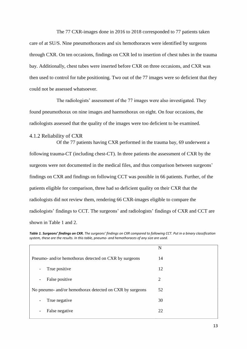
The 77 CXR-images done in 2016 to 2018 corresponded to 77 patients taken
care of at SU/S. Nine pneumothoraces and six hemothoraces were identified by surgeons
through CXR. On ten occasions, findings on CXR led to insertion of chest tubes in the trauma
bay. Additionally, chest tubes were inserted before CXR on three occasions, and CXR was
then used to control for tube positioning. Two out of the 77 images were so deficient that they
could not be assessed whatsoever.
The radiologists’ assessment of the 77 images were also investigated. They
found pneumothorax on nine images and haemothorax on eight. On four occasions, the
radiologists assessed that the quality of the images were too deficient to be examined.
4.1.2 Reliability of CXR
Of the 77 patients having CXR performed in the trauma bay, 69 underwent a
following trauma-CT (including chest-CT). In three patients the assessment of CXR by the
surgeons were not documented in the medical files, and thus comparison between surgeons’
findings on CXR and findings on following CCT was possible in 66 patients. Further, of the
patients eligible for comparison, three had so deficient quality on their CXR that the
radiologists did not review them, rendering 66 CXR-images eligible to compare the
radiologists’ findings to CCT. The surgeons’ and radiologists’ findings of CXR and CCT are
shown in Table 1 and 2.
Table 1. Surgeons’ findings on CXR. The surgeons’ findings on CXR compared to following CCT. Put in a binary classification system, these are the results. In this table, pneumo- and hemothoraces of any size are used.
N
Pneumo- and/or hemothorax detected on CXR by surgeons 14
- True positive 12
- False positive 2
No pneumo- and/or hemothorax detected on CXR by surgeons 52
- True negative 30
- False negative 22

14
Tabell 2. Radiologists’ findings on CXR. The radiologist’ findings on CXR compared to following CCT. Put in a binary classification system, these are the results. In this table, pneumo- and hemothoraces of any size are used.
As presented above, surgeons diagnosed 14 pneumo- and/or haemothorax based
on CXR. However, two of these findings were false positive, rendering 12 as true positive
findings. Also, surgeons missed pneumo- and/or hemothoraces on 22 occasions, thus
rendering in 11 false negatives. Radiologists, on the other hand, found 16 pneumo- and/or
hemothoraces, of which 13 were true positive, and missed 11 pneumo- and/or hemothoraces.
These results are calculated based on pneumo- and/or hemothoraces of any size.
When findings on CCT were categorized in CR and CNR, surgeons found 11 of
the 22 CR findings on CXR and radiologist found 13 of the same. Surgeons missed 11 CR
findings and radiologists missed 5. Through comparative statistics, sensitivity, specificity,
positive predictive value (PPV) and negative predictive value (NPV) were calculated for
surgeons’ and radiologists’ assessment of CXR. The results are presented in Table 3.
All findings on CCT 35
Clinically relevant findings on CCT 22
Pneumo- and/or hemothorax detected on CXR by radiologist
N
16
- True positive 13
- False positive 3
No pneumo- and/or hemothorax detected on CXR by radiologists 50
- True positive 39
- False negative 11
All findings on CCT 35
Clinically relevant findings on CCT 22
15
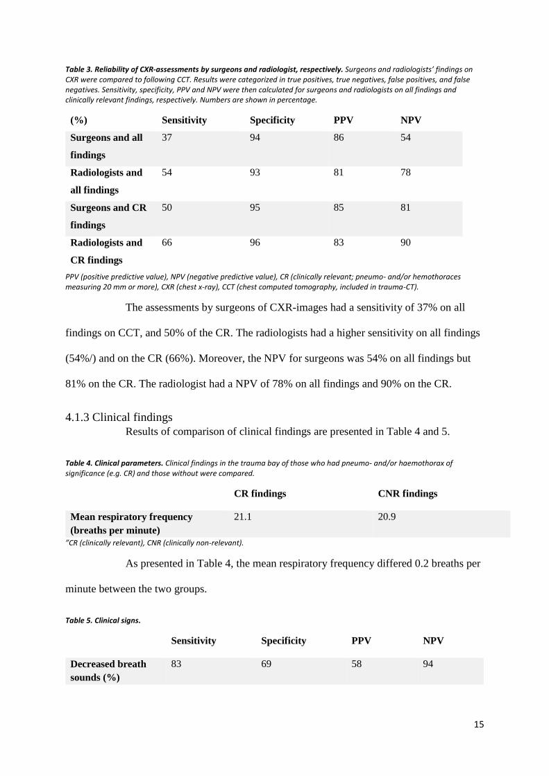
Table 3. Reliability of CXR-assessments by surgeons and radiologist, respectively. Surgeons and radiologists’ findings on CXR were compared to following CCT. Results were categorized in true positives, true negatives, false positives, and false negatives. Sensitivity, specificity, PPV and NPV were then calculated for surgeons and radiologists on all findings and clinically relevant findings, respectively. Numbers are shown in percentage.
(%) Sensitivity Specificity PPV NPV
Surgeons and all
findings
37 94 86 54
Radiologists and
all findings
54 93 81 78
Surgeons and CR
findings
50 95 85 81
Radiologists and
CR findings
66 96 83 90
PPV (positive predictive value), NPV (negative predictive value), CR (clinically relevant; pneumo- and/or hemothoraces measuring 20 mm or more), CXR (chest x-ray), CCT (chest computed tomography, included in trauma-CT).
The assessments by surgeons of CXR-images had a sensitivity of 37% on all
findings on CCT, and 50% of the CR. The radiologists had a higher sensitivity on all findings
(54%/) and on the CR (66%). Moreover, the NPV for surgeons was 54% on all findings but
81% on the CR. The radiologist had a NPV of 78% on all findings and 90% on the CR.
4.1.3 Clinical findings
Results of comparison of clinical findings are presented in Table 4 and 5.
Table 4. Clinical parameters. Clinical findings in the trauma bay of those who had pneumo- and/or haemothorax of significance (e.g. CR) and those without were compared.
CR findings CNR findings
Mean respiratory frequency
(breaths per minute)
21.1 20.9
”CR (clinically relevant), CNR (clinically non-relevant).
As presented in Table 4, the mean respiratory frequency differed 0.2 breaths per
minute between the two groups.
Table 5. Clinical signs.
Sensitivity Specificity PPV NPV
Decreased breath
sounds (%)
83 69 58 94

16
Subcutaneous
emphysemas (%)
30 100 100 73
Asymmetry or
instability of the
thorax (%)
11 98 67 71
PPV (positive predictive value), NPV (negative predictive value).
As shown in Table 5, decreased breath sounds had a sensitivity of 83% but a
PPV of only 58%. In contrary, the presence of subcutaneous emphysemas had a PPV and
specificity of 100%.
4.2 Tube thoracostomy
In a review of all patients receiving tube thoracostomies at the trauma ward at
SU/S (n=275), 27 trauma calls were identified where chest tubes were inserted after the
patients had underwent CCT, without previous CXR in the trauma bay. This suggest that in
27 cases, pneumo- and/or haemothorax could have been diagnosed and treated earlier if CXR
would have been used in the trauma bay. Of these 27 patients, two were in pre-chock at the
trauma bay, and would potentially benefit largely of a chest tube prior to CT. The other 25
patients were hemodynamically stable.
In 14 cases the patients received a chest tube in the trauma bay after a CXR.
Four of these patients had CXR difficult to assess; two of these had complementary studies
with eFAST. Two patients had chest tubes inserted based on clinical suspicion. Of the 14
patients, two were in physical distress. One of these two improved upon receiving a chest
tube.
Further, there was a total of 26 trauma calls when the patients received tube
thoracostomies based on clinical findings only, without previous diagnostic studies.
4.3 Survey
A total of five surgeons and one radiologist participated in the survey. The
surgeons’ and radiologist’s results are presented in Figure 3.

17
Figure 3. Result of survey. Five surgeons and one radiologist took the survey consisting of 20 chest x-ray images. Presence of pneumothorax, pleural fluid, subcutaneous emphysemas, mediastinal widening and parenchymal densification was asked. The answers were compared to the assessment by an expert radiologist. Shown is the percentage of correct answer by surgeons and radiologist separately.
Concerning the presence of pneumothorax, surgeons made the correct
assessment in 68% of the answers, and in 63% of the answers concerning the presence of
pleural fluid. Put together, surgeons had a correct assessment rate of 66% concerning
pneumothorax and/or pleural fluid. The radiologist assessed correctly in 90% of the times
regarding the presence of hemothorax and/or pleural fluid.
5. Discussion
5.1 Findings 1
This study attempted to evaluate the usage of CXR in the trauma bay at SU/S. A
total of 77 CXR-images were taken during 2016 to 2018. Of these, 74% had one remark or
more regarding quality. The high proportion of CXR-images of deficient quality, and
especially being pruned, suggests that the current equipment is difficult to handle and that the
personnel doing so needs more frequent practice. However, considering that 26% of the
0
0,1
0,2
0,3
0,4
0,5
0,6
0,7
0,8
0,9
1
Clinicians Radiologist
Pneumothorax Pleural fluid Subcutaneuos emphysemas
Mediastinal widening Parenchymal densificationParenchymal opacification

18
images were of excellent quality, it is not the equipment that is deficient but rather the
infrequent practice in handling it. That fact that only around 25 images are taken per year
indicates that this examination is rare. However, the time delay between exposure and access
to the images could be minimized with newer equipment. For example, Boffard et al. (14)
presented a time delay of just 13 seconds when investigating the Lodox/Statscan. Such a rapid
examination would probably increase the popularity of conventional x-ray in the trauma bay
immensely.
Furthermore, the sensitivity of surgeons’ interpretation of the CXR-images was
only 50% regarding diagnosing pneumo- and hemothoraces of clinical relevance. This is in
line with previous research (8), while other studies have presented an even lower sensitivity
(9, 10). Radiologists had a slightly higher sensitivity in interpretation (66%). It could
therefore be suggested that a radiologist should be included in the trauma team and present in
the trauma bay. Yet, only 77 images were taken when around 1100 trauma alarms were
issued, which advocates that the cost/benefit of this would be limited.
Moreover, the results of the survey showed that surgeons had a diagnostic
accuracy of 66% concerning the presence of pneumothorax and pleural fluid. This suggest
that a more frequent practise in interpretation of CXR-images is needed. The radiologist had
an accuracy of 90%. Future studies could include more radiologist to investigate this further.
5.2 Findings 2
The study also revealed, somewhat surprisingly, that the difference in mean
respiratory frequency between the two compared groups was small, and thus respiratory
frequency cannot be used to rule in or out pneumo- and/or hemothorax. However, the
presence of decreased breath sounds had a sensitivity of 83%, consequently being a useful
tool. Also, the presence of subcutaneous emphysemas had a specificity of 100%, hence being

19
a valuable indicator. Research on larger number of patients is needed to investigate this
further.
During the three years that the study included, there were two cases where
patients in pre-chock with pneumo- and/or hemothorax underwent CCT and received a chest
tube, without a previous CXR in the trauma bay. Although these cases being few, they point
out the value of the possibility to perform radiographic diagnostic studies in the trauma bay.
However, on ten occasions, findings on CXR were the reason the patient received a chest tube
in the trauma bay. A future study could investigate the number of patients that receive chest
tubes based on clinical findings but not having pneumo- and/or hemothoraces. These chest
tubes are not indicated and could have been avoided if previous CXR would have been
performed.
5.3 Limitations
A main weakness of the study is that there is a time lapse of around 20 minutes
between exposure of CXR and CCT. During this period, any pneumo- or hemothorax can
increase in size. Potentially, it could be smaller than 20 mm when CXR is performed, and
then expand to be larger and thus be categorized as clinically relevant. This is especially
relevant if the patient has been intubated and subjected to positive pressure ventilation during
this time.
Other limitations of the study are the small sample size of only 77 CXR-images,
most of them caused by blunt trauma. Thus, the value of CXR in penetrating chest injuries
cannot be assessed. Moreover, only five surgeons and one radiologist participated in the
survey.
6. Conclusions and Implications This study set out to evaluate the usage of CXR in the trauma bay at SU/S. The
most obvious findings were that the images often were of poor quality, that the sensitivity was

20
low and that surgeons at SU/S need training in interpretation of CXR-images. The findings
suggest that in general the current radiography equipment and routines are of limited value.
The question about what kind of equipment to replace the current remains.
Populärvetenskaplig sammanfattning Christoffer Örtenwall, examensarbete, läkarprogrammet, Göteborgs Universitet, 2021.
Titel: Utvärdering av konventionell lungröntgen på traumarummet.
Bakgrund: Personer som varit med om olyckor (trauma) omhändertas på Sahlgrenska
Universitetssjukhus (SU/S) enligt internationella riktlinjer. Dessa riktlinjer rekommenderar att
patienterna genomgår en ”vanlig” (konventionell) röntgen av lungorna på akuten (i
traumarummet) för att utesluta förekomst av luft eller blod i lungsäcken. På senare tid har
skiktröntgen och ultraljud fått en växande roll i omhändertagande av traumapatienter, och
användbarheten av vanlig röntgen har blivit ifrågasatt.
Syfte: Den här studien syftar till att undersöka användbarheten av lungröntgen på
traumarummet på SU/S.
Metod: Studien bestod av tre delar: en erfaren röntgenläkare bedömde kvalitén av alla
röntgenbilder som togs på traumarummet under 2016 till 2018, och de fynd som gjordes med
vanlig röntgen på traumarummet jämfördes med de fynd som gjordes med efterföljande
skiktröntgen; en registergenomgång av alla thoraxdrän (slang i lungsäcken för att dränera luft
eller blod) som sattes SU/S under samma treårs-period; en enkätundersökning bland kirurger
på SU/S beståendes av vanliga lungröntgenbilder.
Resultat: Studien visade att 74% av alla röntgenbilder som togs på traumarummet hade minst
en anmärkning i kvalitén. Dessutom framgick att bara hälften av alla fall där patienterna hade
luft eller blod i lungsäcken av betydande storlek upptäcktes på traumarummet med
lungröntgen. Dock ledde röntgen på traumarummet till att patienter fick thoraxdrän vid tio

21
tillfällen. Slutligen visade enkätundersökningen att kirurger på SU/S behöver mer träning i att
granska lungröntgenbilder.
Slutsats: Studiens resultat indikerar att befintlig utrustning och rutiner är av begränsat värde.
Acknowledgements I would like to thank my supervisor, Ragnar Ang, for support during the study.
Likewise, I very much appreciate the support by Åse Johnsson, who assessed all CXR-
images, reviewed the article and gave important feed-back.

22
References 1. American College of Surgeons Committee on T. Advanced trauma life support
for doctors. ATLS Student Course Manual. 2004;1:7.
2. Duane TM, Tan BB, Golay D, Cole FJJ, Weireter LJJ, Britt LD. Blunt Trauma
and the Role of Routine Pelvic Radiographs: A Prospective Analysis. Journal of Trauma and
Acute Care Surgery. 2002;53(3):463-8.
3. Saragiotto BT, Maher CG, Lin CWC, Verhagen AP, Goergen S, Michaleff ZA.
Canadian C‐spine rule and the National Emergency X‐Radiography Utilization Study
(NEXUS) for detecting clinically important cervical spine injury following blunt trauma.
Cochrane Database of Systematic Reviews. 2018(4).
4. Oikonomou A, Prassopoulos P. CT imaging of blunt chest trauma. Insights into
Imaging. 2011;2(3):281-95.
5. Dearden CH. Manual of definitive surgical trauma care. K. D. Boffard (ed.). 188
× 245 mm. Pp. 222. Illustrated. 2003. Arnold: London. British Journal of Surgery.
2004;91(8):1075-.
6. Lindelius A, Törngren S, Pettersson H, Adami J. Role of surgeon-performed
ultrasound on further management of patients with acute abdominal pain: a randomised
controlled clinical trial. Emergency Medicine Journal. 2009;26(8):561-6.
7. Sears WB, Luchette AF, Esposito JT, Dickson LE, Grant MM, Santaniello RJ,
et al. Old Fashion Clinical Judgment in the Era of Protocols: Is Mandatory Chest X-Ray
Necessary in Injured Patients? The Journal of Trauma: Injury, Infection, and Critical Care.
2005;59(2):324-32.
8. Exadaktylos AK, Sclabas G, Schmid SW, Schaller B, Zimmermann H. Do we
really need routine computed tomographic scanning in the primary evaluation of blunt chest
trauma in patients with “normal” chest radiograph? Journal of Trauma and Acute Care
Surgery. 2001;51(6):1173-6.
9. Langdorf MI, Medak AJ, Hendey GW, Nishijima DK, Mower WR, Raja AS, et
al. Prevalence and Clinical Import of Thoracic Injury Identified by Chest Computed
Tomography but Not Chest Radiography in Blunt Trauma: Multicenter Prospective Cohort
Study. Annals of Emergency Medicine. 2015;66(6):589-600.
10. Kirkpatrick WA, Sirois BM, Laupland GK, Liu MD, Rowan AK, Ball RC, et al.
Hand-Held Thoracic Sonography for Detecting Post-Traumatic Pneumothoraces: The
Extended Focused Assessment With Sonography For Trauma (EFAST). The Journal of
Trauma: Injury, Infection, and Critical Care. 2004;57(2):288-95.
11. Gentry Wilkerson R, Stone MB. Sensitivity of Bedside Ultrasound and Supine
Anteroposterior Chest Radiographs for the Identification of Pneumothorax After Blunt
Trauma. Academic Emergency Medicine. 2010;17(1):11-7.
12. Ianniello S, Giacomo V, Sessa B, Miele V. First-line sonographic diagnosis of
pneumothorax in major trauma: accuracy of e-FAST and comparison with multidetector
computed tomography. La radiologia medica. 2014;119(9):674-80.
13. Ding W, Shen Y, Yang J, He X, Zhang M. Diagnosis of pneumothorax by
radiography and ultrasonography: a meta-analysis. Chest. 2011;140(4):859.
14. Boffard KD, Goosen J, Plani F, Degiannis E, Potgieter H. The use of low dosage
X-ray (Lodox/Statscan) in major trauma: comparison between low dose X-ray and
conventional x-ray techniques. The Journal of trauma. 2006;60(6):1175.

23
Appendices Survey form

24
Ethical approval by the Swedish Ethical Review Authority