Embed Size (px)
Citation preview

Evaluation of Children with Blunt
Abdominal Trauma
James F. Holmes, MD, MPH
UC Davis School of Medicine
Objectives
� Epidemiology of intra-abdominal injury (IAI)
� Physical examination findings with IAI
� Laboratory abnormalities associated with IAI
� Diagnostic testing for IAI
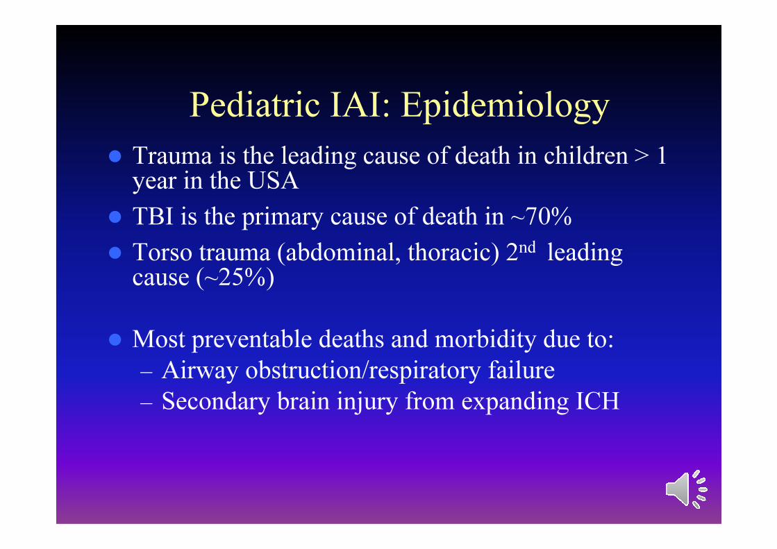
Pediatric IAI: Epidemiology
� Trauma is the leading cause of death in children > 1 year in the USA
� TBI is the primary cause of death in ~70%
� Torso trauma (abdominal, thoracic) 2nd leading cause (~25%)cause (~25%)
� Most preventable deaths and morbidity due to:
– Airway obstruction/respiratory failure
– Secondary brain injury from expanding ICH
– Unrecognized/under-treated IAI

Pediatric IAI: Epidemiology
� Most common mechanisms of injury: – MVA, auto vs. pedestrian, falls
� Frequency of injured organs:– Spleen: 40% – Spleen: 40%
– Liver: 40%
– Kidney: 30%
– GI: 15%
– Pancreas: 2%
� Many children will have more than one injured organ

Pediatric IAI: Epidemiology
� More than 600,000 children with blunt abdominal trauma evaluated annually in U.S. EDs
� 15-25% of these undergo abdominal CT� 15-25% of these undergo abdominal CT
However…
� < 10% of abdominal CTs demonstrate injury
� Relatively few patients with IAI require specific therapy: blood transfusion most common

Pediatric IAI: Epidemiology
� Compared to adults: relatively larger organs, less abdominal wall protection
� Chest wall more flexible:
– energy transferred to the spleen and liver
� The evaluation of abdomen particularly difficult in preverbal children
� ~25% of children with IAIs will have no abdominal tenderness
Childhood Abdominal Trauma:
Controversies
� Controversy over:
– Reliability of the physical examination
– Role of laboratory tests– Role of laboratory tests
– Ultrasound: Utility in children?
� Abdominal CT is the Gold Standard, but has
risks.

Patient History
�Mechanistic injury patterns helpful:
– MVA with seat belt sign: bowl and mesenteric injuries
– MVA: high speed & no seat belt– MVA: high speed & no seat belt
– Auto v. Ped: > 30 Km/hour
– Fall > 3 meters
– Handlebar injury: pancreas and duodenum
– Abuse: liver and spleen

Handlebar Injury

Handlebar Injury

Abdominal Examination
� Abdominal tenderness:
– ↑risk of IAI after adjusting for other findings
(Taylor 1994; Holmes 1999, 2002, 2013)(Taylor 1994; Holmes 1999, 2002, 2013)
� Adjusted odds ratio = 5.8 (95% CI 3.2, 10.4)
– ~75% of alert patients with IAI have
abdominal tenderness
� Gastric distention may complicate exam

Abdominal ExaminationAdelgais, Acad Emerg Med 2013 (Abstract)
� Abdominal tenderness (relative risk of IAI):
– Mild: RR=3.0 (95% CI 2.3, 4.0)
– Moderate: RR=9.4 (95% CI 7.6, 11.6)
– Severe: RR=19.4 (95% CI 15.4, 24.4)
� Location of abdominal tenderness (RR of IAI):
– Diffuse: RR=9.0 (95% CI 7.4, 11.0)
– Above umbilicus: RR=7.0 (95% CI 5.7, 8.6)
– Below umbilicus: RR=2.7 (2.0, 3.7)
All compared to no abdominal tenderness
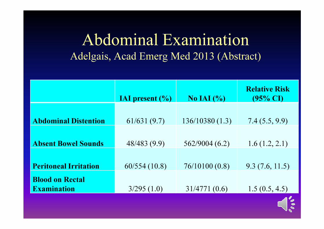
Abdominal ExaminationAdelgais, Acad Emerg Med 2013 (Abstract)
IAI present (%) No IAI (%)
Relative Risk
(95% CI)
Abdominal Distention 61/631 (9.7) 136/10380 (1.3) 7.4 (5.5, 9.9)
Absent Bowel Sounds 48/483 (9.9) 562/9004 (6.2) 1.6 (1.2, 2.1)
Peritoneal Irritation 60/554 (10.8) 76/10100 (0.8) 9.3 (7.6, 11.5)
Blood on Rectal
Examination 3/295 (1.0) 31/4771 (0.6) 1.5 (0.5, 4.5)


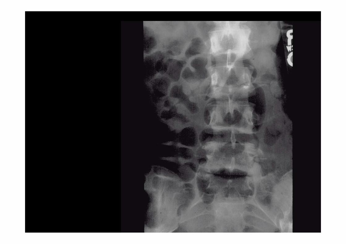
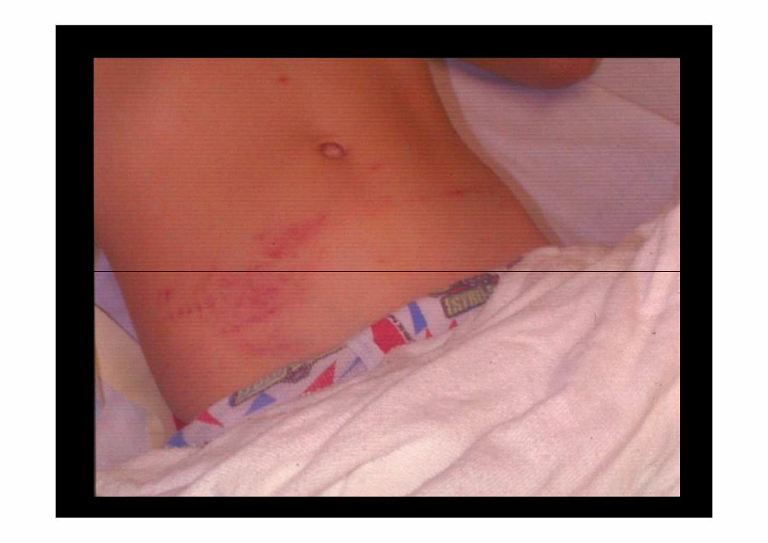
Seat Belt Injury
Injury pattern seen most in children, also in adults
Patient flexes over the lap belt
– May occur despite use of shoulder harness
Lumbar spine fracture
Chance fracture
Gastrointestinal injury

Chance
Fracture ↑ ↑ ↑


↑↑
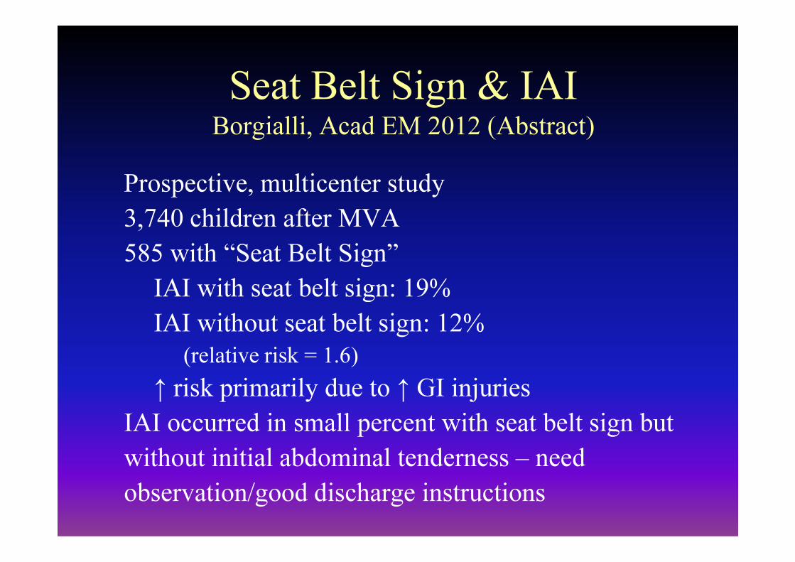
Seat Belt Sign & IAIBorgialli, Acad EM 2012 (Abstract)
Prospective, multicenter study
3,740 children after MVA
585 with “Seat Belt Sign”
IAI with seat belt sign: 19%IAI with seat belt sign: 19%
IAI without seat belt sign: 12%
(relative risk = 1.6)
↑ risk primarily due to ↑ GI injuries
IAI occurred in small percent with seat belt sign but
without initial abdominal tenderness – need
observation/good discharge instructions

Mental Status and IAI
� Children with decreased LOC have impaired ability to
perceive abdominal pain (Beaver, J Ped Surg 1987)
� Physical exam therefore unreliable in these patients� Physical exam therefore unreliable in these patients
� Mental status in patients with IAI:
– GCS < 14 in ~ 30% (Holmes, Ann Emerg Med 2002)
– GCS < 15 in ~ 45% (Holmes, AEM 1999)
– GCS = 15 in 55%

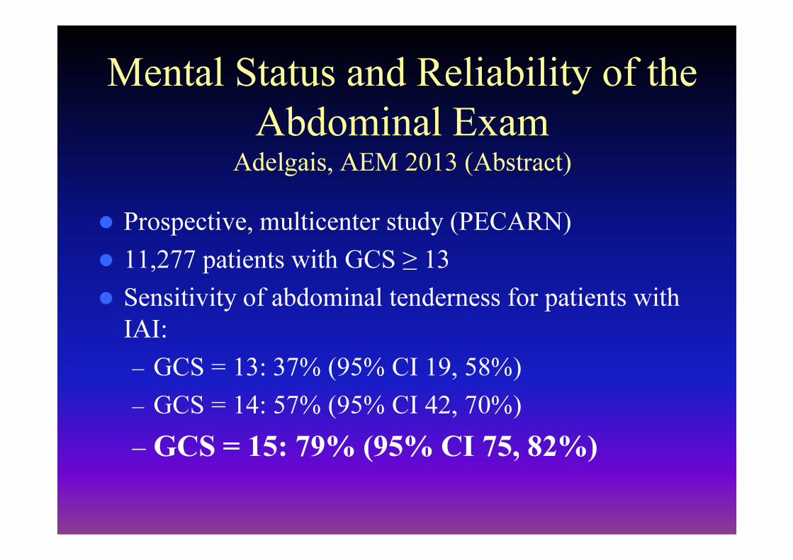
Mental Status and Reliability of the
Abdominal ExamAdelgais, AEM 2013 (Abstract)
� Prospective, multicenter study (PECARN)
� 11,277 patients with GCS ≥ 13
� Sensitivity of abdominal tenderness for patients with
IAI:

Mental Status and Reliability of the
Abdominal ExamAdelgais, AEM 2013 (Abstract)
� Prospective, multicenter study (PECARN)
� 11,277 patients with GCS ≥ 13
� Sensitivity of abdominal tenderness for patients with
IAI:
– GCS = 13: 37% (95% CI 19, 58%)

Mental Status and Reliability of the
Abdominal ExamAdelgais, AEM 2013 (Abstract)
� Prospective, multicenter study (PECARN)
� 11,277 patients with GCS ≥ 13
� Sensitivity of abdominal tenderness for patients with
IAI:
– GCS = 13: 37% (95% CI 19, 58%)
– GCS = 14: 57% (95% CI 42, 70%)
Mental Status and Reliability of the
Abdominal ExamAdelgais, AEM 2013 (Abstract)
� Prospective, multicenter study (PECARN)
� 11,277 patients with GCS ≥ 13
� Sensitivity of abdominal tenderness for patients with
IAI:
– GCS = 13: 37% (95% CI 19, 58%)
– GCS = 14: 57% (95% CI 42, 70%)
– GCS = 15: 79% (95% CI 75, 82%)

Chest Injury and IAI
� Be aware of tenderness/injury to costal margin as these ribs protect the spleen and liver
� Association with IAI in prospective study of adults:Association with IAI in prospective study of adults:
– 3% with isolated left lower ribs had IAI (Holmes, Ann Emerg Med 2005)
� Association with IAI in retrospective pediatric studies: (Taylor, Radiology 1994)

Chest Injury
� In prospective pediatric study, chest tenderness ↑ risk of IAI (univariate), but not in multivariate analysis (Holmes Ann Emerg Med 2002)
Limitations as costal margin injury not – Limitations as costal margin injury not specifically addressed
� PECARN study
– RR of IAI in patients with costal margin tenderness: 3.7 (95% CI 3.2, 4.2)

Laboratory Analysis and IAI
� Multiple laboratory tests historically utilized to
screen patients for possible IAI
– AST, ALT, hematocrit, lipase, amylase, – AST, ALT, hematocrit, lipase, amylase,
bicarbonate, urinalysis
� Prior studies have conflicting results and are
limited in design:
– small, retrospective, or univariate analysis
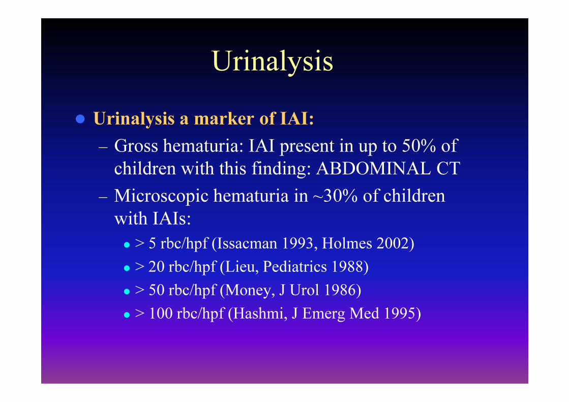
Urinalysis
� Urinalysis a marker of IAI:
– Gross hematuria: IAI present in up to 50% of
children with this finding: ABDOMINAL CT
– Microscopic hematuria in ~30% of children – Microscopic hematuria in ~30% of children
with IAIs:
� > 5 rbc/hpf (Issacman 1993, Holmes 2002)
� > 20 rbc/hpf (Lieu, Pediatrics 1988)
� > 50 rbc/hpf (Money, J Urol 1986)
� > 100 rbc/hpf (Hashmi, J Emerg Med 1995)
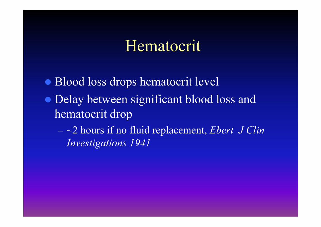
Hematocrit
� Blood loss drops hematocrit level
� Delay between significant blood loss and
hematocrit drop hematocrit drop
– ~2 hours if no fluid replacement, Ebert J Clin
Investigations 1941

Hematocrit
� Hematocrit < 30% significant predictor of IAI
– Taylor. Radiology 1994 (retrospective study: 1000 pts)
– Holmes, Ann Emerg Med 2002 (prospective study: 1095 pts)study: 1095 pts)
� Serial hematocrit levels associated with IAI
– IAI results in hematocrit drop
– No evidence of benefit in obtaining serial hematocrit levels to screen for otherwise unsuspected IAI

Liver (AST/ALT) Enzymes
� AST/ALT (SGOT, SGPT) rise immediately after
hepatic injury
� Degree of elevation does not always correlate � Degree of elevation does not always correlate
with grade of liver injury
� Elevations of 3-4x normal should generate
concern for hepatic injury:
AST >200 or ALT >125 (Holmes, Ann Emerg Med 2002)
� ALT > AST with Liver injury indicates injury >
12 hours old (Baxter, Child Abuse & Neglect 2007)

Amylase/Lipase
� Used to identify pancreatic or bowel injury
� Elevated amylase often salivary
� In pancreatic injury, enzymes increase 24 – 48 hours
after the injury
� Not shown to be a useful predictor of IAI (poor
sensitivity and PPV) in pediatric trauma patients
(Buechter 1990, Simon 1994, Holmes 1999)

Radiologic Imaging in Children
with Blunt Trauma
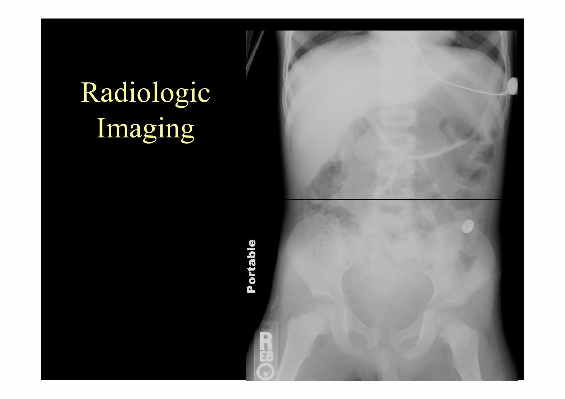
Radiologic
Imaging
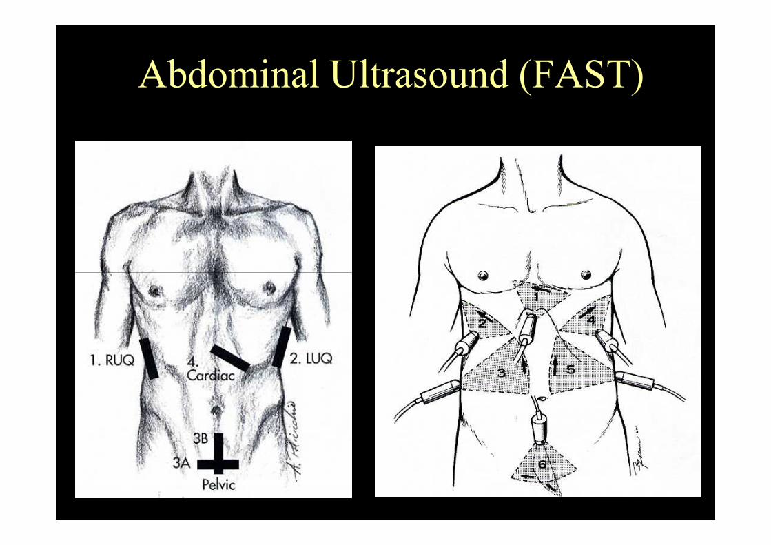
Abdominal Ultrasound (FAST)

Abdominal Ultrasound in Trauma
� Evaluate:
– Hemoperitoneum (FAST examination)
– Solid organ injury and hemoperitoneum
� Rapidly performed at patient bedside� Rapidly performed at patient bedside
� Frequently used in evaluation of adult trauma
patients:
– Two RCTs ↓ abdominal CT use (Rose J Trauma, Melniker Ann Emerg Med)
� Less frequently for pediatric patients
– USA pediatric centers: 15%

Abdominal Ultrasound in Trauma
� Not as sensitive as CT for IAI
� Meta-analysis of Pediatric studies (Holmes, J Ped Surg 2007):� Meta-analysis of Pediatric studies (Holmes, J Ped Surg 2007):
– Sensitivity for hemoperitoneum: 80% (95% CI 76, 84)
– Sensitivity for all IAIs: 66% (95% CI 56, 75%)
– LR (+): 14.5
– LR (-): 0.36

Abdominal Ultrasound in Trauma
� May allow risk stratification for CT scan
� Best performance in hypotensive children
– Sensitivity: 100% for children hypotensive from abdominal
blood loss (Holmes, J Ped Surg 2001)blood loss (Holmes, J Ped Surg 2001)
� Clinical implications unclear in children considered at
significant risk for IAI
– Ultrasound (+) → straight to Abdominal CT
– Ultrasound (-) → Abdominal CT if at moderate/high risk
� A negative FAST exam may alleviate abdominal CT in
lower risk children (<10% risk of IAI)

Abdominal Ultrasound in Trauma d
Menaker, Acad Emerg Med 2012 (Abstract)
� PECARN multicenter (n=20) study
� FAST used in 14% of children with blunt torso trauma
� Risk of IAI based on clinician suspicion
� Determined rate of abdominal CT use in patients with
and without FAST exam stratified by clinician
suspicion

Abdominal Ultrasound in Trauma d
Menaker, Acad Emerg Med 2012 (Abstract)
Clinical Suspicion
Rate of
FAST use
RR of abdominal CT
(FAST vs. no FAST)
<1% risk of IAI 11.0% 0.83 (0.67, 1.03)
1 – 5% risk of IAI 13.5% 0.81 (0.72, 0.91)
6 – 10% risk of IAI 20.5% 0.85 (0.78, 0.94)
11 – 50% risk of IAI 23.2% 0.99 (0.94, 1.05)
> 50% risk of IAI 30.7% 0.97 (0.91, 1.05)
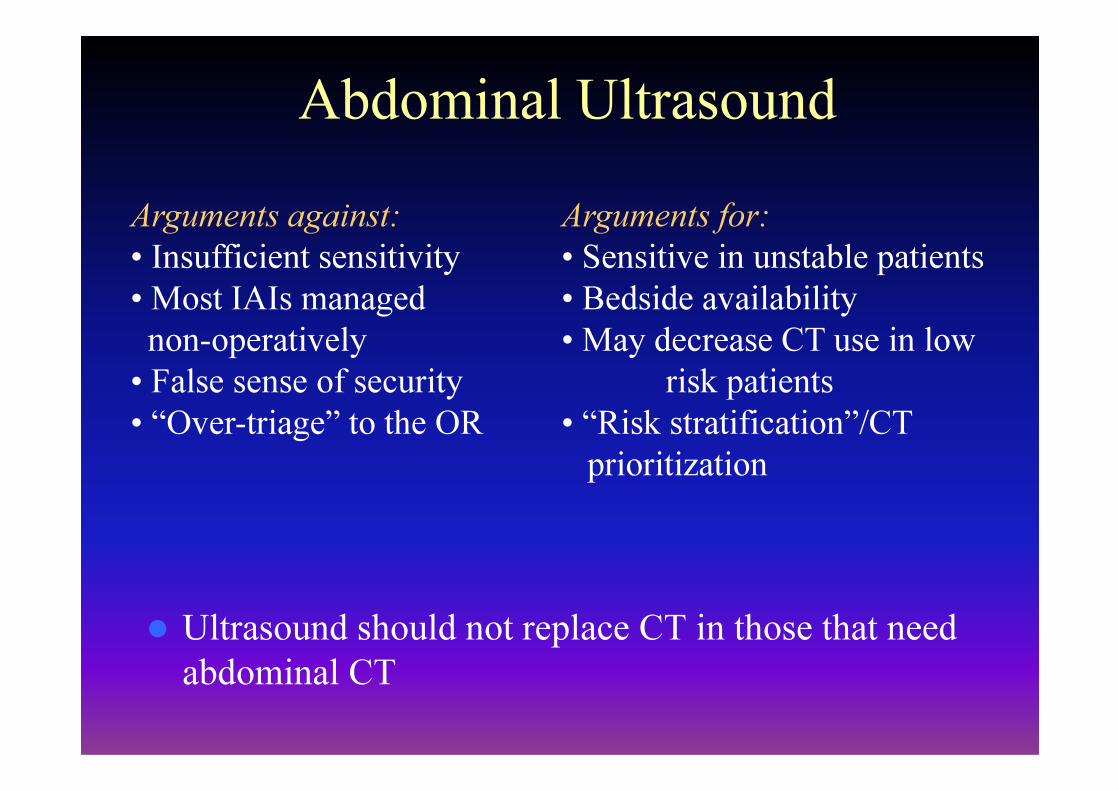
Abdominal Ultrasound
Arguments against:
• Insufficient sensitivity
• Most IAIs managed
non-operatively
• False sense of security
Arguments for:
• Sensitive in unstable patients
• Bedside availability
• May decrease CT use in low
risk patients
� Ultrasound should not replace CT in those that need
abdominal CT
• False sense of security
• “Over-triage” to the OR
risk patients
• “Risk stratification”/CT
prioritization

Abdominal CT for Pediatric Trauma
� Gold standard for diagnosis of IAI
� IV contract needed but no oral contrast
– Ellison, AEM 2013 (abstract)– Ellison, AEM 2013 (abstract)
� Excellent sensitivity for solid organ injuries
� New generation (Helical CT) scanners:
– Good sensitivity (85-95%) for GI injuries
– Limited (~50%) sensitivity for pancreatic injuries
– Consider admitting patient if high risk for GI/pancreatic
injury despite normal CT

Abdominal CT Scan – Risks
� Sedation: patient must be still for the CT,
potential complications from sedation
� Transfer outside the ED
� Charges for abdominal CT …….
� Radiation exposure

Radiation Exposure from CT
� CT scan exposes the child to 500X the compared to a chest
radiograph
� Radiation exposure may cause a malignancy
� Children at increased risk compared to adults
� Risk of death from radiation-induced malignancy from one
abdominal CT scan (Migloretti. JAMA Peds 2012)
– Child < 5 year old: < 1/ 300 - 670 CT scans
– Child 5-14 years old: 1/ 370 – 700 CT scans
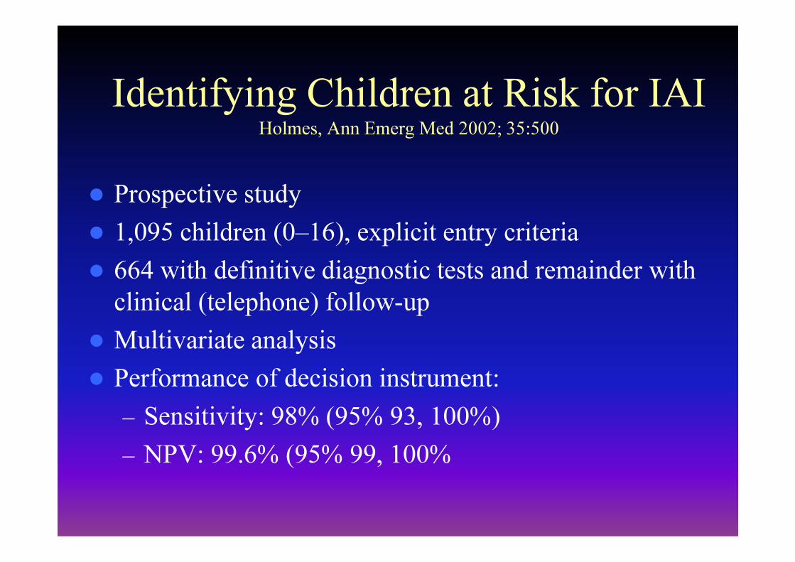
Identifying Children at Risk for IAI Holmes, Ann Emerg Med 2002; 35:500
� Prospective study
� 1,095 children (0–16), explicit entry criteria
� 664 with definitive diagnostic tests and remainder with � 664 with definitive diagnostic tests and remainder with
clinical (telephone) follow-up
� Multivariate analysis
� Performance of decision instrument:
– Sensitivity: 98% (95% 93, 100%)
– NPV: 99.6% (95% 99, 100%

Variables placing Child at Risk:
� Variables in the Decision Instrument:
– Low systolic blood pressure
– Abdominal tenderness
– Femur fracture
– Elevated liver enzymes:
� AST > 200 U/L or ALT > 125 U/L
– Urinalysis > 5 rbc/hpf
– Initial hematocrit < 30%

PECARN Abdominal Injury
Decision Instrument Holmes, Ann Emerg Med 2013; 62:107
� Prospective multicenter study May 2007 - Jan 2010
� < 18 years w/ blunt abdominal trauma evaluated in ED (explicit exclusion criteria)
� Clinical data recorded before abdominal CT (if done)
� Follow-up obtained on all patients:
– Discharged patient: Telephone follow-up
– Admitted patients: medical record review
� Primary outcome: IAI undergoing therapy (IAIAI)
� Analysis: Recursive Partitioning (CART)

Results: Prediction Rule for IAI AI
(n=12,044)
1,963 patients
112 (5.7%) IAIAI
No
GCS < 14826 patients
38 (4.6%) IAIAI
Abdomen tender 2,532 patients
36 (1.4%) IAIAI
Abdominal Wall
Trauma
No
No
Sensitivity = 197/203 (97.0%)
Specificity = 5028/11841 (42.5%)
NPV = 5028/5034 (99.9)
Thoracic Trauma
Abdominal pain
↓ Breath Sounds
Emesis
955 patients
6 (0.6%) IAIAI
305 patients
2 (0.7%) IAIAI
34 patients
1 (2.9%) IAIAI
395 patients
2 (0.5%) IAIAI
No
No
No
No
5,034 patients
No
6 (0.1%) IAIAI1,234 CT scans (25%)

Evidence of abdominal wall trauma/seatbelt sign
or GCS score < 14 with blunt abdominal trauma 23% of population
5.7% risk of IAI-
intervention
Yes
Abdominal CT vs. Observation
with ancillary testing:
FAST exam, laboratory screening,
serial abdominal exams
No
Yes
Figure 3: Suggested algorithm for evaluation of children
with blunt torso trauma
Moderate/Severe
Abdominal tenderness
No
Further evaluation needed,
Strongly consider abdominal CT
(especially in those with abdominal
wall trauma)
21% of population
1.4% risk of IAI-intervention
serial abdominal exams
Thoracic wall trauma, abdominal pain,
decreased breath sounds, vomiting, mild
abdominal tenderness
Observation, based on other
clinical factors including:
Physician experience
Multiple versus isolated
findings
No improvement in symptoms or
signs after ED observation
Parental preference
Laboratory screening tests
FAST exam
No
CT generally not recommended(consider laboratory/FAST screening if concern
for IAI remains)
14% of population
0.7% risk of IAI-
intervention
42% of population
0.1% risk of IAI-intervention
Yes
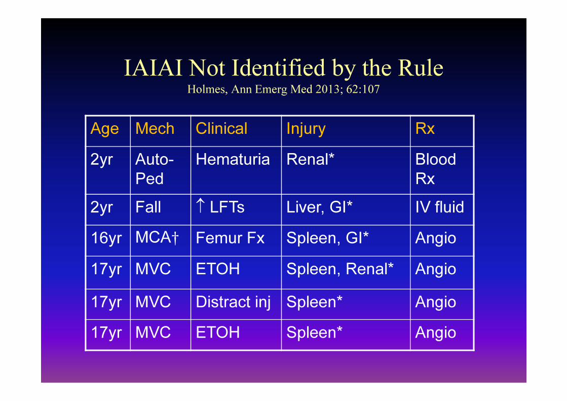
IAIAI Not Identified by the RuleHolmes, Ann Emerg Med 2013; 62:107
Age Mech Clinical Injury Rx
2yr Auto-
Ped
Hematuria Renal* Blood
Rx
2yr Fall ↑ LFTs Liver, GI* IV fluid2yr Fall ↑ LFTs Liver, GI* IV fluid
16yr MCA† Femur Fx Spleen, GI* Angio
17yr MVC ETOH Spleen, Renal* Angio
17yr MVC Distract inj Spleen* Angio
17yr MVC ETOH Spleen* Angio
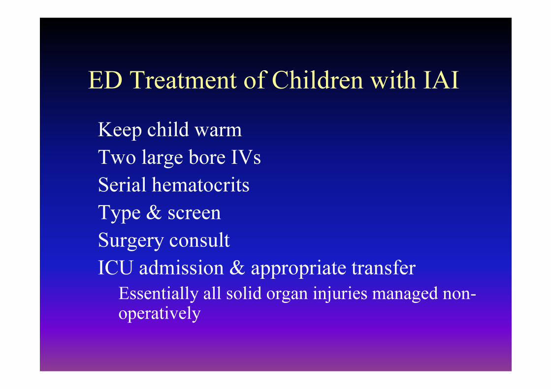
ED Treatment of Children with IAI
Keep child warm
Two large bore IVs
Serial hematocritsSerial hematocrits
Type & screen
Surgery consult
ICU admission & appropriate transfer
Essentially all solid organ injuries managed non-operatively

Summary: Exam
� High risk physical examination findings for IAI:
– GCS < 14: unable to evaluate the abdomen
– Abdominal wall trauma: contusion/abrasion/seat belt sign
– Abdominal tenderness– Abdominal tenderness
� Severe/moderate tenderness >>>> mild tenderness
– Additional variables (PECARN rule) to stratify into very low risk category
Summary: Labs
�High risk laboratory findings for IAI:
–Elevated AST/ALT (3-4x normal)
–Hematuria: especially gross hematuria–Hematuria: especially gross hematuria
–Low hematocrit: <30%
–Amylase/Lipase ↑ over 24/48 hours

Summary: Imaging
�No role for plain x-rays of the abdomen
�Abdominal Ultrasound:
– Use if hypotensive to direct management– Use if hypotensive to direct management
– May risk stratify children for CT scan
– May alleviate the need for CT in lower risk children
� Child with 1-10% risk of IAI and normal FAST exam at very low risk for therapy
Summary: CT
� Abdominal CT:
– Gold standard for diagnosing IAI
– Variables available to risk stratify patients� Strongly consider in patients with high risk findings, but be cognizant of radiation risks