Embed Size (px)
Citation preview

EVALUATION OF BONE MINERAL DENSITY AMONG PATIENTS WITH DEPRESSION IN PENANG ISLAND,
MALAYSIA
By
JAAFER MOSADEK KURMANJI
UNIVERSITI SAINS MALAYSIA
Thesis submitted in fulfillment of the
requirements for the degree of
Master of Science
December 2009

Dedication
This research work is dedicated to my country, my father, my mother, my wife and my children.

ACKNOWLEDGEMENTS
First of all, I am great full of ALLAH, who always gave me hope when I am
disappointed and show the light when I am in the dark. I am thankful to many
individuals for bringing out this piece of work. I would be akin to express my
gratitude to my supervisor Associate Professor Dr. Syed Azhar Bin Syed Sulaiman
for his supervision, advice and essential throughout my research.
I would like to thank my field supervisors Dr. Lau Kim Kah and Dr. Prem Kumar
Chandrasekaran for their help and recommendations, the doctors, staff and
directories in Penang General Hospital and Penang Adventist Hospital for their
facilitation and cooperation.
My appreciation to All eight Co. Malaysia, for their support in achieve this research
by lending the bone measurement instrument.
Special thanks to Dr. Siti Ruhana Abdulhadi for her helping in patients interviewing,
and Mr .Ahmed Awaisu for his help in statistical information.
My sincere love and thank to my parents who always pray for my success, My wife
and children who coloring my life, best wishes to my friends Mr. Harith, Mr.
Muhannad, Miss Hafsa and Mr. Taher for their support and encouragement.
Jaafer Mosadek Kurmanji

TABLE OF CONTENTS
ACKNOWLEDGEMENT ............... · ..................................................... .iii
TABLE OF CONTENTS ..................................................................... .iv
LIST OF TABLES ........................................................................... viii
LIST OF FIGURES ............................................................................. x
LIST OF ACRONYMS ....................................................................... xi
APPENDICES ................................................................................. xiii
CONFERENCE PRESENTATIONS ....................................................... xiii
ABSTRAK ..................................................................................... xiv
ABSTRACT ................................................................................... xvi
TABLE OF CONTENT:
1.0 <:~1r~ll ()~~= 1~1rlt()J)1J<:1rl()~ .....•.......•.........................•••.••.•.. l
1.1.0 Background ............................................................................. 1
1.1.1 Osteoporosis .......................................................................... 1
1.1.2 Bone Mineral Density Measurement ............................................. .4
1.1.3 Bone physiology ..................................................................... 6
1.1.4 Osteoporosis pathophysiology and risk factors ................................. 11

1.1.5 Depression ........................................................................... l8
1.1.6 Pathophysiological effect of depression on bone .............................. 19
1.2 Literatures review ....................................................................... 22
1.3 Problem statement and motivation ................................................... 28
1.4 Objectives ................................................................................ 30
2.0 CHAPTER TWO: MATERIALS AND METHOD ............................ 31
2.1 Research design ......................................................................... 31
2.2 Research setting ......................................................................... 31
2.3 Study population ........................................................................ 31
2.4 Outcomes measurements ............................................................... 32
2.5 Study approval ........................................................................... 33
2.6 Data collection form .................................................................... 33
2.7 Patients information sheet ............................................................ 33
2.8 Informed consent form ................................................................ 33
2.9 Data Collection procedure ............................................................. 34
2.10.0 Data analysis procedure ........................................................... .36
2.10.1 Dataentry ........................................................................... 36
2.10.2 Statistical analysis ................................................................. 36

3.0 CHAPTER THREE: RESULTS ................................................... ... 3 7
3.1.0 Data description ....................................................................... 38
3 .1.1 Characteristics of the study respondents demographic data .................. 3 8
3.1.2 Life style among the respondents .............................................................. .41
3.1.3 Data description upon medical conditions analysis in depressed patients .. .45
3.2.0 Bone Mineral Density assessments ................................................ .47
3.2.1 Bone Mineral Density assessment according to depression exposure ...... .47
3.2.2 Bone Mineral Density assessment according to demographic ............... Sl
3.2.3 Bone Mineral Density assessments according to life style .................... 58
3.2.4 Assessing the relationship between risk factors and BMD .................... 60
3.2.5 Bone Mineral Density assessment according to medical records of
depressed patients .................................................................. 63
4.0 CHAPTER FOUR: DISCUSSION ................................................... 65
4.1 Introduction .............................................................................. 65
4.2 Demographic distribution .............................................................. 67
4.3 Life style distribution ................................................................... 71
4.4 Medical conditions of the depressed patients ........................................ 74
4.5 Prevalence oflow Bone Mineral Density ............................................. 75
4.6 Bone Mineral Density comparison .................................................... 78

4.7 Study conclusion ........................................................................ 91
4.8 Study limitations ....................................................................... 93
4.9 Study recommendations ............................................................... 94
~1?~~~<:~~ .............................................................................. 95

LIST OF TABLES
Table Title Page No.
1.1 Osteoporosis risk factors ................................................................. 12
1.2 Cross-sectional studies assessed the relationship of depression with BMD ...... 23
1.3 Case- control studies assessed the relationship of depression with BMD ........ .24
1.4 Longitudinal studies assessed the relationship of depression with BMD ......... 25
1.5 Studies assessed the relationship of BMD and Antidepressant medications ..... .26
3.1 Demographic distributions with depression .......................................... .39
3.2 Mean Age in depressed and control groups ......................................... .40
3.3 Body Mass Index in depressed and control groups .................................. .40
3.4 Life styles of the respondents .......................................................... .42
3.5 Medical history of the respondents .................................................... .43
3.6 Menopause and pregnancy among female respondents .............................. .44
3. 7 Frequency of signs and symptoms suffered by the depressed patients ............ .45
3.8 Duration of depression in depressed patients ........................................................ .45
3 .9 Medications used by the depressed patients .......................................... .46
3.10 Low BMD in depressed and control groups ........................................... .48
3.11 Means ofT-scores and Z-scores of depressed and control groups ................... .49
3.12 Means ofT-scores and Z-scores of depressed and control males ................... .49
3 .13 Means ofT -scores and Z-scores of depressed and control female ................... 50
3.14 Mean T -scores between races ............................................................ 51
3.15 Mean Z-scores between races ............................................................ 51
3.16 Mean T -score and Z-scores of depressed and control among Malay
respondents ................................................................................. 52
3 .17 Mean T -score and Z-score of depressed and control among Chinese
respondents ................................................................................. 52
3.18 Mean T -score and Z-score of depressed and control among Indian
respondents ................................................................................ 53

3.19 Low BMD among respondents based on age .......................................... 53
3 .20 Means ofT -score of depressed patients and controls based on age groups ....... 54
3.21 Means ofT -score of depressed patients and controls based on age groups among
males only ................................................................................... 54
3.22 Means ofT-score of depressed patients and controls based on age groups among
females only ................................................................................ 55
3.23 Means of Z-score of employment groups .............................................................. 55
3.24 Means of Z-scores of married or single subjects ....................................... 56
3.25 Means of Z-score among religion groups ............................................... 56
3.26 Means of Z-score of education levels groups ....................................................... .56
3.27 Means of Z-scores based on family history information ..................................... 57
3 .28 Means of Z-scores based on back pain .................................................. 57
3.29 Means of Z-scores based on gender and the presence of depression ............... 57
3.30 Means ofZ-scores based on Multivitamins intake ..................................... 58
3.31 Means of Z-scores based on Calcium supplement intake ................................... 58
3.32 Mean Z-score based on the smoking habit .............................................. 59
3.33 Mean Z-score based on the regular exercise habit ..................................... 59
3.34 Mean Z-score based on the alcohol consumption ...................................... 60
3.3 5 Correlation of smoking with bone mineral density .................................... 60
3.36 Correlation of alcohol intake with bone mineral density .............................. 61
3.3 7 Correlation of exercise with bone mineral density ..................................... 61
3.3 8 Correlation of dairy intake with bone mineral density ................................. 61
3.39 Correlation of exercise with bone mineral density .................................... 62
3.40 Correlation of number of pregnancy with bone mineral density ..................... 62
3.41 Correlation of number of abortion with bone mineral density ........................ 62
3.42 Linear regression analysis of exercise times and T-score ............................ 63
3.43 Linear regression analysis of exercise times and Z-score ............................ 63
3.44 Correlation of symptoms number with bone mineral density ......................... 63
3.45 Correlation of depression duration with bone mineral density ....................... 63

•
Figure
2.1
2.2
LIST OF FIGURES
Title Page No.
Data collection among control group .................................... 34
Data collection among depressed group ................................. .35

BMD
BMI
BUA
dB
DEXAorDXA
DSM-111
DSM-IV
HPA
GDS
GHQ
IGF-1
IL
M-CSF
MDD
NHANES III
OPG
pDEXA
PTH
QCT
QUS
RA
RANK
RANKL
RR
SE-QCT
sos
SSRI
TCA
ABBREVIATIONS
Bone Mineral Density
Body Mass Index
Broadband Ultrasound Attenuation
Decibels
Dual Energy X-ray Absorptiometry
Diagnostic and Statistical Manual of mental disorder 3rd edition
Diagnostic and Statistical Manual of mental disorder 4th edition
Hypothalamus-Pituitary-Adrenal
Geriatric Depression Scale
General Health Questionnaire
Insulin Like Growth Factor-!
Interleukin
Macrophage-Colony Stimulating Factor
Major Depressive Disorder
Third National Health and Nutrition Examination Survey
Osteoprotegerin
Peripheral Dual Energy X -ray Absorptiometry
Parathyroid Hormone
Quantitative Computed Tomography
Quantitative Ultrasound
Radiographic Absorptiometry
Receptor Activator ofNuclear Factor-kB
Receptor Activator ofNuclear Factor- kB Ligand
Relative Risk
Single-Energy Quantitative Computerized Tomography
Speed of Sound
Selective Serotonin Re-uptake Inhibitor
Tricyclic Antidepressant

TGF-B
TNF- a
WHO
Transforming Growth Factor- B
Tumor Necrosis Factor- a
World Health Organization

APPENDICES
Appendix A Patient information sheet
Appendix B Informed consent form
Appendix C Data collection form
Appendix D Request letter for permission to conduct research at Penang General Hospital
Appendix E Request letter for permission to Psychiatric clinic in Penang General Hospital
Appendix F Approval from NIH
Appendix G Approval from MREC
Appendix H Request letter for permission to Penang Adventist Hospital
Appendix I Letter of appoint Dr. Lau Kim Kah as a field supervisor
Appendix J Letter of appoint Dr. Prem Kumar as a field supervisor
Appendix K Permission from Penang Adventist Hospital
Appendix L Ultrasound Bone Densitometer CM200
Appendix M Simplified operator's manual
Appendix N Pre viva presentation certificate
Appendix 0 Confirmation of thesis edition
CONFERENCE PRESENTATIONS
Abstract 1
Abstract 2
Abstract 3
Low bone mineral density of the calcaneus bone in men with
depressive illnesses. (Poster in 4th AASP-MPS Pharmacy Scientific
Conference, Penang, Malaysia)
Association of depressive illness with low bone density in
Malaysian population. (Oral in 9th ACCP, Seoul, Korea)
Evaluation of the morbidity of depression in lowering bone density
between sexes. (Poster in 9th ACCP, Seoul, Korea)

PENILAIAN KETUMPAT AN MINERAL TULANG DALAM KALANGAN PESAKIT DEPRESI DI PULAU PINANG, MALAYSIA
ABSTRAK
Pengenalan: Depresi merupakan penyakit mental yang digambarkan melalui kesedihan,
hilang minat, tidur dan selera makan yang terganggu serta tahap tenaga yang rendah. Ia juga
berhubung kait dengan beberapa perubahan paras harmon endogen serta tingkah laku sosial
yang teruk yang mengakibatkan perubahan dalam metabolisme tulang dan proses
pembentukan semula tulang. Kajian membuktikan bahawa wujud hubungan ketara antara
depresi dengan ketumpatan mineral tulang (BMD) yang rendah dan ini menggalakkan lagi
usaha untuk menilai perkaitan di atas dalam konteks penduduk Malaysia melalui dalam
Pulau Pinang, Malaysia. Metodologi: Kajian ini menggunakan sampel kohort 420 peserta -
140 daripadanya mempunyai rekod penyakit depresi yang diperolehi daripada klinik psikiatri
Hospital Besar Pulau Pinang dan Hospital Adventist Pulau Pinang, manakala 280 lagi ialah
kumpulan bukan depresi iaitu sebagai kelompok kawalan yang terdiri daripada komuniti
dengan julat umur antara 25-70 tahun. BMD dinilai pada kalsanus tumit dengan mengukur
T -skor dan Z-skor derivatif ultrabunyi kuantitatif menggunakan densitometri tulang
Ultrabunyi Furuno CM200 bagi kesemua sampel. Keputusan: Peratusan peserta yang
mempunyai BMD yang rendah seperti yang dijelaskan oleh T-skor derivatif ultrabunyi
kuantitatif ( < -1) adalah lebih tinggi pada pesakit depresi (P < 0.001) dengan risiko relatif
sebanyak 1.72 (95% CI 1.40-2.11). dan signifikan dalam kumpulan hanya laki-laki dengan
risiko relatif 3,0 kali ganda (95% CI 1 ,99-4,68) dan kumpulan-satunya perempuan dengan
risiko relatif 1,37 kali ganda (95% CI 1,08-1,74) pada wanita. Setelah menyesuaikan variable
pengganggu dengan menggunakan regresi logistik biner, dijumpai persatuan BMD rendah
dengan kemelesetan dalam kumpulan kedua-dua jenis kelamin pada nisbah ganjil 2, 76 (95%
CI 1,61-4,73) dan P-nilai (<0,001) dan menunjukkan hubungan yang signifikan dengan
kemurungan BMD rendah pada kumpulan Iaki-laki hanya dengan P-nilai (<0,001) dan aneh
juadah 19,82 (95% CI 5,64-69,63). Min T-skor derivatif ultrabunyi pada pesakit depresi
temyata lebih rendah berbanding kumpulan kawalan bagi kedua-dua jantina sementara
min Z-skor derivatif ultrabunyi kuantitatif adalah lebih rendah pada kumpulan depresi
berbanding kumpulan sihat dalam kalangan lelaki. Ujian ANOV A tidak menunjukkan
sebarang pengaruh ketara bangsa, pekerjaan, tahap pendidikan, agama dan gaya hidup yang
lain terhadap BMD. subjek yang mempunyai amalan senaman yang teratur mempunyai min
Z-skor lebih tinggi berbanding kumpulan yang tidak bersenam iaitu P-nilai (0.020) serta
perkaitan yang ketara dengan tempoh masa bersenam per minggu dengan P-nilai (0.002).
Ujian Spearman mendapati tiada perkaitan antara Z-skor dengan jumlah simptom yang
dihidap oleh pesakit depresi P-nilai (0.036). Tidak ada perkaitan antara Z-skor dengan

tempoh depresi pada pesakit yang menghidapnya. Ujian ANOV A tidak menunjukkan
perbezaan ketara dalam min Z-skor pada pesakit depresi yang menggunakan ubat
antidepresan yang berbeza. Kesimpulan: Depresi mungkin boleh dikaitkan dengan BMD
yang rendah seperti yang ditunjukkan oleh T -skor derivatif ultrabunyi kuantitatif ( <-1) bagi
populasi Malaysia. Kajian ini membuktikan bahawa Ielaki lebih cenderung dipengaruhi oleh
depresi berbanding wanita dari segi kereputan tulang. Kajian ini menilai bukti pengaruh
perlindungan ke atas BMD. Ia negatif menunjukkan perhubungan ketara antara jumlah
simptom dengan kereputan tulang. Penelitian ini melaporkan tidak ada keuntungan dari
penggunaan ubat antidepresan atas lain terhadap BMD ini. menyarankan perhatian
selanjutnya daripada pakar psikiatri berhubung dengan osteoporosis atau ketumpatan mineral
tulang yang rendah semasa menangani pesakit-pesakit depresi.

EVALUATION OF BONE MINERAL DENSITY AMONG PATIENTS WITH
DEPRESSION IN PENANG ISLAND, MALAYSIA
ABSTRACT
Introduction: Depression a mental illness described by sadness, loss of interest, troubled
sleep and appetite and low energy levels. It is also correlating with some alterations in
endogenous hormonal levels as well as poor socio-behaviors, resulting in modification in a
bone metabolism and bone remodeling process. Studies have shown that there is a significant
association between depression and low Bone Mineral Density (BMD) and this prompt the
effort to evaluate this reiationship in a Malaysian population in Penang, Malaysia.
Methodology: The study employed a cohort sample of four hundred and twenty participants,
140 had depressive illnesses obtained from psychiatric clinics in Penang General Hospital
and Penang Adventist Hospital, and 280 were non-depressed subjects as controls from the
community, with their age ranged from 25-70 years old. BMD assessed at the heel's
calcaneus by measure the quantitative ultrasound derivatives T-and Z-scores using Furuno
Ultrasound bone densitometry CM200 for the whole sample. Result: The percentage of
participant who had low BMD as defined by quantitative ultrasound derivatives T -scores ( < -
I) was significantly higher in patients with depressive illnesses (P < O.OOI) with relative risk
of I. 72 (95% CI 1.40-2.II ), and significant in male only group with relative risk 3.0 folds
(95% CI 1.99 to 4.68) and female only group with relative risk 1.37 fold (95% CI 1.08 to
1.74) in females. After adjusting the confounding variables by applying binary logistic
regression, found association of low BMD with depression in group of both sexes at odd
ratio 2.76 (95% CI 1.61-4.73) and P-value (<0.001) and showed significant association of
depression with low BMD in males group only with P-value (<0.001) and odd ration 19.82
(95% CI 5.64-69.63). The mean ultrasound derivatives T-score in the depressed patients was
significantly lower than in control group in both sexes while the mean quantitative
ultrasound derivatives Z-score was significantly lower in depressed group than in healthy
group among males only. ANOV A test showed no significant effect of race, employment,
education level, religion and other life styles on the BMD. Subjects bad regular exercise
habit showed had significantly high mean Z-score comparing to those who had not with P
value (0.020). This study showed significant correlation ofBMD in term ofT- and Z-scores
with the times of exercise doing per week with P-value (0.002). Spearman test significant
negative correlation of Z-score with the number of symptoms suffered by the depressed
patients P-value (0.036). No correlation recorded between Z-score and the duration of
depression in the depressed patients. ANOV A showed no significant difference in the mean
Z-score between the depressed patients using different antidepressant medications.

Conclusion: Depression may be associated with low BMD represented by quantitative
ultrasound derivatives T -score (<-I) in a Malaysian population. This study concluded that
males are highly affected by depression than females regarding bone loss. The study
estimated the evidence of protective effect of exercise on the BMD. It showed a significant
relationship of the number of symptoms with bone loss. This study reported no advantage of
use antidepressant medication over the other toward the BMD. The study recommends
further attention from the psychiatrist with regard to the incidence of osteoporosis or low
bone mineral density when dealing with their depressed patients.

1.0 INTRODUCTION
1.1.0 Background
CHAPTER ONE
Bone fracture has been included along with other most essential health problems,
especially hip or spine fracture, which costs the government and health organizations
many of the human and financial resources. Bone fracture occurs due to some types
of trauma to the bone as a consequence of fall, physical abuse, and vehicle accidents,
or it might be due to diseases such as osteoporosis that weakens the bone and
increases its fragility (Becker, 2006).
Osteoporosis occurs due to several reasons, which are either physiological changes
happened normally during growing up age or pathological effect of some diseases.
Some reasons related to pharmacological adverse effects of some medications.
Several diseases approve to have negative effect on the bone such as
hyperparathyroidism while others still have unrecognized risk effect on the bone
such as depression and that what this study tries to investigate.
1.1.1 Osteoporosis
The World Health Organization (WHO) defines osteoporosis as " a disease
characterized by low bone mass and micro architectural deterioration of bone tissue,
leading to ameliorate bone fragility and a consequent increase in fracture risk"
(WHO, 1994).
Osteoporosis is known as a "silent disease" where the bone loss occurs with no
symptoms. Lots of people may not be aware that they have osteoporosis until their
skeletons become too fragile that any unexpected trauma or fall will cause a

vertebrae collapse, hip or wrist fracture. Distorted vertebrae may firstly be felt or
observed in the form of loss of height, severe back pain, or spinal deformities such as
kyphosis (severe stooped posture) (NRC, 2007).
It is a case in which the bone loses its proper level of mineral density and increases
its porosity, which has a negative effect on the skeleton which elevates the possibility
of bone fractures, leading to an elevation in morbidity, mortality and a decrease in
quality oflife (Lau, 2001).
According to a study by C. Cooper in 1999 with regard to worldwide prevalence of
osteoporosis among 200 millions people, the prevalence is directly related to elderly
population. In other words, human age has a clear intensive effect on bone tissue
structure and its mineral contents where the ageing factor is enormously responsible
for the incidence of osteoporosis (C. Cooper, 1999, Reginster and Burlet, 2006).
The prevalence of these osteoporotic fractures increases with time where the total
number of hip fractures in both genders in 1990 was reported to be 1.26 million
(Johnell and Kanis, 2006, Gullberg et al., 1997). Accordingly, in 2000, the annual
worldwide fracture due to osteoporosis was more than 8.9 million cases, in which
more than 4.5 million cases occurred in Europe and America. An estimated number
of fractures due to osteoporosis, both in men and women within 50 years old and
above, is shown by regional study by WHO. The highest number of fracture is in
Europe with an estimation of 3.1 million cases. South East Asia is considered the
third of the regional survey with 1.55 million cases estimated while Africa is
considered to have a lower level with the estimation of 0.075 million cases {WHO,
2004, Johnell and Kanis, 2006).
.. ...

Osteoporosis is not a rare disease in Malaysia. The prevalence of osteoporosis in
Malaysia in 2005 was reported at 24.1 %, usually affecting the hip (Loh and Shong,
2007).
Osteoporotic hip or vertebral fractures have cost health institutions a lot of expenses
during hospitalization, where direct costs of fractures due to osteoporosis for health
care and hospital services in the year 2000 in European Union were estimated at RM
133 billion (Kanis and Johnell, 2005). In USA, 63 billion RM has been spent for the
cost of osteoporotic fractures estimated in 2005, which is predicted to rise to
approximately 84 billion RM within 2025 as a consequence of this increase parallel
with the time of the prevalence of those osteoporotic fractures (NOF, 2008). In
Malaysia, direct hospitalization cost for hip fractures in 1997, according to
Malaysian Guidelines on Management of Osteoporosis, was estimated at RM22
million (Khir and Lee, 2007).
Osteoporosis is classified into two main classifications; primary osteoporosis and
secondary osteoporosis. Primary osteoporosis is sub-classified into two types; Type I
i.e. postmenopausal osteoporosis which relates to women after their menopause.
Wrist fractures and vertebral crush fractures are usually associated with Type I
osteoporosis. The second sub-classification of primary osteoporosis is Type II i.e.
age-related or senile osteoporosis, which impinges on men and women of older age
than 70 and is usually associated with hip breaks and vertebral wedge fractures
(Riggs, 1991). Secondary osteoporosis occurs due to the effect of diseases such as
hyperparathyroidism and type 2 diabetic mellitus or due to medication such as
glucocorticoids, chemotherapy and insulin .
..,

1.1.2 Bone mineral density measurement
The bone mineral density is estimated by the amount of bone mineral content in unit
(gram) in the surface area in a square centimeter (cm2) to be {g/cm2
). In evaluating
bone density, a standard deviation called T-scores indicates that the amount of one's
bone mineral density varies from the mean. It is scaled from positive scores, which
indicate good bone density and is reduced to negative scores, which indicate low
bone density. WHO puts a criteria in the diagnosis of normal bone density,
osteopenia and osteoporosis i.e. the T-scores > -1 means normal bone density, while
osteopenia is estimated when the T-scores is within -1 to -2.5, and if it is less than
-2.5, then this indicates the osteoporosis.
In addition to the T -scores, there are Z-scores, which are the value of standard
deviations in patients where their bone density differs from the normal BMD
according to the age, sex, and race of these patients. Z-scores are used in
premenopausal women and men under the age of 50 as well as in children.
The bone density is measured by different manners, which include Dual Energy X
ray Absorptiometry (DEXA or DXA), spinal Quantitative Computed Tomography
(QCT), Peripheral Dual Energy X-ray Absorptiometry (pDEXA), Quantitative
Ultrasound (QUS) and Radiographic Absorptiometry (RA). In comparing these
techniques, the RA is considered to be the earliest way in measuring bone density,
which has many limitations like high expense, time-consuming measurement and
radiation in addition to the low accuracy. DEXA is a highly accurate absorptiometry
technique of time-saving measurement, which undertakes the measurement in central
axial, hip and other peripheral skeletons. DEXA is usually used in the diagnosis of
osteoporosis and to approve the effect of new drug therapy. pDEXA is similar to
DEXA in these advantages, but it is used only to measure bone density in peripheral

axis. The disadvantage of DEXA and pDEXA is the high cost and the radiation. For
QCT, it has high expanse as well as high radiation. However, it is different from
DEXA in that it estimates BMD in accurate parameter of cubic centimeters (cm3)
which is a volumetric parameter while DEXA estimates in unit area square
centimeter ( cm2) and has an ability to differentiate between cortical and trabecular
bones.
While osteoporosis is a systemic disease, bone density loss occurs in the whole
skeleton but not at the same rate, that a study estimated one-half of those who
experienced a significant decrease in spine BMD at 5 years showed no significant
fall in forearm. It showed a significant contract for total hip with spine and whole
body BMD than for the femoral neck concluding that changes at the commonly
measured sites are discordant (Abrahamsen et al., 2001).
Most QUS measure bone density on heel, forearm and fingers where this technique
has its own advantages i.e. no radiation is included in this process while the machine
is small, lightweight and portable in addition to the short time quantify the duration.
However, the restriction of this type of measurement is that it is not highly accurate
in comparison to DEXA or QCT.
Mostly, types of Q US determine the T -scores and Z-scores in calcaneus bone, which
consists of 95% of trabecular bone, which is similar to the type of the bone in spin
and hip, and it is located between relatively flat faces. This type of bone causes
scatting of the sound wave. The idea of ultrasound absorptiometry is by measuring
broadband ultrasound attenuation (BUA) in a unit decibel (dB) which is decreased in
high porous bone tissue. Another technique of QUS is to measure the speed of the
sound (SOS) which travels through the calcaneus tissue where this speed reduces in
low bone density. The QUS is usually used in most studies as well as in the screening

of bone density and osteoporosis (Blake and Fogelman, 2002, Faulkner, 2001,
Miller, 2002, Mautalen and Oliveri, 1999, Sterkel and Miller, 2000).
1.1.3 Bone physiology
In order to understand physiology and pathophysiology of osteoporosis and bone
fractures as the outcome of osteoporosis and how and why they occur in most
people's life, first bone structure and matrix of the bone tissue in the human body
must be defined as well as get a review on the included components in the bone
tissue, bone metabolic processes such as bone resorption and reformation activities
and biochemical compounds that manage these activities.
Bone is a live connective tissue, which has growing ability where the skeletons are
composed of cavity bones in which a solid cortical shell covers a marrow space
containing variety amounts of trabecular bone. The vertebrae, pelvis, skull, and
scapulae are filled with continuous trabecular network while in long bones, the
trabecular bone is found only at the end of these bones. In other parts of the long
bone, there is cortical bone, which concentrates on the areas under compression or
subject to impact loading, where the long bones have a function as major weight \
bearing bone, carrying on large bending and torsion forces produced during the
movement while the trabecular bone sustains lauding of the axial compression.
(Bono and Einhorn, 2003, NRC, 2005).
Chemically, bone is a complex structure which consists of collagen protein matrix,
upon which the crystals of calcium and phosphate are lain down in a prepared way.
In addition to collagen and minerals, a large number of non-collagen proteins are
present in the skeleton. These proteins play a role in signaling between cells and

matrix as well as to regulate the distribution of minerals on the collagen scaffold
(Raisz et al., 2005).
In addition to the supporting function of the bone and the protection of vital organs,
it has a protected chamber of blood formation marrow and serves as a store for
mineral ions (Ca+2, P04-3
, Mg+2). Bone has a rule in protecting the body from
acidosis, i.e. it can adsorb heavy metals and toxins, which lead to a decrease in their
harmful effect on other body tissues (Turner, 1998).
Since the skeleton is a basic organ that supports standing and mobility of the human
body, it must have two properties; first, it must have proper rigidity that improves
supporting function of bone, which is obtained through the way of arrangement of
minerals in bone structure, and second, it must have suitable flexibility that protects
itself from being broken by any trauma and this is obtained by collagen, which is
responsible for tensile strength. Therefore, to preserve these two properties and
maximize bone protection, the bone is exposed to an essential process called bone
remodeling process (Boskey et a/., 2006). Remodeling is a local removing process of
the aged bone by replacing it with newly formed bone. On the other hand, the
resorption activity will release calcium for physiological requirements and improve
the bone structure in order to make it better for its physical role while the formation
activity is to restore the bone which is lost (Martinet al., 2001).
Bone remodeling is undertaken by two types of bone cells:
(a) Osteoblast cells which are responsible for forming the bone matrix in which they
are usually called bone-forming cells.
(b) Osteoclast cells which are responsible for resorbing and degrading the existing
bone. Even the osteoblast cells and mast cells in bone marrow are responsible for
stimulating the differentiation and activating osteoclast cells.

There are another type of bone cell which do not have any role in bone remodeling
process called osteocytes which are mature osteoblast cells surrounded by bone
matrix.
The first "activation" stage starts with osteoclast cells which attach to the mineralized
bone surface and begin the resorption by releasing hydrogen ions and lysosomal
enzymes, mainly cathepsin K, which has the ability in acidic environment, to
dissolve all bone matrices compounds as well as collagen protein. This resorption
activity of the osteoclast cells forms irregular cavities in the cortical bone called
Cylindrical Haversian Canals as well as on the trabecular bone surface called
Howship lacunae. Directly, after the osteoclast completes the bone removal, the
second "reversal" stage starts where in this phase, the mononuclear cells are lined on
the bone surface. They dissolve collagen and deposit of proteoglycans to form a cell
layer called cement line, followed by the release of a growth . enzyme to start a
"formation" phase. During the third "formation" stage, a final phase of the
remodeling sequence, the cavities created by resorption activity, can be completely
filled in by the prepared layers of osteoblast cells and release a mineralizable matrix.
Once the osteoblasts have completed their work of matrix synthesis, they can become
flattened lining cells on the bone surface and be hidden in the bone as an osteocyte.
The osteocyte cells are vital cells for regulating fluid flow through the bone and the
variations in this fluid flow may provide a signal for cellular feedback to mechanical
forces such as impact loading (Raisz, 1999, Eriksen, 1986).
There are endogenous factors and biological mediators, which have massive effects
on this mentioned process such as parathyroid hormones, vitamin D, estrogen, leptin
and cytokines (Khovidhunkit et al., 1997, Bilezikian et a/., 2001, Hodsman et a/.,
2002). Any changes in the levels of these mediators lead to the modification of this

remodeling process maintenance, causing changes in regular levels of mineral ions in
the bone which vary the required bone properties where this mostly decreases bone
mineral density and increases the incidence of osteoporosis.
Most of the systemic calcium regulation mediators are parathyroid hormone (PTH)
which is responsible for regulating serum calcium level by resorbing calcium from
bone. It is a strong stimulator of bone resorption. In spite of PTH considered as an
anabolic factor, PTH at high serum concentration causes acute inhibition of collagen
synthesis but prolonged irregular administration of this hormone leads to the increase
in bone formation.
In general, the parathyroid hormone stimulates bone resorption and formation but
due to the slow bone formation process, it leads to the degradation in bone density.
Ageing is one of the elevating factors of serum PTH where this may increase bone
resorption which mostly occurs in cortical bone (Dempster et al., 1993)
Calcitriol 1,25-dehydroxy-vitamin-D, the second calcium regulating hormone, is a
metabolite of vitamin D which is synthesized from cholesterol found in the skin by
the action of ultraviolet sun light. The metabolism process occurs in the liver and
kidney to form active hormone of 1,25-dehydroxy-vitamin-D. This active hormone is
responsible for absorbing calcium and phosphorus from intestine as well as
stimulating bone resorption as a protection mechanism to provide calcium and
phosphorus from the skeleton in case of deficiency of these two minerals in the
intestine. The elevation in serum calcitriollevel increases feedback in PTH in which
it has resorption effect on the bone (Christakos et al., 2006).
The third major calcium maintaining hormone is calcitonin. It is secreted from C-
cells in thyroid gland as a response to the elevation in a serum calcium level where
this calcitonin inhibits resorption activity of osteoclast cells (lkegame et al., 2004).
cytokine called RANKL ( Receptor Activator of Nuclear factor-kB Ligand). This
protein is generated from preosteoblast and activates stromal cell found in the bone
marrow. These ligands are bound to the activated receptors called RANK located on
osteoclast precursors and initiate differentiation of these precursors to mature
multinuclear functional osteoclast cells which start the bone resorption. The
interaction of RANKL and RANK is prevented by decoy receptor osteoprotegerin
(OPG) which has ability to bind with RANKL and inhibits osteoclast activation
where OPG is considered as anti-resorptive cytokine. Another ligand produced by
preosteoblast is macrophage-colony stimulating factor (M-CSF) that binds with
c-fms. A receptor is also found on the precursor of osteoclast which also induces
osteoclastogenesis (Khosla, 2001 ). This osteoclastogenesis process is enhanced by
some local cytokines and prostaglandins such as prostaglandin E2, interleukin-1,
interleukin-6, tumor necrosis factor-a (1NF- a), in addition to local growth factors
such as Insulin like Growth Factor-1 and transforming growth factor (TGF-.8) which
have important additional function for bone remodeling including bone development
(Roux and Orcel, 2000).
1.1.4 Osteoporosis pathophysiology and risk factors
In osteoporosis, there is a highly diminish of density and a mineralized matrix of the
bone which increase the porosity of bone (cylindrical Haversian canals and Howship
lacunae) and elevate its fragileness. This fragileness is approved through the
imbalance of remodeling processes when the resorption activity of the osteoclast
exceeds the reforming activity of the osteoblast within two ways, either by excessive
bone resorption or defect, in reforming the damaged and resorbed bone influenced by

biological factors which rule osteoclast number and activity of these mediators
systemic or local (Raisz and Rodan, 2003).
The most common osteoporosis related risk factors are listed in Table 1.1:
Table 1.1 Osteoporosis risk factors.
1. ageing
2. gender
3. race
4. Body Mass Index (BMI)
5. smoking
6. alcohol consumption
7. exercise
8. food intake
9. family history
Ageing is considered as one of the most essential causative factors in osteoporosis
incidence. Bone mass modifies over the life of human being. Bone mass raises fast
from puberty period until mid 20s to mid 30s when the peak of bone mass is
achieved. Then, the bone loss starts at 1% rate of bone per annum (Kenny and
Prestwood, 2000, Reginster and Burlet, 2006).
Osteoporosis is influenced by many factors which relate directly or inversely to the
incidence of this disease. Individual ageing is first and important factor which affects
the bones through different manners in which elder individuals are estimated at 20%-
I 30% of malnutrition. This malnutrition decreases the required protein-calories intake.
I;

Protein is one of the competent of bone tissue in addition to the calories required in
the process of bone formation. On the other hand, deficiency of protein-calories
intake reduces the production of IGF-1 where it is produced by osteoblast and is a
major anabolic factor for bone when the serum and skeletal levels of IGF -1 decline
with the ageing that may contribute to the pathogenesis of age-related osteoporosis
(Rizzoli et al., 200 I, Rosen et al., 2006). Other associations of ageing on bone
density are the reduction in calcium intake and deficiency of vitamin D level which
also reduce the efficient calcium intestinal absorption and lead to secondary
hyperparathyroidism.
A rise in PTH is triggered by a fall in serum 25-hydroxy-vitamin-D and low dietary
calcium intake in elders which can result to enhance the calcium mobilization from
bone stores and diminish the bone formation and give clarification for bone loss in
vitamin D insufficient and deficiency of calcium intake (Bouillon et al., 1997).
Ageing enhances osteoporosis through hormonal changes which occur with ageing. It
is accompanied with a reduction in the secretion of gonadotropin releasing hormone
(GnRH), gonadotropin luteinizing hormone (LH), testosterone and somatotropin
(growth hormone) in human which play important roles in bone formation (V eldhuis,
2007).
Second risk factor is gender which has different effects on the incidence of age
related fractures. In other word, the incidence of fractures in women increases with
the increase in their ages as compared to men, in addition to high prevalence of
osteoporosis among women. In women, the elevation of bone fragility usually
coincides with the beginning of menopause and estrogen deficiency. Bone loss
relates to the estrogen deficiency, initially takes place in early rate as compared to
other age-related reasons of bone loss. Therefore, bone loss is hastened in women,

leading to earlier expansion of osteoporosis in women's life span in addition to the
incidence of bone fracture as compared to men. As a result, hospitalization cost after
osteoporotic fractures becomes higher in female than in male. However, the
morbidity is equal in both sexes while the mortality is high among men after the bone
fracture (Geusens and Dinant, 2007).
Third risk factor is race where it differs in terms of its effect on the incidence of
osteoporosis, depending on sub-factors - cultural, religious, dietary, geographic as
well as other differences among races which are recognized as ethnicity and
acculturation. Acculturation is a scale to measure how much an ethnic group
assimilates the language, habits and cultural values of the country or area to which it
migrates (Villa et al., 2001).
Smoking has been proved to have negative effect on bone density There are
evidences that smoking increases the risk of bone degradation in both men and
women which increases the possibility of fractures later in their life. A meta-analysis
study by Law and Hackshaw (1997) including 29 cross-sectional designed studies
and 19 case-control and cohort studies report significant negative effect of smoking
on postmenopausal women by accelerating bone loss in an additional 0.2% in each
year, but not a significant association on premenopausal women. In general, smoking
increases the possibility of hip fracture cumulatively with age where smokers at the
age of 60 have possibility of 17% higher which increases with the age reaching 71%
higher at 80 (Hollenbach et al., 1993, Law and Hackshaw, 1997). The harmful effect
of smoking - direct or indirect- is similar to the nicotine compromises bone
reformation and perhaps by motivating ischemia and straight inhibitory effect on
osteoblastic cells, where smoking reduces calcium absorption, elevates parathyroid

if l
I
hormones serum level (Rapuri et al., 2000) and is approved to increase circulating
cortisol levels (Steptoe and Ussher, 2005).
Besides, alcohol intake has been varying the outcome on bone density but the vast
majority of these outcomes is negative on the bone density and elevates the
possibility of lower extremity fractures. Low to moderate alcohol consumption per
week (1-27 drinks for male and 1-13 drinks for female) is not related to the
incidence of hip fracture. In men, relative risk of hip break is gradually large for
those who consume 28 drinks or further per week. Bone loss has been revealed to be
increased in men with alcohol ingestion above the median and the latest conclusions
record that alcohol abuse may even be correlated with a rise in the relative risk of hip
fracture while it is announced that women who weekly drink 14-27 times have the
relative risk of hip breakage depending on age (Malnick et al., 1999). In other meta-
analysis studies, it is found that there is an elevation in the risk of hip fractures
directly with daily consumption of alcohol starting with low risk of 0.5 drinks per
. day to a higher when it reaches and passes 2 drinks per day (Berg et al., 2008). This
degradation effect of alcohol on bone occurs due to different manners which may be
direct or by contribution of other diseases related to bone density in which alcohol
approves causative factor of liver cirrhosis and chronic liver diseases which are
associated with low bone mineral density (BMD) (Davies et al., 2005, Uretmen et
al., 2005). Alcoholism contributes to hypogonadism by affecting reproductive axis
and elevating cortisol level (Warren and Vu, 2003). Alcohol directly affects the bone
density by diminishing osteocalcin level which is a vitamin K dependent protein
synthesized by osteoblast. Serum osteocalcin concentration is raised in states of high
osteoblastic activity and declined in phases of diminished bone synthesis.
Furthermore, low serum level of osteocalcin for the period of acute alcohol
intoxication and in chronic alcohol consumption may point that alcohol reduces
osteoblastic activity (Labib et a/., 1989). In addition to this harmful effect of alcohol
on bone, there are several studies supporting a defensive consequence of moderate
alcohol intake opposing hip fracture to moderate consumption of alcohol and this
appears to be associated with lower risk of fractures and is statistically not
significant. They suggest that bone destruction and fracture risk will only be
significantly on rise in men with alcohol abuse and not in the condition of moderate
alcohol consumption (Nguyen et al., 1996).
Other risk factor on bone density which is the most impor&.ant one is dairy and
calcium-contained product intake alone and in combination with exercise and
physical mobility. There are lots of evidences exhibiting the relationship between
calcium intake and bone density and the role of exercise in decreasing bone porosity.
A cross-sectional study in Japan includes male and female high school students. The
BMD of the girls who exercise has improved with an increase in the times of milk
ingestion to a certain peak but decreases when the milk is taken daily. Bone density
of the girls who do not exercise is unaffected by the frequency of milk intake. While
in the boys, bone density is intensified in both groups who doing exercise and other
who do not when the milk consumption increases. These outcomes mention that the
intake of milk is useful to pick up BMD in the high school boys (Y oshiia et al.,
2007).
A new study provides evidence on positive effects of regular childhood dairy intake
on adolescent bone density. These useful effects of dairy intake on bone health are
supported by diets with high meat and other non-dairy proteins which stimulate the
secretion and action of IGF-1 which plays an important role in children's growing

and holds up bone formation and assists in reducing renal losing of calcium which
may occur as an outcome of protein intake (Moore et al., 2008).
Women should be educated to change their behaviors towards the ways of preventing
osteoporosis. This means that when postmenopausal women improve the ageing with
proper calcium intake and weight-bearing exercise conducts, it may be useful in
hindering osteoporosis (Rachelle et al., 2008).
Bone mineral density varies from person to another depending on family history. A
study conclusion shows that the arrangement of early postmenopausal women with
family fracture history at any age has occurred in mothers or sisters due to low
trauma where this is one of the best expectance of low BMD and a fracture in that
group of women (Grainge et al., 1999). A meta-analysis estimates that the risk of
fracture is obtained from international cohorts population-based studies which
conclude that parental history of fracture, especially when we are talking about
history of hip fractures, confirms an increased risk of fracture which is independent
ofBMD (Kanis et al., 2004).
As for secondary osteoporosis, it is developed by the effect of either diseases,
medication, or both of them. Recent studies show that there is unclear as to the risk
consequence of depression on the bone mineral density and it might be considered as
an unrecognized risk factor of secondary osteoporosis or low bone density (Cizza et
al., 2001) and several studies have established an involvement between
antidepressant drugs usage and osteoporotic fracture (Liu et al., 1999).

r ~ ~ ~
~ 1.1.5 Depression
WHO defmes 'depression' as "a common mental disorder, characterized by sadness,
loss of interest or pleasure, feelings of guilt or low self-worth, disturbed sleep or
appetite, low energy and poor concentration, affecting about 121 million people
worldwide" (WHO, 2008).
In order to achieve good clinical outcome treatment, it is necessary, during
assessment and monitoring, to consider a wide spectrum of Major depressive
disorder (MDD) in which it is composed of a number of emotional, cognitive,
behavioral and physical symptoms (Truax et al., 2006).
Today, depression is highly common experienced and diagnosed as mental disorder
in the United States. It is estimated that 16.9% of the population shows a significant
life span as well as currently exposure to depressive disorder, where up to 13.2% of
men and 20.2% of women have an active MDD while higher prevalence is within the
age range of 30-59 years (NCS, 2007).
While Australian national survey estimates lower than the USA survey, the total
frequency rate of Diagnostic and Statistical Manual of mental disorder, 4th edition
(DSM-IV) proves that the prevalence of MDD is around 3.2% among Australian,
which highly occurred among women with 3.9% than in men about 2.4% (Wilhelm
et al., 2003).
In Malaysia, a study by Ministry of Health and Malaysian Psychiatric Association on
rural survey reports that depressive disorder is highly frequent psychiatric illness
identified with prevalence rate of 3.6%. The prevalence is also shown to be highly
occurrence among people with physical problems and during the postpartum period
(Majeed and Pereira, 2007). Depression has important risk morbidity effect and it is

correlated with the elevation of mortality 2-3 times more particularly in males
I I
(Zheng et al., 1997).
1.1.6 Pathophysiological effect of depression on bone
There are many intervening processes that may conduce to the relationship between
depression and bone mineral density. Two important ways in which depression is
linked to straight affect BMD and risk of osteoporotic fracture are physiological
changes (e.g., modification in hormonal system) and the assumption of bad health
behaviors (e.g., smoking and physical immobility). It is also speculated t!tat
depression itself is not causally linked to bone strength but is associated with other
conditions related to co-morbid medical conditions as well as the use of psychiatric
medication.
Physiologically, and as mentioned, many endocrine hormones affect bone formation
and/or bone resorption. Elevated level of hormones, either increases osteoclast (bone
resorption) activity (e.g. cytokines interleukin-6 (IL-6) and (IL-l), tumor necrosis
factor-alpha ('INF-a) and PTH) or inhibits osteoblast (bone formation) activity (e.g.,
leptin and cortisol) are envisaged to be correlated with lower BMD. Levels of many
hormones that organize bone metabolism are altered in depression.
The elevation of cortisol level is the most common consequence of hypothalamus-
pituitary-adrenal axis (HPA axis) activation during depression and this elevation
persists for long period even after effective treatment of major depression especially
among old age where this hypercortisolism has potent effects on bone metabolism
(Arborelius et al., 1999, Peeters et al., 2004, Burke et al., 2005, Beluche et al.,
2009). Cytokines Levels such as interleukin-6 and other inflammatory markers are
also increased during depression and this elevated level of pro-inflammatory markers
r '
is associated with low BMD (Bob et al., 2009). Depression is associated with
reduced levels of gonadal hormones estrogen and testosterone (Rajewska and
Rybakowski, 2003, Rehman and Masson, 2005, Mcintyre et al., 2006), which are
diminished in reproductive hormones, leading to mood disturbance (Rubinow and
Schmidt, 1996, Carnahan and Perry, 2004). These sex hormones play a regulator role
in bone formation and remodeling process (K.eles et al., 2006, Kung, 2008).
During depression, there is alteration in biochemical markers of a bone remodeling
process estimated through several studies. In 2000, Herran and colleagues have
compared the markers of bone turnover between first-episode subjects of MDD and
controls which show that the markers of bone turnover such as osteocalcin and
telopeptide are significantly higher in a patient with depression as compared to
control, and it estimates a high level of cortisol among depressed patients but there
are no changes regarding interlukin-6 (Herran et al., 2000).
In 1996, Michelson and colleagues have estimated high cortisol urinary excretion
and low serum osteocalcin among women of past and current exposure to depression
(Michelson et al., 1996).
Kahl and colleagues in (2005) reported elevated levels of osteocalcin, cortisol and
interleukin-6 among cases of comorbid MDD and borderline personality disorder in
comparison with the controls (K.ahl et al., 2005). Another study in (2007) by
Altindag and colleagues estimated higher level of plasma cortisol and lower level of
osteocalcin among outpatients depression as compared to the controls (Altindag et
al., 2007). Depression is associated with poor health behaviors which physiologically
affect BMD. Depression is associated with smoking, alcohol abuse, less physical
activity and sedentary life style which have negative effect on the bone density.

Smoking is reported to elevate depression among patients which is associated with
lower BMD by inhibiting estrogen activity and calcium absorption by intestines
(Hagiwara and Tsumura, 1999, Malnick et al., 1999, Almeida and Pfaff, 2005,
Husky et al., 2008, Kuo et al., 2008).
Apart from that, depression is reported to have an association with high alcohol
consumption (Grant and Harford, 1995, Dixit and Crum, 2000). Alcohol dependence
and heavy alcohol users are also associated with low BMD through the reduction of
bone cell generation and function (Chakkalakal et al., 2005, Berget al., 2008).
Depression is inversely associated with physical inactivity where low physical
activity is associated with high depression scores and vice versa. Physical activity
and exercise, especially resistance exercise, play an important role in the
improvement ofthe BMD (Harris et al., 2006, Korpelainen et al., 2006, Adami et al.,
2008, Ku et al., 2009).
Another way to raise the possibility of having low bone mineral density among
depressed patients is antidepressant medications. Recent studies show that there is a
relationship between depression and low BMD by the influence of antidepressant
medication use (Schwan and Hallberg, 2009).

1.2.0 Literature review
There are several studies attempted to investigate the association of major depressive
disorder with the BMD in different reigns around the world.
The first study under took the association of depression with BMD was by Schweiger
and colleagues in 1994 in Germany. They used single-energy quantitative
computerized tomography (SE-QCT) to measure BMD at lumber spine It was found
that after age adjusting, the average of BMD values among depressed group was
15% lower than the non-depressed group (Schweiger et al., 1994b ).
Most of the following studies repeated the initial finding of Schweiger et al.;
however some studies estimated no significant association between depression and
the BMD. The researchers conducted this investigation through different manners,
the difference either in the sampled population, design of study or the utility of BMD
measurement.
The majority of the studies undertook women only like a study by Coelho et al. 1999
and Petronijevic et al. 2008 (Coelho et al., 1999, Petronijevic et al., 2008), while are
there few studies carried out males only like a study by Wong et al. 2005 (Wong et
al., 2005).
Some studies tried to employed cross-sectional design which demonstrated in Table
1.2 while other studies employed a cases and matched with controls that shown in
Table 1.3.
Most of the studies utilized DEXA in detecting BMD; one study only utilized
quantitative ultrasound densitometer which is conducted by Alice et al. (2008)(Alice
et al., 2008). Table 1.4 demonstrated the longitudinal studies that relied on a
prospective follow up for the sample subjects to compare the rate of bone density
loss between cases and controls.
) l
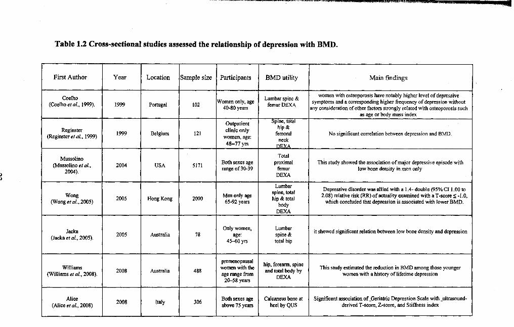
Table 1.2 Cross-sectional studies assessed the relationship of depression with BMD.
First Author Year Location Sample size Participants BMD utility Main findings
Coelho Lumbar spine & women with osteoporosis have notably higher level of depressive
(Coelho eta/., 1999). 1999 Portugal 102 Women only, age
femurDEXA symptoms and a corresponding higher frequency of depression without 40-80 years any consideration of other factors strongly related with osteoporosis such
as age or body mass index
Outpatient Spme, total
Reginster clinic only hip& 1999 Belgium 121 femoral No significant correlation between depression and BMD. (Reginster eta/., 1999) women, age: neck
48-77yrs DEXA
Mussolino Total
(Mussolino eta/., 2004 USA 5171 Both sexes age proximal This study showed the association of major depressive episode with
2004). range of30-39 femur low bone density in men only DEXA
Lumbar Depressive disorder was allied with a 1.4- double (95%CI 1.00 to spine, total Wong 2005 Hong Kong 2000 Men only age hip & total 2.08) relative risk (RR) of actuality examined with aT-scoreS -1.0,
(Wong eta/., 2005) 65-92 years body
which concluded that depression is associated with lower BMD.
DEXA
Jacka Only women, Lumbar
it showed significant relation between low bone density and depression (Jacka eta/., 2005).
2005 Australia 78 age: spine & 45-60 yrs total hip
premenopausal hip, forearm, spine Williams
2008 Australia 488 women with the and total body by This study estimated the reduction in BMD among those younger
(Williams eta/., 2008). age range from DEXA
women with a history of lifetime depression 20-58 years
Alice 2008 Italy 306 Both sexes age Calcaneus bone at Significant association of _Geriatric Depression Scale with _ultrasound-
(Alice eta/., 2008) above 75 years heel byQUS derived T -score, Z-score, and Stiffness index

Table 1.3 Case and control studies assessed the relationship of depression with BMD.
First Author Location No. of No. of
Participants BMD utility Main Finding Year Cases control
Schweiger Inpatient clinic, single-energy quantitative after age adjusting, the average ofBMD values (Schweiger eta/., 1994 Germany 70 80 community controls, computerized tomography
among depressed group was 15% lower than the non· 1994) both sexes age: 4D-95 (SE-QCT) to measure depressed group. years BMD at lumber spine
Michelson Lumbar BMD in depressed women was 6.5 %at the spine (Michelson et al.,
1996 USA 24 24 Women only mean age
spine, hip & while at the femoral neck, it was 13.6 %lower than 1996). 4lyears radius DEXA normal women
Amsterdam 1998 USA 6 5 Outpatient clinic, Lumbar no significant association between depression and (Amsterdam and community controls, spineDEXA bone density Hooper, 1998) age: 27-53 yrs
Vrkljan Inpatient clinic, good correlation between the duration of depression
(Vrkljan et al., 2001) 2001 Croatia 31 17 community controls, Unknown and the reduction in the bone mineral density a~te: 29-45.years Outpatient clime,
Yazici 2003 Turkey 25 15
community controls lumbar spine and proximal Significant association of depression with low BMD
(Yaz1c1 et al., 2003) only women), mean femur by DEXA aee: 31 vrs
outpatient clime, Lumbar spine Yazici
2005 Turkey 35 30 community controls
& femoral neck No assoc between depression and BMD (Yazlcl et al., 2005) (only women), mean
age·4~ veal'!l DEXA
Outpatient clime, Lumbar spine the mean BMD of the women with depression was
Altindag 2007 Turkey 36 41 community controls
& femoral neck significantly lower at the lumbar spine in addition to (Aitindag et al., 2007) (only women), age: 26- DEXA all sites of the proximal femur
56vrs
Petronljevic premenopausal women, lumbar spine and femoral premenopausal women with unipolar depression have
(Petronljevic eta/., 2008 Serbia 73 47 age40 years neckbyDEXA significantly lower bone density than the control
2008) sample.