Embed Size (px)
Citation preview

1
Evaluation of AnemiaEvaluation of Anemia
Mark Wurster, M.D., F.A.C.P.The Ohio State University y
Anemia - DefinitionAnemia - Definition• Most common hematologic disorder• Decrease from normal levels of Hgb, Hct, RBC:
Females – Mean Hgb = 14 g/dl; -2SD = 12 g/dlMales – Mean Hgb = 15.5 g/dl; -2SD = 13.5 g/dlg
• Caveat – Anemia is a syndrome, not a disease. An abnormal Hgb or Hct should ALWAYS be investigated if confirmed on repeat testing.

2
Anemia - DefinitionAnemia - Definition• National Health and Nutrition Examination
SurveySurvey (NHANES III) data-
10-28% of patients over 65 years are anemic
One third of these are due to iron, folate, B12 deficiency alone or in combination
One third are due to renal disease, or other chronic inflammatory response
One third are due to various primary marrow disorders, malignancies or other disorders
• A simplified approach to anemia,
AnemiaClassification Schemes
AnemiaClassification Schemes
s p ed app oac to a e a,emphasizing information already included in the CBC:
• Mean Cellular Volume (MCV)• Red Cell Distribution Width (RDW)( )• Retic count

3
AnemiaClassification Schemes
AnemiaClassification Schemes
• Mean Cellular Volume (MCV)• Decreased MCV (microcytic); < 80 fL• Normal MCV (normocytic); 80 – 99 fL• Increased MCV (macrocytic); > 100 fL
AnemiaClassification Schemes
AnemiaClassification Schemes
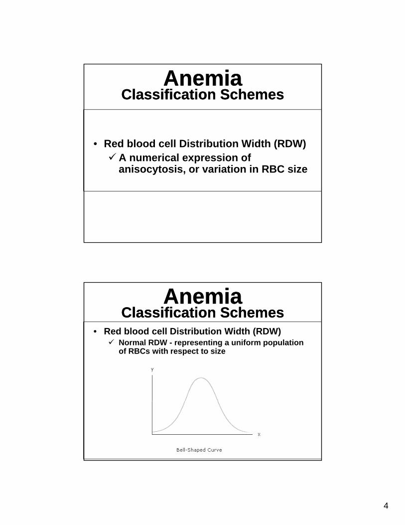
• Red blood cell Distribution Width (RDW)(actually the standard deviation of red blood cell volume divided by the mean volume)
Normal; < or = to app. 14 Elevated; > 14
4
AnemiaClassification Schemes
AnemiaClassification Schemes
• Red blood cell Distribution Width (RDW)A numerical expression of anisocytosis, or variation in RBC size
• Red blood cell Distribution Width (RDW)Normal RDW representing a uniform population
AnemiaClassification Schemes
AnemiaClassification Schemes
Normal RDW - representing a uniform population of RBCs with respect to size

5
• Red blood cell Distribution Width (RDW)El d RDW i RBC i h i
AnemiaClassification Schemes
AnemiaClassification Schemes
Elevated RDW - representing RBCs with varying sizes
AnemiaMCV, RDW classification
AnemiaMCV, RDW classification
• Microcytic indices (MCV < 80)With normal RDW:• Anemia of chronic
disease/inflammationdisease/inflammation• Thalassemia trait

6
AnemiaMCV, RDW classification
AnemiaMCV, RDW classification
• Microcytic indices (MCV < 80)With elevated RDW:• Iron deficiency• Sickle- Beta thalassemiaSickle- Beta thalassemia• Thalassemia major
( C )
AnemiaMCV, RDW classification
AnemiaMCV, RDW classification
• Normocytic indices (MCV 80-99)With normal RDW:• Acute blood loss• Anemia of chronicAnemia of chronic
disease/inflammation• Anemia of chronic renal disease

7
Normocytic indices (MCV 80 99)
AnemiaMCV, RDW classification
AnemiaMCV, RDW classification
Normocytic indices (MCV 80-99)With elevated RDW:• Early iron, folate, B12 deficiency• Combined deficiency states• Sickle cell anemia• Chronic liver disease
• Macrocytic indices (MCV > 99)
AnemiaMCV, RDW classification
AnemiaMCV, RDW classification
• Macrocytic indices (MCV > 99)With elevated RDW:• Folate, B12 deficiency• Immune hemolytic anemia (also, y ( ,
other anemias with elevated Retic counts)
• Myelodysplastic syndromes

8
M ti i di (MCV > 99)
AnemiaMCV, RDW classification
AnemiaMCV, RDW classification
• Macrocytic indices (MCV > 99)With normal RDW:• Alcohol• Myelodysplastic disordersMyelodysplastic disorders• Aplastic anemia• Chemotherapy
Anemia Laboratory Evaluation
Anemia Laboratory Evaluation
R ti l t• Reticulocytes• Immature RBCs, released in response to
decreased Hgb concentration. Increased numbers suggest ongoing RBC loss or destruction; reticulocytes show marrow compensation.
• Lab measures can include:• Reticulocyte Percentage• Absolute Reticulocyte count per flow cytometry• Reticulocyte Index (RI)

9
ReticulocytesReticulocytes• Reticulocyte Percentage• Reticulocyte Percentage• Normally, RBCs live about 120 days, so a
‘normal’ retic count is about 0.8 - 1.0 %• An elevated Retic percentage is
suggestive of hyperproliferative anemiagg yp p• A normal or decreased Retic percentage is
suggestive of hypoproliferative anemia
Anemia Laboratory Evaluation
Anemia Laboratory Evaluation
• Reticulocyte Index• Reticulocyte Index < 2.0 suggests a
hypoproliferative anemiaR ti l t I d > 2 0 t• Reticulocyte Index > 2.0 suggests a hyperproliferative anemia
10
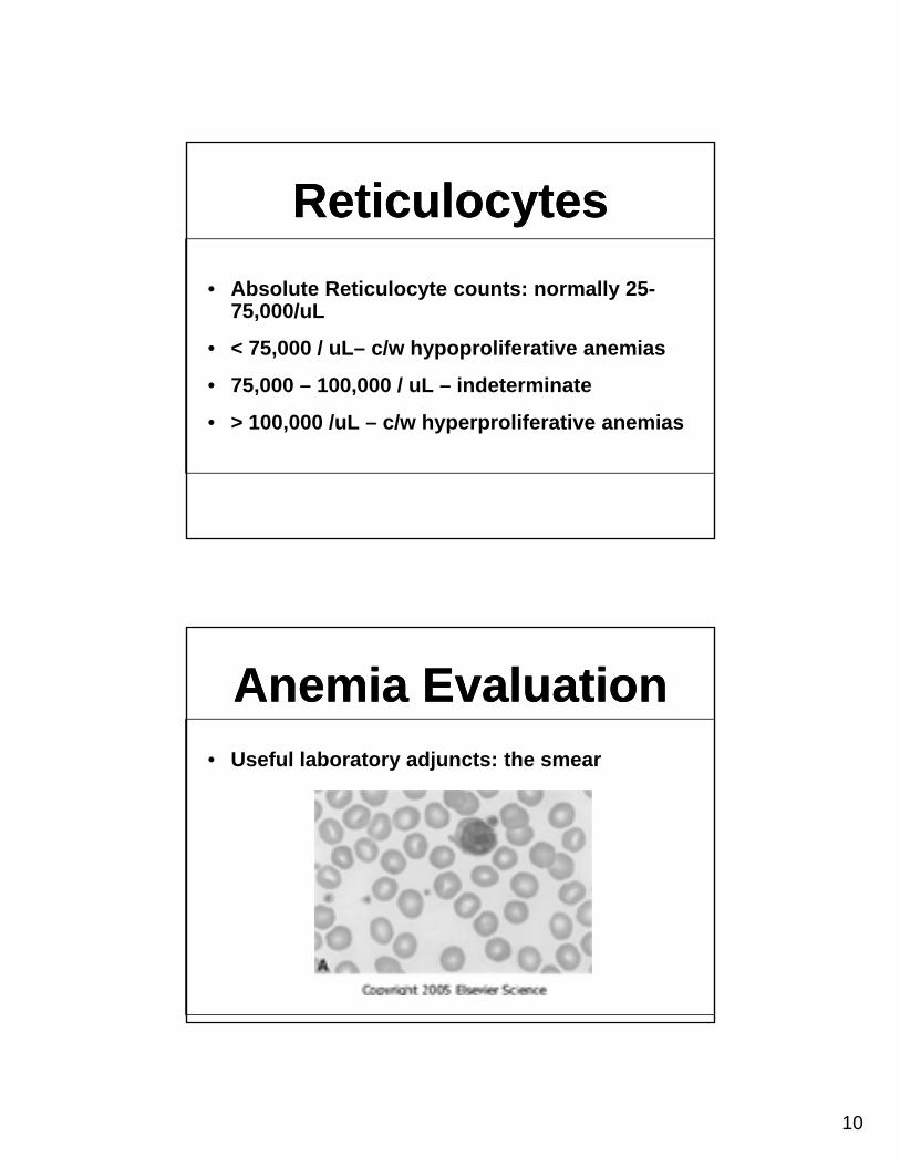
ReticulocytesReticulocytes• Absolute Reticulocyte counts: normally 25-
75,000/uL
• < 75,000 / uL– c/w hypoproliferative anemias
• 75,000 – 100,000 / uL – indeterminate
• > 100,000 /uL – c/w hyperproliferative anemias
Anemia EvaluationAnemia Evaluation• Useful laboratory adjuncts: the smear• Useful laboratory adjuncts: the smear

11
Anemia EvaluationAnemia Evaluation• Useful laboratory adjuncts:Useful laboratory adjuncts:
Technician comments: microcytosis, hypochromia
Anemia EvaluationAnemia Evaluation• Useful laboratory adjuncts:Useful laboratory adjuncts:
Technician comments: macrocytosis, aniso and poikilocytosis

12
Anemia EvaluationAnemia Evaluation• Useful laboratory adjuncts:• Useful laboratory adjuncts:
Technician comments: elliptocytosis, anisocytosis
Anemia EvaluationAnemia Evaluation• Useful laboratory adjuncts:• Useful laboratory adjuncts:
Technician comments: burr cells, acanthocytosis

13
Anemia EvaluationAnemia Evaluation• Useful laboratory adjuncts:• Useful laboratory adjuncts:
Technician comments: sickle cells, aniso- and poikilocytosis
Anemia EvaluationAnemia Evaluation• Useful laboratory adjuncts:• Useful laboratory adjuncts:
Technician comments: spherocytosis
14
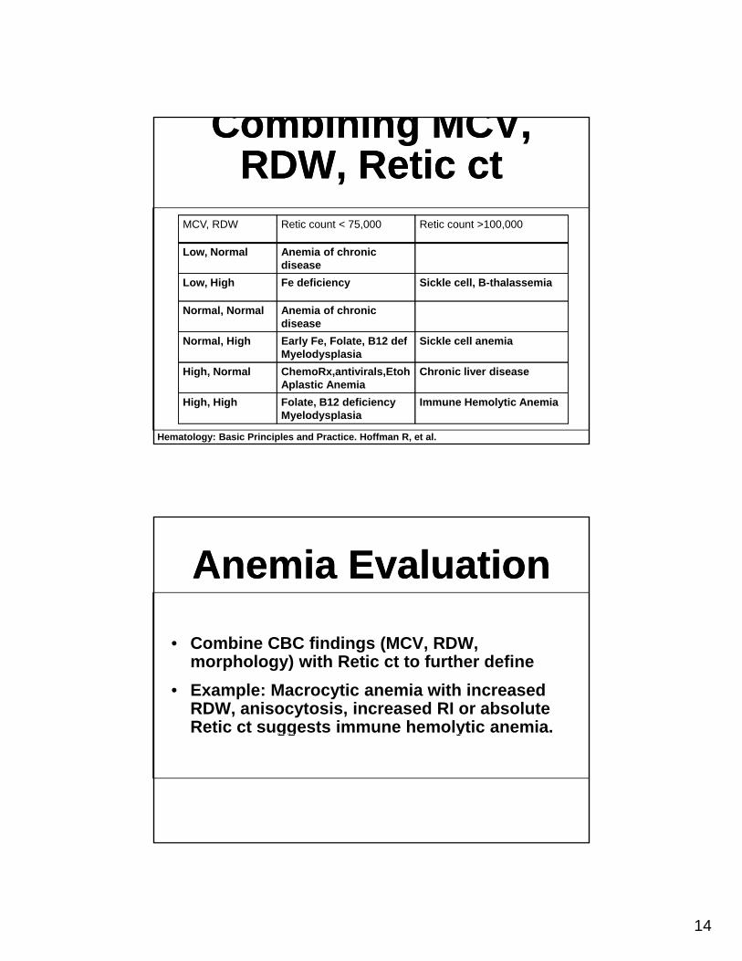
Combining MCV, RDW, Retic ct
Combining MCV, RDW, Retic ct
MCV RDW Retic count < 75 000 Retic count >100 000MCV, RDW Retic count < 75,000 Retic count >100,000
Low, Normal Anemia of chronic disease
Low, High Fe deficiency Sickle cell, B-thalassemia
Normal, Normal Anemia of chronic disease
Normal, High Early Fe, Folate, B12 defMyelodysplasia
Sickle cell anemia
High, Normal ChemoRx,antivirals,EtohAplastic Anemia
Chronic liver disease
High, High Folate, B12 deficiencyMyelodysplasia
Immune Hemolytic Anemia
Hematology: Basic Principles and Practice. Hoffman R, et al.
Anemia EvaluationAnemia Evaluation
• Combine CBC findings (MCV, RDW, morphology) with Retic ct to further define
• Example: Macrocytic anemia with increased RDW, anisocytosis, increased RI or absolute Retic ct suggests immune hemolytic anemia.gg y

15
Anemia EvaluationAnemia Evaluation• Once initial classification established,Once initial classification established,
further lab studies can be used to confirm diagnosis
• Example, if hemolysis is suspected:• Consider LDH, T and D Bilirubin, , ,
haptoglobin, Coomb’s
In SummaryThe End!
In SummaryThe End!
A i i th t h t l i• Anemia is the most common hematologic lab abnormality
• Appropriate evaluation usually demonstrates a treatable cause
• Initial evaluation can be as simple as t a e a uat o ca be as s p e asexamination of CBC diff and plt ct.
• Use Retic count, adjunct tests to confirm initial impression

16
Case 1Case 1• 34 year old Indian female with history of• 34 year old Indian female with history of
heavy periods and recent pregnancy one year ago referred for anemia
• Admits to chewing ice daily
No other significant personal or family• No other significant personal or family history of medical problems
Case 1Case 1
• Physical exam negative for varicosities or telengectasia
• No evidence of organomegaly or adenopathy

17
Case 1Case 1• Lab
Hgb 8.0 g/dL, hct 28%, MCV 75 fL, RPI of 1, RDW of 15
• What is the morphologic and pathophysiologic type of anemia ?
• What are the causes of this?
• What lab studies should be done?
Case 1Case 1• Microcytic Hypochromic Anemia
a) Iron Deficiencyb) Chronic Inflammationc) Thalassemiad) Lead Poisoning
) Sid bl ti A ie) Sideroblastic Anemia• Iron 10, TIBC 450, Ferritin 3• Guaiac negative

18
Case 1Case 1• Iron deficiency in young menstruating female
recently pregnantrecently pregnant
• No reason for GI workup unless guaiac positive
• Evaluate also for celiac disease if no response to oral iron
• Ice craving good clinical sign
• Response to oral iron follow count and ferritin
Case 2Case 2• A 44 year old white female with history of
h t id th iti f irheumatoid arthritis sees you for anemia
• Disease activity is moderate and patient is on intermittent steroids and has received an inhibitor to TNF.
N th di l bl t• No other medical problems are present
• Physical exam unremarkable except for joint deformity

19
Case 2Case 2
• Lab studies include hgb of 8.2 g/dL, hct of 25%, MCV of 75fL, RPI of 1, RDW of 12
• Characterize the anemia according to prior criteria and decide on appropriate labs
Case 2Case 2• The anemia is microcytic with• The anemia is microcytic with
hypoproliferative state
• Patient had a sed rate drawn of > 140, iron of 20, TIBC of 140, saturation of 14%, ferritin of 100, and Erythropoietin level of 30 (nl 0-19)

20
Case 2Case 2• Anemia of Chronic Inflammation
• Due to inability to release iron from macrophages (relative iron deficiency)
• Treatment of underlying disease
• Erythropoietin approved for certain• Erythropoietin approved for certain inflammatory states
Case 3Case 3A 33 ld M i k i• A 33 year old Mexican worker comes in with a week history of dyspnea and fatigue
• No prior history of significant medical problems or family history
Ph i l i iti f t h di• Physical exam is positive for tachycardia and scleral icterus

21
Case 3Case 3
• Lab studies drawn show a Hgb of 6.0 g/dL, Hct of 18.0%, MCV of 100 fL, RPI of 6.0, RDW of 20
• What steps are important next?
Case 3Case 3E al ation of peripheral blood smear• Evaluation of peripheral blood smear
• It shows spherocytes without fragments
• Lab studies for hemolytic anemia including coomb’s test, LDH, bilirubin, haptoglobin,
i h id iurine hemosiderin
22
Case 3Case 3
• Coomb’s test both direct and indirect are positive for IgG and C3
• LDH is slightly elevated to 250 (< 200), bilirubin in 4.0mg/dl with 3.0 indirect, haptoglobin is normal as is urine p ghemosiderin
Case 3Case 3• Patient has autoimmune hemolytic anemia
d h ld b k d f SLEand should be worked up for SLE, lymphoma, and CLL
• Initial treatment is steroids and be cautious about transfusing RBC’s
R t k 1 2 k d• Response may take 1-2 weeks and documented by increasing hemoglobin and clearance of positive coomb’s test

23
Case 4Case 4
• A 45 year old white female had gastric bypass surgery 5 years before. She notes marked fatigue and numbness in her hands and feet.
• The patient has no medical problems and is p pon no supplemental medications
Case 4Case 4• Physical exam is unremarkable except for• Physical exam is unremarkable except for
some gait unsteadiness
• Initial lab studies include a hgb of 9.0 g/dL, hct of 27%, WBC of 2.8, platelets of 100,000/ul, MCV of 110 fL, RPI of 1, and RDW of 18RDW of 18
24
Case 4Case 4What lab studies do you wish to get?• What lab studies do you wish to get?
• Is this an expected problem?
• What therapy is appropriate?
Case 4Case 4• Peripheral blood smear• Serum and RBC Folate• Serum and RBC Folate• Serum Homocysteine and Methylmalonic
acid• Serum B12• Parietal cell Antibodies• Intrinsic Factor Antibodies• Schilling Test

25
Case 4Case 4• Peripheral blood smear shows macrocytic
red cells and hypersegmented neutrophilsyp g p
• Red cell and serum folate normal
• Serum homocysteine and methylmelonic acid are elevated
S B12 170 ( l 250 /dl)• Serum B12 170 (nl> 250 ug/dl)
• Parietal and intrinsic factor antibodies normal
Case 4Case 4S hilli t t t d• Schilling test not done
• Malabsorption of B12 common after gastric bypass and most patients should be on B12

26
Case 5Case 5• Patient is 52 year old male with diabetes
d h t iand hypertension
• Patient noted to have mild fatigue and dyspnea
• Patient has required 2 units of packed RBC’s in the last t o monthsRBC’s in the last two months
• Physical exam is unremarkable
Case 5Case 5P ti t’ h b i 8 0 /dL h t 24% MCV i• Patient’s hgb is 8.0 g/dL, hct 24%, MCV is 85 fL, RPI is 1, and RDW of 12
• The WBC and platelets are normal
• The peripheral blood smear is k blunremarkable

27
Case 5Case 5
• What lab studies are appropriate?
• What treatment should be considered?
Case 5Case 5• The patient has a normocytic anemia with• The patient has a normocytic anemia with
hypoproliferation
• In a patient with diabetes and hypertension chronic renal disease is common
• His creatinine is 2.5 mg/dl andHis creatinine is 2.5 mg/dl and erythropoietin level is 40
• Patient may go on erythropoietin

28
Case 6Case 6• Patient is 65 year old white male with• Patient is 65 year old white male with
history of ischemic heart disease
• Patient has noted increasing angina and dyspnea
• He has no other medical problems and hisHe has no other medical problems and his blood counts were normal 2 years ago
Case 6Case 6H h bl d k d hi h b i 7 5 /dL• He has blood work and his hgb is 7.5 g/dL, hct 20%, MCV 106 fL, RPI of 2, RDW of 22, WBC of 3.0 with an ANC of 1000, and platelets of 100,000.
• His differential also shows a monocyte t f 1500 d h lcount of 1500 and hypogranular
neutrophils

29
Case 6Case 6
• What blood work do you order now?
• What procedures should be considered?
Case 6Case 6• He has a macrocytic anemia but evidence
of possible MDSof possible MDS
• His iron studies show an iron 200, TIBC of 350, iron saturation of 57%, and ferritin of 500
• He has normal folic acid and B12.He has normal folic acid and B12.
• A bone marrow aspirate and biopsy are done showing sideroblastic anemia and MDS


![Home [] Qualitative description of the Wurster-based… · Wurster-based coating process is very different from the top-spray coating process, and optimization should be treated from](https://img.pdfslide.us/doc/110x75/5f5518eae53bae6b0f2357b0/home-qualitative-description-of-the-wurster-based-wurster-based-coating-process.jpg)