Embed Size (px)
Citation preview
1
Nitin Bhatt, MDAssociate Professor of Internal Medicine
Division of Pulmonary, Allergy, Critical Care,and Sleep Medicine
Department of Internal Medicine The Ohio State University Wexner Medical Center
Evaluation and Treatment of Idiopathic Pulmonary Fibrosis
CaseCase• 57 yo WM • SOB over the past 6 months• Throat clearing, dry cough for 3 years
• DOE at work, difficulty climbing steps• Not feeling better after cath/PTCA 2 months
prior• Abnormal CXR showing fibrosis
• PMHx: CAD, GERD• Meds: ASA, Plavix, metoprolol, PPI• PSHx: 15PY tob, quit 20 years ago
2
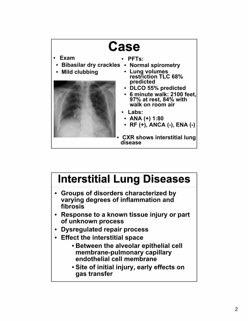
CaseCase• Exam• Bibasilar dry crackles• Mild clubbing
• PFTs: • Normal spirometry• Lung volumes
restriction TLC 68% predicted
• DLCO 55% predicted• 6 minute walk: 2100 feet,
97% at rest, 84% with walk on room air
• Labs:• ANA (+) 1:80• RF (+), ANCA (-), ENA (-)
• CXR shows interstitial lung disease
Interstitial Lung DiseasesInterstitial Lung Diseases• Groups of disorders characterized by
varying degrees of inflammation and fibrosis
• Response to a known tissue injury or part of unknown process
• Dysregulated repair process• Effect the interstitial space
• Between the alveolar epithelial cell membrane-pulmonary capillary endothelial cell membrane
• Site of initial injury, early effects on gas transfer
3
Interstitial Lung DiseasesInterstitial Lung Diseases• Can also effect areas outside the alveoli,
such as the bronchioles, larger airways and pulmonary vasculature
• Diffuse parenchymal lung diseases
Interstitial Lung DiseaseInterstitial Lung Disease• Over 150 etiologies
• Symptoms nonspecific
• SOB/DOE and cough
• Diagnosis requires combination of:
• Clinical presentation
• Radiology (high resolution chest CT)
• Pathology
• Prognosis and treatment dependent on diagnosis
4
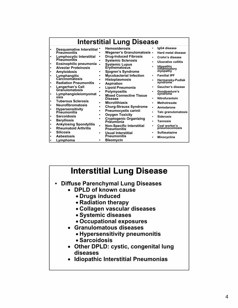
• Desquamative Interstitial Pneumonitis
• Lymphocytic Interstitial Pneumonitis
• Eosinophilic pneumonia• Alveolar Proteinosis• Amyloidosis• Lymphangitic
Carcinomatosis• Radiation Pneumonitis• Langerhan’s Cell
Granulomatosis• Lymphangioleiomyomat
osis• Tuberous Sclerosis• Neurofibromatosis• Hypersensitivity
Pneumonitis• Sarcoidosis• Berylliosis• Ankylosing Spondylitis• Rheumatoid Arthritis• Silicosis• Asbestosis• Lymphoma
• Hemosiderosis• Wegener’s Granulomatosis• Drug-Induced Fibrosis• Systemic Sclerosis• Systemic Lupus
Erythematosus• Sjogren’s Syndrome• Mycobacterial Infection• Histoplasmosis• Aspiration• Lipoid Pneumonia• Polymyositis• Mixed Connective Tissue
Disease• Microlithiasis• Churg-Strauss Syndrome• Pneumocystis carinii• Oxygen Toxicity• Cryptogenic Organizing
Pneumonia• Non-Specific Interstitial
Pneumonitis• Usual Interstitial
Pneumonitis• Bleomycin
• IgG4 disease
• Hard metal disease
• Crohn’s disease
• Ulcerative collitis
• Idiopathic inflammatory myopathy
• Familial IPF
• Hermansky-Pudlaksyndrome
• Gaucher’s disease
• Goodpasture’ssyndrome
• Nitrofurantoin
• Methotrexate
• Amiodarone
• Talc granulomatosis
• Siderosis
• Tannosis
• Coal worker’s pneumoconiosis
• Sulfasalazine
• Minocycline
Interstitial Lung Disease
Interstitial Lung DiseaseInterstitial Lung Disease
• Diffuse Parenchymal Lung Diseases DPLD of known cause
Drugs inducedRadiation therapyCollagen vascular diseasesSystemic diseasesOccupational exposures
Granulomatous diseasesHypersensitivity pneumonitisSarcoidosis
Other DPLD: cystic, congenital lung diseases
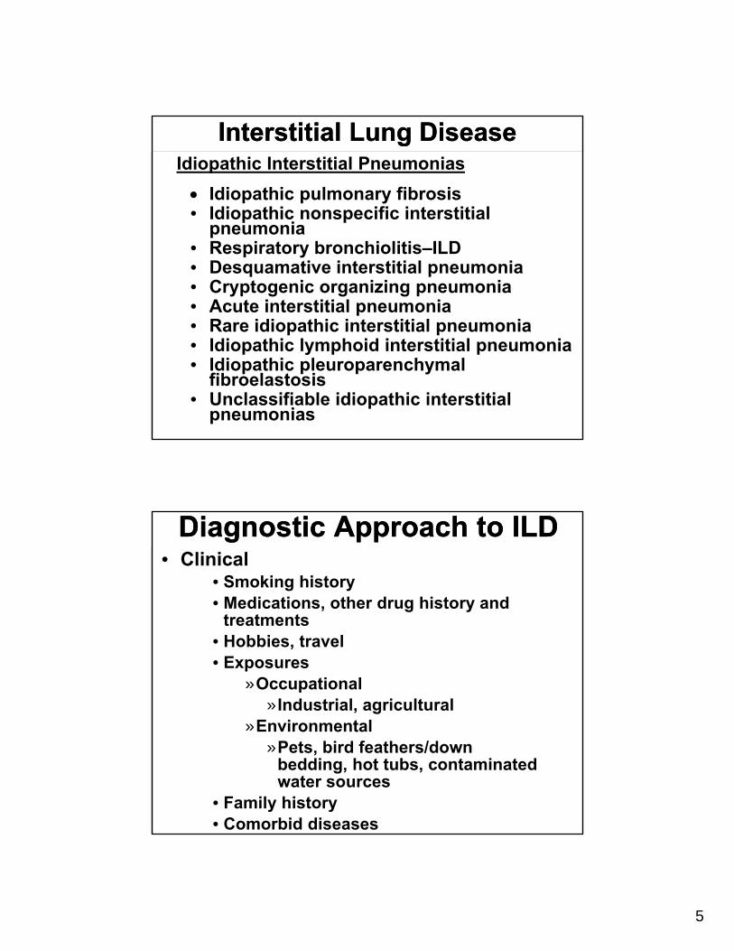
Idiopathic Interstitial Pneumonias
5
Interstitial Lung DiseaseInterstitial Lung DiseaseIdiopathic Interstitial Pneumonias
Idiopathic pulmonary fibrosis• Idiopathic nonspecific interstitial
pneumonia• Respiratory bronchiolitis–ILD• Desquamative interstitial pneumonia• Cryptogenic organizing pneumonia• Acute interstitial pneumonia• Rare idiopathic interstitial pneumonia• Idiopathic lymphoid interstitial pneumonia• Idiopathic pleuroparenchymal
fibroelastosis• Unclassifiable idiopathic interstitial
pneumonias
Diagnostic Approach to ILDDiagnostic Approach to ILD• Clinical
• Smoking history• Medications, other drug history and
treatments• Hobbies, travel• Exposures
»Occupational» Industrial, agricultural
»Environmental »Pets, bird feathers/down
bedding, hot tubs, contaminated water sources
• Family history• Comorbid diseases

6
Diagnostic Approach to ILDDiagnostic Approach to ILD• Physical Examination
• Crackles, dry or velcro
• Clubbing
• Signs of right heart strain/failure
• Signs of systemic disease (vasculitis, connective tissue diseases)
»Potential biopsy sites (rashes)
Diagnostic Approach to ILDDiagnostic Approach to ILD• Pulmonary Function Testing
• Interstitial inflammation and scarring results in restrictive defect
• Impaired gas exchange with a reduced diffusing capacity
• Measures of O2 saturation with exercise»6 Minute walk
• Not diagnostic but characterizes severity
• Obstructive physiology not typical features of ILD
»May be present with coexisting COPD
7
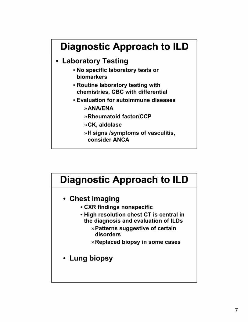
Diagnostic Approach to ILDDiagnostic Approach to ILD• Laboratory Testing
• No specific laboratory tests or biomarkers
• Routine laboratory testing with chemistries, CBC with differential
• Evaluation for autoimmune diseases
»ANA/ENA
»Rheumatoid factor/CCP
»CK, aldolase
» If signs /symptoms of vasculitis, consider ANCA
Diagnostic Approach to ILDDiagnostic Approach to ILD
• Chest imaging• CXR findings nonspecific• High resolution chest CT is central in
the diagnosis and evaluation of ILDs»Patterns suggestive of certain
disorders »Replaced biopsy in some cases
• Lung biopsy
8
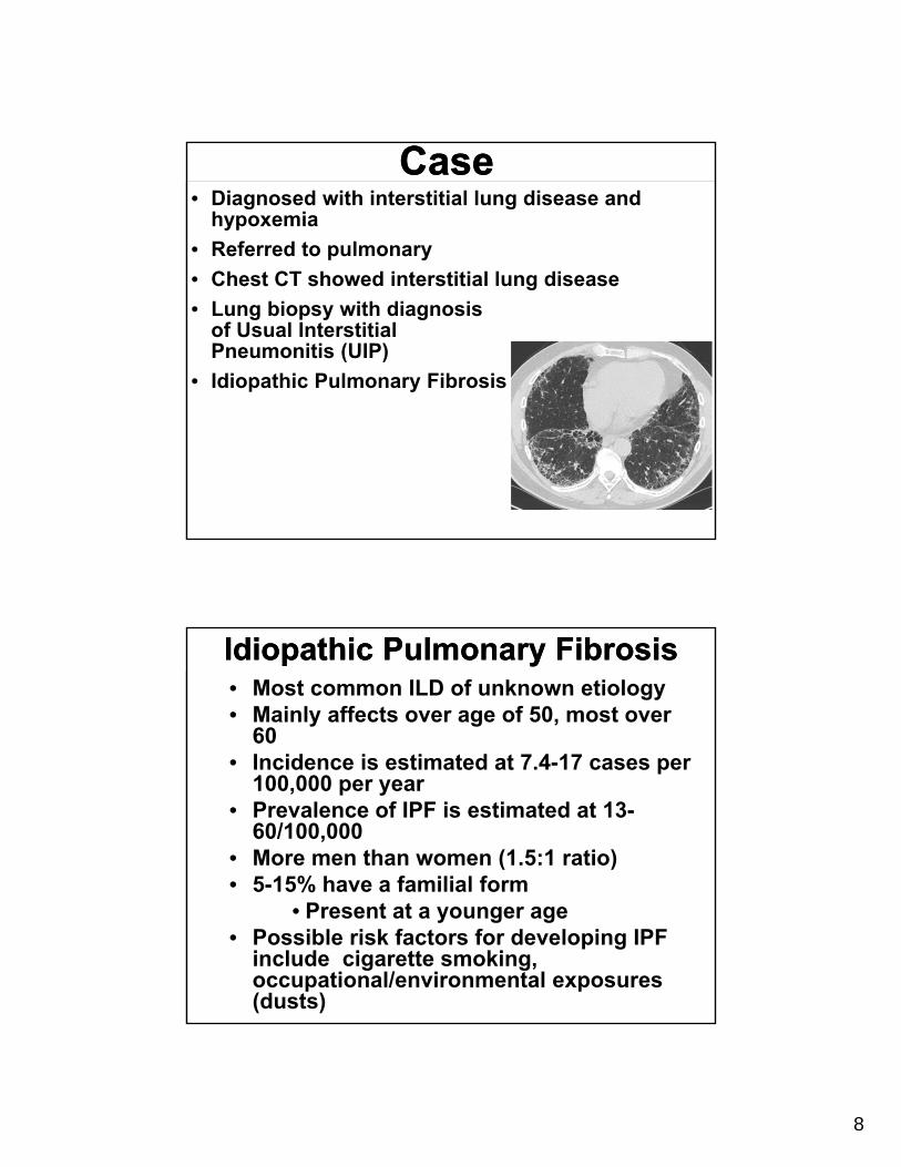
CaseCase• Diagnosed with interstitial lung disease and
hypoxemia
• Referred to pulmonary
• Chest CT showed interstitial lung disease
• Lung biopsy with diagnosis of Usual Interstitial Pneumonitis (UIP)
• Idiopathic Pulmonary Fibrosis
Idiopathic Pulmonary FibrosisIdiopathic Pulmonary Fibrosis• Most common ILD of unknown etiology• Mainly affects over age of 50, most over
60• Incidence is estimated at 7.4-17 cases per
100,000 per year • Prevalence of IPF is estimated at 13-
60/100,000 • More men than women (1.5:1 ratio)• 5-15% have a familial form
• Present at a younger age• Possible risk factors for developing IPF
include cigarette smoking, occupational/environmental exposures (dusts)
9
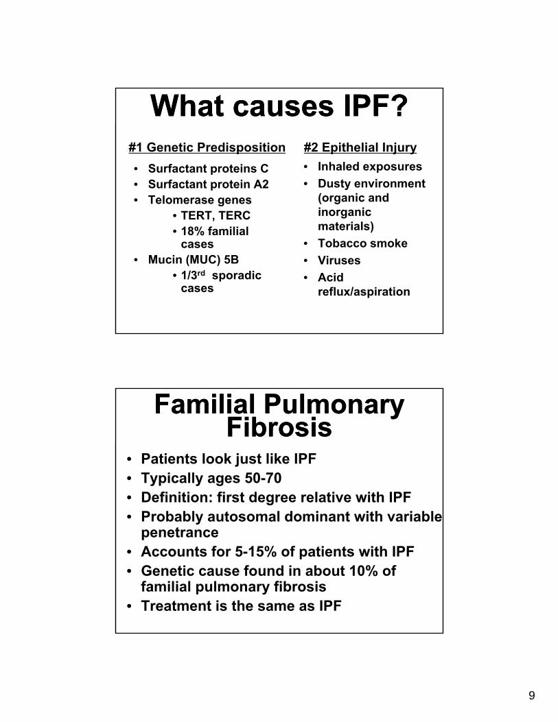
What causes IPF?What causes IPF?#1 Genetic Predisposition
• Surfactant proteins C• Surfactant protein A2• Telomerase genes
• TERT, TERC• 18% familial
cases• Mucin (MUC) 5B
• 1/3rd sporadic cases
#2 Epithelial Injury
• Inhaled exposures
• Dusty environment (organic and inorganic materials)
• Tobacco smoke
• Viruses
• Acid reflux/aspiration
Familial Pulmonary Fibrosis
Familial Pulmonary Fibrosis
• Patients look just like IPF• Typically ages 50-70• Definition: first degree relative with IPF• Probably autosomal dominant with variable
penetrance• Accounts for 5-15% of patients with IPF• Genetic cause found in about 10% of
familial pulmonary fibrosis• Treatment is the same as IPF
10
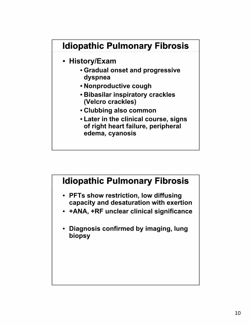
Idiopathic Pulmonary FibrosisIdiopathic Pulmonary Fibrosis
• History/Exam• Gradual onset and progressive
dyspnea • Nonproductive cough• Bibasilar inspiratory crackles
(Velcro crackles) • Clubbing also common• Later in the clinical course, signs
of right heart failure, peripheral edema, cyanosis
Idiopathic Pulmonary FibrosisIdiopathic Pulmonary Fibrosis
• PFTs show restriction, low diffusing capacity and desaturation with exertion
• +ANA, +RF unclear clinical significance
• Diagnosis confirmed by imaging, lung biopsy
11
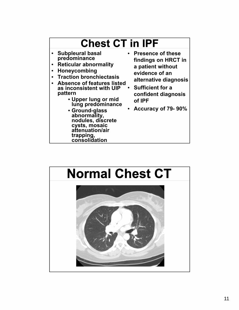
Chest CT in IPFChest CT in IPF• Subpleural basal
predominance• Reticular abnormality • Honeycombing • Traction bronchiectasis• Absence of features listed
as inconsistent with UIP pattern
• Upper lung or mid lung predominance
• Ground-glass abnormality, nodules, discrete cysts, mosaic attenuation/air trapping, consolidation
• Presence of these findings on HRCT in a patient without evidence of an alternative diagnosis
• Sufficient for a confident diagnosis of IPF
• Accuracy of 79- 90%
Normal Chest CTNormal Chest CT
12
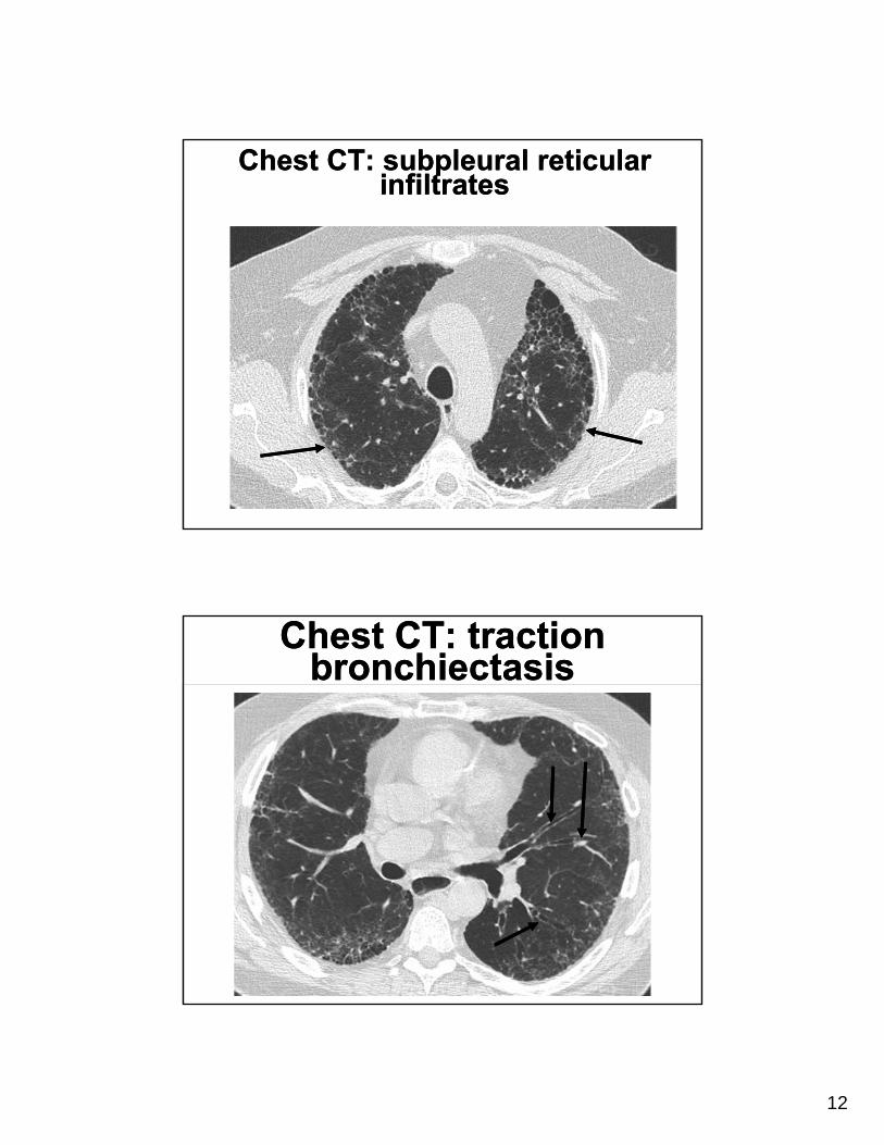
Chest CT: subpleural reticular infiltrates
Chest CT: subpleural reticular infiltrates
Chest CT: traction bronchiectasis
Chest CT: traction bronchiectasis
13
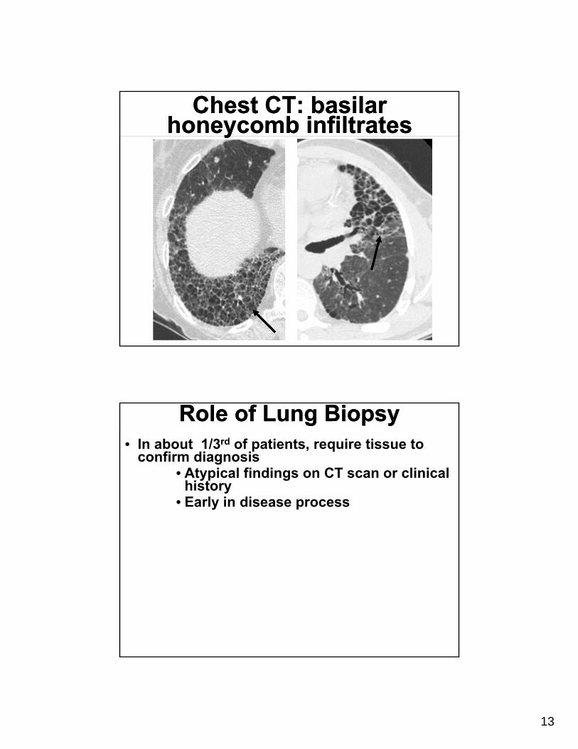
Chest CT: basilar honeycomb infiltrates
Chest CT: basilar honeycomb infiltrates
Role of Lung BiopsyRole of Lung Biopsy• In about 1/3rd of patients, require tissue to
confirm diagnosis• Atypical findings on CT scan or clinical
history• Early in disease process
14
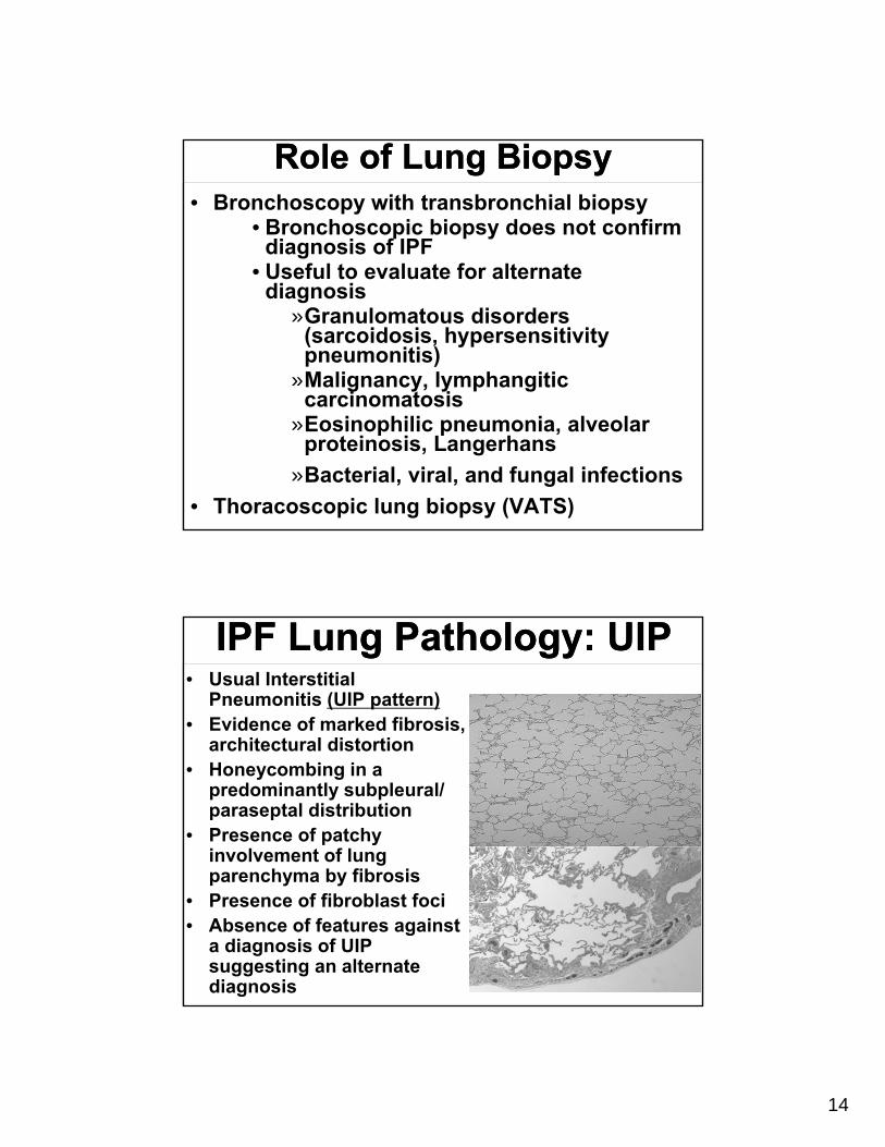
Role of Lung BiopsyRole of Lung Biopsy• Bronchoscopy with transbronchial biopsy
• Bronchoscopic biopsy does not confirm diagnosis of IPF
• Useful to evaluate for alternate diagnosis
»Granulomatous disorders (sarcoidosis, hypersensitivity pneumonitis)
»Malignancy, lymphangitic carcinomatosis
»Eosinophilic pneumonia, alveolar proteinosis, Langerhans
»Bacterial, viral, and fungal infections
• Thoracoscopic lung biopsy (VATS)
Collagen deposition
IPF Lung Pathology: UIPIPF Lung Pathology: UIP• Usual Interstitial
Pneumonitis (UIP pattern)• Evidence of marked fibrosis,
architectural distortion• Honeycombing in a
predominantly subpleural/ paraseptal distribution
• Presence of patchy involvement of lung parenchyma by fibrosis
• Presence of fibroblast foci• Absence of features against
a diagnosis of UIP suggesting an alternate diagnosis
15
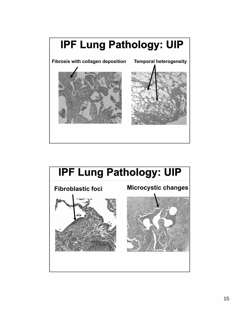
Fibrosis with collagen deposition Temporal heterogeneity
IPF Lung Pathology: UIPIPF Lung Pathology: UIP
Microcystic changesFibroblastic foci
IPF Lung Pathology: UIPIPF Lung Pathology: UIP
16
Causes Of Usual Interstitial Pneumonitis
Causes Of Usual Interstitial Pneumonitis
• Idiopathic pulmonary fibrosis (IPF)• Collagen vascular disease
• Rheumatoid arthritis• Drug toxicity, radiation-induced• Post-inflammatory pulmonary fibrosis• Chronic hypersensitivity pneumonitis
• May see granulomas or other clues of HP
• Occupational exposures• Asbestosis
• Familial idiopathic pulmonary fibrosis• Hermansky–Pudlak syndrome
Clinical Course of IPFClinical Course of IPF• Unpredictable course for an individual
patient
• Progressive disease
• Median survival of about 3-5 years
• Cause of death in about ½ related to IPF and respiratory failure
• Others: CAD/MI, infection, strokes
• Limited treatment options in the past
• Lung transplant
17
Coexisting conditions with IPFCoexisting conditions with IPF• Pulmonary hypertension
• In about 1/3 patients and most with advanced disease
• Associated with worse pulmonary function, hypoxemia
• Decreased exercise capacity and worse survival
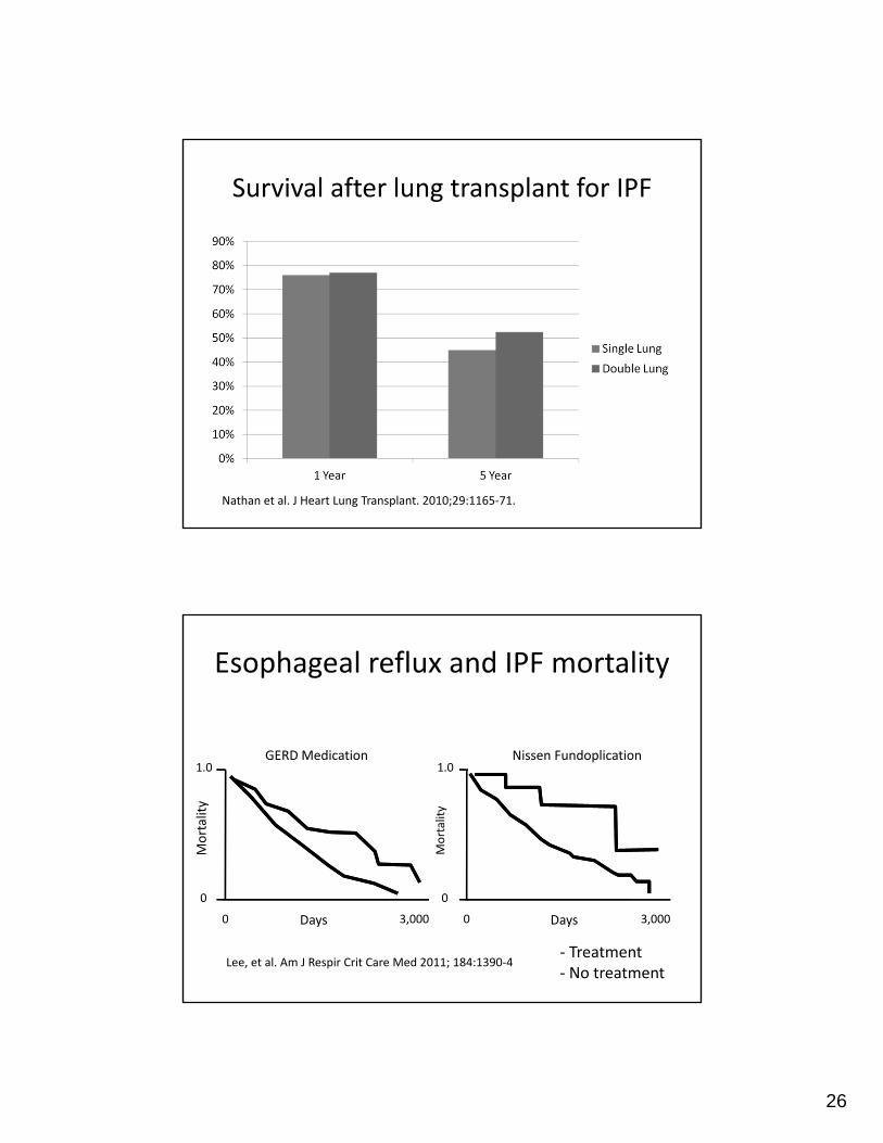
• GERD• Common in IPF (65-94%)• Potential causal relationship between GERD
and IPF through microaspiration of gastric contents
• Acid-suppression therapy was associated with a slower rate of decline in pulmonary function and longer survival
Coexisting conditions with IPFCoexisting conditions with IPF• Combined Pulmonary Fibrosis and
Emphysema • ~8% IPF cases, male, smoking history• Disproportionately low DLCO and gas
exchange• Chest CT upper lobe emphysema, lower
lobe fibrosis • High incidence of pulmonary
hypertension, lung cancer and worse prognosis
• Lung Cancer• Increased risk in IPF patients,
independent of other risks (smoking)• OSA, CAD, depression
18
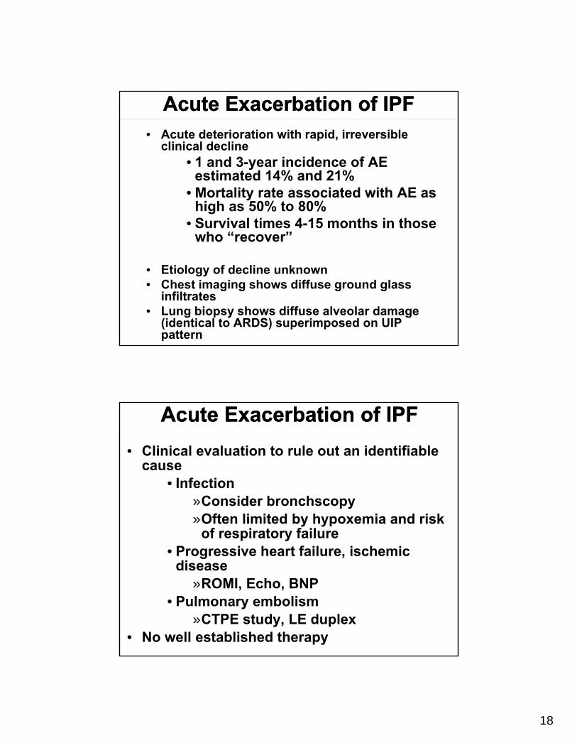
Acute Exacerbation of IPFAcute Exacerbation of IPF• Acute deterioration with rapid, irreversible
clinical decline• 1 and 3-year incidence of AE
estimated 14% and 21%• Mortality rate associated with AE as
high as 50% to 80%• Survival times 4-15 months in those
who “recover”
• Etiology of decline unknown• Chest imaging shows diffuse ground glass
infiltrates• Lung biopsy shows diffuse alveolar damage
(identical to ARDS) superimposed on UIP pattern
Acute Exacerbation of IPFAcute Exacerbation of IPF
• Clinical evaluation to rule out an identifiable cause
• Infection»Consider bronchscopy»Often limited by hypoxemia and risk
of respiratory failure• Progressive heart failure, ischemic
disease»ROMI, Echo, BNP
• Pulmonary embolism»CTPE study, LE duplex
• No well established therapy
19
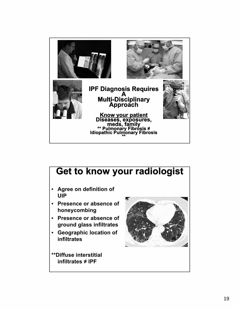
IPF Diagnosis Requires A
Multi-Disciplinary Approach
Know your patientDiseases, exposures,
meds, family** Pulmonary Fibrosis ≠
Idiopathic Pulmonary Fibrosis **
IPF Diagnosis Requires A
Multi-Disciplinary Approach
Know your patientDiseases, exposures,
meds, family** Pulmonary Fibrosis ≠
Idiopathic Pulmonary Fibrosis **
Get to know your radiologistGet to know your radiologist
• Agree on definition of UIP
• Presence or absence of honeycombing
• Presence or absence of ground glass infiltrates
• Geographic location of infiltrates
**Diffuse interstitial infiltrates ≠ IPF
20
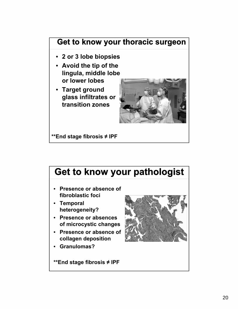
Get to know your thoracic surgeonGet to know your thoracic surgeon
• 2 or 3 lobe biopsies
• Avoid the tip of the lingula, middle lobe or lower lobes
• Target ground glass infiltrates or transition zones
**End stage fibrosis ≠ IPF
Get to know your pathologistGet to know your pathologist
• Presence or absence of fibroblastic foci
• Temporal heterogeneity?
• Presence or absences of microcystic changes
• Presence or absence of collagen deposition
• Granulomas?
**End stage fibrosis ≠ IPF
21
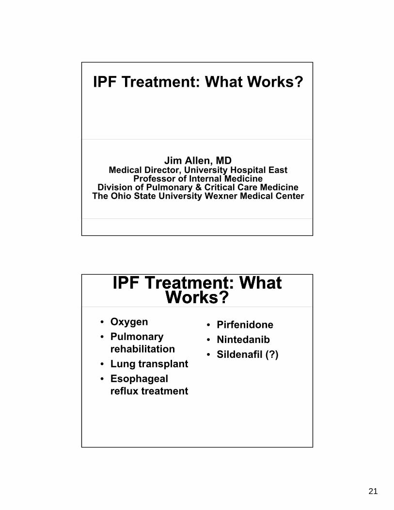
Jim Allen, MDMedical Director, University Hospital East
Professor of Internal MedicineDivision of Pulmonary & Critical Care Medicine
The Ohio State University Wexner Medical Center
IPF Treatment: What Works?
IPF Treatment: What Works?
IPF Treatment: What Works?
• Oxygen
• Pulmonary rehabilitation
• Lung transplant
• Esophageal reflux treatment
• Pirfenidone
• Nintedanib
• Sildenafil (?)
22
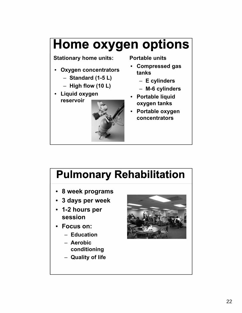
Home oxygen optionsHome oxygen optionsStationary home units:
• Oxygen concentrators
– Standard (1-5 L)
– High flow (10 L)
• Liquid oxygen reservoir
Portable units
• Compressed gas tanks
– E cylinders
– M-6 cylinders
• Portable liquid oxygen tanks
• Portable oxygen concentrators
Pulmonary RehabilitationPulmonary Rehabilitation
• 8 week programs
• 3 days per week
• 1-2 hours per session
• Focus on:– Education
– Aerobic conditioning
– Quality of life
23
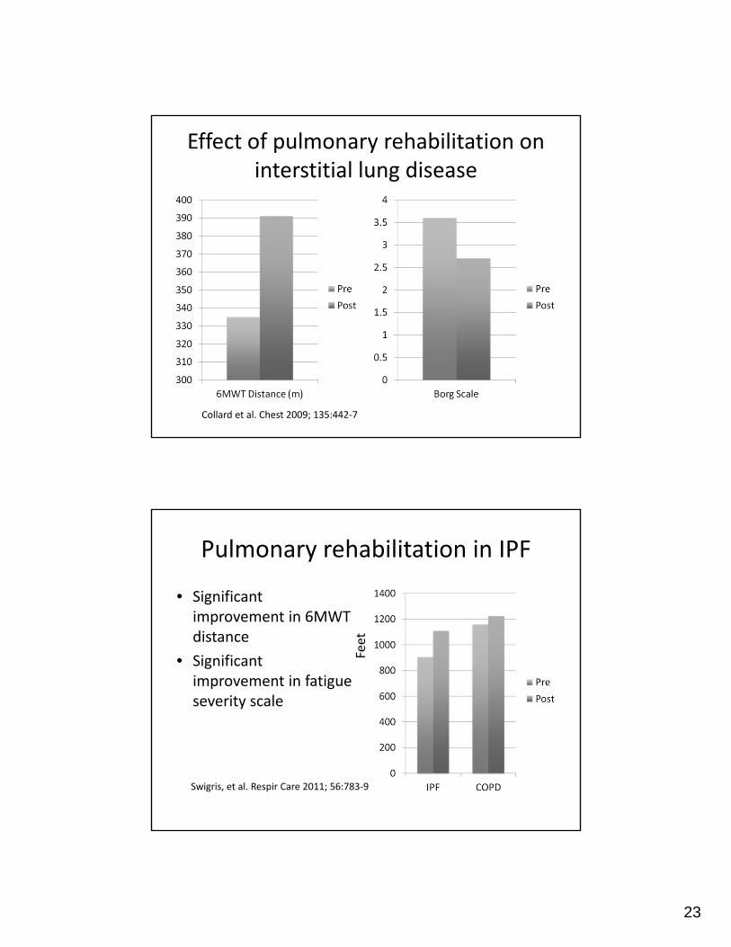
Effect of pulmonary rehabilitation on interstitial lung disease
Collard et al. Chest 2009; 135:442‐7
Pulmonary rehabilitation in IPF
• Significant improvement in 6MWT distance
• Significant improvement in fatigue severity scale
Swigris, et al. Respir Care 2011; 56:783‐9
Feet
24
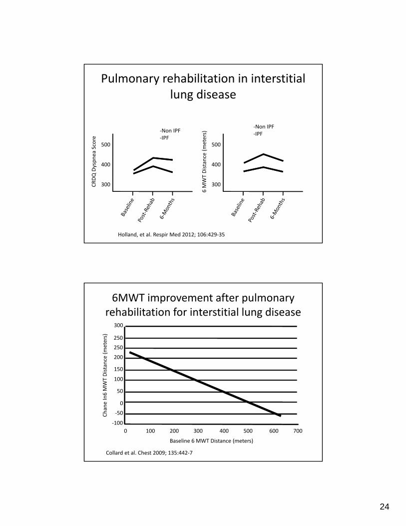
Pulmonary rehabilitation in interstitial lung disease
Holland, et al. Respir Med 2012; 106:429‐35
300
400
500
6 M
WT Distance (meters)
300
400
500
CRDQ Dyspnea
Score
‐Non IPF‐IPF
‐Non IPF‐IPF
6MWT improvement after pulmonary rehabilitation for interstitial lung disease
Collard et al. Chest 2009; 135:442‐7
0 100 200 300 400 500 600 700
‐100
‐50
0
50
100
150
200
250
250
300
Baseline 6 MWT Distance (meters)
ChaneIn6 M
WT Distance (meters)
25
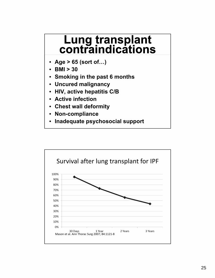
Lung transplant contraindicationsLung transplant
contraindications• Age > 65 (sort of…)• BMI > 30• Smoking in the past 6 months• Uncured malignancy• HIV, active hepatitis C/B• Active infection• Chest wall deformity• Non-compliance• Inadequate psychosocial support
Survival after lung transplant for IPF
Mason et al. Ann Thorac Surg 2007; 84:1121‐8
26
Survival after lung transplant for IPF
Nathan et al. J Heart Lung Transplant. 2010;29:1165‐71.
Esophageal reflux and IPF mortality
Lee, et al. Am J Respir Crit Care Med 2011; 184:1390‐4
3,0000
0
1.0
3,0000
0
1.0
Mortality
Mortality
Days Days
GERD Medication Nissen Fundoplication
‐ Treatment‐ No treatment
27
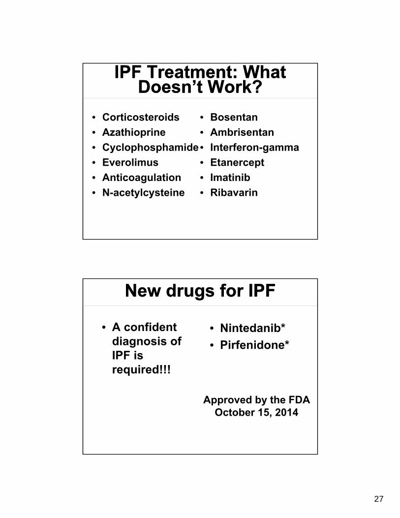
IPF Treatment: What Doesn’t Work?
IPF Treatment: What Doesn’t Work?
• Corticosteroids
• Azathioprine
• Cyclophosphamide
• Everolimus
• Anticoagulation
• N-acetylcysteine
• Bosentan
• Ambrisentan
• Interferon-gamma
• Etanercept
• Imatinib
• Ribavarin
New drugs for IPFNew drugs for IPF
• A confident diagnosis of IPF is required!!!
• Nintedanib*
• Pirfenidone*
Approved by the FDA October 15, 2014
28
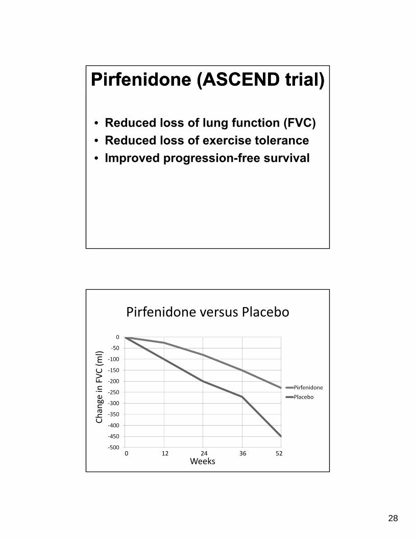
Pirfenidone (ASCEND trial)Pirfenidone (ASCEND trial)
• Reduced loss of lung function (FVC)
• Reduced loss of exercise tolerance
• Improved progression-free survival
Pirfenidone versus Placebo
Weeks0 12 24 36 52
Change in
FVC (ml)
29
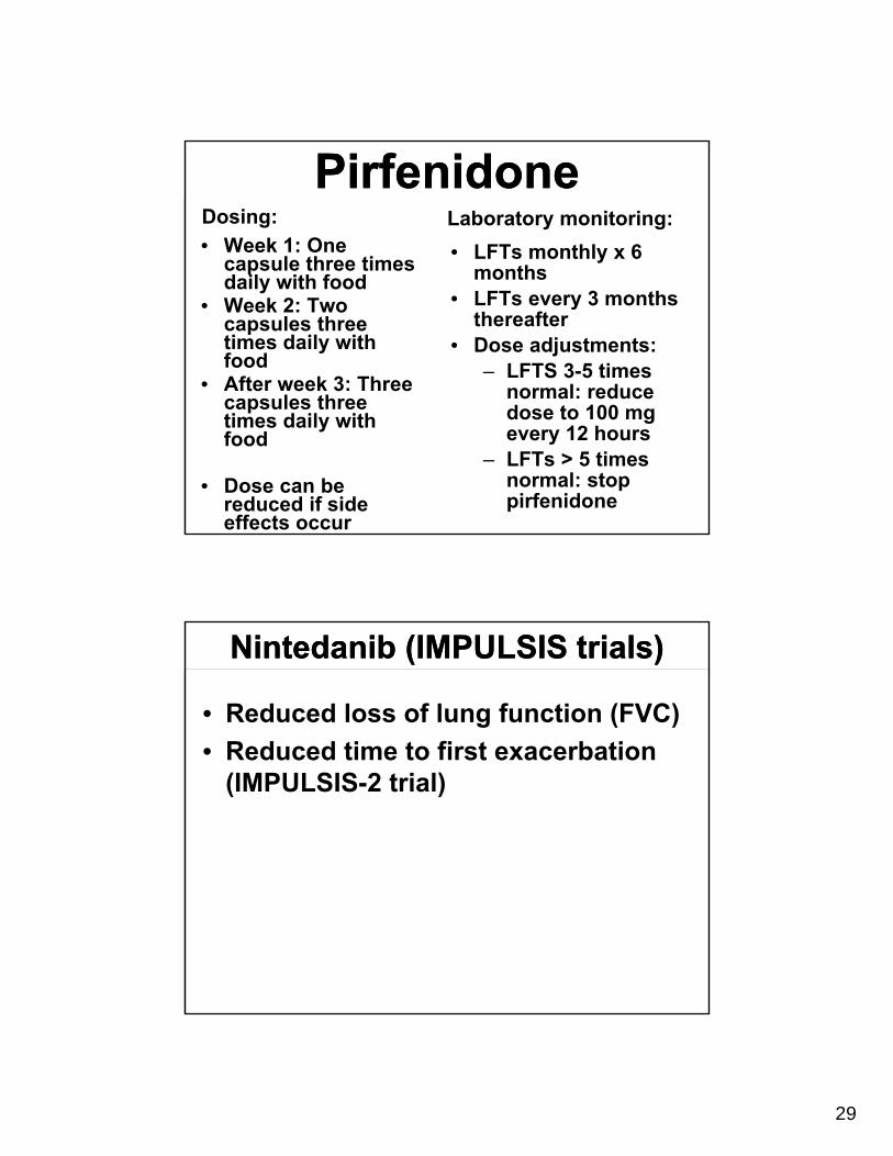
PirfenidonePirfenidoneDosing:
• Week 1: One capsule three times daily with food
• Week 2: Two capsules three times daily with food
• After week 3: Three capsules three times daily with food
• Dose can be reduced if side effects occur
Laboratory monitoring:
• LFTs monthly x 6 months
• LFTs every 3 months thereafter
• Dose adjustments:– LFTS 3-5 times
normal: reduce dose to 100 mg every 12 hours
– LFTs > 5 times normal: stop pirfenidone
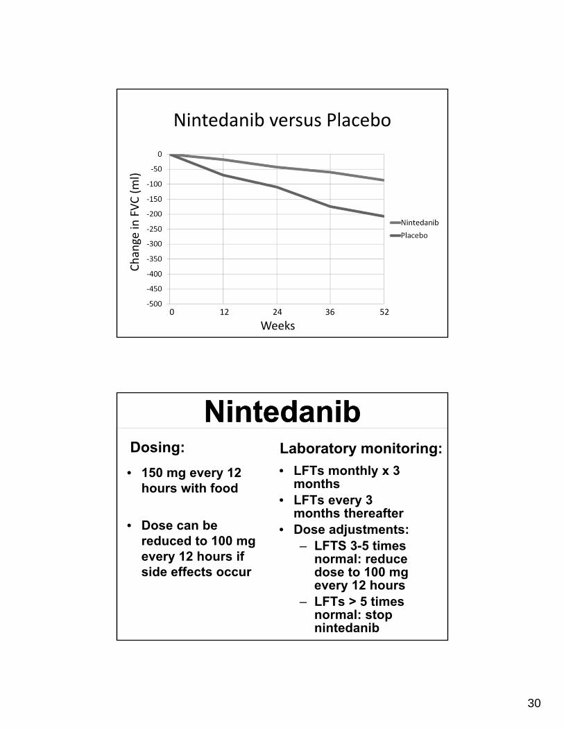
Nintedanib (IMPULSIS trials)Nintedanib (IMPULSIS trials)
• Reduced loss of lung function (FVC)
• Reduced time to first exacerbation (IMPULSIS-2 trial)
30
Nintedanib versus Placebo
Weeks0 12 24 36 52
Change in
FVC (ml)
NintedanibNintedanibDosing:
• 150 mg every 12 hours with food
• Dose can be reduced to 100 mg every 12 hours if side effects occur
Laboratory monitoring:
• LFTs monthly x 3 months
• LFTs every 3 months thereafter
• Dose adjustments:– LFTS 3-5 times
normal: reduce dose to 100 mg every 12 hours
– LFTs > 5 times normal: stop nintedanib
31
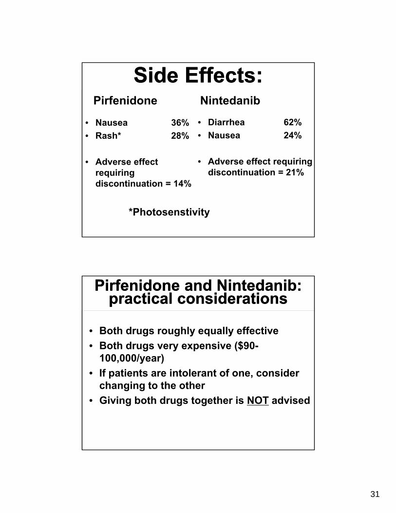
Side Effects:Side Effects:Pirfenidone
• Nausea 36%
• Rash* 28%
• Adverse effect requiring discontinuation = 14%
Nintedanib
• Diarrhea 62%
• Nausea 24%
• Adverse effect requiring discontinuation = 21%
*Photosenstivity
Pirfenidone and Nintedanib: practical considerations
Pirfenidone and Nintedanib: practical considerations
• Both drugs roughly equally effective
• Both drugs very expensive ($90-100,000/year)
• If patients are intolerant of one, consider changing to the other
• Giving both drugs together is NOT advised
32
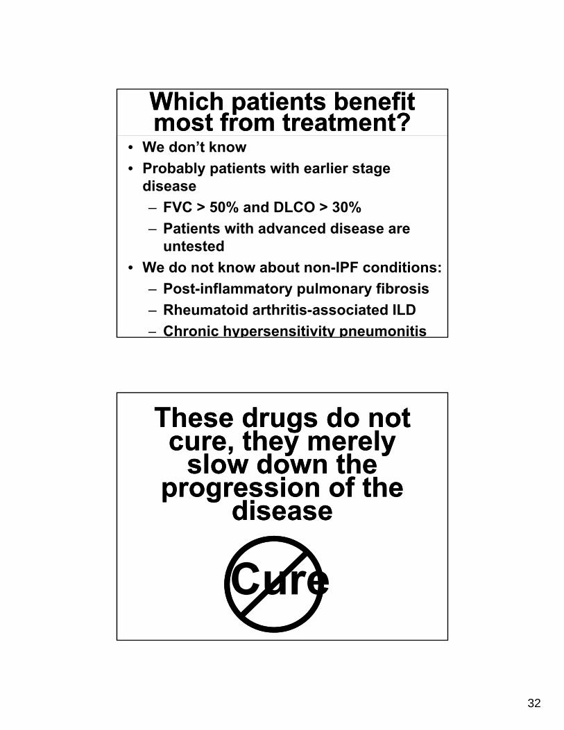
Which patients benefit most from treatment?Which patients benefit most from treatment?
• We don’t know
• Probably patients with earlier stage disease
– FVC > 50% and DLCO > 30%
– Patients with advanced disease are untested
• We do not know about non-IPF conditions:
– Post-inflammatory pulmonary fibrosis
– Rheumatoid arthritis-associated ILD
– Chronic hypersensitivity pneumonitis
Cure
These drugs do not cure, they merely
slow down the progression of the
disease
These drugs do not cure, they merely
slow down the progression of the
disease
33
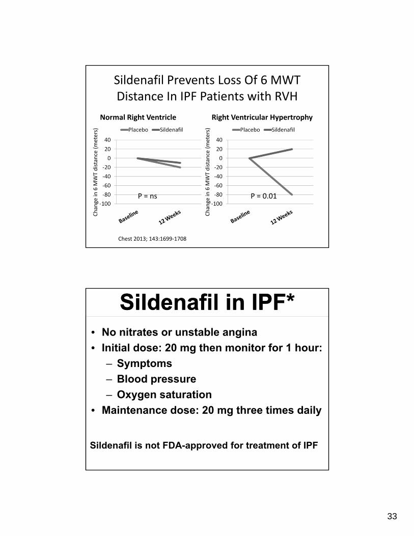
Sildenafil Prevents Loss Of 6 MWT Distance In IPF Patients with RVH
Normal Right Ventricle Right Ventricular Hypertrophy
Change in
6 M
WT distance (meters)
Change in
6 M
WT distance (meters)
P = ns P = 0.01
Chest 2013; 143:1699‐1708
Sildenafil in IPF*Sildenafil in IPF*• No nitrates or unstable angina
• Initial dose: 20 mg then monitor for 1 hour:
– Symptoms
– Blood pressure
– Oxygen saturation
• Maintenance dose: 20 mg three times daily
Sildenafil is not FDA-approved for treatment of IPF
34
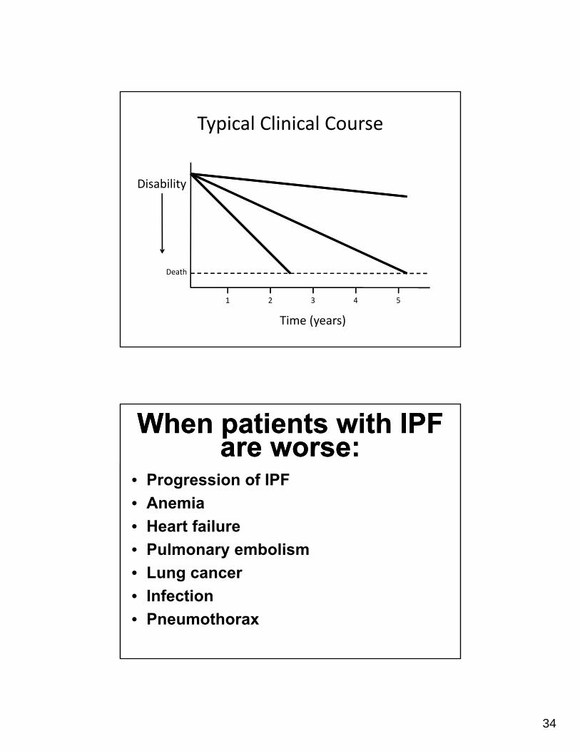
Typical Clinical Course
Disability
Time (years)
Death
1 2 3 4 5
When patients with IPF are worse:
When patients with IPF are worse:
• Progression of IPF
• Anemia
• Heart failure
• Pulmonary embolism
• Lung cancer
• Infection
• Pneumothorax
35
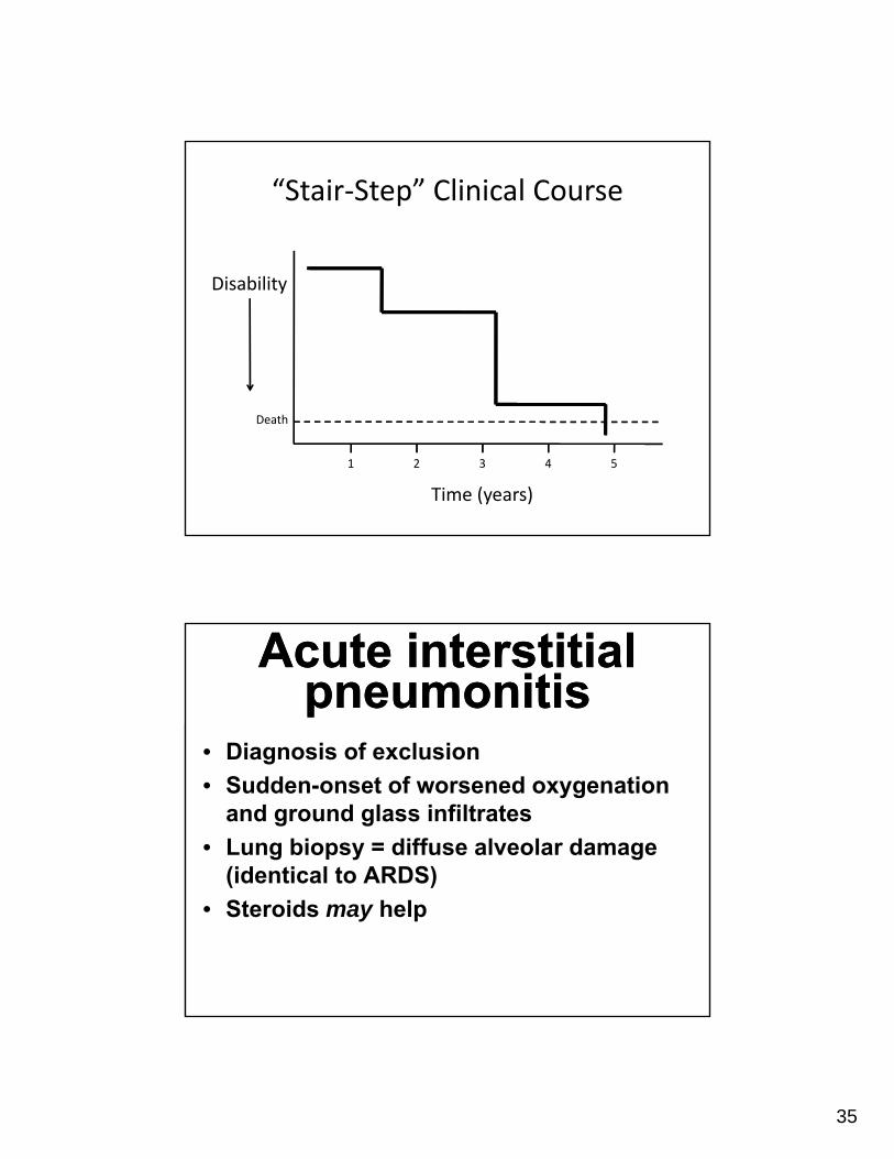
“Stair‐Step” Clinical Course
Disability
Time (years)
Death
1 2 3 4 5
Acute interstitial pneumonitis
Acute interstitial pneumonitis
• Diagnosis of exclusion
• Sudden-onset of worsened oxygenation and ground glass infiltrates
• Lung biopsy = diffuse alveolar damage (identical to ARDS)
• Steroids may help
36
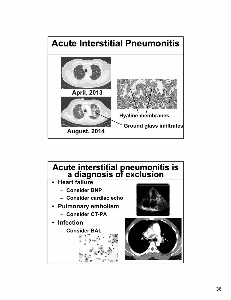
Acute Interstitial PneumonitisAcute Interstitial Pneumonitis
Hyaline membranes
Ground glass infiltrates
April, 2013
August, 2014
Acute interstitial pneumonitis is a diagnosis of exclusion
Acute interstitial pneumonitis is a diagnosis of exclusion
• Heart failure– Consider BNP
– Consider cardiac echo
• Pulmonary embolism– Consider CT-PA
• Infection– Consider BAL
37
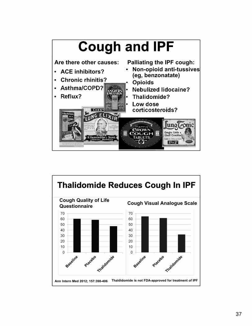
Cough and IPFCough and IPFAre there other causes:
• ACE inhibitors?
• Chronic rhinitis?
• Asthma/COPD?
• Reflux?
Palliating the IPF cough:• Non-opioid anti-tussives
(eg, benzonatate)• Opioids• Nebulized lidocaine?• Thalidomide?• Low dose
corticosteroids?
Thalidomide Reduces Cough In IPFThalidomide Reduces Cough In IPF
Cough Quality of Life Questionnaire
Cough Visual Analogue Scale
Ann Intern Med 2012; 157:398-406 Thaldidomide is not FDA-approved for treatment of IPF
38
Fatigue and IPFFatigue and IPF
• Anemia?
• Thyroid disease?
• Sleep apnea?
• Heart failure?
• Exertional hypoxemia?
Sleep apnea is common in IPF:Sleep apnea is common in IPF:
• Incidence* = 88%!!!
– 20% mild
– 68% moderate-severe
• Undiagnosed sleep apnea contributes to fatigue
• Quality of life can improve with CPAP
*Chest 2009; 136:772-778
39
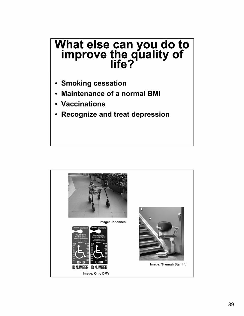
What else can you do to improve the quality of
life?
What else can you do to improve the quality of
life?
• Smoking cessation
• Maintenance of a normal BMI
• Vaccinations
• Recognize and treat depression
Image: JohannesJ
Image: Stannah Stairlift
Image: Ohio DMV
40
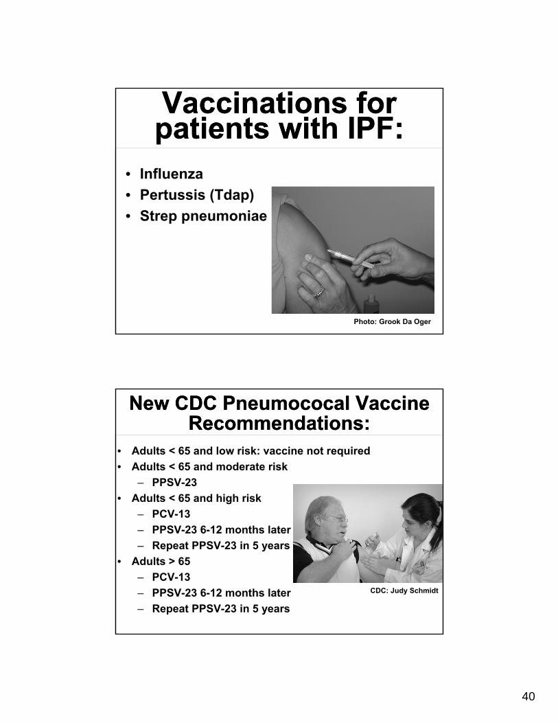
Vaccinations for patients with IPF:Vaccinations for patients with IPF:
• Influenza
• Pertussis (Tdap)
• Strep pneumoniae
Photo: Grook Da Oger
New CDC Pneumococal Vaccine Recommendations:
New CDC Pneumococal Vaccine Recommendations:
• Adults < 65 and low risk: vaccine not required
• Adults < 65 and moderate risk
– PPSV-23
• Adults < 65 and high risk
– PCV-13
– PPSV-23 6-12 months later
– Repeat PPSV-23 in 5 years
• Adults > 65
– PCV-13
– PPSV-23 6-12 months later
– Repeat PPSV-23 in 5 years
CDC: Judy Schmidt
41
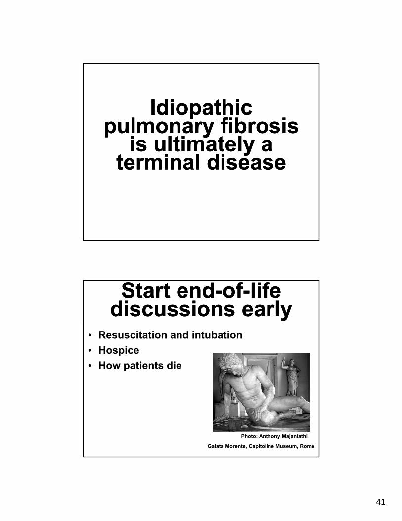
Idiopathic pulmonary fibrosis
is ultimately a terminal disease
Idiopathic pulmonary fibrosis
is ultimately a terminal disease
Start end-of-life discussions earlyStart end-of-life
discussions early• Resuscitation and intubation
• Hospice
• How patients die
Galata Morente, Capitoline Museum, Rome
Photo: Anthony Majanlathi
42
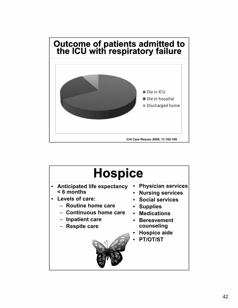
Outcome of patients admitted to the ICU with respiratory failure
Outcome of patients admitted to the ICU with respiratory failure
Crit Care Resusc 2009; 11:102-109
HospiceHospice• Anticipated life expectancy
< 6 months• Levels of care:
– Routine home care– Continuous home care– Inpatient care– Respite care
• Physician services• Nursing services• Social services• Supplies• Medications• Bereavement
counseling• Hospice aide• PT/OT/ST
43
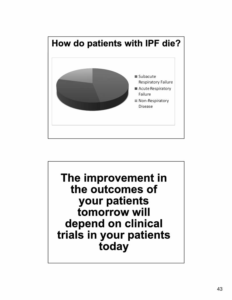
How do patients with IPF die?How do patients with IPF die?
The improvement in the outcomes of
your patients tomorrow will
depend on clinical trials in your patients
today
The improvement in the outcomes of
your patients tomorrow will
depend on clinical trials in your patients
today
44
IPF Treatment: Summary
IPF Treatment: Summary
• Establish a confident diagnosis!
• New drugs: nintedanib & pirfenidone
• Don’t do things that don’t work
• Consider clinical trials
• Never miss an opportunity for transplant
• The little things make a big difference in quality of life
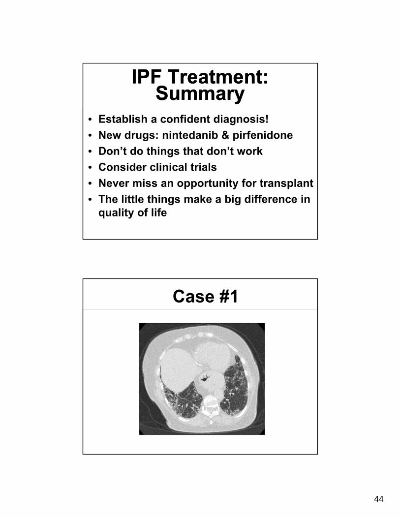
Case #1

45
Case #1
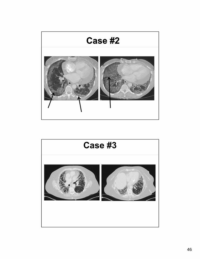
Case #2Case #2
46
Case #2Case #2
Case #3