Embed Size (px)
Citation preview
Türkiye Acil Tıp Dergisi - Tr J Emerg Med 2013;13(2):75-80 doi: 10.5505/1304.7361.2013.01488
Submitted (Geliş tarihi): 27.10.2012 Accepted (Kabul tarihi): 09.05.2013 Published online (Online baskı): 06.06.2013
Correspondence (İletişim): Dr. Ahmet İmerci. Erzurum Palandöken Devlet Hastanesi, Ortopedi ve Travmatoloji Kliniği, 25000 Erzurum, Turkey.
e-mail (e-posta): [email protected]
75ORIGINAL ARTICLE KLİNİK ÇALIŞMA
1Department of Orthopaedics and Traumatology, Erzurum Palandöken State Hospital, Erzurum;2Department of Orthopaedics and Traumatology, Tepecik Training and Research Hospital, İzmir;
3Department of Orthopaedics and Traumatology, Mardin Derik State Hospital, Mardin;4Department of Radiology, Çanakkale 18 Mart University Faculty of Medicine, Çanakkale;
5Department of Orthopaedics and Traumatology, Muğla Sıtkı Kocman University Faculty of Medicine, Muğla
Ahmet İMERCİ,1 Ahmet KAYA,2 Muhammet BOZOĞLAN,3
Gürhan ADAM,4 Umut CANBEK,5 Ahmet SAVRAN2
Evaluating the Use of Computed Tomography forOrthopedic Trauma Patients in the Emergency Department
Acil Ortopedik Travma HastalarındaBilgisayarlı Tomografi Kullanımının Değerlendirilmesi
SUMMARYObjectivesThe purpose of this study is to evaluate the necessity of computed tomog-raphy (CT) scans requested to examine the spine, extremity, or pelvis of or-thopedic trauma patients in the emergency department.
MethodsWe retrospectively screened the medical records of all patients who had a CT scan during their emergency department (ED) evaluation. All data were classified as either child (aged 0-14 years) or adult (aged >14 years).
ResultsOf the 32, 685 patients examined in the child and adult emergency trauma unit over one year, 1, 664 were recommended for an extremity, pelvis, or spine CT (7.02%). The mean age of the patients was 38.6 years (range 2-94 years). Of these patients, 145 of the computed tomography scans (CTs) (80.1%) in the child group and 1, 108 CTs (74.7%) in adult group were negative.
ConclusionsThe unnecessary use of CT in the emergency department to examine ortho-pedic trauma patients has drawn attention. Considering the risk of radia-tion to the patient, it is necessary to develop protocols to determine which emergency department patients should undergo computed tomography.
Key words: Computed tomography; emergency room; radiation; trauma.
ÖZETAmaçBu çalışmanın amacı acil ortopedik travma hastalarında omurga, pelvis ve ekstremitenin değerlendirilmesi için istenilen bilgisayarlı tomografi-nin (BT) gerekliliğini değerlendirmektir.
Gereç ve YöntemGeriye dönük olarak acil servisimizde değerlendirme esnasında BT çe-kilen hastaların tıbbi kayıtları tarandı. Bütün veriler çocuk (0-14) ve eriş-kin yaş grubu (>14) olarak gruplandı.
BulgularÇocuk ve erişkin acil travma bölümünde bir yılda muayene olan 32.685 hastadan 1.664 tanesinden ekstremite, pelvis veya vertebra BT (%7.02) istenmiştir. Hastaların yaş ortalaması 38.6 (2-94) idi. Bütün hastalar içinden çocuklarda çekilen BT’lerin 145’i (%80.1) ve erişkinlerin 1.108’i (%74.7) negatif olarak bulundu.
SonuçAcil serviste ortopedik travma hastalarının değerlendirmesinde ge-reksiz BT kullanımı dikkat çekmektedir. Radyasyon riski de göz önüne alındığında hangi hastaya tomografi çekileceği hususu ile ilgili proto-kollerin geliştirilmesinin gerekli olduğu düşünülmektedir.
Anahtar sözcükler: Bilgisayarlı tomografi; acil servis; radyasyon; travma.
Presented at the 7th Turkish Congress on Emergency Medicine (October 13-16, 2011, Trabzon, Turkey).
IntroductionWhile computed tomography (CT) in addition to direct radiography is of great importance for the diagnosis and treatment of certain orthopedic patients in the emergency room, it is regarded as unnecessary for others.[1-3] The recent advancements in the quality of CT imaging and the ability to obtain CT sections at different planes have resulted in or-thopedists demanding CT more often.[4,5] For example, a CT scan is a very useful imaging method in the evaluation of the posterior wall in acetabular fractures, the posterior el-ements, the loss of alignment and intra-canal fragments in spinal fractures, joint surfaces in intra-articular fractures, and especially the multi-planar epiphysiolisis in children.[5-7]
The initial diagnosis, classification, and planning the treat-ment of spinal fractures in the Emergency Room (ER) is im-portant.[8] Although pelvic fractures constitute only 3 % of all fractures, they are destructive traumas requiring careful attention in the ER. Since the mortality rate is high in pelvic traumas, their treatment and diagnosis should be aggres-sive.[9] For intra-articular fractures and infant fractures, the accurate diagnosis of the fracture is very important in order to accurately plan the treatment.[7]
Though direct radiography can be used as an initial imaging method, CT is often used, especially in certain complicated locations. Now, CT is a reliable and safe method that can be used to examine spine traumas.[9] Yet, CT must be considered as a method to be used in addition to direct radiography. CT is indicated in instances especially when direct radiography is insufficient and findings are doubtful. In addition, even if there were no abnormal findings found with direct radiog-raphy, the presence of neurological deficiency, head trauma, persistent serious pain, and suspicion for serious injury indi-cate the need for CT.[1,6,10] Another benefit of CT is its capa-bility to properly image the craniocervical, cervicothoracic, and posterior pelvic ring, which cannot be imaged via direct radiography.[9,10]
The purpose of this study is to evaluate the necessity of CT scans requested for spine, extremity and pelvis examina-tions of emergency orthopedic trauma patients, and to de-termine the rates of positive and negative examinations.
Materials and MethodsThe records for the application of CT on children (aged 0-14 years) and adults (aged >14 years) visiting the emergency trauma section of the hospital over one year (1 January 2010-1 January 2011) were analyzed. During this analysis, we used emergency examination cards, the computer-based hospital registration system, and pre-CT direct radiographies and CT images taken from the computer archive (PACS). The analy-
ses were performed by one orthopedist and one radiolo-gist. Patients that did not have direct radiographic images, a trauma history, or recorded examination findings were not included in the study. All patients who had CT scans due to their trauma history and their related direct radiographies were included in the study. The reasons for demanding a CT and the findings of the physical examination were record-ed. The analyses that yielded no relevant findings from CT, those that contained irrelevant findings, and those analyses that did not yield more information than that obtained from the direct radiography of the relevant region and did not af-fect the method of treatment were considered negative CT. Those analyses which yielded findings consistent with the request, more information than that given by the direct ra-diography and therefore changed the method of treatment, and CTs that were requested to plan the surgical treatment were considered positive CT. Those fractures seen as normal in direct radiography but evaluated as occult fractures with CT were considered necessary CT. The CTs were grouped as either extremity, pelvic, cervical, thoracic or lumbar spine. Iliac bone, sacrum, acetabulum and hip joint CTs were clas-sified as pelvis CT. All data were also classified as either child (aged 0-14 years) or adult (aged >14 years).
The data were analyzed with the SPSS version 15.00 pack software program. Chi square and two sided analyses were used where appropriate, and the alpha value was accepted as 0.05.
ResultsThe numbers of patients examined in the adult and child ERs of our hospital in 2010 were 183, 552 and 171, 450, respec-tively. Of the 32, 685 patients examined in the child and adult emergency trauma unit over one year, 1, 943 underwent an extremity, pelvis, or spine CT (5.9%). Both CT and plain ra-diographic imaging methods were used in 1, 664 patients. A total of 279 patients (14.3%), including those who had a CT scan but not a plain radiographic scan, and those whose ex-amination findings were not recorded or no trauma history could be found were excluded from the study. The mean age of the adult patients was 42.06 years (15-94 years) and the mean age of the child patients was 10.2 years (2-14). Table 1 shows the distribution of the CTs according to anatomic lo-cations. The number of occult fractures determined with CT scanning for adults and children, whose plain radiographic images were negative, were 102 and 12, respectively.
Out of the 493 extremity CTs requested for adults, a total of 176 (35.6%) were positive CT, with 37 detected to be occult. Out of the 56 extremity CTs requested for children, a total of 18 (32.1%) were positive, with 2 detected to be occult. Out of the 169 pelvis CTs requested for adults, a total of 63 (37.2%)
Türkiye Acil Tıp Dergisi - Tr J Emerg Med 2013;13(2):75-8076
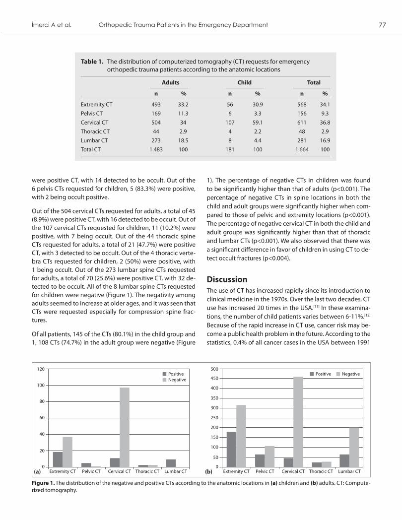
were positive CT, with 14 detected to be occult. Out of the 6 pelvis CTs requested for children, 5 (83.3%) were positive, with 2 being occult positive.
Out of the 504 cervical CTs requested for adults, a total of 45 (8.9%) were positive CT, with 16 detected to be occult. Out of the 107 cervical CTs requested for children, 11 (10.2%) were positive, with 7 being occult. Out of the 44 thoracic spine CTs requested for adults, a total of 21 (47.7%) were positive CT, with 3 detected to be occult. Out of the 4 thoracic verte-bra CTs requested for children, 2 (50%) were positive, with 1 being occult. Out of the 273 lumbar spine CTs requested for adults, a total of 70 (25.6%) were positive CT, with 32 de-tected to be occult. All of the 8 lumbar spine CTs requested for children were negative (Figure 1). The negativity among adults seemed to increase at older ages, and it was seen that CTs were requested especially for compression spine frac-tures.
Of all patients, 145 of the CTs (80.1%) in the child group and 1, 108 CTs (74.7%) in the adult group were negative (Figure
1). The percentage of negative CTs in children was found to be significantly higher than that of adults (p<0.001). The percentage of negative CTs in spine locations in both the child and adult groups were significantly higher when com-pared to those of pelvic and extremity locations (p<0.001). The percentage of negative cervical CT in both the child and adult groups was significantly higher than that of thoracic and lumbar CTs (p<0.001). We also observed that there was a significant difference in favor of children in using CT to de-tect occult fractures (p<0.004).
Discussion The use of CT has increased rapidly since its introduction to clinical medicine in the 1970s. Over the last two decades, CT use has increased 20 times in the USA.[11] In these examina-tions, the number of child patients varies between 6-11%.[12] Because of the rapid increase in CT use, cancer risk may be-come a public health problem in the future. According to the statistics, 0.4% of all cancer cases in the USA between 1991
İmerci A et al. Orthopedic Trauma Patients in the Emergency Department 77
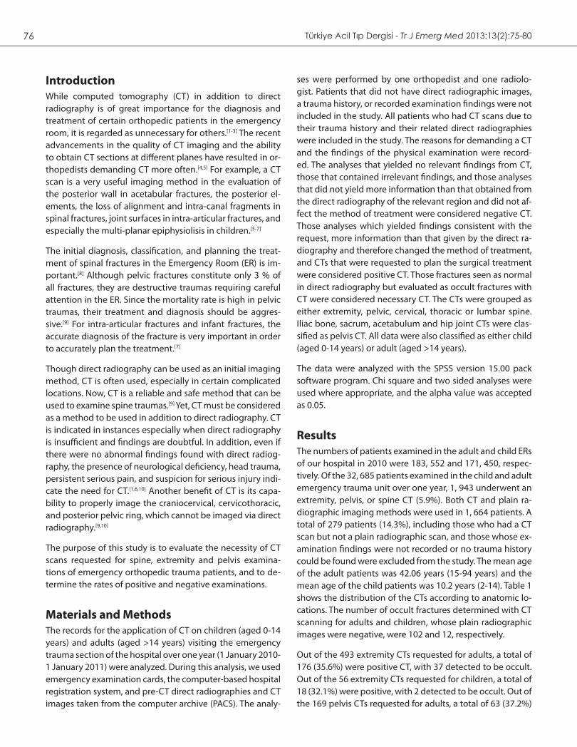
Table 1. The distribution of computerized tomography (CT) requests for emergency orthopedic trauma patients according to the anatomic locations
Adults Child Total
n % n % n %
Extremity CT 493 33.2 56 30.9 568 34.1
Pelvis CT 169 11.3 6 3.3 156 9.3
Cervical CT 504 34 107 59.1 611 36.8
Thoracic CT 44 2.9 4 2.2 48 2.9
Lumbar CT 273 18.5 8 4.4 281 16.9
Total CT 1.483 100 181 100 1.664 100
Figure 1. The distribution of the negative and positive CTs according to the anatomic locations in (a) children and (b) adults. CT: Compute-rized tomography.
100
120
Extremity CT Pelvic CT Cervical CT Thoracic CT Lumbar CT
80
60
40
20
0
NegativePositive
400
350
300
250
200
150
100
50
500
450
Extremity CT Pelvic CT Cervical CT Thoracic CT Lumbar CT0
NegativePositive
(a) (b)
and 1999 were caused by CT use.[13] This suggests that the benefits of a CT examination should be carefully considered before requesting one. However, CT examination should be used if it is medically necessary. Scans have to be carried out with the smallest dose required to obtain adequate infor-mation.[14] In our study, we retrospectively examined the 1, 664 CTs requested by the child and adult emergency room trauma department during 2010. In our country, there have not been any extensive epidemiological studies about the risk of cancer associated with CT. We hope that our study can inspire more of these studies in the future.
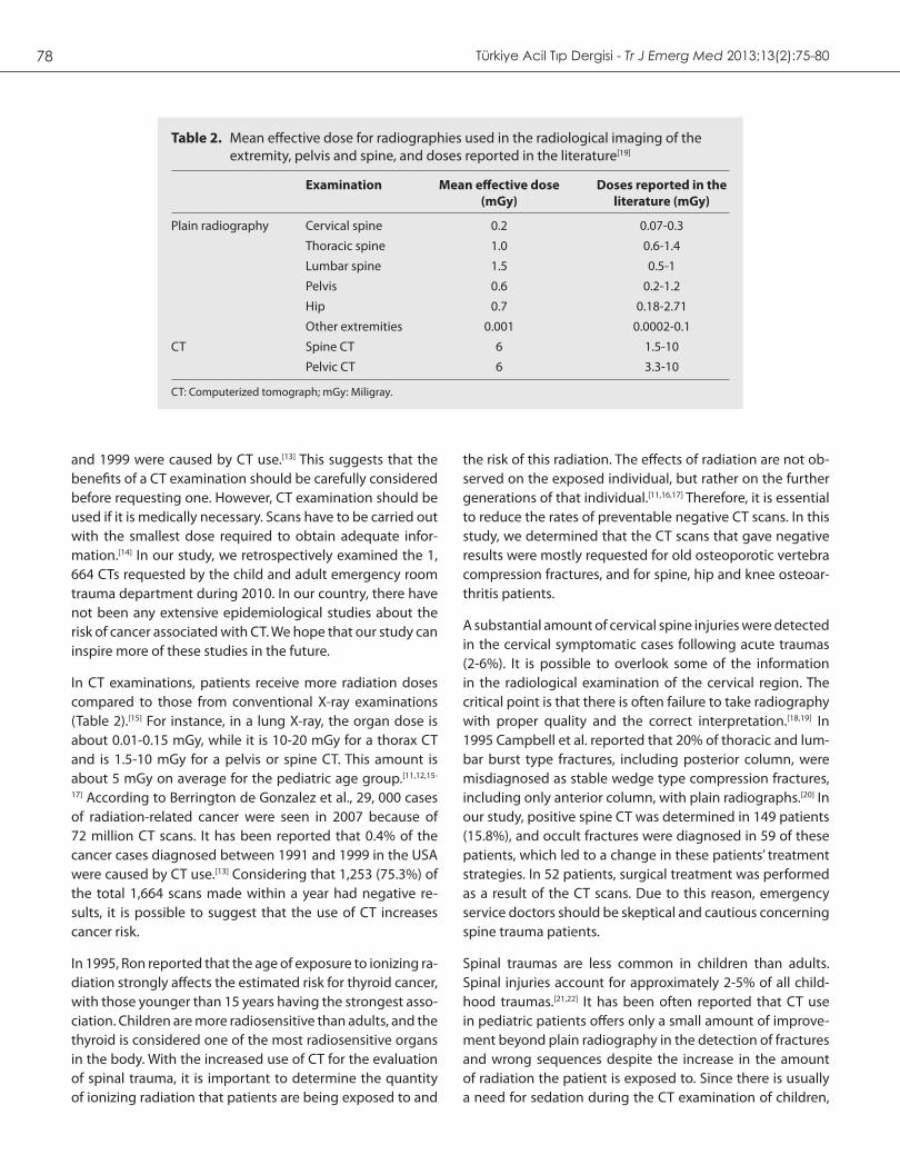
In CT examinations, patients receive more radiation doses compared to those from conventional X-ray examinations (Table 2).[15] For instance, in a lung X-ray, the organ dose is about 0.01-0.15 mGy, while it is 10-20 mGy for a thorax CT and is 1.5-10 mGy for a pelvis or spine CT. This amount is about 5 mGy on average for the pediatric age group.[11,12,15-
17] According to Berrington de Gonzalez et al., 29, 000 cases of radiation-related cancer were seen in 2007 because of 72 million CT scans. It has been reported that 0.4% of the cancer cases diagnosed between 1991 and 1999 in the USA were caused by CT use.[13] Considering that 1,253 (75.3%) of the total 1,664 scans made within a year had negative re-sults, it is possible to suggest that the use of CT increases cancer risk.
In 1995, Ron reported that the age of exposure to ionizing ra-diation strongly affects the estimated risk for thyroid cancer, with those younger than 15 years having the strongest asso-ciation. Children are more radiosensitive than adults, and the thyroid is considered one of the most radiosensitive organs in the body. With the increased use of CT for the evaluation of spinal trauma, it is important to determine the quantity of ionizing radiation that patients are being exposed to and
the risk of this radiation. The effects of radiation are not ob-served on the exposed individual, but rather on the further generations of that individual.[11,16,17] Therefore, it is essential to reduce the rates of preventable negative CT scans. In this study, we determined that the CT scans that gave negative results were mostly requested for old osteoporotic vertebra compression fractures, and for spine, hip and knee osteoar-thritis patients.
A substantial amount of cervical spine injuries were detected in the cervical symptomatic cases following acute traumas (2-6%). It is possible to overlook some of the information in the radiological examination of the cervical region. The critical point is that there is often failure to take radiography with proper quality and the correct interpretation.[18,19] In 1995 Campbell et al. reported that 20% of thoracic and lum-bar burst type fractures, including posterior column, were misdiagnosed as stable wedge type compression fractures, including only anterior column, with plain radiographs.[20] In our study, positive spine CT was determined in 149 patients (15.8%), and occult fractures were diagnosed in 59 of these patients, which led to a change in these patients’ treatment strategies. In 52 patients, surgical treatment was performed as a result of the CT scans. Due to this reason, emergency service doctors should be skeptical and cautious concerning spine trauma patients.
Spinal traumas are less common in children than adults. Spinal injuries account for approximately 2-5% of all child-hood traumas.[21,22] It has been often reported that CT use in pediatric patients offers only a small amount of improve-ment beyond plain radiography in the detection of fractures and wrong sequences despite the increase in the amount of radiation the patient is exposed to. Since there is usually a need for sedation during the CT examination of children,
Türkiye Acil Tıp Dergisi - Tr J Emerg Med 2013;13(2):75-8078
Table 2. Mean effective dose for radiographies used in the radiological imaging of the extremity, pelvis and spine, and doses reported in the literature[19]
Examination Mean effective dose Doses reported in the (mGy) literature (mGy)
Plain radiography Cervical spine 0.2 0.07-0.3
Thoracic spine 1.0 0.6-1.4
Lumbar spine 1.5 0.5-1
Pelvis 0.6 0.2-1.2
Hip 0.7 0.18-2.71
Other extremities 0.001 0.0002-0.1
CT Spine CT 6 1.5-10
Pelvic CT 6 3.3-10
CT: Computerized tomograph; mGy: Miligray.
the advantages the CT examination has for adults, such as short and effective use of time, do not apply.[17,23] In our study, 89.8% of patients who underwent CT for cervical trauma were evaluated normally. The percentage of nega-tive CTs in children was found to be significantly higher than that of adults (p<0.001). Due to these differences between adult and pediatric patients, a trauma protocol should be designed specifically for pediatric patients.
In a research study involving radiologists and ER doctors in the USA, it was reported that the radiation dose caused by CT examinations was disregarded by 75% of both of these groups. Fifty three percent of radiologists, 91% of ER doc-tors, and 97% of patients questioned do not believe the fact that CT examinations increase cancer risk. In addition, 93% of 18 patients questioned said that the benefits and risks of CT were not explained to them before the procedure. Almost all patients stated they were not informed about the radia-tion dose. Despite the fact that the radiation dose of CT is much higher than that of other radiological methods, both doctors and patients disregard this fact.[24] Due to the high rates of negative findings from those undergoing CT that were obtained in our study, the attitudes of both doctors and patients concerning CT should be taken into consideration.
Other than one study regarding unnecessary CT use for the evaluation of the spine and pelvis in the ER, little research has been reported about unnecessary computerized to-mography.[1,3] Daglar et al.[1] found examples of negative CT examinations (51.2%) in the evaluation of spine and pelvic regions in orthopedics clinics. Slovis et al.[3] found that one third of all CTs were unnecessary. Based on this information, they reported that about 20 million unnecessary CTs are requested in the USA each year. In the present study, nega-tive CT was found in 80.2% of children and 74.8% of adults, which is remarkably higher than the results of the cited study.[1] Daglar et al. determined that the negativity in spine CT examinations is significantly higher than that for pelvic CT examinations, and a similar finding was also obtained in our study (p<0.001).
One limitation of our study is that it has a retrospective de-sign. The second limitation is the fact that evaluations of the sensitivity and specificity of plain radiographs and CT examinations were not carried out. While extremity, pelvis, thoracic, and lumbar spine CT evaluations were carried out by an orthopedist, cervical vertebra evaluations were made by a radiologist. However, these experts cannot always per-form the evaluations in the ER due to the high density of patients. A reason for the high negativity ratio in our study may be the high patient density of our hospital. This single center study exhibits the tendencies of our institution, and therefore, generalization of these to the entire universe may
not be accurate.
In conclusion, although CT has significant diagnostic medi-cal benefits, it has a great risk of causing cancer due to ex-posure to radiation. Before using CT, a careful consideration should be made about its pros and cons. Also, other imaging methods should be considered.
As can be seen from our study, further retrospective and pro-spective studies should be done on the use of CT for trauma patients in order to establish guidelines of when to use CT and to prevent its superfluous utilization. Emergency service doctors should be adequately trained on CT and should con-sult the orthopedist first when needed.
Conflict of Interest
The authors declare that there is no potential conflicts of in-terest.
References1. Daglar B, Delialioğlu OM, Ceyhan E, Ozdemir G, Taşbaş BA,
Bayrakci K, et al. Superfluous computed tomography utiliza-tion for the evaluation of the pelvis and spinal column in an orthopedic emergency department. Acta Orthop Traumatol Turc 2008;42:59-63. [CrossRef]
2. Dogan Z, Guven FMK; Cankorkmaz L, Korkmaz I, Coskun A, Doles KA. Evaluation of the child trauma cases applied to our university hospital department of emergency. Turk Arch Ped 2011;46:164-7.
3. Slovis TL, Berdon WE. Perfect is the enemy of the very good. Pediatr Radiol 2002;32:217-8. [CrossRef]
4. Broder J, Fordham LA, Warshauer DM. Increasing utilization of computed tomography in the pediatric emergency de-partment, 2000-2006. Emerg Radiol 2007;14:227-32. [CrossRef]
5. Berg EE, Chebuhar C, Bell RM. Pelvic trauma imaging: a blind-ed comparison of computed tomography and roentgeno-grams. J Trauma 1996;41:994-8. [CrossRef]
6. France JC, Bono CM, Vaccaro AR. Initial radiographic evalua-tion of the spine after trauma: when, what, where, and how to image the acutely traumatized spine. J Orthop Trauma 2005;19:640-9. [CrossRef]
7. Lemburg SP, Lilienthal E, Heyer CM. Growth plate fractures of the distal tibia: is CT imaging necessary? Arch Orthop Trauma Surg 2010;130:1411-7. [CrossRef]
8. Bagley LJ. Imaging of spinal trauma. Radiol Clin North Am 2006;44:1-12. [CrossRef]
9. Alost T, Waldrop RD. Profile of geriatric pelvic fractures pre-senting to the emergency department. Am J Emerg Med 1997;15:576-8. [CrossRef]
10. Roberge RJ. Facilitating cervical spine radiography in blunt trauma. Emerg Med Clin North Am 1991;9:733-42.
11. Hall EJ, Brenner DJ. Cancer risks from diagnostic radiology. Br J Radiol 2008;81:362-78. [CrossRef]
12. Mettler FA Jr, Wiest PW, Locken JA, Kelsey CA. CT scanning:
79İmerci A et al. Orthopedic Trauma Patients in the Emergency Department
patterns of use and dose. J Radiol Prot 2000;20:353-9. [CrossRef]
13. Berrington de González A, Mahesh M, Kim KP, Bhargavan M, Lewis R, Mettler F, et al. Projected cancer risks from computed tomographic scans performed in the United States in 2007. Arch Intern Med 2009;169:2071-7. [CrossRef]
14. Charles M. UNSCEAR report 2000: sources and effects of ion-izing radiation. United Nations Scientific Comittee on the Ef-fects of Atomic Radiation. J Radiol Prot 2001;21:83-6. [CrossRef]
15. Mettler FA Jr, Huda W, Yoshizumi TT, Mahesh M. Effective dos-es in radiology and diagnostic nuclear medicine: a catalog. Radiology 2008;248:254-63. [CrossRef]
16. Scaife ER, Rollins MD. Managing radiation risk in the evalu-ation of the pediatric trauma patient. Semin Pediatr Surg 2010;19:252-6. [CrossRef]
17. Ron E. Let’s not relive the past: a review of cancer risk af-ter diagnostic or therapeutic irradiation. Pediatr Radiol 2002;32:739-44. [CrossRef]
18. Grogan EL, Morris JA Jr, Dittus RS, Moore DE, Poulose BK, Diaz JJ, et al. Cervical spine evaluation in urban trauma centers: lowering institutional costs and complications through heli-
cal CT scan. J Am Coll Surg 2005;200:160-5. [CrossRef]
19. Blackmore CC, Ramsey SD, Mann FA, Deyo RA. Cervical spine screening with CT in trauma patients: a cost-effectiveness analysis. Radiology 1999;212:117-25.
20. Campbell SE, Phillips CD, Dubovsky E, Cail WS, Omary RA. The value of CT in determining potential instability of simple wedge-compression fractures of the lumbar spine. AJNR Am J Neuroradiol 1995;16:1385-92.
21. Adelgais KM, Grossman DC, Langer SG, Mann FA. Use of heli-cal computed tomography for imaging the pediatric cervical spine. Acad Emerg Med 2004;11:228-36. [CrossRef]
22. Hamilton MG, Myles ST. Pediatric spinal injury: review of 174 hospital admissions. J Neurosurg 1992;77:700-4. [CrossRef]
23. Hernandez JA, Chupik C, Swischuk LE. Cervical spine trauma in children under 5 years: productivity of CT. Emerg Radiol 2004;10:176-8. [CrossRef]
24. Lee CI, Haims AH, Monico EP, Brink JA, Forman HP. Diagnostic CT scans: assessment of patient, physician, and radiologist awareness of radiation dose and possible risks. Radiology 2004;231:393-8. [CrossRef]
Türkiye Acil Tıp Dergisi - Tr J Emerg Med 2013;13(2):75-8080