Embed Size (px)
Citation preview

EVALUATING THE EFFECT OF SOUTH AFRICAN HERBAL
EXTRACTS ON BREAST CANCER CELLS
Mpho Susan Choene
A dissertation submitted in fulfilment of the requirements for the degree of Masters in Science in
the School of Molecular and Cell Biology, University of the Witwatersrand
Johannesburg, 2012

ii
DECLARATION
I declare “EVALUATING THE EFFECT OF SOUTH AFRICAN HERBAL EXTRACTS
ON BREAST CANCER CELLS’’ to be my work which has not been submitted for any degree
or examination at any other University and that all the sources I have used or quoted have been
indicated and acknowledged by complete references.
Mpho Susan Choene
Signature…………………………………………………………………………………………
On this…19…………………………….day of ………July..……………………………2012

iii
RESEARCH OUTPUT
Conference Outputs
Poster presentations
Mpho S. Choene and Lesetja R. Motadi. Evaluating the anti-tumour properties of South African
herbal extracts on breast cancer. SASBMB/FASBMB congress, Drakensberg, 29-1 February,
2012
Mpho S. Choene and Lesetja R. Motadi. Evaluating the effect of South African herbal extracts
on breast cancer cells. ASCB Annual Meeting, Denver, Colorado, 3-7 December, 2011
(International Conference)
Mpho S. Choene and Lesetja R. Motadi. Identifying the network pathway between p53, BRCA2
and RBBP6 in breast cancer using gene silencing technology. SASBMB congress, Bloemfontein,
17-20 January, 2010
Original publications and Reviews
Mpho S. Choene and Lesetja R. Motadi
Anti-proliferative effects of the methanolic extracts of Kedrostis foetidissima in breast cancer
cells lines. Molecular Biology, 2012, 1:107
Nonkululeko N. Mthembu, Mpho S. Choene, Lesetja R. Motadi
Wisdom from the forefathers’ traditional medicine can induce apoptosis and inhibit cell
proliferation. (Review) Biological and Biomedical Reports, 2012, 2(3), 180-189

iv
DEDICATION
This dissertation is dedicated to my mother, Ntshebo, grandmother, Alinah, grandfather, James
and to my little brother, Tiisetso who have continuously supported me throughout the course of
my studies. You encouraged me even when I felt like giving up.
Thank you!

v
ACKNOWLEDGEMENTS
I would like to express my gratitude to my supervisor Dr. Lesetja Raymond Motadi for the
opportunity to embark on this topic. I am grateful for all the support, motivation and intellectual
input throughout my research and write-up phase.
My gratitude is extended to the National Research Foundation (NRF) for the financial support of
this project. I would also like to thank Farhahna van Schalk for her assistance and patience in
helping me carry out real-time PCR.
My boundless appreciation goes to the following people: Maabo Moralo, Xolisiwe Maputsoe,
Palesa Seele and Obakeng Ntshudisane for all their support.
Lastly, a very heartfelt thank you to my mother for all the sacrifices she has made, her
encouragement and continuous support have helped me to complete this research.

vi
ABSTRACT
In this research we aimed to investigate the anti-proliferative properties of three South African
plants: Kedrostis foetidissima, Euphorbia mauritanica and Elytropappus rhinocerotis against
breast cancer cells. This was done on the basis of their documented ethno-medicinal use against
cancer and other ailments. The plant extracts were screened for cytotoxicity and pro-apoptotic
activity against two breast cancer cell lines MCF-7 and YMB-1. With an IC50 ~ 100 µg/ml, K.
foetidissima was the only extract that exhibited significant cytotoxicity on both cell lines, whilst
E. mauritanica was cytotoxic to MCF-7 cells only. The cytotoxicity assay was followed by the
Annexin-V detection assay to evaluate the occurrence of apoptosis. The results observed
suggested that K. foetidissima was inducing significant apoptosis on both YMB-1 and MCF-7
cells, whilst E. mauritanica was inducing significant apoptosis on MCF-7 cells.
Since both K. foetidissima and E. mauritanica crude extracts induced apoptosis to MCF-7 cells,
they were selected for gene expression studies on MCF-7 using real-time PCR. This was done
with the aim of investigating if these extracts were having an effect on the tumour suppressors
p53 and RBBP6, which were shown in previous studies to be deregulated in up to 50% of
cancers. From the real-time PCR data we observed no changes in the expression levels of these
genes following treatment with the herbal extracts. This may suggest that these plants have an
effect on other components of the apoptotic pathway other than the tumour suppressors p53 and
RBBP6.
The antiproliferative activity observed whilst treating these particular cell lines with K.
foetidissima and E. mauritanica suggests that these South African herbal plants present

vii
themselves as potential future cancer therapeutic agents; however, further studies on these herbal
plants need to be performed to validate these results.
KEYWORDS: Apoptosis
Breast cancer
Euphorbia mauritanica
Kedrostis foetidissima
p53

viii
TABLE OF CONTENTS
DECLARATION ............................................................................................................................ ii
RESEARCH OUTPUT .................................................................................................................. iii
DEDICATION ............................................................................................................................... iv
ACKNOWLEDGEMENTS ............................................................................................................ v
ABSTRACT ................................................................................................................................... vi
KEYWORDS ................................................................................................................................ vii
TABLE OF CONTENTS ............................................................................................................. viii
LIST OF FIGURES ....................................................................................................................... xi
LIST OF TABLES ....................................................................................................................... xiii
CHAPTER 1 INTRODUCTION AND LITERATURE REVIEW ................................................ 1
1.1 Traditional medicine ............................................................................................................. 1
1.2 Breast cancer ......................................................................................................................... 2
1.2.1 Statistics .......................................................................................................................... 2
1.2.2 Epidemiology.................................................................................................................. 3
1.3 BRCA 1 and 2 ....................................................................................................................... 4
1.4 Cell cycle ............................................................................................................................... 6
1.5 Apoptosis ............................................................................................................................... 8
1.6 Tumour suppressor genes .................................................................................................... 10
1.6.1 p53 ................................................................................................................................ 11
1.6.2 Retinoblastoma tumour suppressor gene (RB) ............................................................. 14
1.6.3 RBBP6 .......................................................................................................................... 15
1.7 Plant extracts as anti-cancer drugs ...................................................................................... 15
1.7.1 Taxol ............................................................................................................................. 16
1.7.2 Camptothecin ................................................................................................................ 17

ix
1.7.3 Podofilox ...................................................................................................................... 17
1.7.4 Velban (vinblastine), vincristine, vinorelbine and vindesine ....................................... 18
1.8 AIMS AND OBJECTIVES ................................................................................................. 19
CHAPTER 2 MATERIALS AND METHODS ........................................................................... 20
2.1 Materials .............................................................................................................................. 20
2.1.1 Breast cancer cell lines ................................................................................................. 20
2.1.2 Plant collection ............................................................................................................. 21
2.1.3 Plant extraction ............................................................................................................. 21
2.1.4 Cell culture expansion .................................................................................................. 21
2.2 Experimental assays ............................................................................................................ 22
2.2.1 MTT assay .................................................................................................................... 22
2.2.2 Flow cytometry ............................................................................................................. 23
2.2.3 RNA extraction ............................................................................................................. 24
2.2.4 Reverse transcription .................................................................................................... 24
2.2.5 Real Time PCR (RT-PCR) ........................................................................................... 25
CHAPTER 3 SCREENING INDIGENOUS SOUTH AFRICAN PLANTS FOR
CYTOTOXICITY ON BREAST CANCER ................................................................................ 27
3.1 Introduction ......................................................................................................................... 27
3.2 Kedrostis foetidissima ......................................................................................................... 28
3.2.1 Classification ................................................................................................................ 28
3.2.2 Botanical description .................................................................................................... 28
3.2.3 Geographical distribution ............................................................................................. 28
3.2.4 Traditional medicinal use ............................................................................................. 29
3.3 Euphorbia mauritanica ....................................................................................................... 30
3.3.1 Classification ................................................................................................................ 30
3.3.2 Geographical distribution ............................................................................................. 30
3.3.3 Botanical description .................................................................................................... 31

x
3.3.4 Traditional medicinal use ............................................................................................. 31
3.4 Results and discussion ......................................................................................................... 33
CHAPTER 4 SCREENING OF HERBAL EXTRACTS FOR MODE OF DEATH INDUCTION
(APOPTOSIS OR NECROSIS) .................................................................................................... 39
4.1 Introduction ......................................................................................................................... 39
4.2 Results and discussion ......................................................................................................... 43
CHAPTER 5 TUMOUR SUPPRESSOR GENES ....................................................................... 48
5.1 Introduction ......................................................................................................................... 48
5.2 Primer efficiency assessment using standard curves........................................................... 49
5.3 Relative quantitative real-time polymerase chain reaction ................................................. 53
5.4 Results and discussion ......................................................................................................... 55
CHAPTER 6 GENERAL DISCUSSION and conclusion ............................................................ 64
6.1 General discussion............................................................................................................... 64
6.2 Conclusion and future perspectives..................................................................................... 69
CHAPTER 7 REFERENCES ....................................................................................................... 71
Appendix A ................................................................................................................................... 90
Appendix B ................................................................................................................................... 91
Appendix C ................................................................................................................................... 94
Appendix D ................................................................................................................................... 95

xi
LIST OF FIGURES
Figure 1.1: Schematic representation of cell cycle regulation indicating G1, S and G2
checkpoints. .................................................................................................................................... 8
Figure 1.2: A simplified diagrammatic representation of the p53 pathway. ................................ 13
Figure 3.1: Kedrostis foetidissima (Curcubitaceae). ..................................................................... 30
Figure 3.2: Euphorbia mauritanica (Euphorbiaceae). .................................................................. 32
Figure 3.3: Cytotoxicity effects of the herbal extracts Kedrostis foetidisima A) and Euphorbia
mauritanica B) on MCF-7 breast cancer cells. ............................................................................. 34
Figure 3.4: Cytotoxicity effects of the herbal extracts Kedrostis foetidisima A) and Euphorbia
mauritanica B) on YMB-1 breast cancer cells. ............................................................................ 35
Figure 4.1: Characteristic features that distinguish between apoptotic and necrotic cell death (van
der Meer 2010). ............................................................................................................................. 41
Figure 4.2: Scatter analysis diagrams for YMB-1 cells. ............................................................... 43
Figure 4.3: Scatter analysis diagrams for MCF-7 cells................................................................. 44
Figure 5.1: Standard curves for Beta-actin (A) and GADPH (B) primers which served as
housekeeping genes. ..................................................................................................................... 51
Figure 5.2: Standard curves for the tumour suppressors RBBP6 (A) and p53 (B) primers. ........ 52
Figure 5.3: Amplification curves for the housekeeping genes. .................................................... 55
Figure 5.4: Amplification curves for RBBP6 expression. ........................................................... 56
Figure 5.5: RT-PCR products resolved on a gel following RBBP6 amplification. ...................... 56
Figure 5.6: Amplification curves for p53 expression. .................................................................. 57
Figure 5.7: RT-PCR products resolved on a gel following p53 amplification. ............................ 57
Figure 5.8: Relative expression of p53 versus that of β-actin housekeeping gene. ...................... 59
Figure 5.9: Relative expression of p53 versus that of GADPH housekeeping gene. ................... 60
Figure 5.10: Relative expression of RBBP6 versus that of β-actin housekeeping gene. .............. 61
Figure 5.11: Relative expression of RBBP6 versus that of GADPH housekeeping gene. ........... 62
Figure A.1: Cytotoxicity of Elytropappus Rhinocerotis on MCF-7 breast cancer cells. .............. 90

xii
Figure A.2 Cytotoxicity of Elytropappus Rhinocerotis on YMB-1 breast cancer cells. .............. 90
Figure B.1: Melting curves for GADPH ....................................................................................... 94
Figure D.1: Cytotoxicity of Euphorbia mauritanica and Kedrostis hirtella extracts. .................. 95
Figure D.2 : Cytotoxicity of Euphorbia mauritanica and Kedrostis hirtella extracts. ................. 95

xiii
LIST OF TABLES
Table 2.1: Real Time PCR parameters. ........................................................................................ 26
Table 4.1: Differences between apoptotic and necrotic cellular death pathways ......................... 40
Table B.1: RNA extraction protocol ............................................................................................. 91
Table B.2: The cocktail for cDNA synthesis ................................................................................ 92
Table B.3: Reverse transcription conditions ................................................................................. 93
Table B.4: Real-time PCR cocktail ............................................................................................... 93

1
CHAPTER 1 INTRODUCTION AND LITERATURE REVIEW
1.1 Traditional medicine
For generations, mankind has believed and still believes in the healing powers of plants, using
them as medicine to treat various common infectious diseases. Some of these plants have been
used for centuries and are still used even today as a primary source of healthcare. In spite of the
increasing usage of modern medicines in some areas, there are still countries that don’t have
regular access to essential drugs. The use of traditional plants as a great source of medicine has
been greatly documented in African countries and other developing countries such as India and
China. According to the World Health Organisation (WHO), 70-80% of the world’s population
use herbal sources as medicine (Chan 2003).
It is only in the 20th
century that there has been increasing interest in the use of natural resources,
especially herbal medicine, as an alternative to orthodox medicine in industrialized countries.
This has thus increased interest from pharmaceuticals, and phytotherapeutic research. Scientists
started developing an interest in medicinal plants due to their documented antibacterial,
antifungal, antiviral and even anti-amoebic properties (McGaw et al. 2000). Today, up to 50% of
the world drugs are made from natural products or their derivatives (Gurib-Fakim 2006). Some
of these drugs include aspirin, quinine and morphine amongst others (Singh and Barrett 2006).
The recent surge in natural plants research has also led to production of drugs such as taxol,
camptothecin and podofilox which are now being used for chemotherapy. These drugs were
derived from parts of various plants (Itokawa et al. 2008; Pezzuto 1997). This has been a
remarkable progress in elucidating the possible anti-apoptotic properties of plants. As researchers
2
worldwide continue to explore plants for their anti-cancer properties, breast cancer should also
be at the forefront of medicinal plant research, since it is now the leading cause of cancer in
females.
1.2 Breast cancer
1.2.1 Statistics
Breast cancer is a malignant tumour that starts in the breast tissue; it usually starts in the ducts or
in lobules. It can occur in both males and females; however, male breast cancer is rare. It is the
second leading cancer in the world following lung cancer, with an estimated 1.15 million cases a
year in 2002 (Parkin et al. 2005). The American Cancer Society estimated 182 460 cases (which
accounted for 26% of all new cancer cases) for 2008 in the U.S.A alone, resulting in 40 480
deaths (Jemal et al. 2008). Breast cancer is primarily a postmenopausal women’s disease, with
the American National Cancer Institute’s Cancer Review estimating the average age of women at
risk to be 61 years of age. Approximately 0 cases have being reported for women below 20, but
the figures are gradually increasing with every decade (Altekruse et al. 2009).
In South Africa, between 1986 and 1992, cervical cancer was the most leading cause of cancer
amongst women, but recently breast cancer seems to have overtaken and is now the most
common cancer in females (Vorobiof et al. 2001; Siegel et al. 2011). Compared to the rest of the
world, breast cancer cases are relatively low in Africa, with an estimated 65 000 new cases being
reported in 2002. The highest incidence was observed in South Africa with about 6474 (or 35 per
105) cases being reported (Parkin et al. 2008).

3
1.2.2 Epidemiology
The lifetime risk of an individual developing breast cancer cannot be attributed to one factor
alone. A number of variables play a role in the development of breast malignancies. Even though
it is fairly difficult to pinpoint one cause as the main risk factor for breast cancer, studies have
shown there are certain genetic, reproductive and lifestyle factors that play a major role in its
induction and progression. One of these factors is age, with studies finding that the incidence
rates double with every 10 years up until menopause, where the rates start to dramatically decline
(McPherson et al. 2000). Exposure to radiation and use of oral contraceptives may be associated
with induction of breast cancer (Ronckers et al. 2005; Urban et al. 2012).
Very small fraction of breast cancers could be attributed to preventable lifestyle behaviour such
as obesity, alcohol consumption, lack of physical activity and long term use of hormone
replacement therapy. In several studies conducted to determine the role these factors have on
breast cancer incidence, the data appears to be inconsistent, hence it is hard to establish the
severity these factors play in breast cancer induction that is if they even do at all (Chen et al.
2006; Lorincz and Sukumar 2006; Zhang et al. 2007). In developed countries, incidence rates are
higher relative to developing countries. However, incidence rates are steadily increasing in
developing countries (Key et al. 2001). This increase may be attributed to the adoption of a more
westernized lifestyle which encompasses a more sedentary lifestyle, delayed childbearing, high
alcohol use and a diet high in fat (Bray et al. 2004; Porter 2008).
In a lot of breast malignancies, hormones play a major role. It is known that estrogen starts breast
tissue proliferation, and studies have now shown that the longer a woman is exposed to estrogen,
the greater the risk of developing breast cancer. Hence, women with an early age at menarche
4
(<12 years), were found to be 2.2 times more likely to develop breast cancer in the future than
those with late menarche (Peeters et al. 1994). In a different study, women who started
menopause late i.e. at the age of 55, were found to have 44% higher breast cancer incidence rates
than women who started menopause at 45. This may be due to a 10 year increase in the amount
of estrogen the women are exposed to. Exposure to excessive exogenous estrogen plays a role in
hyperestrogenia. Hyperestrogenia could trigger breast cancer in two ways: either by breast
epithelium tumour promotion or by DNA damage which could be caused by estrogen
metabolites directly (Butler et al. 2000; Imyanitov and Hanson 2004).
Another important factor which may increase susceptibility to breast cancer is inheritance of
susceptibility genes, it has been estimated that 7-10% of breast cancers arise due to inherited
mutated genes. The major predisposing factors to breast cancer are breast susceptibility genes,
known as BRCA1 and BRCA 2, which are important for early onset of breast cancer (Hedau et
al. 2004; Imyanitov and Hanson 2004). An initial germline mutation in one of these genes
followed by a second mutation leading to the loss of function of the second allele is required for
induction of tumouriginesis (Powell and Kachnic 2003). These genetic factors mostly show an
autosomal dominant pattern of inheritance, with germline mutations rendering susceptibility to
the following generation (Loubser et al. 2008; Ponzone and Baum 1998).
1.3 BRCA 1 and 2
The BRCA genes are thought to be tumour suppressors since the wild type allele is often lost in
tumours of heterozygous people (Tutt and Ashworth 2002). It has been suggested that BRCA 1
and BRCA2 genes play a role in a common pathway which controls DNA damage repair and
DNA replication fidelity (Powell and Kachnic 2003). They are thought to be involved in repair

5
of damaged DNA associated with replication by homologous recombination. This they do by
interacting with a recombinational repair protein Rad51, thereby maintaining genomic integrity
(Tutt and Ashworth 2002; Welsch and King 2001). In accordance with their role in DNA repair;
a cell with a mutant BRCA gene will not be able to have a functional protein product thereby
leading to defects in DNA repair mechanisms. It has been speculated that germline mutations in
cancer susceptibility genes BRCA1 and BRCA2 led to an increased risk of developing breast
cancer.
Breast cancer patients with mutant BRCA 1 were found to contain up to 2 to 3 fold more
chromosomal rearrangements than sporadic cancers (Thangaraju 2000). Abnormalities that arise
due to mutant BRCA2 include broken chromosomes and chromatids as well as formation of
abnormal structures which mark an abnormal mitotic recombination which has been linked to
predisposition to diseases such as Blooms syndrome and also leaves the person with high
susceptibility to various types of cancers (Venkitaraman 2002). Ford et al. (1998) showed that
up to 84% of hereditary breast cancers were linked to mutations in either BRCA1 or BRCA2
whilst only 16% of breast cancer cases were not linked to BRCA mutations.
BRCA1 tumour suppressor gene encodes an 1863-amino acid and is located at 17q21. BRCA1 is
implicated in many cellular pathways including transcription, cell cycle, checkpoint control,
apoptosis and DNA repair (Venkitaraman 2002). BRCA2 is a breast cancer susceptibility gene
located at 13q12-q13 which the product is thought to be involved in monitoring genome integrity
and cell cycle progression. BRCA 2-null mice have been reported to have a defect in embryonic
cellular proliferation and die in the uterus (Cheung et al. 2002).

6
Though BRCA mutations have been implicated in a lot of breast cancer cases, there are still
breast cancers which occur due to deregulation of other genes. A substantial proportion of
susceptibility to breast cancer can be attributed to mutations in cell cycle regulation and
apoptosis genes.
1.4 Cell cycle
The cell cycle is an important regulator of DNA replication, cell growth and cell division
(Schwartz and Shah 2005). It entails a series of highly coordinated steps, these are divided into 4
distinct stages: G1-phase - gap phase, it represents the stage of entry into the cell cycle whereby
cells prepare for the S-phase; S-phase- DNA synthesis; G2- the second gap phase, a period of
rapid cell growth where cells prepare for mitosis and M phase which represents mitosis, which
entails cell division resulting in the formation of 2 daughter cells from each cell (Caldon et al.
2006; Schwartz and Shah 2005). It is essential for a cell to complete one phase before
proceeding to another to ensure orderly transitions.
One of the most important things is for cells to ensure repair of damaged DNA and prevention of
uncontrolled proliferation whilst going through the cell cycle. The most important mechanisms
that mammalian cells have developed to deal with constant DNA damage is the development of
cell cycle control checkpoints. This is a mechanism in which cell progression through the cell
cycle is arrested, to prevent cells with damaged DNA from undergoing DNA replication or
mitosis and this is mainly mediated by cyclin-dependent kinases (CDKs) (Kastan and Bartek
2004; Malumbres and Barbacid 2005). CDKs require specific regulatory proteins known as
cyclins to be positively regulated and they are negatively regulated by CDK inhibitors (Schwartz

7
and Shah 2005). Depending on which stage of the cell cycle the cell is in will be the determining
factor on which particular CDK will be expressed by its respective cyclin.
During the G1 phase, a signal will induce synthesis of D-cyclins which will interact with
CDK4/6, these will form complexes and phosphorylate the tumour suppressor retinoblastoma
(Rb) (Malumbres and Barbacid 2005; Potten and Wilson 2004). To bring about G1 cell cycle
arrest, Rb protein interacts with E2F family of transcription factors (Weinberg 1995).
Association between CDK2 and cyclin E drives the cell to the S-phase. Cyclin A’s interaction
with CDK2 will drive the cell through the S-phase. Whilst the cyclin A-CDK1 and cyclin B-
CDK1 will get the cells through the S-phase to mitosis transition (Schwartz and Shah 2005) (see
Figure 1.1). The two most important checkpoints being the G1 and the G2 checkpoints, the G1
checkpoint ensures that cells with a damaged DNA do not undergo DNA replication and the G2
checkpoint that unreplicated and damaged DNA do not undergo mitosis.
There has been increasing evidence that implicates cell cycle control disruption as a common
pathway in the development of breast tumouriginesis. The fidelity of the cell cycle requires a
coordinated complex between DNA repair, metabolic adjustments, cell death, chromatin
remodeling which are controlled by regulatory surveillance systems (Elledge 1996). Mutations
in genes involved in DNA damage responses and mitotic checkpoints pathways, may allow
continued proliferation of genes with aberrant DNA, thus leading to increased chances of cancer
(Kastan and Bartek 2004). The two most important genes that control the cell cycle that have
been implicated in tumouriginesis being the tumour suppressors p53 and pRb, and these will be
discussed further in the following section.

8
Figure 1.1: Schematic representation of cell cycle regulation indicating G1, S and G2
checkpoints.
The phase specific cyclin-dependent kinases (CDKs) and their regulatory proteins known as
cyclins are shown. The negative regulators of the CDKs are p16, p21 and p27. The tumour
suppressor protein pRb in its phosphorylated form is required for progression through the G1
phase whilst p53 is essential for both the G1 and G2 checkpoints (Stewart et al. 2001).
1.5 Apoptosis
Apoptosis is a fundamental process that occurs throughout the lives of most organisms, from
humans to simple organisms such as hydra, to insects, amphibian and fish (Potten and Wilson
2004). It is an active form of programmed cell death which is important for maintaining
p16/p21/p27
pRb-E2F; p53
Cyclin A- Cdk2
Cyclin D – Cdk4/6 Cyclin E- Cdk2
Cyclin A- Cdk1 Cyclin B- Cdk1
G0 p21
p53

9
homeostasis by ensuring a balance between proliferation of normal cells and non-inflammatory
death of damaged cells in adult multicellular organisms (Kerr et al. 1972). Deregulation of
apoptosis could lead to induction of certain human disease including neurodegenerative
disorders such as Alzheimer’s and Parkinson’s diseases, autoimmune disorders e.g., multiple
sclerosis and rheumathoid arthritis and various types of cancers (Fadeel et al. 1999). It also plays
a crucial role in embryonic development (Kerr et al. 1972). It is important for mammalian
development and patterning of structures such as organs (brains, eyes) and limbs. Failure of
apoptosis regulation could lead to an observable phenotype characterized by various defects
(Lang 1997; Haydar et al. 1999; Potten and Wilson 2004).
Even though apoptosis may be triggered by various stimuli, it is initiated by two major pathways
the intrinsic and the extrinsic pathways (Boatright and Salvesen 2003).
(i) Intrinsic pathway
The intrinsic pathway is regulated by pro- and anti-apoptotic members of the Bcl-2 family,
which will either induce or prevent permeabilization of the outer mitochondrial membrane. Once
an apoptotic signal is received, it will lead to activities of signal inducing pro-apoptotic members
of the Bcl-2 family leading to permeabilization of the outer mitochondrial membrane (Green and
Kroemer 2004). This will lead to release of cytochrome c into the cell cytoplasm, which had
been sequestered in the mitochondrial intermembrane space (Schuler et al. 2000). Cytochrome c
will then bind to the protein apoptosis-protease-activating-factor 1 (Apaf-1), this complex will
also require the binding of dATP as a cofactor to form the apoptosome complex which will be
responsible for activation of caspase-9 (Purring-Koch and McLendon 2000). Active caspase-9

10
will lead to protease cascade which will eventually lead to cleavage of caspase-3 causing
apoptosis (Li et al. 1997)
(ii) Extrinsic pathway
The extrinsic pathway involves cell members of the tumour necrosis factor-alpha (TNF-α)
superfamily. These are receptors found on the cell surface, several members have been identified
so far and they include TRAIL-1 and TRAIL-2, TNFR-1, CD-95, Fas, DR6 and NGFR (Chen
and Wang 2002; Lavrik et al. 2005). An appropriate ligand has to bind to one of the receptors on
the cell surface; this will lead to recruitment and activation of caspase-8 and -10 which are part
of a proteinase family called caspases to form the death inducing signalling complex (Boatright
and Salvesen 2003). This will then lead to activation of effector caspases or indirectly activate
downstream caspases which will then lead to apoptosis (Schuler and Green 2001).
There are a number of regulatory proteins that have been implicated in influencing cells to
undergo apoptosis. However, there are two key regulatory proteins that have been largely
conserved in species; they exist as multigene families with multiple homologues. They are
known as Bcl-2 (family of inhibitors and promoters of apoptosis) and the p53 tumour suppressor
gene and both have been extensively researched in breast cancer (Parton et al. 2009). DNA
damage could activate p53 which may act as an inducer of apoptosis (Kopper and Petak 2008).
1.6 Tumour suppressor genes
The occurrence of cancer is usually owed to gain of function mutation of proto-oncogenes,
converting them to oncogenes, or loss of function mutations which occur in tumour suppressor
genes (Garg 2007). The first to be discovered was RB1, whose mutations were found to be the

11
main cause of retinoblastoma (Friend et al. 1986). Another one which has been highly studied is
p53, both these will be discussed in the following sections.
1.6.1 p53
p53, one of the most highly studied tumour suppressor, is important for preventing proliferation
of cancer cells thereby playing an important role in maintaining genomic integrity. It is due to
this crucial role that it is also known as the guardian of the genome. This role is vital in
preventing development of various cancers (Barnes and Camplejohn 1996; Potten and Wilson
2004). The gene p53 encodes a protein p53; it is this protein that plays an important role in
cellular processes involved in DNA damage recognition and repair, thereby preventing
proliferation of abnormal cells. The p53 protein is normally present in low concentrations and in
some cells; it may even be latent or inactive. This is mediated by Mdm2 which binds to p53 and
decreases its stability and half-life through the ubiquitin-proteasome pathway, requiring some
kind of signal for it to be active (Lee and Lozano 2006; Levine 1997).
Following DNA damage, expression of activated oncogenes or other stressful events in normal
cells, p53 is rapidly activated in high amounts which are proportional to the severity of the DNA
damage and acts as a checkpoint factor, allowing cells to either undergo apoptosis or cell cycle
arrest at the G1-phase (Gasco et al. 2002; Giono and Manfredi 2006). p53 may also act as an
effector protein, allowing expression of genes involved in cell damage control and repair by
interacting with proteins involved in DNA damage recognition and sending signals to those
repair genes (Potten and Wilson 2004). The importance of having a functional p53 protein was
further shown in mice with a null mutation that was introduced in the p53 gene and it was found

12
that mice with a homozygous null mutation in p53 had a high incidence of spontaneous tumours
from a very early age (Donehower et al. 1992).
Of over 2500 human cancer tumours that have been analyzed, p53 has been found to be mutated
in over 50% of them (Potten and Wilson 2004). Humans with a mutant germ line p53 gene
develop Li-Fraumeni syndrome and are more easily prone to developing cancer (Hainaut and
Hollstein 2000).

13
(Constructed using protein lounge)
Figure 1.2: A simplified diagrammatic representation of the p53 pathway.
In normal cells, p53-mdm2 autoregulatory feedback loop regulates amounts of p53 and keeps
them low. Upon exposure to DNA damage, mdm2 is inhibited. This triggers p53, leading to
transcription of its downstream targets, e.g. Bax, Bcl2 and cytochrome c, eventually resulting in
apoptosis.

14
1.6.2 Retinoblastoma tumour suppressor gene (RB)
Retinoblastoma tumour suppressor gene (RB) encodes the retinoblastoma protein (pRb or Rb)
which is dysfunctional in many types of cancer. It belongs to a ―protein pocket‖ family whose
members possess a highly conserved sequence in that pocket domain; they interact with specific
cellular proteins to mediate certain effects (Poznic 2009). The main function of the pRb is
believed to be a signal transducer between the cell cycle and its transcriptional machinery (Zhu
2005). This protein regulates the G1 to S transition by binding to E2F and inhibiting promoters
of expression genes required for transition through this phase (Poznic 2009). It functions to
inhibit cell proliferation and is important for regulation of transcriptional factors that play a role
in cellular differentiation, and ensures permanent withdrawal from the cell cycle once cell is
differentiated (Poznic 2009; Weinberg 1995).
The protein Rb is thought to be a tumour suppressor; however, the mechanism of how it controls
apoptosis is not yet clear. By genetically engineered mouse models, it has been speculated that it
may trigger apoptosis by different pathways, the most studied being that as a regulator of E2F
(Lee 2002; Zhu 2005). E2F transcriptional factors not only have a role in the cell cycle, but also
regulate expression of pro-apoptotic proteins, then due to Rb’s ability to repress E2F, and then it
can repress expression of these pro-apoptotic proteins such as cyclin E, CDC6 and CDK1
(MacLeod 2010).
Mutations in RB were first established to be the sole cause of retinoblastoma. Mutations in RB
gene leads to production of a dysfunctional protein which may lead to increased uncontrolled
proliferation and cancer. It has been further studied and has been found to be mutated in a third

15
of human cancers (Lee 2002).The Rb tumour suppressor pathway has been found to be one of
the most common disrupted pathways in hepatocellular carcinoma (Mayhew et al. 2007).
1.6.3 RBBP6
Retinoblastoma binding protein (RBBP6), also known as potential related protein (P2P-R) or
p53-associated cellular protein-testes derived (PACT) is a 250-kDa multidomain protein (Chibi
et al. 2008; Li et al. 2007). The tumour suppressors p53 and Rb require specific protein to
protein interactions to mediate their respective functions. RBBP 6 protein is one of very few
proteins known to interact with both p53 and Rb tumour suppressors (Ntwasa 2008; Simons et
al. 1997). RBBP6 consists of a conserved N-terminal domain known as the DWNN, a zinc finger
and a conserved ring finger like motif (Pugh et al. 2006). It was identified by human genomic
libraries for its binding properties to the tumour suppressor Rb. It binds to unphosphorylated Rb
but not phosphorylated Rb on a region near the C-terminus (Sakai et al. 1995).
Even though the physiological role of p53-RBBP6 interaction has not been clearly understood,
recent studies have given better insight on this interaction. Li et al. (2007) looked at the p53-
RBBP6 interaction in mice. They found that RBBP6 is a negative regulator of p53, and it does
this through physical interaction with mdm2, its primary regulator leading to p53 ubiquitination.
Overall, they found that RBBP6 could inhibit p53 by promoting its degradation.
1.7 Plant extracts as anti-cancer drugs
Over the years, research on plants as anti-cancer agents has involved collection and
identification of plants, which is done by a botanist, and then crude extracts are prepared from
the plant material for biological screening done by phytochemists, followed by isolation of the

16
active compound or compounds (Balunas and Kinghorn 2005). Even though interest in
medicinal plants may only have been for a couple of decades, a number of important drugs have
stemmed from the different researchers and today a lot of these drugs made from natural
products are widely used worldwide. Of over 50 FDA approved drugs that are used for
chemotherapy, many are derived from natural sources e.g., Taxol, Camptothecin, Podofilox,
Oncovin and Velban etc. which are derived from the plants Taxus brevifolia, Camptotheca
acuminate, Podophyllum peltatum and Catharanthus roseus, respectively (Itokawa et al. 2008;
Pezzuto 1997).
1.7.1 Taxol
Taxol, a diterpene alkaloid was first discovered and isolated in the 1960s from the bark of a tree
Taxus brevifolia (also known as Pacific yew or Western yew) (Guchelaar et al. 1994; Nicolaou et
al. 1994). It became highly talked about in the 1980s and 1990s in America because it
demonstrated efficacy as an anti-cancer agent. It has been approved for use in the treatment
against metastatic breast and ovarian cancer in most countries around the world and is currently
the best-selling anti-cancer drug. In America, it has also been approved for use in treating
Kaposi’s sarcoma and non-small lung cancer, its potential to treat others cancers continues to
grow (Goodman and Walsh 2001).
Taxol’s antitumour mechanism derives from its activity on microtubules. Microtubules are
crucial in the cell since they play an important role in the cell’s mitotic functions, cellular
motility and cell shape. Taxol acts by binding to the microtubules promoting their assembly and
stabilising them (Rowinsky et al. 1990). The stabilising of the microtubules prevents them from
17
depolymerising, and thus, this blocks the G1 or M stage of the cell cycle, preventing cells from
undergoing cell division (Arnal and Wade 1995).
1.7.2 Camptothecin
Camptothecin is an alkaloid that can be isolated from the leaves, bark and fruit of Camptotheca
acuminate tree (also known as Nyssaceae) (van Hengel et al. 1992). This plant is native to India
but was introduced into the U.S.A. in the 1950s to test for potential anti-tumour, anti-viral and
antibiotic properties, it was later found to have high anti-tumour properties (Potmesil 1994; Wall
and Wani 1996). Camptothecin acts by binding to and strongly inhibiting DNA topoisomerase I,
an enzyme important in DNA replication, thus, preventing DNA and RNA synthesis (Kessel et
al. 1972; Kjeldsen 1992). The problem with this drug is that it is poorly soluble. It has shown
anti-tumour activity on colon and gastric tumours, but it also showed hematological toxic effects.
This lead to an interest in producing camptothecin analogues which are more soluble. To date,
two camptothecin analogues have been approved by the FDA. Topotecan is one such analogue, it
is water soluble and has been found to be effective against many tumours including ovarian and
colon tumours. The second is irinotecan, which is a more potent camptothecin analogue; it is
primarily used in the treatment of colorectal cancers (Hind 2008; Pizzolato and Saltz 2003).
1.7.3 Podofilox
Podofilox (podophyllotoxin), and its derivatives etoposide and teniposide are extracted from
rhizomes of Berberidaceae family Podophyllum peltatum, Podophyllum emodi or Podophyllum
hexandrum (Giri and Narasu 2000). Podofilox has shown to be effective in the treatment of
genital warts (Condyloma acuminatum). These warts are as a result of a sexually transmitted

18
disease caused by the human papillomavirus (HPV), which has strains that have the potential to
cause cervical and genital cancer (Beutner et al. 1989; Bonnez 1994). Etoposide is used to treat
leukemia, ovarian cancer and Hodgkin’s disease. Teniposide is used to treat leukemia, brain and
bladder tumours (Giri and Narasu 2000). Podophyllotoxin acts by inhibiting microtubule
assembly at the beginning of metaphase thereby preventing cell division. The derivatives act in a
different manner, they prevent DNA replication at the S-phase and they also bind to and inhibit
DNA topoisomerase II (Giri and Narasu 2000).
1.7.4 Velban (vinblastine), vincristine, vinorelbine and vindesine
Vincristine, and vinorelbine and their synthetic derivatives vindesine and vinblastine are vinca
alkaloids isolated from the periwinkle plant Catharanthus roseus (Hait et al. 2007; Ngan et al.
2000). These vinca alkaloids are potent inhibitors of cell proliferation and have been found to
exhibit different cytotoxicities to different cancers. Vinblastine is used to treat non-Hodgkin’s
lymphoma, testicular and breast cancer. Vincristine is effective against Hodgkin’s disease and
paediatric cancers. Vinorelbine is effective against metastatic breast cancer and ovarian cancer
and has been approved for use against non-small lung cancer. (Aniszewski 2007; Hait et al.
2007). Vindesine has been found to be effective against metastatic breast cancer (Hansen and
Brickner, 1984). Vinca alkaloids act by binding to depolymerized mitotic spindle microtubules,
preventing their assembly (Hait et al. 2007; Ngan et al. 2000). This then brings about mitotic
arrest at early metaphase (Weaver and Cleveland 2005).

19
1.8 AIMS AND OBJECTIVES
The aim of this study was to investigate the anti-tumour properties of 3 southern African plants
on breast cancer cell lines. For those that exhibit a significant level of cytotoxicity, their
mechanism of death induction to the cells was evaluated, to determine whether death was
occurring via apoptosis or necrosis. Since apoptosis is the ideal method of death induction, we
aimed to see if these plant extracts had an effect on some of the major genes involved in
apoptosis being p53 and RBBP6. This was done with the aim of assisting in developing future
targeted therapeutic strategies that prevent deregulation of these genes, thereby allowing their
regulated expression to induce apoptosis in potential breast cancer cells.
Objectives of the study
To evaluate the antiproliferative activity of three crude plant extracts: Kedrostis
foetidissima, Euphorbia mauritanica and Elytropappus rhinocerotis against two
human breast cancer cell lines namely: MCF-7 and YMB-1.
To determine whether apoptosis is the induced mode of cell death by the extracts on
the breast cancer cell lines.
To determine if the herbal extracts that exhibit cytotoxicity on the breast cancer cell
lines affect the apoptotic genes p53 and RBBP6.

20
CHAPTER 2 MATERIALS AND METHODS
2.1 Materials
2.1.1 Breast cancer cell lines
The following breast cancer cell lines were used in this study: YMB-1 (human breast carcinoma,
polygonal epithelial cell line) and MCF-7 (human breast adenocarcinoma, mammary epithelial
cell line). YMB-1 cells are aggressive squamous cells whilst MCF-7 cells are less aggressive
adenocarcinoma, both cell lines were obtained from Japan Health Resource Centre. The two cell
lines were included to test if the herbal extracts would induce the same effect on different cell
lines.
In a study of this nature, it would have been ideal to include a normal breast cell line. However,
this was not possible for the following reasons. Normal breast cells undergo a limited number of
cell divisions and readily enter senescence under in vitro conditions. This clearly poses a
problem for i) the maintenance of large quantities of uniform cell populations and ii) when the
primary goal of the analyses is to assess programmed cell death. In attempting to address this
issue, some researchers have made use of oncogenic (viral/cellular) or carcinogenic e.g.,
benzo[a]pyrene (B[a]P) transformation to give the cells ―extended life‖. It can then be argued,
however, that this defeats the whole purpose of keeping the cells ―normal‖ (Wazer et al. 1995).
Nevertheless, the herbal extracts were screened for cytotoxicity on MRC5 fibroblast cells in the
lab by Thafeni (unpublished) and results are shown in appendix D.

21
2.1.2 Plant collection
The three herbal extracts used in this study: Kedrostis foetidissima, Euphorbia mauritanica and
Elytropappus rhinocerotis were collected from various locations around South Africa. The
specimens were obtained from the School of Molecular and Life Sciences, University of
Limpopo.
2.1.3 Plant extraction
The stems and leaves were collected, washed and frozen. The frozen plant material was ground
to a fine powder in liquid nitrogen using a warring blender. Once ground, the plant material was
weighed and extracted using absolute methanol (1 g/10 ml, w/v) at room temperature for 24
hours. The resulting extract was filtered through a Whatman filter paper, and then the filtrate was
dried at 40°C under low pressure using a Büchi rotavapor R-205 (Büchi Labortechnik AG,
Switzerland). Once dried, the extract was weighed and dissolved in 100% dimethyl sulfoxide
(DMSO, Sigma) to the desired concentration and stored as a stock solution in an airtight
container at -20°C until use.
2.1.4 Cell culture expansion
MCF-7 breast cancer cells were routinely cultured in 25 cm2
tissue culture flasks with minimum
essential media (MEM with L-glutamine, Sigma) supplemented with 10% fetal bovine serum
(FBS, Sigma) and 1% penicillin/streptomycin (Sigma). YMB-1 cells were cultured in 25 cm2
tissue culture flasks with RPMI-1640 (Sigma) media supplemented with 10% FBS and 1%
penicillin/streptomycin. The cells were incubated at 37ºC, 95% humidity and 5% CO2.

22
2.2 Experimental assays
2.2.1 MTT assay
The MTT {3-(4,5-dimethylthiazol-2-yl)-2,5-diphenyl tetrazolium bromide} assay is a simple
colorimetric assay used to measure cell cytotoxicity, proliferation or viability. MTT is a pale
yellow substrate which enters live cells and passes into the mitochondria to produce a dark blue
insoluble formazan. It is converted to formazan by cleavage of the tetrazolium ring by
dehydrogenase enzymes of viable cells. The formazan requires an organic solvent to be
solubilised; the solubilised formazan can then be measured on a spectrophotometer (Mosmann,
1983). Since MTT can only be reduced in metabolically active cells, it can thus be used to
determine the amount of viable cells versus the dead ones. For this study, this assay was used to
measure the effect of liquid plant extract on breast cancer cell viability.
Ninety six well tissue culture plates were used to culture cells for the MTT assay. Into each well,
5 x 10³ cells in 90 µl of media were seeded. These were then incubated overnight. Cells were
treated with varying concentrations of the herbal extracts (10, 30, 50 and 100 µg/ ml, w/v); these
were prepared from 40 ng/ml stock solution by diluting with either supplemented MEM or
RPMI-1640 media. A non-treatment control (cells, media and later MTT) and a blank (only cells
and media) were included. Cells were treated over a period of 24 hours. This was followed by
addition of 10 µl of MTT (Sigma) (prepared to 5 mg/ml 1x PBS at 37°C) into each well except
for the blank, these were further incubated for 4 hours. To dissolve the formazan crystals formed,
90 µl of DMSO was added into each well and the plate was read using Bio-Rad Microplate
reader at an absorbance of 570 nm.

23
Percentage Cell Viability was calculated as follows:
% Cell viability = Absorbance of treated cells – Absorbance of blank X 100
Absorbance of untreated cells – Absorbance of blank
2.2.2 Flow cytometry
The flow cytometer is highly sophisticated equipment used to analyse individual cells in
heterogeneous populations. It allows thousands of cells to pass through a light beam every
second (Potten and Wilson 2004), and then it can distinguish these cells based on size and health
amongst other parameters. This technique was employed following the MTT assay to investigate
whether the herbal extracts were inducing cytotoxity to the cells by either necrosis or apoptosis.
Annexin V (Ambion Annexin V-FITC Apoptosis Detection Kit) was used to quantitate the
percentage of cells undergoing apoptosis, necrosis and viable cells. Annexin V has high calcium
dependent affinity to phosphatidylserine residues; these residues are normally embedded in the
cytoplasmic plasma membrane in healthy cells but are translocated to the surface of the cells
during apoptosis. Therefore, Annexin V can bind to these residues acting as a probe to detect and
measure apoptosis.
MCF-7 and YMB-1 cells (1x10⁵) were incubated overnight. They were then treated with plant
extract at a concentration that induced ~50% cell death and incubated for 24 hours. Suspended
YMB-1 cells were collected by centrifugation whilst attached MCF-7 cells were detached by
trypsinization (Trypsin-EDTA, Sigma). MCF-7 cells were rinsed twice with 2 ml MEM and
YMB-1 with RPMI-1640 at 37°C. The cells were resuspended in 500 µl of 1x Annexin V

24
Binding Buffer. This was followed by addition of 5 µl of Annexin V-FITC and 5 µl propidium
iodide and 5 minutes incubation. The cells we then analysed by flow cytometry.
2.2.3 RNA extraction
Since RNA is highly susceptible to degradation, safety measures were taken throughout the
processing steps to minimize its degradation. Working surfaces, glassware, pipettors and gloves
were sprayed with RNaseZAP (Sigma) to eliminate RNase. RNA was isolated from treated and
untreated cells using the High Pure RNA Isolation Kit (Roche). This kit is designed to extract
total RNA from cultured cells; it includes a DNAse digestion step which aims to remove all
contaminating DNA from the RNA. Before RNA extraction, cultured cells were rinsed twice
with 2 ml 1x PBS (Sigma) at 37ºC then RNA was extracted using RNA Isolation Kit according
to manufacturer’s specifications, (see Appendix A1). Resulting RNA was quantified using a
nanodrop (NanoDrop technologies, USA), reading was taken at 260A, A260/A280 ratio of ~2.0
was regarded as pure.
2.2.4 Reverse transcription
For one step Real time PCR (RT-PCR), mRNA had to first be reverse transcribed to cDNA. This
process was conducted following RNA extraction and it employs the use of specific primers with
sequence complementary to the mRNA site where reverse transcription is meant to initiate.
Extracted RNA was reverse transcribed using the ImProm-II Reverse Transcription System
(Promega). Sequence specific primers were selected for use to generate specific cDNAs from the
RNA target. The reverse transcription cocktail (see Appendix A2) was prepared in PCR tubes to
a final volume of 20 µl. The tubes with the reverse transcription cocktail were briefly vortexed

25
then placed into the Multigene Gradient Thermal cycler system (Labnet International, Inc). The
RNA was reverse transcribed under the conditions explained in Appendix A3. The resulting
cDNA was used for one-step Real Time (RT)-PCR.
2.2.5 Real Time PCR (RT-PCR)
Real time polymerase chain reaction is the most sensitive technique for simultaneous mRNA
detection and quantification. It has been the preferred method of choice for quantitating changes
in gene expression patterns. It can be used to quantify mRNA levels from much smaller samples,
such as mRNA from a single cell.
To monitor cDNA amplification in RT-PCR, SYBR Green JumpStart Taq Ready Mix (Sigma)
was used in our study. It contains JumpStart Taq DNA polymerase and only requires addition of
template, primers and water. It is a fluorescent intercalating dye which presents the simplest and
cheapest way of detecting PCR product in real time. The dye will only fluoresce when bound to
double stranded DNA, thus allowing for fluorescence to increase as the number of double
stranded DNA increases, this allows for DNA concentration to be quantified at each cycle. The
real-time PCR cocktail is explained in Appendix A4.
Quantitative analysis of the mRNA transcripts was carried out using the Roche lightcycler. The
parameters on the machine were set as follows:

26
Table 2.1: Real Time PCR parameters.
Analysis Cycle Segment Temperature in °C Hold time
Enzyme
activation
1 1 95 2 minutes
Quantification 35- 45 Amplification
Denaturation
Annealing
Extension
95
55
72
45 seconds
45 seconds
45 seconds
None 1 Final extension 72 10 minutes
Melting 1 Melting curve
Denaturation
Annealing
Extension
95
65
95
10 seconds
15 seconds
0.1°C/Sec
None 1 Cooling 40 30 seconds

27
CHAPTER 3 SCREENING INDIGENOUS SOUTH AFRICAN PLANTS
FOR CYTOTOXICITY ON BREAST CANCER
3.1 Introduction
South Africa has a remarkably rich flora biodiversity, comprising of about 8% (20 000) of the
world’s plant species. Of these, about 20% (3689) are used as medicine, and from these, about
350 are commonly used and traded medicinal plants (Cherry 2005; Van Wyk et al. 2008). A
great proportion of black South Africans rely on traditional medicine, with about 70-80%
consulting with traditional healers as a source of primary healthcare. Traditional healers use
various traditional indigenous plants to heal different ailments. They either use the roots, stems
or leaves of individual plants or combinations of a number of these plants.
However, a great number of people harvest their own plants or obtain them from local muthi
vendors. This vast usage of medicinal plants stems from the fact that these plants are easily
accessible or can be obtained at a very affordable price. Indigenous knowledge on the use of
these medicinal plants has been passed down from generation to generation, providing great
expertise on their ethnobotanical use. This has allowed for these natural plant medicines to have
been tested for efficacy and side effects throughout human history. The obstacle faced however,
is little or no knowledge on the molecular mechanisms of active compounds and the chemical
profiles of these plants (Lee et al. 2010). Hence, it is imperative for those plants which have been
used traditionally as medicine for treatment of various diseases, that they undergo
pharmacological and biological studies to test for their activities and efficacy.

28
3.2 Kedrostis foetidissima
3.2.1 Classification
Kedrostis foetidissima, also called Utuvishe in Xhosa belongs to a group of plants known as
cucurbitacins. Cucurbitacins are a group of bitter tasting plants mostly found in the plant family
Curcubitaceae. There are however, a few other cucurbitacins that may be found in other plant
kingdoms (Miro 1995). Cucurbitacins are a fairly large family comprising about 130 genera and
900 species (Jeffrey 1980). The characteristic constituent of this family of plants are
cucurbitacins, these are tetracyclic triterpenoids derived from the skeleton of these plants.
Cucurbitacins are all named after successive letters of the alphabet from A to R. Cucurbitacins B
and D are the most common in the plant kingdom. A previous study identified cucurbitacins B,
D, E and I as present in K. foetidissima (Miro 1995).
3.2.2 Botanical description
Kedrostis foetidissima is a leguminous herb. It is a succulent perennial climber with a very
unpleasant smell. It grows to a height of between 0, 3-3 metres (Figure3.1).
3.2.3 Geographical distribution
The distribution of K. foetidissima stems in the southern African region including Namibia,
Botswana through to Gauteng, KwaZulu-Natal, Limpopo, Mpumalanga, Northwest and Northern
Cape [South African National Biodiversity Institute (SANBI)].

29
3.2.4 Traditional medicinal use
Fruits belonging to the cucurbitacins family have been used for centuries due to the wide range
of biological activities they exhibit. They are useful for human health due to their ability to act as
natural laxatives, to treat liver diseases and inflammatory response such as rheumatism amongst
others. Other species have been found to exhibit insecticidal, antifungal and antibacterial
properties. These plants have been of great interest in scientific research and a number of
compounds from this group have been investigated for antitumour activities and positive effects
on cardiovascular health (Miro 1995; Rahman 2008).
Kedrostis foetidissima has been used by tribes throughout Africa to treat various human and
livestock ailments. It is used by the Masai people in Kenya to treat cattle suffering from bloat;
crushed plant is feed to the ailing cattle; they also use it to treat children suffering from measles,
the crushed plant is added to bath water of the sick children (Ole-Miaron 2003). The Zay people
in Ethiopia use it to treat chest pains (Giday et al. 2003). Tanzanians use it to treat measles
(Otieno et al. 2007). In South Africa, especially in the Xhosa culture, an infusion of the tuber is
used as a ritual wash to bring luck, a piece can also be put under the tongue during times of
trouble e.g., in court cases (Cocks and Dold 2006)

30
Figure 3.1: Kedrostis foetidissima (Curcubitaceae).
A perennial climber with a simple tendril (left). Geographical distribution of K. foetidissima in
Southern Africa (right). (http://www.bihrmann.com/audiciforms/SUBS/ked-foe-sub.asp and
SANBI)
3.3 Euphorbia mauritanica
3.3.1 Classification
Euphorbia mauritanica belongs to a genus called Euphorbia. This genus belongs to a family of
plants known Euphorbiaceae. Euphorbiaceae is the most diverse genera with over 2000 species.
Of these 2000 species over 200 can be found in South Africa alone (Pienaar 1984; Webster 1994;
Zimmermann 2010).
3.3.2 Geographical distribution
The Euphorbia genus can be found all over the world, e.g., Arabia, Morocco, Canary Islands and
South Africa amongst others (Jacobsen 1946). They occur mostly in temperate regions, with

31
most of the succulent species being indigenous to South Africa. Euphorbia mauritanica is mostly
dominant in KwaZulu-Natal, Northern Cape, Western Cape, Eastern Cape, Free State as well as
Namibia and Lesotho (Pienaar 1984).
3.3.3 Botanical description
Euphorbiaceae display a wide range of growth morphologies ranging from annual and perennial
herbs, succulents to trees and shrubs. Euphorbia mauritanica, also known as Yellow milk bush,
Gifmelkbos and Jakkalskos is a freely branched succulent shrub that produces yellow flowers in
spring. It can grow to a height of between 0,07 -2,74 metres (Figure 3.2). The leaves are up to 1,
3 cm long, which are only on young growing stems. It has smooth green branches which produce
white milky latex. It produces fruit with a three chambered capsule, with each capsule containing
a seed (Pienaar 1984; Webster 1994; Botha and Venter 2002; Zimmermann, 2010).
3.3.4 Traditional medicinal use
Plants of the Euphorbiaceae family have a long history of use as traditional medicine. In Africa
and Fiji, a decoction or maceration of some Euphorbiaceae plants are used to treat asthma,
cough, diabetes, cure ear-ache, bowel complaints and infertility (Singh 1986; Igoli et al. 2005).
Some have been used for centuries as traditional medicine against cancers and tumours (Rizk
1987). In countries like Egypt, Euphorbia species are widely used in the treatment of stomach,
uterus, liver and kidney cancer (Sayed 1980). The root of E. mauritanica is infused in boiling
water and used to treat toothache. Euphorbia mauritanica’s latex is used as treatment for warts
(van Wyk et al. 2008).

32
Figure 3.2: Euphorbia mauritanica (Euphorbiaceae).
A shrub producing yellow flowers in spring (left). Geographical distribution of E. mauritanica in
southern Africa (right) (http://sophy.u-3mrs.fr/Afriqsud/Photo-cpAFS /E/
Euphorbia_mauritanica and SANBI)
From section 1.5, it is evident that plants have shown great potential in anti-cancer therapeutics.
South Africa with its huge biodiversity is also playing a role in production of medicine used
around the world. Some of the most popular medicine such as Cape aloe (Aloe ferox) and devil’s
claw (Harpagophytum procumbens) are indigenous to South Africa (van Wyk 2008). It is now
up to scientists to explore other indigenous plants, especially those that have been used for
centuries by South African people to treat different ailments for potential anti-cancer activities.
For this study, we aimed to investigate the possible antiproliferative properties of the 3 southern
African herbal extracts, namely: Kedrostis foetidissima, Euphorbia mauritanica and
Elytropappus rhinocerotis against breast cancer cells. This was done by conducting an MTT (3-
(4,5-dimethylthiazol-2-yl)-2,5-diphenyl tetrazolium bromide) assay, and the IC50 was

33
determined. All experiments were repeated at least five times (five assays with eight replicates)
to ensure accuracy of the results.
3.4 Results and discussion
MTT assay is a cell cytotoxicity or viability assay. The three herbal extracts were screened for
their possible antiproliferative effect against YMB-1 and MCF-7 breast cancer cell lines. The
MTT assay method used is described in Chapter 2, Section 2.2.1. Results obtained suggest the
crude extracts of the plant Elytropappus rhinocerotis did not exhibit any antiproliferative activity
against either MCF-7 or YMB-1 breast cancer cell lines, showing very minimal activity at the
highest concentration tested of 100 µg/ml, following 24 hour treatment (see Appendix A).
Results observed for E. rhinocerotis in this study correlate with those obtained in a study by
Essack (2006) whereby the cytotoxicity of E. rhinocerotis on MCF-7 cells was tested; the results
obtained in that study suggested that the herbal extract exhibited insignificant antiproliferative
activity. At a concentration of 5 mg/ml, cell death of less than 20% would be considered
negligible (Essack 2006). It is interesting to note that they studied the herbal extract at high
concentrations of between 0,625-5 mg/ml whilst in our study the concentrations ranged between
10-100 µg/ml. This may suggest that E. rhinocerotis generally does not have any
antiproliferative properties on MCF-7 and YMB-1 cell lines; however, further studies testing
much higher concentrations of this plant extract may be required to completely rule out its
cytotoxicity against MCF-7 and YMB-1 breast cancer cells.

34
Figure 3.3: Cytotoxicity effects of the herbal extracts Kedrostis foetidisima A) and
Euphorbia mauritanica B) on MCF-7 breast cancer cells.
Cells were treated for 24 hours at increasing concentrations of 10, 30, 50 and 100 µg/ml using
the plant aqueous extracts. 25 nM-S represents the positive control, staurospaurine, at a
concentration of 25 nM. n = 5, p < 0.05.
B
A
35
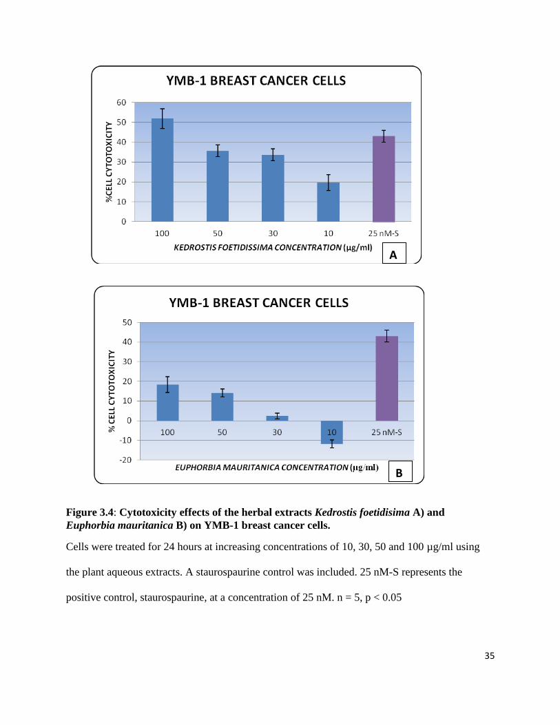
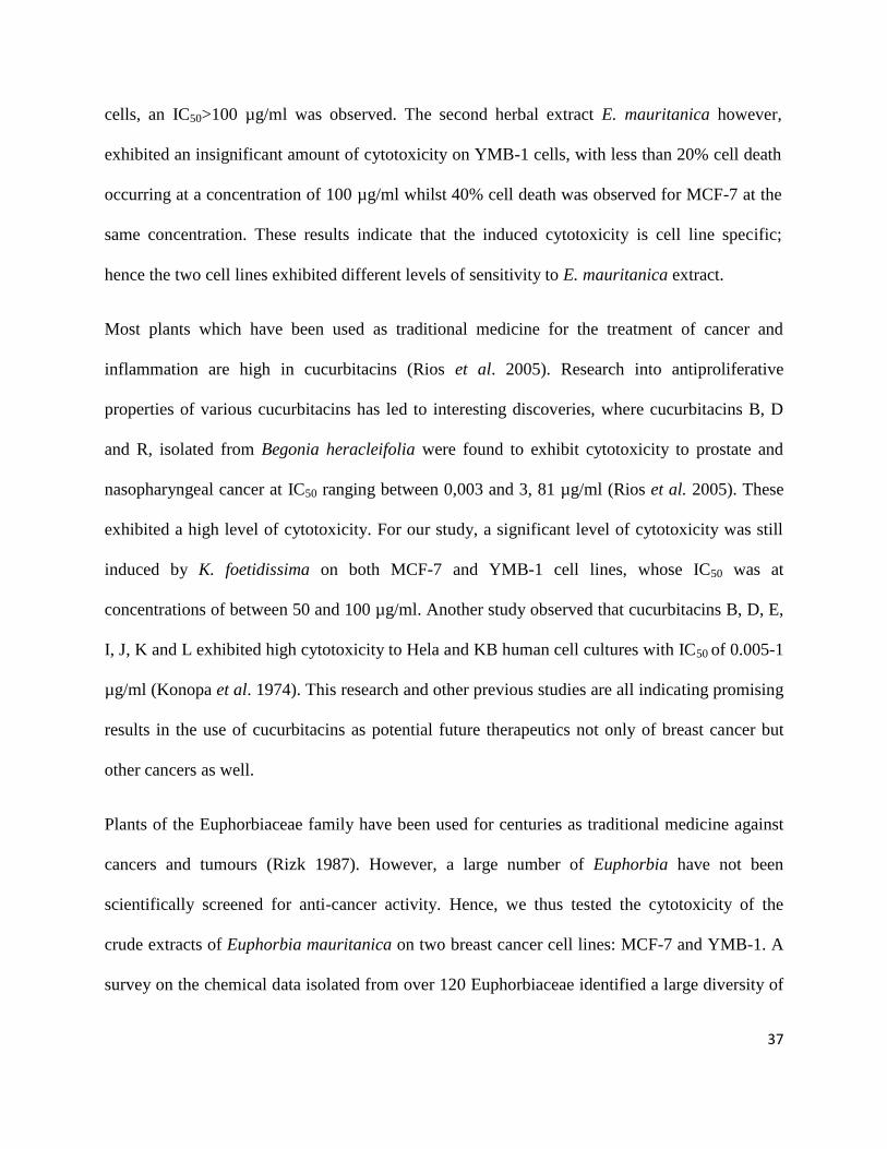
Figure 3.4: Cytotoxicity effects of the herbal extracts Kedrostis foetidisima A) and
Euphorbia mauritanica B) on YMB-1 breast cancer cells.
Cells were treated for 24 hours at increasing concentrations of 10, 30, 50 and 100 µg/ml using
the plant aqueous extracts. A staurospaurine control was included. 25 nM-S represents the
positive control, staurospaurine, at a concentration of 25 nM. n = 5, p < 0.05
A
B

36
The antiproliferative activity of the herbal plant extracts on the breast cancer cell lines was
measured using the IC50 value principle, which is a principle based on the concentration of the
plant extract that causes 50% cell death. The lower the IC50 value of an extract on a cell line, the
more potent it is considered to be. The aqueous extracts of both the herbal plants Kedrostis
foetidissima and Euphorbia mauritanica exhibited considerable levels of antiproliferative
activity against MCF-7 breast cancer cells presented in Figure 3.3.
Treatment with K. foetidissima at a concentration of 100 µg/ml induced 48% cell death (IC50
>100 µg/ml) (Figure 3.3 A). Euphorbia mauritanica induced 40% cell death at a concentration
of 100 µg/ml (IC50 >100 µg/ml) (Figure 3.3. B). The positive control, staurospaurine induced
43% cell death at a concentration of 25 nM. In a previous study aimed at investigating the
possible antiproliferative properties of E. mauritanica on MCF-7 cells (Essack 2006), no
significant cytotoxicity could be observed, with less than 10% cell death being observed even at
a high concentration of 5 mg/ml. Results were contrary to what was observed in this study with
40% cell death resulting from a much lower concentration of 100 µg/ml.
In addition to the cell cytotoxicity assay for K. foetidissima and E. mauritanica on MCF-7 breast
cancer cells, we aimed to investigate the possible antiproliferative effects of these herbal extracts
on a different breast cancer cell line. Hence their cytotoxicity was tested on YMB-1 cell lines.
The positive control, staurospaurine, induced 42% cell death at a concentration of 25nM on the
YMB-1 cells. On comparing to the cytotoxicity of K. foetidissima on YMB-1 to MCF-7, it was
observed that K. foetidissima extract exhibited a slightly higher cytotoxicity on YMB-1 cells,
with 53% cell cytotoxicity being observed at a concentration of 100 µg/ml and 37% cytotoxicity
at a concentration of 50 µg/ml, meaning 50 µg/ml<IC50>100 µg/ml (Figure 4.2. A). For MCF-7
37
cells, an IC50>100 µg/ml was observed. The second herbal extract E. mauritanica however,
exhibited an insignificant amount of cytotoxicity on YMB-1 cells, with less than 20% cell death
occurring at a concentration of 100 µg/ml whilst 40% cell death was observed for MCF-7 at the
same concentration. These results indicate that the induced cytotoxicity is cell line specific;
hence the two cell lines exhibited different levels of sensitivity to E. mauritanica extract.
Most plants which have been used as traditional medicine for the treatment of cancer and
inflammation are high in cucurbitacins (Rios et al. 2005). Research into antiproliferative
properties of various cucurbitacins has led to interesting discoveries, where cucurbitacins B, D
and R, isolated from Begonia heracleifolia were found to exhibit cytotoxicity to prostate and
nasopharyngeal cancer at IC50 ranging between 0,003 and 3, 81 µg/ml (Rios et al. 2005). These
exhibited a high level of cytotoxicity. For our study, a significant level of cytotoxicity was still
induced by K. foetidissima on both MCF-7 and YMB-1 cell lines, whose IC50 was at
concentrations of between 50 and 100 µg/ml. Another study observed that cucurbitacins B, D, E,
I, J, K and L exhibited high cytotoxicity to Hela and KB human cell cultures with IC50 of 0.005-1
µg/ml (Konopa et al. 1974). This research and other previous studies are all indicating promising
results in the use of cucurbitacins as potential future therapeutics not only of breast cancer but
other cancers as well.
Plants of the Euphorbiaceae family have been used for centuries as traditional medicine against
cancers and tumours (Rizk 1987). However, a large number of Euphorbia have not been
scientifically screened for anti-cancer activity. Hence, we thus tested the cytotoxicity of the
crude extracts of Euphorbia mauritanica on two breast cancer cell lines: MCF-7 and YMB-1. A
survey on the chemical data isolated from over 120 Euphorbiaceae identified a large diversity of

38
structurally unique chemical compounds. These include methyl esters, their derivatives and
diterpene polyesters. Other compounds identified include alkaloids, phenolic compounds,
cyanogenic glucosides and glucosinolates (Rizk 1987). Some of these chemical compounds
have been shown to exhibit antiproliferative activity against various cancer cells. A study by
Engi et al. (2007) looked at the effect of diterpenes isolated from methanol extracts of nine
Euphorbia species on human colon cancer cells and observed a moderate antiproliferative effect
of IC50< 3.5 μg/ml- 17.46 μg/ml.
Studies by Whelan and Ryan (2003), investigated the effect of ethanolic extracts of Euphorbia
species: E. grandidens, E. grandicorni, E. latea, E. coerulescens, E. trigona1, E. istigy, E.
candelabrum, E. pentagona, E. triangularis against HEp-2 (Human epidermoid cancer) cells at
concentrations of 8.53 μg/ml, 85.3 μg/ml and 853.9 μg/ml. They found that of the nine plant
extracts tested, four extracts resulted in reduced cell viability at concentrations of 85.3 μg/ml and
853.9 μg/ml, with the most potent extract causing IC50 at 57 μg/ml. Interestingly, for our study,
we observed increase in proliferation for YMB-1 cells that were treated with E. mauritanica at a
concentration of 10 µg/ml (Figure 3.4. B), in their study, they observed a similar trend with
seven of their extracts also promoting cellular proliferation at a concentration of 8.53 μg/ml. This
may indicate that some Euphorbia species, including E. mauritanica, may actually induce cell
proliferation to some cancers at low doses.
It was evident from our study that the crude extracts of the two plants E. mauritanica and K.
foetidissima were exhibiting antiproliferative properties on the breast cancer cells, but the next
important issue was to determine if the observed cytotoxicity was as a result of apoptosis or
necrosis.

39
CHAPTER 4 SCREENING OF HERBAL EXTRACTS FOR MODE OF
DEATH INDUCTION (APOPTOSIS OR NECROSIS)
4.1 Introduction
Apoptosis and necrosis are two different types of cell death characterised by different modes or
morphologies of death (Figure 4.1). Apoptosis is a form of programmed cell death, whereby
cells undergo orderly typical morphological changes whereas necrosis involves rapid lysis of a
cell (Kumar 1998; Raffray and Cohen 1997). Necrosis is generally not the ideal death pathway
since it leads to leakage of damaged internal components of the dying cell into surrounding
tissue, which may be harmful to the surrounding cells, and leads to induction of an inflammatory
response (Potten and Wilson 2004).

40
Table 4.1: Differences between apoptotic and necrotic cellular death pathways
Apoptosis Necrosis
Morphological features
Early loss of cell to cell contact and involves single cells (d) Late loss of cell to cell contact and involves clusters (d)
Anoikis- detachment from the surface No anoikis
Cell shrinkage (e) Cell swelling (e)
Mitochondrial condensation, no swelling (a) Mitochondria swelling (a)
DNA fragmentation- nuclear DNA breaks down in large, then
small nucleosomal fragments (b)
Nuclear swelling (e)
Formation of apoptotic bodies No apoptotic bodies
Rapid recognition, uptake and degradation of apoptotic bodies by
phagocytes (c)
No cellular uptake by phagocytes, attraction of inflammatory
cells (c,e)
No leakage of cell contents (a) Cells swell up and suddenly collapse, leading to spilling of cell
contents (b)
Apoptosis Necrosis
Biochemical and molecular features
ATP levels are preserved (e) Rapid intracellular ATP depletion (e)
Oxidative stress (e) ___
Cytochrome C released from mitochondria (e) ___
Protein degradation mediated by specific proteolytic enzymes
known as caspases (a)
No regulated protein degradation (a)
Plasma membrane remains intact almost until the end, then losses
asymmetry and integrity. Phosphatidyl serine residues located in
the inner plasma membrane translocate to the cell surface. (a,b)
Plasma membrane losses integrity rapidly (b)
Key: a) Kroemer et al. 1998; b) Fiers et al. 1999; c) Savill et al. 1993; d) Potten and Wilson 2004; e) Wlodkowic et al. 2009

41
Figure 4.1: Characteristic features that distinguish between apoptotic and necrotic cell
death (van der Meer et al. 2010).
Necrotic cells are characterised by swelling up of the cell, loss of membrane integrity and
eventually the cells lyse releasing their internal components into the surrounding tissue.
Apoptotic cells shrink, chromatin condenses, nuclear DNA breaks down and apoptotic bodies
form which are then phagocytosized by either macrophages or surrounding cells (van der Meer et
al. 2010).
Normal Cell
Necrosis
Formation of apoptotic bodies
Cellular shrinkage, chromatin condensation
Phagocytosis
Cell swells up Cell disintegration Cell membrane becomes
leaky
Apoptosis

42
When working with potential therapeutic agents, it is important that they induce high
cytotoxicity to their target cells whilst showing none or very minimal toxicity to the surrounding
unaffected healthy tissue. From Table 4.1, it is clear that apoptosis would be the desired method
of death since it is more regulated, considering the fact that dying cells are taken up by
phagocytes and do not lyse to release their internal components to neighbouring cells, which
would otherwise prove to be catastrophic.
The purpose of this section was to screen the mode of death induction of the crude extracts of K.
foetidissima on MCF-7 and YMB-1 cells and E. mauritanica on MCF-7 cells since they showed
significant cytotoxicity on the cells. MCF-7 and YMB-1 breast cancer cells, treated with the
herbal extract concentrations that induced about 50% cell death were incubated with Annexin V
according to manufacturer’s specifications (see section 2.2.2). A negative control of untreated
cells and positive control of cells treated with 25 nM of staurospaurine were included.
Staurospaurine is a known inducer of apoptosis in many different cancer cell types (Motadi
2009). Flow cytometry was employed to analyse the percentage of cells that underwent
apoptosis compared to the percentage of necrotic and live cells following treatment with the
herbal extracts.

43
4.2 Results and discussion
The graphs below are representative of the flow cytometry apoptosis analysis
Figure 4.2: Scatter analysis diagrams for YMB-1 cells.
A) Untreated cells, B) Staurospaurine (25 nM) treated cells and C) Kedrostis foetidissima (100
µg/ml) treated cells.
A B
C
2.29% 0.26%
95.03%
2.42%
0.59% 2.77%
57% 39.63%
1.98% 1.58%
60.26% 36.17%

44
Figure 4.3: Scatter analysis diagrams for MCF-7 cells.
Cells were treated with A) Kedrostis foetidissima (100 µg/ml), B) Euphorbia mauritanica (100
µg/ml), C) Staurospaurine (25 nM) and D) Untreated.
A B
C B
D
B
0.90% 5.21%
68.40% 25.49%
0.60% 6.27%
59.10% 34.10%
1.04% 7.54%
57.68% 33.75%
0.59% 1.21%
90.70% 7.50%
45
The cells were scattered into one of four quadrants: Quadrant B1 contains cells undergoing
necrosis, quadrant B2 contains cells undergoing late apoptosis, quadrant B4 contains cells
undergoing early apoptosis and quadrant B3 contains live cells (Figures 4.2 & 4.3).
Since K. foetidissima seemed to be the only extract that exhibited cytotoxicity on YMB-1 cells,
it was then further assessed for possible pro-apoptotic activity on this cell line. The YMB-1 cells
were treated with K. foetidissima at a concentration of 100 µg/ml for 24 hours and tested for
markers of apoptosis. On observing results for the positive control, staurospaurine (Figure 4.2 B)
40% of the cells stained positive for externalised phosphatidylserine residues, indicating that
40% of the cells underwent apoptosis whilst 57% remained alive. The cells treated with K.
foetidissima extract demonstrated a similar percentage of phosphatidylserine externalisation to
that of staurospaurine, with about 40% of cells undergoing apoptosis and about 60% remaining
alive (Figure 4.2 C). A negative control of untreated cells was included which from flow
cytometer analysis indicated that 95% of the cells were alive and less than 5% were dying
naturally either via necrosis or apoptosis (Figure 4.2 A). The flow cytometer results suggested
that the herbal extract K. foetidissima mainly induced cell death to YMB-1 cells via an apoptotic
pathway.
MCF-7 cells exhibited significant sensitivity to both herbal extracts E. mauritanica and K.
foetidissima hence we then aimed to investigate if these herbal extracts were inducing death to
the cells via necrosis or apoptosis. Similarly to the previous experiments, staurospaurine (25 nM)
was used as a positive control, and the flow cytometry results suggested that over 40% of cells
stained positive for externalised phosphatidylserine. MCF-7 cells were treated with both K.
foetidissima and E. mauritanica at concentrations of 100 µg/ml. Flow cytometry analysis

46
indicated that treatment with K. foetidissima resulted in about 30% cells staining positive for
externalised phosphatidylserine residues i.e. undergoing apoptosis and with almost 70%
remaining alive. Euphorbia mauritanica treatment resulted in about 40% cells undergoing
apoptosis whilst about 60% remained viable (Figure 4.6). A no treatment control was included
which showed that over 90% of the cells were still alive after 24 hours and the rest of the cells
had died mainly due to apoptosis (Figure 4.3).
Extracts of K. foetidissima were assayed for markers of pro-apoptotic activity on MCF-7 and
YMB-1 cell lines whilst E. mauritanica was assayed for markers of pro-apoptotic activity on
MCF-7 cell lines only. On assessing cells in the second and fourth quadrant representing cells
undergoing late and early apoptosis respectively, it was observed that K. foetidissima induced the
highest apoptotic activity in YMB-1 than in MCF-7 cells (Figures 4.3C & 4.4A). This difference
was also observed in the cytotoxicity assay. It is promising however, that the majority of cells
were undergoing apoptosis in both cell lines. In a study by Essack (2006), contradictory results
were obtained, with no phosphatidylserine residues externalisation being observed for MCF-7
cells treated with E. mauritanica.
In summary, the Annexin V apoptotic assay has demonstrated that the aqueous extracts of two
South African herbal plants K. foetidissima and E. mauritanica induce their cytotoxic effects by
an apoptotic mechanism. This indicates that these plants possess compound/s which can trigger
an apoptotic pathway, which is promising in that induction of apoptosis correlates with anti-
proliferation of cancer cells. Even though regulation of apoptosis is controlled by a huge network
comprising a number of genes, the two most important genes which have been found to be
highly deregulated in cancer are p53 and RBBP6. Following the flow cytometry apoptotic assay,

47
we then aimed to investigate if these herbal extracts have any effect on these two tumour
suppressor genes, using real-time PCR.

48
CHAPTER 5 TUMOUR SUPPRESSOR GENES
5.1 Introduction
The majority of breast cancers arise from deregulation of genes involved in the cell cycle or
apoptosis. As with all drugs that are manufactured to treat different diseases, it is important for
potential natural anti-cancer agents to exhibit a certain level of specificity on their target. The
FDA approved chemotherapeutic drugs as mentioned in section 1.5 all exhibit specific activity
on their target at various steps of the cell cycle. Their activity ranges from inhibiting microtubule
assembly, to preventing DNA and RNA synthesis whilst some work by preventing DNA
replication (Giri and Narasu 2000; Kessel et al. 1972; Kjeldsen 1992).
Tumour suppressor genes have now become of great interest in understanding molecular
pathogenesis of cancer. The tumours suppressors which have been highly found to be
deregulated in breast cancer include p53 and RB genes. p53, a tumour suppressor is very crucial
in the control of cell proliferation. Deregulation of p53 has been implicated in over 50% of
cancers (Potten and Wilson 2004). This may stem from its role as a sequence specific DNA
binding transcription factor, whose downstream targets are mostly gene products which control
cell cycle arrest, DNA repair and apoptosis (Picksley and Lane 1994). Retinoblastoma (Rb), the
first tumour suppressor to be discovered, acts as a crucial component of the cell cycle
checkpoint. It functions to prevent excessive cell growth, by inhibiting progression of aberrant
DNA along the cell cycle through G1 into the S-phase (Garg 2007; Mayhew et al. 2007). Rb has
been found to be mutated in a third of human cancers (Lee 2002). From the extensive amount of
research done on p53 and Rb deregulation, it can be said that they are arguably the most
deregulated pathways in human cancer. The tumour suppressors p53 and Rb require specific

49
protein to protein interactions to mediate their respective functions. RBBP 6 protein is one of
very few proteins known to interact with both p53 and Rb.
In this study, the cytotoxicity assay showed that MCF-7 exhibited sensitivity to both E.
mauritanica and K. foetidissima whilst YMB-1 cells were sensitive to E. mauritanica only
(Figures 3.3 & 3.4). The flow cytometric assay further indicated that death was induced by an
apoptotic pathway (Figures 4.3 & 4.4). With the high rate of p53 and Rb mutations that have
been shown to occur in cancer and the fundamental role these genes play in tumour suppression,
we thus aimed to investigate if the herbal extracts could restore or upregulate the levels of p53
and reduce expression levels of RBBP6 in MCF-7 breast cancer cells thereby increasing
apoptosis. This was done by means of real-time PCR, which can accurately measure changes in
gene expression levels.
Real-Time PCR is a highly sensitive and powerful method used for quantitating small changes in
gene expression. Its accuracy and the rapid quantification of results make it the ideal method of
nucleic acid quantification (Klein 2002; Pfaffl 2001). Real-Time PCR was employed to (i)
investigate the expression levels of the above mentioned tumour suppressors: p53 and RBBP6 in
MCF-7 breast cancer cells and (ii) to investigate if the herbal extracts which were inducing
apoptosis to these cells were having any effect on the expression levels of these tumour
suprressors.
5.2 Primer efficiency assessment using standard curves
To answer the above questions, real-time PCR specific primers were designed using NCBI for
collection of gene sequences, primer3plus for designing primers and NCBI Blast for ensuring
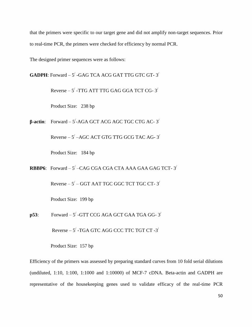
50
that the primers were specific to our target gene and did not amplify non-target sequences. Prior
to real-time PCR, the primers were checked for efficiency by normal PCR.
The designed primer sequences were as follows:
GADPH: Forward – 5/ -GAG TCA ACG GAT TTG GTC GT- 3
/
Reverse – 5/ -TTG ATT TTG GAG GGA TCT CG- 3
/
Product Size: 238 bp
β-actin: Forward – 5/-AGA GCT ACG AGC TGC CTG AC- 3
/
Reverse – 5/ –AGC ACT GTG TTG GCG TAC AG- 3
/
Product Size: 184 bp
RBBP6: Forward – 5/ –CAG CGA CGA CTA AAA GAA GAG TCT- 3
/
Reverse – 5/ – GGT AAT TGC GGC TCT TGC CT- 3
/
Product Size: 199 bp
p53: Forward – 5/ -GTT CCG AGA GCT GAA TGA GG- 3
/
Reverse – 5/ -TGA GTC AGG CCC TTC TGT CT -3
/
Product Size: 157 bp
Efficiency of the primers was assessed by preparing standard curves from 10 fold serial dilutions
(undiluted, 1:10, 1:100, 1:1000 and 1:10000) of MCF-7 cDNA. Beta-actin and GADPH are
representative of the housekeeping genes used to validate efficacy of the real-time PCR

51
experiments. The real-time PCR cocktail was prepared as explained in appendix A4 and the real-
time PCR running parameters are explained in Table 2.1.
Figure 5.1: Standard curves for Beta-actin (A) and GADPH (B) primers which served as
housekeeping genes.
The amplification efficiencies of the slopes were 1.834 and 1.941, respectively. n= 2
A
B

52
Figure 5.2: Standard curves for the tumour suppressors RBBP6 (A) and p53 (B) primers.
The amplification efficiencies of the slopes were 1.981 and 1.932, respectively. n= 2
A
B

53
5.3 Relative quantitative real-time polymerase chain reaction
Relative quantification, which is based on the relative expression of the target gene versus that of
a housekeeping gene, was the preferred mode of quantification for this study. Housekeeping
genes are constitutively expressed in all nucleated cells since they are required for basic cell
survival. Their mRNA is considered stable even under changing experimental conditions (Pfaffl
2001). Housekeeping genes such as glyceraldehyde-3-phosphate dehydrogenase (GADPH),
actins, tubulins, cyclophilin, 18S rRNA and 28S rRNA are an absolute prerequisite for use in
real-time RT-PCR experiments to ensure accurate normalisation of the experiments (Schmittgen
and Zakrajsek 2000). For this study, mRNA expression levels were normalised to the levels of
GADPH and β-actin transcripts, which served as our housekeeping or reference genes.
One-step real-time PCR requires a separate step of reverse transcription of mRNA to cDNA
before running RT-PCR. This method is ideal in that the starting amount of cDNA can be
equalised to ensure the difference in expression level of genes is not due to experimental error.
The following mRNA templates were reverse transcribed as in section 2.2.3 in preparation of
RT-PCR:
i. Untreated MCF-7 cells
ii. MCF-7 cells treated with K. foetidissima
iii. MCF-7 cells treated with E. mauritanica
iv. Standard ( Prepared from untreated cells at one of the concentrations correlating with one
of the points or concentrations on the standard curve)
v. Negative control (-ve)
vi. No template control (NTC)

54
To validate if the selected housekeeping genes were the best possible for normalisation of these
experiments, we had to test if they were stably expressed under changing experimental systems.
Real-time PCR amplification curves were constructed using (1) GADPH and (2) β-actin primers.
Each run comprised of (i), (ii) and (iii), all experiments were conducted in duplicates with (iv),
(v) and (vi) also included.
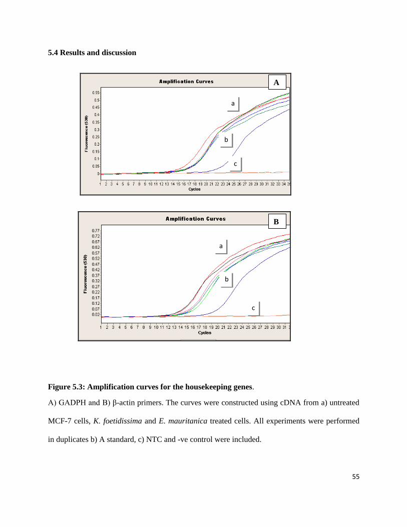
55
5.4 Results and discussion
Figure 5.3: Amplification curves for the housekeeping genes.
A) GADPH and B) β-actin primers. The curves were constructed using cDNA from a) untreated
MCF-7 cells, K. foetidissima and E. mauritanica treated cells. All experiments were performed
in duplicates b) A standard, c) NTC and -ve control were included.
A
B
a
b, b, c
c, b, c
a
b
c

56
Figure 5.4: Amplification curves for RBBP6 expression.
a) untreated MCF-7 cells, K. foetidissima and E. mauritanica treated MCF-7 cells b) A standard,
c) NTC and -ve control were included.
Figure 5.5: RT-PCR products resolved on a gel following RBBP6 amplification.
M represents the molecular weight marker (MassRuler DNA Ladder Mix, Fermentas), U
represents untreated MCF-7 cells, K.F and E.M represent K. foetidissima and E. mauritanica
treated MCF-7 cells, respectively.
M U K.F E.M
a
b
c
~200bp
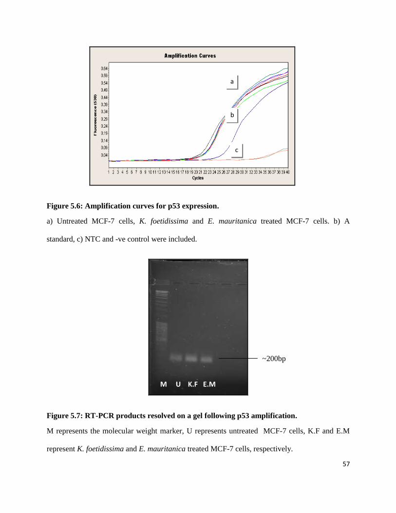
57
Figure 5.6: Amplification curves for p53 expression.
a) Untreated MCF-7 cells, K. foetidissima and E. mauritanica treated MCF-7 cells. b) A
standard, c) NTC and -ve control were included.
Figure 5.7: RT-PCR products resolved on a gel following p53 amplification.
M represents the molecular weight marker, U represents untreated MCF-7 cells, K.F and E.M
represent K. foetidissima and E. mauritanica treated MCF-7 cells, respectively.
M U K.F E.M
a
b
c
~200bp

58
There are currently a number of mathematical models used to analyse real-time PCR data. A
fairly new and simple method by Pfaffl (2001) was used since it seems to provide the most
accurate data quantification. This method compares the ratio of the target gene to that of the
reference (housekeeping) gene. The relative expression ratio of an unknown target is computed
based on its real-time PCR efficiencies, the crossing point difference of the unknown target
versus that of the control in comparison to that of the control (Pfaffl 2001).
Where:
Etarget = real time PCR efficiency of target gene
Eref = real time PCR efficiency of reference gene
ΔCPtarget = Deviation in crossing point of control - crossing point of target gene
ΔCPref = Deviation in crossing point of control - crossing point of reference gene
Relative expression = (Etarget) ΔCPtarget (control – sample)
(Eref) ΔCPref (control – sample)
(Pfaffl 2001)
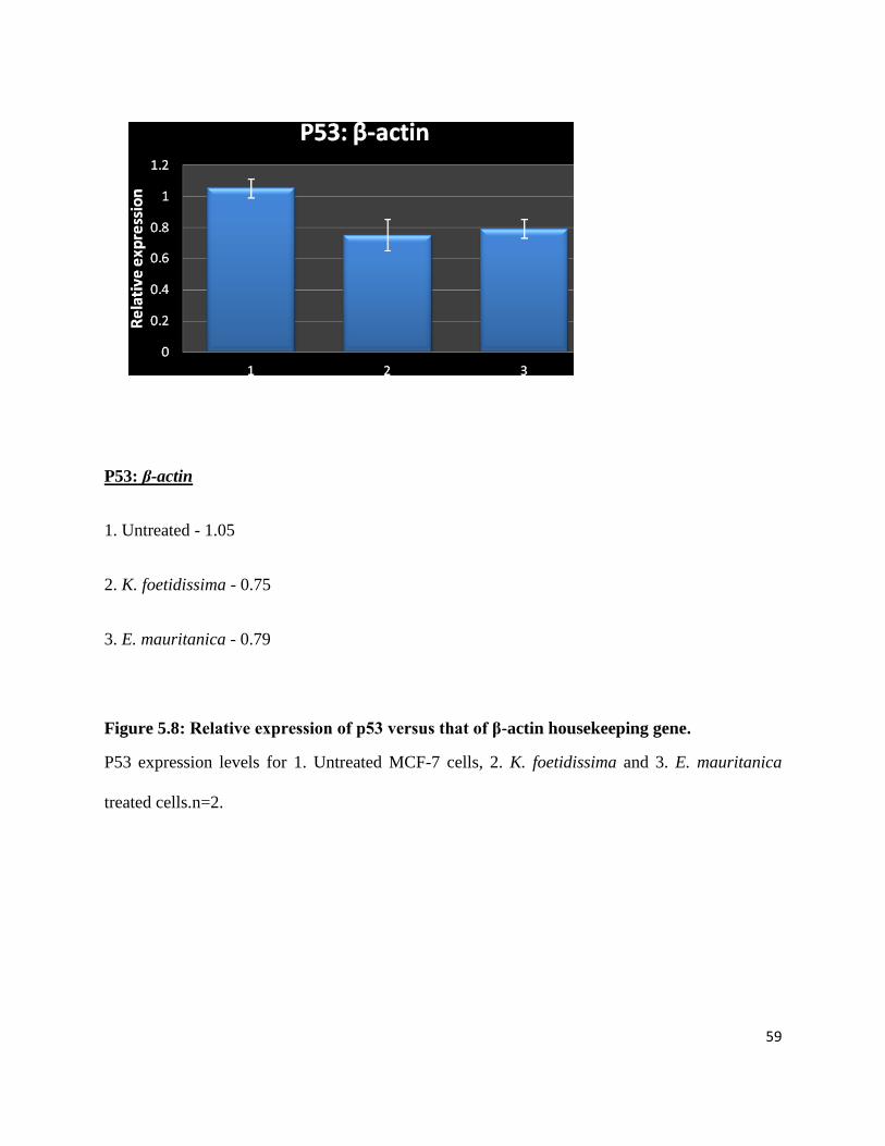
59
P53: β-actin
1. Untreated - 1.05
2. K. foetidissima - 0.75
3. E. mauritanica - 0.79
Figure 5.8: Relative expression of p53 versus that of β-actin housekeeping gene.
P53 expression levels for 1. Untreated MCF-7 cells, 2. K. foetidissima and 3. E. mauritanica
treated cells.n=2.

60
P53: GADPH
1. Untreated – 0.99
2. K. foetidissima - 0.84
3. E. mauritanica - 0.79
Figure 5.9: Relative expression of p53 versus that of GADPH housekeeping gene.
P53 expression levels for 1. Untreated MCF-7 cells, 2. K. foetidissima and 3. E. mauritanica
treated cells. n=2.

61
RBBP6: β-actin
1. Untreated – 1.08
2. K. foetidissima - 0.86
3. E. mauritanica - 0.79
Figure 5.10: Relative expression of RBBP6 versus that of β-actin housekeeping gene.
RBBP6 expression levels for 1. Untreated MCF-7 cells, 2. K. foetidissima and 3. E. mauritanica
treated cells. n=2.
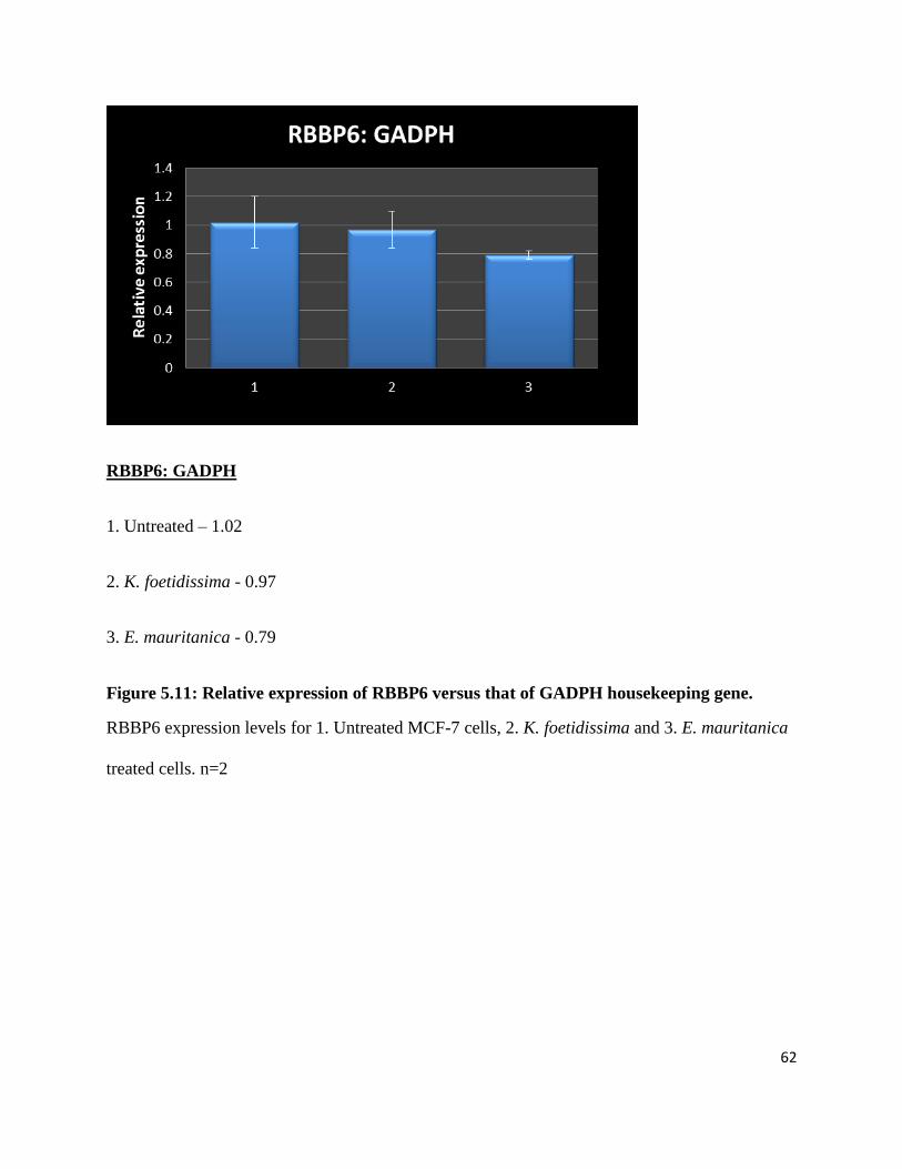
62
RBBP6: GADPH
1. Untreated – 1.02
2. K. foetidissima - 0.97
3. E. mauritanica - 0.79
Figure 5.11: Relative expression of RBBP6 versus that of GADPH housekeeping gene.
RBBP6 expression levels for 1. Untreated MCF-7 cells, 2. K. foetidissima and 3. E. mauritanica
treated cells. n=2

63
Relative quantitative RT-PCR revealed that the levels of the RBBP6 transcript following
normalisation with β-actin were 1.08 in the untreated MCF-7 cells (Figure 5.10). Treatment with
K. foetidissima slightly reduced expression levels to 0.86 and with E. mauritanica it was reduced
to 0.79. Following normalisation with GADPH, the RBBP6 levels seemed to have been slightly
reduced from 1.02 to 0.97 whilst treatment with E. mauritanica led to a slightly higher reduction
of 0.79 (Figure 5.11).
The relative expression levels of p53 in MCF-7 cells following normalisation with β-actin were
1.05 before treatment (Figure 5.8). Treatment with K. foetidissima slightly reduced expression
levels to 0.75 and with E. mauritanica it was reduced to 0.79. Following normalisation with
GADPH, relative expression of p53 in the untreated cells was 0.99, it was slightly reduced to
0.84 following treatment with K. foetidissima. Treatment with E. mauritanica yielded a slight
reduction of 0.79 (Figure 5.9). The herbal extracts K. foetidissima and E. mauritanica exhibit
minimal effect on the expression levels of the tumour suppressors p53 and RBBP6. Both extracts
caused a slight reduction in the expression of both genes.
In summary, the crude extracts of E. mauritanica and K. foetidissima induced apoptosis on
MCF-7 cells, but, RT-PCR analysis indicated that their mode of action is not by upregulating
p53 or downregulating RBBP6. Since apoptosis is controlled by such a huge network of genes
and signalling molecules, it is possible that these plant extracts control other components of the
apoptotic pathway. Further research that incorporates isolation and identification of the active
compounds would be required to better understand their molecular mechanism and the pathway
components they affect.

64
CHAPTER 6 GENERAL DISCUSSION AND CONCLUSION
6.1 General discussion
Historically, plants, herbs and spices were a folkloric source of medicinal agents, and as modern
medicine expanded, many useful drugs were developed from lead compounds discovered from
medicinal plants. This approach has provided leads against various pharmacological targets,
including cancer, malaria and pain, and remains an important route to new pharmaceuticals
(Essack 2006). Recent advances in cytotoxic and phytochemical screening have provided
scientists with insight into the bioactive properties of medicinal plants, which has led to the
development of new medicines. In this study, Kedrostis foetidissima, Euphorbia mauritanica and
Elytropappus rhinocerotis plants which are indigenous to South Africa and its neighbouring
countries were screened for their possible antiproliferative and/or pro-apoptotic effect on two
breast cancer cell lines: MCF-7 and YMB-1.
MTT assay was used to measure significant variations in the inhibitory activity of the 3 plant
extracts on breast cancer cell lines. The best thing about MTT is that it does not have limitations
and one can test as many times as possible. It was used to measure IC50, the concentration of the
plant extract that induces 50% cell death on the breast cell lines. Untreated cells were included as
a control and cells treated with staurospaurine as a positive control. Little to no cell cytotoxicity
was observed when Elytropappus rhinocerotis was used in these two cell lines. From our results
as described in Chapter 3 (Figures 3.3 & 3.4), we observed interesting results with Kedrostis
foetidissima being able to induce up to 48% and 53% cell death in MCF-7 and YMB-1 cells
respectively, at a concentration of 100 µg/ml. Whereas Euphorbia mauritanica induced up to

65
40% cell death in MCF-7 but insignificant cell death of less than 20% in YMB-1 could be
observed. In this case, only MCF-7 showed to be a cell line that can be used to assess the growth
inhibition potential of Euphorbia mauritanica. The results obtained especially for E. mauritanica
treatments, are similar to those observed by Kummalue et al. (2011) who compared inhibition of
cell growth by Sapindusrarax water extract on two lung cancer cell lines and a breast cancer cell
line; they also observed a significant growth inhibition in a dosage dependent manner, but only
in the A549 cell line, and no significant growth inhibition was observed in the other two cell
lines. These results therefore suggest that this plant extract might be cell line specific and cancer
type dependent.
After the confirmation that the two plant extracts indeed managed to induce significant cell death
in the two breast cancer cell lines tested, we had to verify if the type of cell death induced was
due to apoptosis, which is genetically controlled or if it was due to necrosis. One way of
verifying this was by the use of flow-cytometry staining which uses both the propidium iodide
and FITC. Propidium iodide stains cells in their late apoptotic stages as well as G1 cell cycle
arrest and FITC stains cells in the early stages of apoptosis. Kedrostis foetidissima was then
further assessed for possible pro-apoptotic activity using flow cytometer on MCF-7 and YMB-1
cells, whilst E. mauritanica was assessed on MCF-7 cells. Similarly to the cytotoxicity assays,
staurospaurine was used as the positive control whilst cells that were not treated with anything
but media were used as the control.
The results confirmed our earlier findings that indeed cell death had been induced following
treatment with the two plant extracts, and that it was as a result of apoptosis. YMB-1 and MCF-7
cells treated with K. foetidissima at a concentration of 100 µg/ml demonstrated a 40% and 30%

66
phosphatidylserine residue externalization, respectively; whilst MCF-7 cells treated with E.
mauritanica demonstrated a 40% phosphatidylserine residue externalization, which is an
indication of induced apoptosis. Staurospaurine induced just over 40% apoptosis in both cell
lines. This demonstrated that treatment of MCF-7 cells with K. foetidissima and E. mauritanica
and of YMB-1 cells with K. foetidissima resulted in a significant induction of apoptosis, while
showing relatively no significant toxicity to the cells in the form of necrosis.
Research into the development of novel small-molecule plant-derived extracts and compounds,
which may prevent carcinogenesis, curtail its progression, or even cure the disease is still at the
forefront of cancer therapeutics (D’Archivio et al. 2007). With studies by Creemers et al. (1994)
showing compounds such as irinotecan and topotecan derived from Nyssacea Camptotheca
accuminata as the main inducers of apoptosis against colorectal and ovarian cancer. Kedrostis
foetidissima, which we found to be the most potent of the plant extracts tested, has been found to
contain cucurbitacins B, D, E, I, J ,K and L by Konopa et al. (1974). They further went on to
show that K. foetidissima exhibits high cytotoxicity to Hela and KB human (Konopa et al. 1974),
however the synergistic activity of these cucurbitacins need to be studied further to understand
their exact activities on the different cancer cell lines.
On the other hand, E. mauritanica belongs to Euphorbiaceae family, whose extracts include
methyl esters, their derivatives and diterpene polyesters. Other compounds that have been
discovered include alkaloids, phenolic compounds, cyanogenic glucosides and glucosinolates
which have been identified as players in the induction of apoptosis in some cells (Rizk 1987).
This study clearly shows promising results for this plant extract; however, further studies that

67
entail isolation and research into the biological activities of the active compounds from the plant
E. mauritanica would provide better insight.
As with all potential anti-tumour agents, it is crucial to understand the molecular mechanism
underlying their apoptotic activities. In this study, we concentrated on the two genes RBBP6 and
p53. The involvement of p53 and apoptosis in the cucurbitacins-mediated effect has been studied
in various cancer cells (Miro 1995; Rios et al. 2005). In response to various cellular stresses, p53
is phosphorylated on NH2-terminal residues. In particular, the phosphorylation of Ser 15 affects
the interaction with the negative regulator MDM2 oncoprotein and enhances contribution to the
stabilization of p53 (Chen et al. 2008; Suzuki et al. 2007). Phosphorylation of p53 is mediated
by phosphoinositide-3-kinase (PI3K)-related proteins, including Ataxia telangiectasia-mutated
(ATM), ATM- and Rad3- related kinase (ATR) and DNA-dependent protein kinases (DNA-
PKs).
While p53 plays numerous roles in the cell, our experiments focused on distinguishing whether
K. foetidissima and E. mauritanica-induced apoptosis was p53 dependent or independent. On the
other hand RBBP6 is a negative regulator of p53 that results in the ubiquitination of p53 thereby
leading to cell proliferation (Motadi et al. 2011). Kedrostis foetidissima and Euphorbia
mauritanica treated cells revealed that p53 was not significantly increased (Tables 5.1 and 5.2)
following treatment with the extracts. Similarly, RBBP6 levels did not differ among the K.
foetidissima and E. mauritanica treated cells with less than 1/16 fold decrease (Tables 5.3 and
5.4). The biological activities of these plants have not been studied in depth, however, several
studies have looked at the molecular mechanisms of different curcubitacins and Euphorbia
species on different cancer cells. Lee et al. (2010) reviewed the molecular mechanism of

68
cucurbitacins and gathered that they target different oncogenic signalling pathways that have
been implicated in cancer such as the MAPK, JAK-STAT and the Akt-PKB pathways. The JAK-
STAT pathway induces Signal Transducers and Activators of Transcription (STATs) and Janus-
Kinases (JAKs). Cucurbitacin B was found to inhibit downstream phosphorylation of some
STATs and JAKs in pancreatic tumours, thereby inhibiting this pathway affecting several of its
downstream targets involved in apoptosis such as p53 and Bcl-2 (Lee et al. 2010). Escandell et
al. 2008 investigated cucurbitacin R and cucurbitacin I on HCT116 cells harbouring a Ras
mutant and found that they induced expression of p53 and p21. They further reported that
cucurbitacin R resulted in p53 protein overexpression and Bcl-2 downregulation following 8, 12
and 18 hour treatments in RAW 264.7 macrophages.
One of the most prominent compounds that has been identified in Euphorbia species are
jatrophane esters, which belong to Euphorbia semiperfoliata (Cragg and Newman 2005). This
was discovered following research on chemotypes of the drug taxol (explained in section 1.7.1),
which exhibited poor water solubility and side effects. Miglietta et al. (2002) looked at the effect
of these jatrophane polyesters (250 nM) on MCF-7 cells over a period of 16 hours and observed
that they highly induced expression of p53 and also abrogates Bcl-2 probably through the Raf-1
mediated signaling pathway. These results, however, are contradictory to our observations where
expression of p53 was slightly reduced. This could be attributed to the diversity of compounds
which have been identified from Euphorbia species. Hence future studies will require further
isolation of the active compounds within E. mauritanica and molecular mechanism
investigations.

69
In summary, the crude extracts of K. foetidissima induced apoptosis in both MCF-7 and YMB- 1
cells whilst those of E. mauritanica induced apoptosis in MCF-7. RT-PCR indicated that their
mode of action is in a p53 independent manner. Since apoptosis is controlled by such a huge
network of genes and signalling molecules, it is possible that these plant extracts control other
components of the apoptotic pathway. Further research that incorporates isolation and
identification of the active compounds would be required to better understand their molecular
mechanism and the molecular pathway that follows this apoptosis induction.
6.2 Conclusion and future perspectives
Inducers of apoptosis are considered good agents in anti-cancer therapeutics. Small molecules
and natural plant extracts have recently attracted attention to modern medical science research
with their non-lethal activity. In this study, crude extracts of three indigenous South African
plants were screened for their antiproliferative activity against two breast cancer cell lines: MCF-
7 and YMB-1. This study revealed that not only did K. foetidissima and E. mauritanica plant
extracts demonstrate antiproliferative activity to the breast cancer cells, but they induced cell
death by apoptosis, rendering them good agents in anticancer drug discoveries.
It could not be determined in this research which components of the apoptotic pathway are being
regulated. Hence, further research which includes isolation and characterisation of the active
compounds present in these plants is required. Further research into the molecular targets of the
isolated active compounds would also provide better insight on the molecular mechanisms of
action of these plant extracts.
In conclusion, this study has further suggested the importance of using or targeting traditional
plant extracts as potential therapy against several cancers and other illnesses. However, we

70
should continue to approach this with caution as most plant extracts turn to be unsuccessful once
several secondary metabolites are separated.

71
CHAPTER 7 REFERENCES
Altekruse, S.F., Kosary, C.L., Krapcho, M., Neyman, N., Aminou, R., Waldron, W.,
Ruhl, J., Howlader, N., Tatalovich, Z., Cho, H., Mariotto, A., Eisner, M.P., Lewis, D.R.,
Cronin, K., Chen, H.S., Feuer, E.J., Stinchcomb, D.G. & Edwards, B.K. 2009, SEER
Cancer Statistics Review, 1975-2007, National Cancer Institute.
Aniszewski, T. 2007, "Applications", Alkaloids - Secrets of Life, vol. 4, pp. 181-204.
Arnal, A. & Wade, R.H. 1995, "How does taxol stabilize microtubules?", Current
Biology, vol. 5, no. 8, pp. 900-908.
Balunas, M.J. & Kinghorn, A.D. 2005, "Drug discovery from medicinal plants", Life
Sciences, vol. 78, pp. 431-441.
Barnes, D.M. & Camplejohn, R.S. 1996, "p53, apoptosis and breast cancer". Journal of
Mammary Gland Biology and Neoplasia, vol. 1, pp. 163-175.
Beutner, K.R., Friedman-Kien, A.E., Artman, N.N., Conant, M.A., Illeman, M., Thisted,
R.A. & King, D.H. 1989, "Patient-applied podofilox for treatment of genital warts", The
Lancet, vol. 333, no. 8642, pp. 831-834.
Boatright, K.M. & Salvesen, G.S. 2003, "Mechanisms of caspase activation", Current
Opinion in Cell Biology, vol. 15, pp. 725-731.
Bonnez, W., Elswick, R.K., Bailey-Farchion, A., Hallahan, D., Bell, R., Isenberg, R.,
Stoler, M.H. & Reichman, R.C. 1994, "Efficacy and safety of 0.5% podofilox solution in
the treatment and suppression of anogenital warts", The American Journal of Medicine,
vol. 96, pp. 420-424.

72
Botha, C.J. & Venter, E. 2002, Plants Poisonous to Livestock. Southern Africa,
University of Pretoria, Faculty of Veterinary Science, Dept. of Paraclinical Sciences,
Section Pharmacology and Toxicology, Pretoria, South Africa.
Bray, F., McCarron, P. & Parkin, D.M. 2004, "The changing global patterns of female
breast cancer incidence and mortality", Breast Cancer Research, vol. 6, pp. 229-239.
Butler, L.M., Potischman, N.A., Newman, B., Millikan, R.C., Brogan, D., Gammon,
M.D., Swanson, C.A. & Brinton, L.A. 2000, "Menstrual risk factors and early-onset
breast cancer", Cancer Causes and Control, vol. 11, pp. 451-458.
Caldon, C.E., Daly, R.J., Sutherland, R.L. & Musgrove, E.A. 2006, "Cell cycle control in
breast cancer", Journal of Cellular Biochemistry, vol. 97, pp. 261-274.
Chan, K. 2003, "Some aspects of toxic contaminants in herbal medicines", Chemosphere,
vol. 52, pp. 1361-1371.
Chen, L.J., Hsu, C.C., Hong, J.R., Jou, L.K., Tseng, H.C., Wu, J.L., Liou, Y.C. & Her,
G.M. 2008, "Liver-specific expression of p53-negative regulator mdm2 leads to growth
retardation and fragile liver in zebrafish", Developmental Dynamics, vol. 237, no.4,
pp.1070-1081.
Chen, M. & Wang, J. 2002, "Initiator caspases in apoptotis signaling pathways",
Apoptosis, vol. 7, no. 4, pp. 313-319.
Chen, W.Y., Manson, J.A., Hankinson, S.E., Rosier, B., Holmes, M.D., Willett, W.C. &
Condit, G.A. 2006, "Unopposed estrogens therapy and the risk of invasive breast cancer".
Archives of Internal Medicine, vol. 166, pp. 1027-1032
Cherry, M. 2005, "South Africa—serious about biodiversity science.", PLoS Biology, vol.
3, no. 5, pp. 743-747.

73
Cheung, A.M.Y., Hande, M.P., Jalali, F., Tsao, M.S., Skinnider, B., Hirao, A.,
McPherson, J.P., Karaskova, J., Suzuki, A., Wakeham, A., You-Ten, A., Elia, A., Squire,
J., Bristow, R., Hakem, R., & Mak, T.W. 2002, "Loss of Brca2 and p53 synergistically
promotes genomic instability and deregulation of T-cell apoptosis", Cancer Research,
vol. 62, pp. 6194–6204.
Chibi, M., Meyer, M., Skepu, A., Rees, D.J.G., Moolman-Smook, J.C. & Pugh, D.J.R.
2008, "RBBP6 interacts with multifunctional protein YB-1 through its RING finger
domain, leading to ubiquitination and proteosomal degradation of YB-1", Journal of
Molecular Biology, vol. 384, pp. 908-916.
Cocks, M.L. & Dold, A.P. 2006, "Cultural significance of biodiversity: The role of
medicinal plants in urban African cultural practices in the Eastern Cape, South Africa.",
Journal of Ethnobiology, vol. 26, no. 1, pp. 60-82.
Cragg, G.M. & Newman, D.J. 2005, "Plants as a source of anti-cancer agents", Journal of
Ethnopharmacology, vol. 100, pp. 72-79.
Creemers, G.J., Lund, B. & Verweij, J. 1994, "Topoisomerase I inhibitors: topotecan and
irinotecan", Cancer Treatment Reviews, vol. 20, pp.73-76
D’Archivio, M., Filesi, C., Di Benedetto, R. Gargiulo, R., Giovannini, C. & Masella, R.
2007, ―Polyphenols, dietary sources and bioavailability‖, Annali dell’Istituto Superiore
Sanita, vol. 43, pp. 348–361.
De Wilde, W.J.J.O. & Duyfjes, B.E.E. 2004, "Kedrostis medik (Cucurbitaceae) in Asia",
Reinwardtia, vol. 12, no. 2, pp. 129-133.

74
Donehower, L.A., Harvey, M., Slagle, B.L., McArthur, M.J., Montgomery Jr., C.A.,
Butel, J.S. & Bradley, A. 1992, "Mice deficient for p53 are developmentally normal but
susceptible to spontaneous tumours", Nature, vol. 356, pp. 215-221.
Elledge, S.J. 1996, "Cell cycle checkpoints: preventing an identity crisis", Science, vol.
274, pp. 1664-1671.
Engi, H., Vasas, A., Redei, D., Molnar, J. & Hohmann, J. 2007, "New MDR modulators
and apoptosis inducers from Euphorbia species", Anticancer Research, vol. 27, pp. 3451-
3458.
Escandell, J.M., Kaler, P., Recio, M. C., Sasazuki, T., Shirasawa, S., Augenlicht, L.,
Ríos, J. & Klampfer, L. 2008, "Activated kRas protects colon cancer cells from
cucurbitacin-induced apoptosis; the role of p53 and p21", Biochemical Pharmacology,
vol. 76, no. 2, pp. 198-207.
Escandell, J.M., Recio, M.C., Giner, R.M., Manez, S. & Rios, J.L. 2010, "Bcl-2 is a
negative regulator of interleukin 1B secretion in murine macrophages in
pharmacological-induced apoptosis", British Journal of Pharmacology, vol. 160, pp.
1844-1856.
Essack, M. 2006, Screening extracts of indigenous South African plants for the presence
of anti-cancer compounds, Thesis, (MSc), University of the Western Cape.
Fadeel, B., Orrenius, S. & Zhivotovsky, B. 1999, "Apoptosis in human disease: A new
Skin for the Old Ceremony?‖, Biochemical and Biophysical Research Communications,
vol. 266, no. 3, pp. 699-717.
Fiers, W., Beyaert, R., Declercq, W. & Vandenabeele, P. 1999, "More than one way to
die: apoptosis, necrosis and reactive oxygen damage", Oncogene, vol. 18, pp. 7719-7730.

75
Ford, D., Easton, D.F. , Stratton, M., Narod, S., Goldgar, D., Devilee, P., Bishop,
D.T.,Weber, B.,Lenoir, G., Chang-Claude, J., Sobol, H., Teare, M.D., Struewing, J.,
Arason, A., Scherneck, S., Peto, J., Rebbeck, T.R., Tonin, P., Neuhausen, S.,
Barkardottir, R., Eyfjord, J., Lynch, H., Ponder, B.A.J., Gayther, S.A., Birch, J.M.,
Lindblom, A., Stoppa-Lyonnet, D., Bignon, Y., Borg, A., Hamann, U., Haites, N., Scott,
R.J., Maugard, C.M., Vasen, H., Seitz, S., Cannon-Albright, L.A., Schofield, A.,
Zelada-Hedman, M. & Breast Cancer Linkage Consortium, 1998, "Genetic heterogeneity
and penetrance analysis of the BRCA1 and BRCA2 genes in breast cancer families", The
American Journal of Human Genetics, vol. 62, pp. 676−689.
Friend, S.H., Bernards, R., Rogelj, S., Weinberg, R.A., Rapaport, J.M., Albert, D.M. &
Dryja, T.P. 1986, "A human DNA segment with properties of the gene that predisposes to
retinoblastoma and osteosarcoma" Nature, vol. 323, pp. 643-646.
Garg, A.D. 2007, The role of Rb protein in human cancers, Theoretical project,
Certificate course in Molecular Medicinal Biochemistry, RTM Nagpur University
Gasco, M., Shami, S. & Crook, T. 2002, "The p53 pathway in breast cancer", Breast
Cancer Research, vol. 4, pp. 70-76.
Gavrieli, Y., Sherman, Y. & Ben-Sasson, S.A. 1992, "Identification of programmed cell
death in situ via specific labeling of nuclear DNA fragmentation", The Journal of Cell
Biology, vol. 119, no. 3, pp. 493-501.
Giday, M., Asfaw, Z., Elmqvist, T. & Woldu, Z. 2003, "An ethnobotanical study of
medicinal plants used by the Zay people in Ethiopia", Journal of Ethnopharmacology,
vol. 85, no. 1, pp. 43-52.

76
Giono, L.E. & Manfredi, J.J. 2006, "The p53 tumor suppressor participates in multiple
cell cycle checkpoints", Journal of Cellular Physiology, vol. 209, no.1, pp. 13-20.
Giri, A. & Narasu, M.L. 2000, "Production of podophyllotoxin from Podophyllum
hexandrum: a potential natural product for clinically useful anticancer drugs",
Cytotechnology, vol. 34, pp. 17-26.
Goodman, J. & Walsh, V. 2001, The Story of Taxol. Nature and Politics in the Pursuit of
an anti-cancer drug, 1st edn, Cambridge University Press, United Kingdom.
Green, D.R. & Kroemer, G. 2004, "The Pathophysiology of mitochondrial cell death",
Science, vol. 305, pp. 626-629.
Guchelaar, H.J., Napel, C. H. H. T., de Vries, E. G. E. & Mulder, N. H. 1994, "Clinical,
toxicological and pharmaceutical aspects of the antineoplastic drug taxol: A Review",
Clinical Oncology, vol. 6, pp. 40-48.
Gurib-Fakim, A. 2006, "Medicinal plants: traditions of yesterday and drugs of
tomorrow", Molecular Aspects of Medicine, vol. 27, pp. 1-93.
Hainaut, P. & Hollstein, M. 2000, "p53 and human cancer: the first ten thousand
mutations". Advances in Cancer Research, vol. 77, pp. 81-137.
Hait, W.N., Rubin, E., Alli, E. & Goodin, S. 2007, "Tubulin targeting agents", Update on
Cancer Therapeutics, vol. 2, pp. 1-18.
Hansen, P.V. & Brickner, H. 1984, "Vindesine in the treatment of metastatic breast
cancer", European Journal of Cancer, vol. 20, no. 10, pp. 1221-1225.

77
Haydar, T.F., Kuan, C., Flavell, R.A. & Rakic, P. 1999, "The role of cell death in
regulating the size and shape of the mammalian forebrain", Cerebral Cortex, vol. 9, no.
6, pp. 621-626.
Hedau, S., Jain, N. Husain, S.A., Mandal, A.K., Ray, G., Shahid, M., Kant, R., Gupta, V.,
Shukla, N.K., Deo, S.S.V. & Das, B.C. 2004, "Novel germline mutations in breast cancer
susceptibility genes BRCA1, BRCA2 and p53 gene in breast cancer patients from India",
Breast Cancer Research and Treatment, vol. 88, pp. 177–186.
Hind, D., Tappenden, P., Tumur, I., Eggington, S., Sutcliffe, P. & Ryan, A. 2008, "The
use of irinotecan, oxaliplatin and raltitrexed for the treatment of advanced colorectal
cancer: Systematic review and economic evaluation", Health Technology Assessment,
vol.12, no.15, pp.1-40
Hu, Y.F., Lau, K.M., Ho, S.M. & Russo, J. 1998, "Increased expression of estrogen
receptor beta in chemically transformed human breast epithelial cells", International
Journal of Oncology, vol. 12, no.6, pp. 1225-1233.
Igoli, J.O., Ogaji, O.G., Tor-Anyiin, T.A. & Igoli, N.P. 2005, "Traditional medicine
practise amongst the Igede people of Nigeria. Part II", The African Journal of
Traditional, Complementary and Alternative Medicines, vol. 2, no. 2, pp. 134-152.
Imyanitov, E.N. & Hanson, K.P. 2004, "Mechanisms of breast cancer", Discovery Today:
Disease Mechanisms, vol. 1, no. 2, pp. 235–245.
Itokawa, H., Morris-Natschke, S.L., Akiyama, T. & Lee, K.H. 2008, "Plant-derived
natural product research aimed at new drug discovery", Journal of Natural Medicines,
vol. 62, pp. 263-280.
78
Jacobsen, H. 1946, Succulent Plants. Description, Cultivation and Uses of Succulent
Plants, other than Cacti, 2nd edn, Williams and Norgate, London.
Jeffrey, C. 1980, "A review of the Cucurbitaceae", Botanical Journal of the Linnean
Society, vol. 81, pp. 233-247.
Jemal, A., Siegel, R., Ward, E., Hao, Y, Xu, J., Murray, T, & Thun, M.J. 2008, "Cancer
statistics", A Cancer Journal for Clinicians, vol. 58, pp. 71–96.
Kastan, M.B. & Bartek, J. 2004, "Cell-cycle checkpoints and cancer", Nature, vol. 432,
pp. 316-323.
Kerr, J.F.R., Wyllie, A.H. & Currie, A.R. 1972, "Apoptosis: A basic biological
phenomenon with wide ranging implications in tissue kinetics", British Journal of
Cancer, vol. 26, no. 4, pp. 239-257.
Kessel, D., Bosmann, H.B. & Lohr, K. 1972, "Camptothecin effects on DNA synthesis in
murine leukemia cells", Biochimica et Biophysica Acta, vol. 269, pp. 210-216.
Key, T.J., Verkasalo, P.K. & Banks, E. 2001, "Epidemiology of breast cancer", The
Lancet Oncology, vol. 2, no. 3, pp. 133-140.
Kjeldsen, E., Svejstrup, J.Q., Gromova, I.I., Alsner, J. & Westergaard, O. 1992,
"Camptothecin inhibits both the cleavage and relegation reactions of eukaryotic DNA
topoisomerase I", Journal of Molecular Biology, vol. 228, no. 4, pp. 1025-1030.
Klein, D. 2002, "Quantification using real-time PCR technology: applications and
limitations", Trends in Molecular Medicine, vol. 8, no. 6, pp. 257-260.
Konopa, J., Matuszkievicz, A., Hrabowska, N. & Onoszka, K. 1974, "Cucurbitacins,
cytotoxic and antitumor substances from Bryonia alba L.", Arzneimittel-Porsch, vol. 24,
no.11, pp. 1741-1743.

79
Kopper, I. & Peták, I. 2008, "Apoptosis and cancer", Cancer Growth and Progression,
vol. 11, pp. 103-113.
Kroemer, G., Dallaporta, B. & Resche-Rigon, M. 1998, "The mitochondrial death/life
regulator in apoptosis and necrosis", Annual Review of Physiology, vol. 60, pp. 619-642.
Kumar, S. 1998, Apoptosis: Mechanisms and Role in Disease, 1st edn, Springer-Verlag
Berlin Heidelberg, New York.
Kummalue, T, Sujiwattanarat, P. & Jiratchariyakul, W. 2011, "Apoptotic inducibility of
Sapindus rarax water extract on A549 human lung cancer cell line", Journal of Medicinal
Plants Research, vol. 5, no. 7, pp. 1087-1094
Lang, R.A. 1997, "Apoptosis in mammalian eye development: lens morphogenesis,
vascular regression and immune privilege", Cell Death and Differentiation, vol. 4, pp.
12-20.
Lavrik, I., Golks, A. & Krammer, P.H. 2005, "Death receptor signalling", Journal of Cell
Science, vol. 118, pp. 265-267.
Lee, D.H., Iwanski, G.B. & Thoennissen, N.H. 2010, "Cucurbitacin: Ancient compound
shedding new light on cancer treatment", The Scientific World Journal, vol. 10, pp. 413-
418.
Lee, E.Y., Cam, H., Ziebold, U., Rayman, J.B., Lees, J.A. & Dynlacht, B.D. 2002, "E2F4
loss suppresses tumourigenesis in Rb mutant mice", Cancer Cell, vol. 2, no. 6, pp. 463-
472.
Lee, M. & Lozano, G. 2006, "Regulation of the p53-MDM2 pathway by 14-3-3 and other
proteins", Seminars in Cancer Biology, vol. 16, pp. 225-234.
80
Levine, A.J. 1997, "p53, The Cellular Gatekeeper for Growth and Division", Cell, vol.
88, pp. 323-331.
Li, L., Deng, B., Xing, G., Teng, Y., Tian, C., Cheng, X., Yin, X., Yang, J., Gao, X., Zhu,
Y., Sun, Q., Zhang, L., Yang, X. & He, F. 2007, "PACT is a negative regulator of P53
and essential for cell growth and embryonic development", Proceedings of the National
Academy of Sciences, vol. 104, no. 19, pp. 7951-7956.
Li, P., Nijhawan, D., Budihardjo, I., Srinivasula, S.M., Ahmad, M., Alnemri, E.S. &
Wang, X. 1997, "Cytochrome c and dATP-dependent formation of Apaf-1/caspase-9
Complex Initiates an Apoptotic Protease Cascade", Cell, vol. 91, pp. 479-489.
Lorincz, A.M. & Sukumar, S. 2006, "Molecular links between obesity and breast cancer".
Endocrine-Related Cancer, vol. 13 pp. 279–292.
Loubser, F., Edge, J. & Fieggen, K. 2008, "Epidemiology, risk factors and genetics of
breast cancer", Continuing Medical Education, vol. 26, pp. 497-501.
MacLeod, K.F. 2010, "The RB tumour suppressor: A gatekeeper to hormone
independence in prostate cancer", The Journal of Clinical Investigation, vol. 120, no. 12,
pp. 4179-4182.
Malumbres, M. & Barbacid, M. 2005, "Mammalian cyclin-dependent kinases", TRENDS
in Biochemical Sciences, vol. 30, no. 11, pp. 630-641.
Mayhew, C.N., Carter, S.L., Fox, S.R., Sexton, C.R., Reed, C.A., Srinivasan, S.V., Liu,
X., Wikenheiser-Brokamp, K., Boivin, G.P., Lee, J., Aronow, B.J., Thorgeirsson, S.S. &
Knudsen, E.S. 2007, "RB loss abrogates cell cycle control and genome integrity to
promote liver tumourigenesis", Gastroenterology, vol. 133, no. 3, pp. 976-984.

81
McGaw, L.J., Jager, A.K. & van Staden, J. 2000, "Antibacterial, anthelmintic and anti-
amoebic activity in South African medicinal plants", Journal of Ethnopharmacology, vol.
72, no. 1-2, pp. 247-263.
McPherson, K., Steel, C.M. & Dixon, J.M. 2000, "Breast cancer- epidemiology, risk
factors and genetics", British Medical Journal, vol. 321, pp. 624-628.
Miglietta, A., Gabriel, L., Appendino, G. & Bocca, C. 2003, "Biological properties of
jatrophane polyesters, new microtubule-interacting agents", Cancer Chemotheraphy and
Pharmacology, vol. 52, pp. 67-74.
Miro, M. 1995, "Curcubitacins and their Pharmalogical Effects", Phytotheraphy
Research, vol. 9, pp. 159-168.
Mosmann, T. 1983, "Rapid Colorimetric Assay for Cellular Growth and Survival:
Application to Proliferation and Cytotoxicity Assays", Journal of lmmunological
Methods, vol. 65, pp. 55-63.
Motadi, L.R. 2009, Molecular mechanisms of apoptosis in lung cancer: A role for
Retinoblastoma Binding protein 6 (RBBP6) and its protein products, Thesis, (PhD),
University of the Witwatersrand.
Motadi, L.R., Bhoola, K.T. & Dhlamini, Z. 2011, "Expression and function of
retinoblastoma binding protein 6 (RBBP6) in human lung cancer", Immunobiology, vol.
216, no. 10, pp. 1065–1073.
Ngan, V.K., Bellman, K., Panda, D., Hill, B.T., Jordan, M.A. & Wilson, L. 2000, "Novel
actions of the antitumour drugs vinflunine and vinorelbine on microtubules", Cancer
Research, vol. 60, pp. 5045-5051.

82
Nicolaou, K.C., Yang, Z., Liu, J.J., Ueno, H., Nantermet, P.G., Guy, R.K., Claiborne,
C.F., Renaud, J., Couladouros, E.A., Paulvannan, K. & Sorensen, E.J. 1994, "Total
synthesis of taxol", Nature, vol. 367, pp. 630-634.
Ntwasa, M. 2008, "The retinoblastoma binding protein-6 is a potential target for
therapeutic drugs", Biotechnology and Molecular Biology Review, vol. 3, no. 2, pp. 24-
31.
Ole-Miaron, J.O. 2003, "The Maasai ethnodiagnostic skill of livestock diseases: a lead to
traditional bioprospecting", Journal of Ethnopharmacology, vol. 84, pp. 79-83.
Otieno, J.N., Lyaruu, H.V.M. & Hosea, K.M.M. 2007, "Effect of domestication on
bioactivity of medicinal herbs: case of tarime district Tanzania", Discovery and
Innovation, vol. 19, no. 1.2, pp. 85-91.
Parkin, D.M., Bray, F., Ferlay, J. & Pisani, P. 2005, "Global Cancer Statistics, 2002",
CA: A Cancer Journal for Clinicians, vol. 55, pp. 74-108.
Parkin, D.M., Sitas, F., Chirenje, M., Stein, L., Abratt, R. & Wabinga, H. 2008, "Part I:
Cancer in indigenous Africans - burden, distribution and trends", Lancet Oncology, vol.
9, pp. 683-692.
Parton, M., Dowsett, M. & Smith, I. 2009, "Studies of apoptosis in breast cancer", British
Medical Journal, vol. 322, pp. 1528–1532.
Peeters, P. H. M., Verbeek, A. L. M., Krol, A., Matthyssen, M. M. M. & Waard, F. 1994,
"Age at menarche and breast cancer risk in nulliparous women", Breast Cancer Research
and Treatment, vol. 33, pp. 55-61.

83
Pezzuto, J.M. 1997, "Plant-derived anticancer agents", Biochemical Pharmacology, vol.
53, pp. 121-133.
Pfaffl, M.W. 2001, "A new mathematical model for relative quantification in real-time
RT-PCR", Nucleic Acids Research, vol. 29, no. 9, pp. 2002-2007.
Picksley, S.M. & Lane, D.P. 1994, "p53 and Rb: their cellular roles", Current Opinion in
Cell Biology, vol. 6, pp. 853-858.
Pienaar, K. 1984, The South African “What flower is that"? 2nd edn, Struik Publishers,
Cape Town.
Pizzolato, J.F. & Saltz, L.B. 2003, "The camptothecins", The Lancet, vol. 361, pp. 2235-
2242.
Ponzone, R. & Baum, M. 1998, "The BRCA paradox in breast and ovarian cancer",
European Journal of Cancer, vol. 34, no.7, pp. 966 - 967.
Porter, P. 2008. "Westernizing" Women's Risks? Breast cancer in lower-income
Countries", The New England Journal of Medicine, vol. 358, pp. 213-216.
Potmesil, M. 1994, "Camptothecins: From bench research to hospital ward", Cancer
Research, vol. 54, pp. 1431-1439.
Potten, C. & Wilson, J. 2004, Apoptosis: The Life and Death of Cells, 1st edn.,
Cambridge University Press, Cambridge, United Kingdom.
Powell, S.N. & Kachnic, L.A. 2003, "Roles of BRCA1 and BRCA2 in homologous
recombination, DNA replication fidelity and the cellular response to ionizing radiation",
Oncogene, vol. 22, pp. 5784–5791.

84
Poznic, M. 2009, "Retinoblastoma protein: a central processing unit", Journal of
Biosciences, vol. 34, no. 2, pp. 305-312.
Pugh, D.J.R., AB, E., Faro, A., Lutya, P.T., Hoffmann, E. & Rees, D.J.G. 2006, "DWNN,
a novel ubiquitin-like domain, implicaters RBBP6 in mRNA processing and ubiquitin-
like pathways", BMC Structural Biology, vol. 6, no. 1. pp. 1-12.
Purring-Koch, C. & McLendon, G. 2000, "Cytochrome c binding to Apaf-1: The effects
of dATP and ionic strength", The Proceedings of the National Academy of Sciences, vol.
97, no. 22, pp. 11928-11931.
Raffray, M. & Cohen, G.M. 1997, "Apoptosis and necrosis in toxicology: A continuum
or distinct modes of cell death?", Pharmacology and Therapeutics, vol. 75, no. 3, pp.
153-177.
Rahman, A.H.M.M., Anisuzzaman, M., Ahmed, F., Islam R.A.K.M. & Naderuzzaman,
A.T.M. 2008, "Study of nutritive value and medicinal uses of cultivated cucurbits",
Journal of Applied Sciences Research, vol. 4, no. 5, pp. 555-558
Rios, J.L., Escandell, J.M. & Recio, M.C. 2005, "New insights into the bioactivity of
cucurbitacins", Studies in Natural Product Chemistry, vol. 32, part L, pp. 429-469.
Rizk, A.F.M. 1987, "The chemical constituents and economic plants of the
Euphorbiaceae", Botanical Journal of the Linnean Society, vol. 94, pp. 293-326.
Ronckers, C.M., Erdmann, C.A & Land, C.E., 2005 "Radiation and breast cancer: a
review of current evidence" Breast Cancer Research, vol. 7, no. 1, pp. 21-32.
Rowinsky, E.K., Cazenave, L.A. & Donehower, R.C. 1990, "Taxol: A novel
investigational antimicrotubule agent", Journal of the National Cancer Institute, vol. 82,
no. 15, pp. 1247-1259.

85
Sakai, Y., Saijo, M., Coelho, M., Kishino, T., Niikawa, N. & Taya, Y. 1995, "cDNA
sequence and chromosomal localization of a novel human protein, RBQ-1 (RBBP6), that
binds to the retinoblastoma gene product", Genomics, vol. 30, pp. 98-101.
Savill, J., Fadok, V., Henson, P. & Haslett, C. 1993, "Phagocyte recognition of cells
undergoing apoptosis", Immunology Today, vol. 14, no. 3, pp. 131-136.
Sayed, M.D. 1980, "Traditional medicine in healthcare", Journal of Ethnopharmacology,
vol. 2, pp. 19-22.
Schmittgen, T.D. & Zakrajsek, B.A. 2000, "Effect of experimental treatment on
housekeeping gene expression: validation by real-time, quantitative RT-PCR", Journal of
Biochemical and Biophysical Methods, vol. 46, pp. 69-81.
Schuler, M., Bossy-Wetzel, E. Goldstein, J.C. Fitzgerald, P. & Green, D. R. 2000, " p53
induces apoptosis by caspase activation through mitochondrial cytochrome c release",
The Journal of Biological Chemistry, vol. 275, no. 10, pp. 7337–7342.
Schuler, M. & Green, D.R. 2001, "Mechanisms of p53-dependent apoptosis",
Biochemical Society Transactions, vol. 29, pp. 684-688.
Schwartz, G.K. & Shah, M.A. 2005, "Targeting the cell cycle: A new approach to cancer
therapy", Journal of Clinical Oncology, vol. 23, no. 36, pp. 9408-9421.
Siegel, R., Ward, E., Brawley, O. & Jemal, A. 2011, "Cancer Statistics, 2011: The impact
of eliminating socioeconomic and racial disparities on premature cancer deaths", CA: A
Cancer Journal for Clinicians, vol. 61, pp. 212–236.
Simons, A., Melamed-Bessudo, C., Wolkowicz, R., Sperling, J., Sperling, R., Eisenbach,
L. & Rotter, V. 1997, "PACT: Cloning and characterization of a cellular p53 binding
protein that interacts with Rb", Oncogene, vol. 14, pp. 145-155.
86
Singh, S.B. & Barrett, J.F. 2006, "Empirical antibacterial drug discovery- Foundation in
natural products", Biochemical Pharmacology, vol. 71, no. 7, pp. 1006-1015.
Singh, Y.N. 1986, "Traditional medicine in Fiji: Some herbal folk cures used by Fiji
Indians", Journal of Ethnopharmacology, vol. 15, pp. 57-88.
Stewart, C.L., Soria, A.M., Hamel, P.A. 2001, "Integration of the pRB and p53 cell cycle
control pathways", Journal of Neuro-Oncology, vol. 51, pp. 183-204.
Suzuki, E., Umezawa, K., Bonavida, B. 2007, "Rituximab inhibits the constitutively
activated PI3K-Akt pathway in B-NHL cell lines: involvement in chemosensitization to
drug-induced apoptosis", Oncogene, vol. 26, pp. 6184-6193.
Thafeni, A. 2012, Biological effects of South African plant extracts on RBBP6 as anti-
cancer therapy in lung cancer, Thesis, (MSc), University of the Witwatersrand.
Thangaraju, M., Kaufmann , S.H., & Couch F.J. 2000, "BRCA1 facilitates stress-induced
apoptosis in breast and ovarian cancer cell lines", Journal of Biological Chemistry, vol.
275, pp. 33487-33496.
Tutt, A. & Ashworth, A. 2002, "The relationship between the roles of BRCA genes in
DNA repair and cancer predisposition", Trends in Molecular Medicine, vol. 8, no.12, pp.
571–576.
Urban, M., Banks, E., Egger, S., Canfell, K., O’Connell, D., Beral, V., Sitas, F. 2012,
"Injectable and oral contraceptive use and cancers of the breast, cervix, ovary, and
endometrium in black South African women: Case–control study.‖ PLoS Medicine, vol.
9, no. 3, pp. 1-11.

87
van der Meer, F.J., Faber, D.J., Aalders, M.C.G., Poot, A.A., Vermes, I. & van Leeuwen,
T.G. 2010, "Apoptosis- and necrosis-induced changes in light attenuation
measured by optical coherence tomography", Lasers in Medical Science, vol. 25, pp. 259-
267.
van Hengel, A.J., Harkes, M.P., Wichers, H.J., Hesselink, P.G.M. & Buitelaar, R.M.
1992, "Characterization of callus formation and camptothecin production by cell
lines of Camptotheca acuminata", Plant Cell, Tissue and Organ Culture, vol. 28, pp. 11-
18.
van Wyk, B.E. 2008, "A broad review of commercially important southern African
medicinal plants", Journal of Ethnopharmacology, vol. 119, no. 3, pp.342-355
van Wyk, B.E., de Wet, H. & van Heerden, F.R. 2008, "An ethnobotanical survey of
medicinal plants in the southeastern Karoo, South Africa", South African Journal of
Botany, vol. 74, pp. 696-704.
Venkitaraman, A.R. 2002, "Cancer susceptibility and the functions of BRCA1 and
BRCA2", Cell, vol. 108, pp. 171–182.
Vorobiof, D.A., Sitas, F. & Vorobiof, G. 2001, "Breast cancer incidence in South
Africa", Journal of Clinical Oncology, vol. 19, pp. 125-127.
Wall, M.E. & Wani, M.C. 1996, "Camptothecin and Taxol: from discovery to clinic",
Journal of Ethnopharmacology, vol. 51, pp. 239-254.
Wazer, D.E., Liu, X., Chu, Q., Gao, Q. & Band, V. 1995, "Immortalization of distinct
human mammary epithelial cell types by human papilloma virus 16 E6 or E7", Cell
Biology, vol. 92, pp. 3687-3691
88
Weaver, B.A.A. & Cleveland, D.W. 2005, "Decoding the links between mitosis, cancer,
and chemotherapy: The mitotic checkpoint, adaptation, and cell death", Cancer Cell, vol.
8, no. 1, pp. 7-12.
Webster, G.L. 1994, "Classification of the Euphorbiaceae", Synopsis of the Genera and
Suprageneric Taxa of Euphorbiaceae, vol. 81, pp. 33-144.
Weinberg, R.A. 1995, "The Retinoblastoma protein and cell cycle control", Cell, vol. 81,
pp. 323-330.
Welsch, P.L. & King, M. 2001, "BRCA1 and BRCA2 and the genetics of breast and
ovarian cancer", Human Molecular Genetics, vol. 10, pp. 705-713.
Whelan, L.C. & Ryan, M.F. 2003, "Ethanolic extracts of Euphorbia and other
ethnobotanical species as inhibitors of human tumour cell growth", Phytomedicine, vol.
10, pp. 53-58.
Wlodkowic, D., Skommer, J. & Darzynkiewicz, Z. 2009, "Flow cytometry- based
apoptosis detection", Methods in Molecular Biology, vol. 559, pp. 19-32.
Zhu, L. 2005, "Tumour suppressor retinoblastoma protein Rb: A transcriptional
regulator", European Journal of Cancer, vol. 41, pp. 2415-2427.
Zimmermann, N.F.A., Ritz, C.M. & Hellwig, F.H. 2010, "Further support for the
phylogenetic relationships within Euphorbia L. (Euphorbiaceae) from nrITS and trnL–
trnF IGS sequence data", Plant Systematics and Evolution, vol. 286, pp. 39-58.
(http://www.bihrmann.com/audiciforms/SUBS/ked-foe-sub.asp and SANBI)[Date of
access 11/10/2011]

89
(http://sophy.u-3mrs.fr/Afriqsud/Photo-cpAFS /E/ Euphorbia_mauritanica and SANBI)
[Date of access 11/10/2011]

90
APPENDIX A
Figure A.1: Cytotoxicity of Elytropappus Rhinocerotis on MCF-7 breast cancer cells.
Figure A.2 Cytotoxicity of Elytropappus Rhinocerotis on YMB-1 breast cancer cells.
(Figure A1 and A2) Cells were treated for 24 hours at increasing concentrations of 10, 30, 50
and 100 µg/ml using the plant aqueous extracts. A staurospaurine control was included. 25 nM-S
represents the positive control, staurospaurine, at a concentration of 25 nM. n = 5, p < 0.05

91
APPENDIX B
Table B.1: RNA extraction protocol
Step 1 Cells were harvested(106) and resuspended in 200 µl PBS
Step 2 400 µl Lysis/Binding buffer was added, contents were mixed well, then added to
upper reservoir of the High Pure Filter Tube assembly
Step 3 These were centrifuged for 15seconds at 8000 x g, flowthrough was discarded.
Step 4 Each sample was incubated with a mixture of 90 μl DNase I Incubation Buffer
and 10 μl DNase I for 15 minutes at 25°C.
Step 5 Wash Buffer 1(500 μl) was added to each sample, these were centrifuged for
15seconds at 8000 x g. Flowthrough was discarded.
Step 6 Wash Buffer II (500 μl) was added to each sample, these were centrifuged for
15seconds at 8000 x g. Flowthrough was discarded.
Step 7 Wash Buffer II (200 μl) was added to each sample, these were centrifuged for 2
minutes at 13000 x g. Flowthrough was discarded through collection tube.
Step 8 50 μl of Elution Buffer was added then centrifuged for 1minute at 8000 x g

92
Table B.2: The cocktail for cDNA synthesis
Reagent Volume
10X reaction buffer 2.0 µl
25 mM Magnesium Chloride 3.0 µl
Deoxyribonucleotide mix 2.0 µl
Specific primers 2.5 µl
RNase inhibitor 1.0 µl
AMV reverse transcriptase 1.0 µl
sdH2O 7.5 µl
RNA sample 1.0 µl
Total volume 20 µl

93
Table B.3: Reverse transcription conditions
Step Temperature Time (minutes)
Step 1 25 °C 10
Step 2 42 °C 60
Step 3 70 °C 15
Step 4 4 °C ∞
Table B.4: Real-time PCR cocktail
Reagent Volume
SyBr Green 10 µl
Forward primer 1 µl
Reverse primer 1 µl
H2O 7 µl
1µl of the respective cDNAs were added lastly to the capillaries.
Total 20 µl

94
APPENDIX C
Figure C.1: Melting curves for GADPH

95
APPENDIX D
Figure D.1: Cytotoxicity of Euphorbia mauritanica and Kedrostis hirtella extracts.
Cells were treated with herbal extracts at a concentration of 5 µg/ml on MRC5 primary human
fibroblast cells, minimal growth inhibition was observed at low concentrations (Thafeni
unpublished).
Figure D.2 : Cytotoxicity of Euphorbia mauritanica and Kedrostis hirtella extracts.

96
Cells were treated with herbal extracts at a concentration of 10 µg/ml on MRC5 primary human
fibroblast cells, minimal growth inhibition was observed at minimal concentrations (Thafeni
unpublished).
.

![PROFILE ON HERBAL COSMETICS - ::krishna::krishna.nic.in/PDFfiles/MSME/Herbal/herbal cosmetics[1].pdf · 2 SECTION I PRODUCT CHARACTERISTICS Herbal Cosmetics consists of herbal based](https://img.pdfslide.us/doc/110x75/5a9e561f7f8b9a0d7f8d5810/profile-on-herbal-cosmetics-krishna-cosmetics1pdf2-section-i-product-characteristics.jpg)