Embed Size (px)
Citation preview
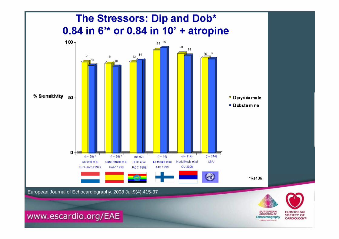
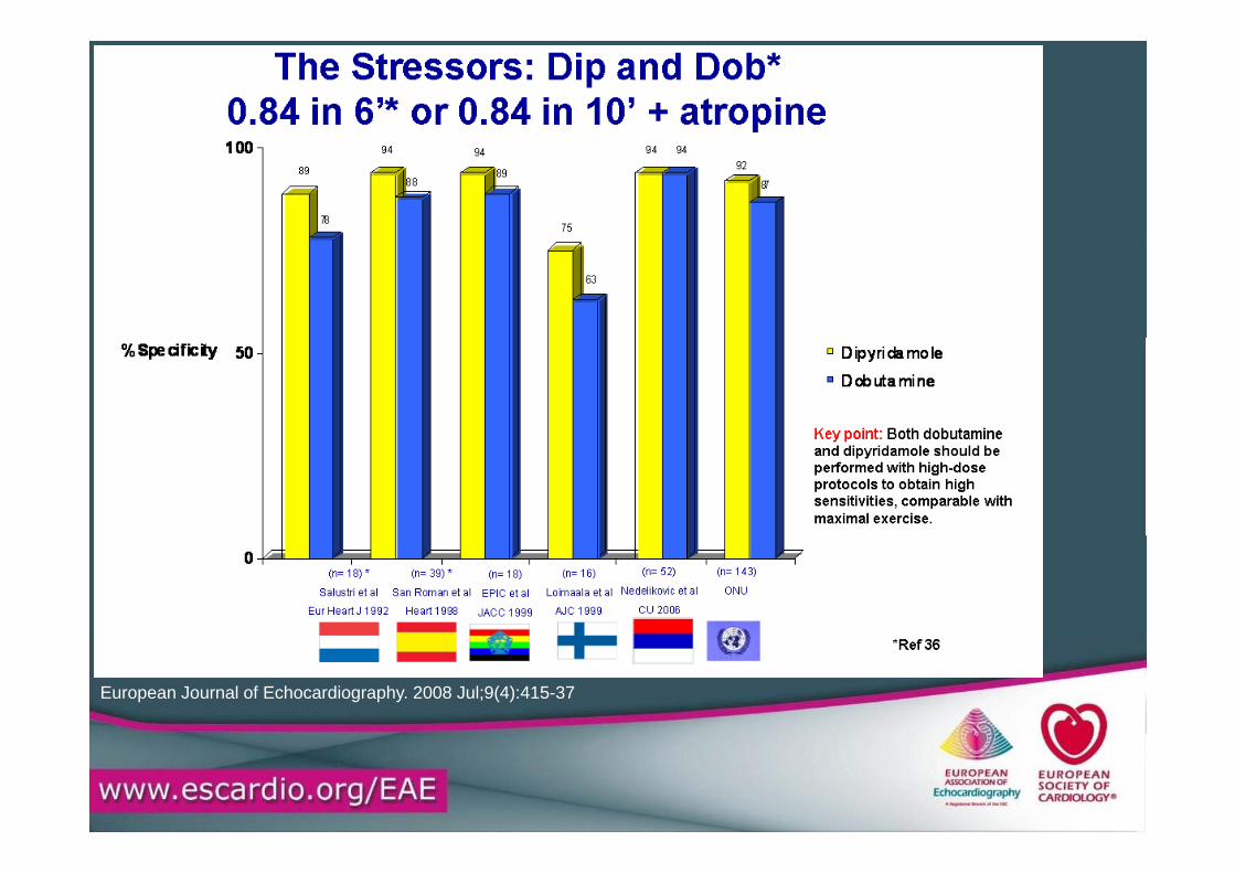
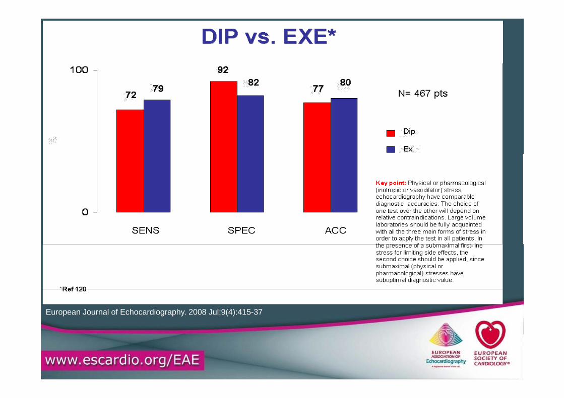
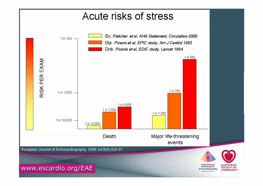
European Journal of Echocardiography. 2008 Jul;9(4):415-37
European Journal of Echocardiography. 2008 Jul;9(4):415-37
European Journal of Echocardiography. 2008 Jul;9(4):415-37
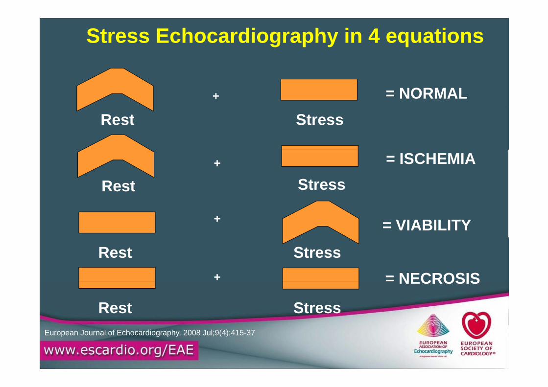
Stress Echocardiography in 4 equations
= NORMAL+
Rest Stress
Rest Stress= ISCHEMIA+
= VIABILITY+
Rest Stress= NECROSIS+
Rest Stress
NECROSIS
European Journal of Echocardiography. 2008 Jul;9(4):415-37
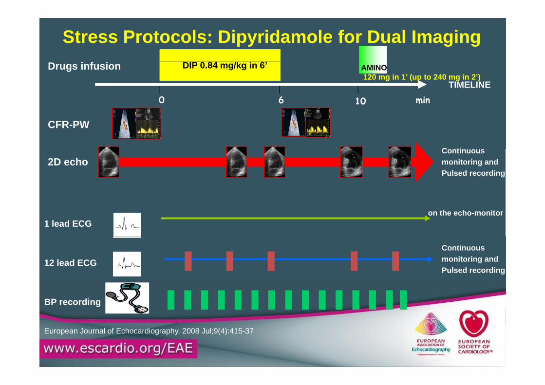
Stress Protocols: Dipyridamole for Dual Imaging
0 6 10 min
DIP 0.84 mg/kg in 6’
TIMELINE
Drugs infusion AMINO 120 mg in 1’ (up to 240 mg in 2’)
CFR-PW
C ti2D echo
Continuous monitoring and Pulsed recording
1 lead ECGon the echo-monitor
12 lead ECGContinuous monitoring and Pulsed recording
BP recording
European Journal of Echocardiography. 2008 Jul;9(4):415-37

Stress Protocols: Dobutamine
European Journal of Echocardiography. 2008 Jul;9(4):415-37
European Journal of Echocardiography. 2008 Jul;9(4):415-37
European Journal of Echocardiography. 2008 Jul;9(4):415-37
Indications for Stress Echo
1 – Coronary artery disease
2 – Prognosis and risk stratification in patients with established diagnosis
3 – Preoperative risk assessment
4 E l ti f di ti l f ti l d4 – Evaluation of cardiac etiology of exertional dypnea
5 – Evaluation after revascularization
6 - Ischemia location
7 – Evaluation of heart valve stenosis severity7 – Evaluation of heart valve stenosis severity
Indications for Stress EchoKey point: Stress echocardiography should not be used as a first-line imaging
technique for diagnostic and prognostic purposes in patients with known or
suspected coronary artery disease but only when exercise ECG stress test is p y y yeither
non-diagnostic or non-interpretable (e.g. for left bundle branch block or g p ( gpacemaker).
The less informative and/or interpretable exercise electrocardiography the higher is the
level of appropriateness to stress echocardiography.

Stress Echo Risk Titration of a Positive Test
European Journal of Echocardiography. 2008 Jul;9(4):415-37

Stress Echo Risk Titration of a Negative TestStress Echo Risk Titration of a Negative Test
European Journal of Echocardiography. 2008 Jul;9(4):415-37
European Journal of Echocardiography. 2008 Jul;9(4):415-37
Stress Echo:The Safety Rules
A id t i di ti
Stress Echo:The Safety Rules
• Avoid contraindications• Never exceed standard dosages• After signed informed consent• Always physician attending y p y g• Outpatients kept for 60’ after
testing• Indications must be class I• Ex whenever possible, Dip first p , p
choice for pharmacological testing
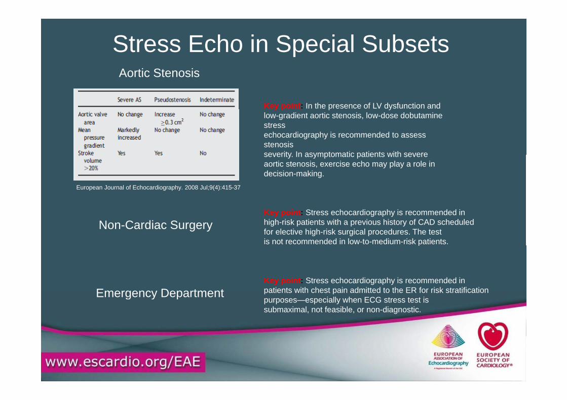
Stress Echo in Special Subsets
Key point: In the presence of LV dysfunction and
Aortic Stenosis
y p p ylow-gradient aortic stenosis, low-dose dobutamine stressechocardiography is recommended to assess stenosisseverity In asymptomatic patients with severeseverity. In asymptomatic patients with severe aortic stenosis, exercise echo may play a role in decision-making.
European Journal of Echocardiography. 2008 Jul;9(4):415-37
Non-Cardiac SurgeryKey point: Stress echocardiography is recommended inhigh-risk patients with a previous history of CAD scheduledfor elective high-risk surgical procedures. The testis not recommended in low-to-medium-risk patients.p
Emergency DepartmentKey point: Stress echocardiography is recommended inpatients with chest pain admitted to the ER for risk stratificationEmergency Department patients with chest pain admitted to the ER for risk stratificationpurposes—especially when ECG stress test issubmaximal, not feasible, or non-diagnostic.

Stress Echo vs. Competing TechniquesKey point: Stress echocardiography should be preferred due to it lower cost, wider availability and—most importantly— for the
f Sradiation-free nature. Stress scintigraphy offers similar information to stress echocardiography,b t ith di ti b d b t 600but with a radiation burden between 600 and 1300 chest X-rays for every single stress scintigraphy. This poses a significant biological risk both for the individual and forbiological risk both for the individual and for the society, since small individual risks multipliedby millions stress tests per year become aby millions stress tests per year become a significant population burden.
“When similar information is obtained with ionizing European Journal of Echocardiography. 2008 Jul;9(4):415-37
and non- ionizing techniques, the latter should be employed”
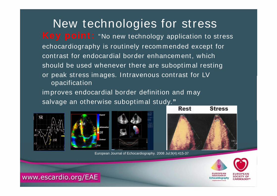
New technologies for stressKey point: “No new technology application to stressechocardiography is routinely recommended except for
t t f d di l b d h t hi hcontrast for endocardial border enhancement, whichshould be used whenever there are suboptimal restingor peak stress images Intravenous contrast for LV or peak stress images. Intravenous contrast for LV
opacificationimproves endocardial border definition and maysalvage an otherwise suboptimal study.”
European Journal of Echocardiography. 2008 Jul;9(4):415-37
European Journal of Echocardiography. 2008 Jul;9(4):415-37
European Journal of Echocardiography. 2008 Jul;9(4):415-37


![WAC 415 - 02 CHAPTER - Washingtonleg.wa.gov/CodeReviser/WACArchive/Documents/2015/WAC 415 - 02... · (2/27/14) [Ch. 415-02 WAC p. 1] Chapter 415-02 Chapter 415-02 WAC GENERAL PROVISIONS](https://img.pdfslide.us/doc/110x75/5ad016617f8b9aca598d40d7/wac-415-02-chapter-415-0222714-ch-415-02-wac-p-1-chapter-415-02.jpg)