Embed Size (px)
Citation preview

B R I E F I N G S
Issue 2
EUROPEANMUSCULOSKELETALREVIEW 2007
www.touchbriefings.com
EXTRACT
Vertebral AxialDecompression
a report by
Frank T i laro
Doctor of Internal Medicine, Utah

a report by
Frank T i laro
Doctor of Internal Medicine, Utah
Reduction of a nuclear protrusion by spinal distraction was practiSed
even before the intervertebral disc was recognised. A 14th-century
translation of Albucasis’s Surgery illustrates lumbar manipulation
during spinal traction.1 Apollonius of Kitium describes a form of
distraction 2,000 years ago. Guidi (1544) illustrates a traction table in
his Chirugia, and one of his tables can be found in the Wellcome
Historical Museum of London. In their book on manipulation past and
present, Cyriax and Schotz2 illustrate the employment of traction by
Hippocrates (400 BCE), Galen (131–202 CE) and the Spanish-Arabian
physician Abu’L Qasim (1013–1106 CE).
Today, two methods of performing traction are practised: the sustained
manner, preferred by Cyriax, and various forms of intermittent traction.
Intermittent traction can be performed electronically, manually (by a
therapist) or by the patient (autotraction). The effects of sustained
traction have been investigated. An increase in body length of
10–30mm was demonstrated in healthy males when a sustained force
of 60kg was applied for one hour, and was lost at 4mm/hr.3 In the
excised spine the greatest separation was in those subjects with wide
disc spaces, and the least in those with evidence of disc degeneration.
Other investigators confirmed an increase in stature over and above
that known to occur when the load is taken off the spine by lying
down.4 The findings suggest that most of the vertebral separation takes
place within the first 30 minutes. During normal traction, the
enlargement between two consecutive lumbar end-plates is between
1 and 1.5mm. Other studies have demonstrated a widening of
the lumbar intervertebral space of between 3 and 8mm, measured
radiographically during gravitational traction.5,6 Anderson et al.7
have shown an increase in intradiscal pressure with certain
traction techniques.
The heavy lumbar paravertebral muscles exert resistance to distraction.
At least 30–35kg of force is required to influence the lumbar spine.5
Other studies have shown that a force of at least 25% of bodyweight
is necessary to achieve lumbar distraction. With the split table,
designed by Dr Allan Dyer, it is estimated that 25% of the traction
force is required for distraction to occur.8
The effects of distraction include tautening of the posterior
longitudinal ligament, which exerts a centripetal force at the back of
the joint. This manoeuvre may be of therapeutic value, particularly
if the protrusion is located anterior to and remains in close contact
with the ligament. On the basis of biomechanical calculations,
significant intradiscal negative pressure may be achieved during
sustained traction.9 One study has shown that a traction load of 30kg
caused the intradiscal pressure to drop from 30 to 10kp in the L3
intervertebral disc.10 Improvement in nutrition, deposition of reparative
collagen and healing of annular tears and fissures have all been
suggested as benefits of axial distraction.
Dr Allan Dyer, former Deputy Minister of Health from Ontario, Canada,
and a pioneer in the development of the external cardiac defibrillator,
designed the vertebral axial decompression (VAX-D) therapeutic table
to apply distraction tension to the patient’s lumbar spine without
eliciting reflex paravertebral muscle contractions. A patented harness
is attached to a tensiometer during separation of the movable part of
the table. The distraction–relaxation cycles are automated or variably
timed. Distraction tensions and rates are continuously monitored and
measured by the tensiometer, and the output is shown on a digital
gauge and captured on a pen-write printout.
Procedure
The VAX-D table utilises pneumatic cylinders coupled with hydraulic
damping as the drivel-damping mechanism for the pre-tension and
therapeutic programme. The technology applies and maintains a
baseline tension of 20–24lb (the pre-tension) to the patient’s pelvis
throughout the treatment session (even during the rest periods), and
the distraction cycles then move from tile-pre-tension range up to a
pre-selected therapeutic tension. The above parameters are absolutely
critical to the success of the treatment. The pneumatic hydraulic
cylinders separate the lower table section from the upper section and
apply the tensions to the patient’s pelvis. The pneumatic hydraulic
drive mechanism allows precise control of the amount of tension and
is able to apply tensions in a logarithmic time/force curve. The
pneumatic hydraulic drive mechanism is applied in both the distraction
and retraction movements of the VAX-D table and provides a smooth,
controlled operation with gradual return of the patient to the starting
position each time. To achieve optimum control of the application of
distractive tensions, it was found essential to develop a harness that
would attach directly to an electronic tensiometer, which continuously
monitors and provides feedback of the tensions being applied to the
Vertebral Axial Decompression
E U R O P E A N M U S C U L O S K E L E T A L R E V I E W 2 0 0 7 E X T R A C T W W W . T O U C H B R I E F I N G S . C O M
Spinal Decompression
2
The effects of distraction include
tautening of the posterior longitudinal
ligament, which exerts a centripetal
force at the back of the joint.
3E U R O P E A N M U S C U L O S K E L E T A L R E V I E W 2 0 0 7 E X T R A C T W W W . T O U C H B R I E F I N G S . C O M
Vertebral Axial Decompression
spinal column. The harness design also facilitates proper placement,
which is necessary to attain reproducible results.
Patients with discogenic low-back pain – with or without radiculopathy –
who have failed conventional therapy become candidates for VAX-D
therapy after six to eight weeks.11 Patients with neurological deficits are
also candidates since outcome studies have shown no difference with
surgical or medical management.12 Patients with fusion or failed back
surgery syndrome are also candidates.
Contraindications for VAX-D therapy include infection, neoplasm,
osteoporosis, bilateral pars defect, unstable grade 2 spondylolisthesis,
fractures and the presence of surgical hardware in the spine.11 The
patient should be evaluated by a therapist or physician prior to
initiating therapy, and routine spine films are necessary to rule out any
contraindications. A computed tomography (CT) or magnetic
resonance imaging (MRI) scan is not a pre-requisite before therapy, but
most patients have undergone neuroimaging. A trained VAX-D
technician administers the daily therapy for approximately 20 sessions.
An occasional patient may require a short maintenance period in
which two to three treatments a week are given for two to four weeks
following initial therapy. The average patient has required 20–25
sessions. Each session is 15 cycles, each cycle being one minute in
distraction and one minute in relaxation.
Patients are instructed to wear loose clothing for each treatment.
The patient is placed prone on the table so that the superior border
of the pelvic harness is at the level of the split. The patient then grasps
the adjustable handgrips, which are positioned to ensure the arms
remain straight without bending the elbows. A roll is placed under the
patient’s ankles – a chin or forehead roll is optional. Patients who have
difficulty lying prone can use a pillow placed under the abdomen.
Patients with shoulder pathology may employ a roll under the axilla.
The patients are instructed to hold tightly to the handgrips, since
motion artifact can be seen on the graph printout if the patients are
pulling with their arms. This manoeuvre inhibits decompression.
Patients are allowed to release their grip during the relaxation phase.
A pre-tension level of 20lb is set and maintained throughout the
resting phase. Ramos13 demonstrated that 50lb of tension was the
threshold tension necessary to develop negative intradiscal pressures.
Women start with 50Ib and work up to 70lb. Men usually start at 60lb
and work up to 80lb. Tension increments are in the order of 5lb every
three to four days, although some patients need to proceed more
slowly. Tension should remain constant for each treatment cycle
(see Figure 1).
If the centralisation phenomenon – the movement of pain pattern
from a distal to a more proximal location – occurs in the early
treatment stages, the patient will most likely respond to physical
therapy and not require further VAX-D. Centralisation may appear at
a later stage of treatment or shortly after completing a full VAX-D
course. In patients with an intact annulus, no researcher has yet
reported on the results of CT discography prior to treatment and
following the onset of centralisation.14
Pain during distraction that lessens with relaxation is probably due to
stretching shortened tissue. If pain persists for more than 30 minutes
after treatment, the tension should be reduced for the next few
sessions. The tension should be lowered or the treatment cycle stopped
for pain that increases with each two-minute cycle. Some patients
require a two- to three-day hiatus from therapy if they have too much
discomfort. The daily response to treatment and any changes made are
recorded in the patient’s chart and reviewed by the physician and
technician every few days.
Patients are encouraged to remain active, but should not engage in
strenuous activities while undergoing therapy. They should not be
receiving any other treatment modalities while receiving VAX-D
therapy. Patients may wear a back support after therapy, but it should
be removed within one to two hours. Once the VAX-D course is
completed, patients are encouraged to enter some form of
rehabilitation programme and learn proper biomechanics.
Discussion
Ramos and Mart13 studied intradiscal pressures during VAX-D
treatment. Five cases with subligamentous disc herniation at L4–5 –
confirmed by MRI and scheduled for percutaneous discectomy – were
chosen. Using lateral and anteroposterior (AP) fluoroscopy, a cannula
was inserted into the nucleus pulposus of the L4–5 intervertebral disc.
The pressure measurements were recorded by an Ohmeda pressure
transducer connected to a Hewlett-Packard pressure monitor via a
saline bridge and a Camino fibre optic intracranial transducer, adapted
for intradiscal measurements. Since the pressure transducers were
designed to measure changes in the positive range, calibration was
necessary. The pressure transducer and monitor for each patient were
individually calibrated, and a correction curve was plotted showing
the transducer readings versus actual pressures to correct for the
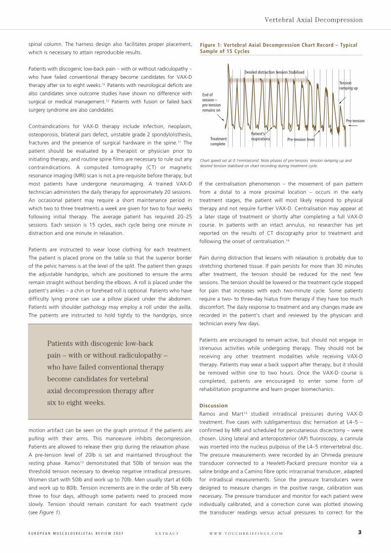
Desired distraction tension Stabilised
End ofsession – pre-tensionremains on
Treatmentcomplete
Tensionramping up
Pre-tension
Patient’srespirations Pre-tension fever
Figure 1: Vertebral Axial Decompression Chart Record – TypicalSample of 15 Cycles
Chart speed set at 0.1mm/second. Note phases of pre-tension, tension ramping up anddesired tension stabilised on chart recording during treatment cycle.
Patients with discogenic low-back
pain – with or without radiculopathy –
who have failed conventional therapy
become candidates for vertebral
axial decompression therapy after
six to eight weeks.
4 E U R O P E A N M U S C U L O S K E L E T A L R E V I E W 2 0 0 7 E X T R A C T W W W . T O U C H B R I E F I N G S . C O M
Spinal Decompression
non-linearity of the instrumentation in the range of the negative
pressures achieved. A pneumatic calibration analyser was employed.
Distraction tensions ranging from 50 to 100lb were monitored on a
digital read-out and recorded on a continuous graph tracing by a chart
printer incorporated in the control console. Intradiscal pressure changes
were observed as a digital read-out on the pressure monitor. Intradiscal
pressures were significantly reduced to negative levels, ranging from a
negative 100mmHg to a negative 160mmHg. Changes in intradiscal
pressure were minimal until a threshold distraction tension was reached.
The relationship between percentage maximum tension and time was a
logarithmic function. If one plots the percentage of the maximum
tension reached in 60 seconds versus time, it takes 17–20 seconds to
reach 50%, 25–28 seconds to reach 70% and 42–45 seconds to attain
90% of the maximum. The retraction phase followed a linear time–
tension relationship and returned to baseline in 25–30 seconds.
The first large-scale retrospective study15 involved over 700 patients
with low-back pain – with and without radicular symptoms. Over 70%
achieved a positive outcome. Even though the study was not a
randomised blinded trial, the majority of patients were suffering
beyond the period where natural resolution would be expected. All
had failed treatment with other modalities and demonstrated a
positive response during treatment and/or immediately thereafter.
Sherry et al.16 conducted a prospective, randomised controlled trial of
VAX-D versus transcutaneous electrical neural stimulation (TENS). All
patients had chronic symptoms (with the average duration of pain
being 7.3 years). TENS was regarded as a placebo. The data revealed
an attributable success rate of 68.4% for VAX-D, significantly superior
compared with TENS (p<0.001).
A study by Ramos13 compared the effects of a subtherapeutic treatment
versus the protocol treatment. All patients had symptoms of sciatica and
were referred to a neurosurgeon after failing conventional therapy. Imaging
studies and the clinical examination were concordant. The protocol group
showed significantly superior results compared with the subtherapeutic
treatment group. Two similar studies evaluating the effect of VAX-D on
sensory nerve dysfunction in cases of low-back pain came to similar
conclusions.17,18 Either a current perception threshold neurometer or
dermatomal somatosensory-evoked potentials protocol was employed. Both
studies demonstrated that VAX-D was capable of positively influencing
sensory nerve dysfunction associated with compressive radiculopathy.
Although compression is a frequent finding in sciatica, compression does
not explain all the observed symptomatology. Other factors include the
force and rapidity of compression, the effect on arterial and venous
circulation and the release of pain, vascular and neural modulators – nitrous
oxide, phospholipase A2, the prostaglandins and leukotrienes.19–22
Summary
VAX-D should not be considered as traction in the traditional sense, but
as decompression: it is the only non-invasive treatment that has been
proved to decompress only the disc. With other traction devices, there
has been indirect proof. The patented therapeutic curve demonstrates
that, when time is plotted against force, one observes a logarithmic
function. Conventional traction devices have a linear time–force
relationship. Non-steroidal anti-inflammatory drugs, steroids and
doxycycline have been given in conjunction with VAX-D therapy to study
possible diffusion into the disc and any beneficial effects. Other concepts
for the future include investigation of immunomodulators, transplanting
live fibroblast and chondrocytes and minimally invasive surgical
techniques in conjunction with VAX-D. The current focus may shift from
treating back pain to repair and healing of the damaged disc. ■
1. MacKinney L, Medical I l lustrations in Medieval Manuscripts,Wellcome, 1965.
2. Schotz EH, Manipulaasjonsbehandling av Columna underMedisinkhistorisk Synsvinkel Tidsskr, Norske Laegeforen, 1958.
3. Worden RE, Humphrey TL, Effect of spinal traction on length ofbody, Arch Phys Med, 1964;45:318.
4. Bridger RS, Ossey S, Furrie G, Effect of lumbar traction onstature, Spine, 1990;15:522–4.
5. Janke AW, Kerkow TA, Griffiths HJ, et al., The biomechanics ofgravity dependent traction of the lumbar spine, Spine, 1997;22:253–60.
6. Tekeoglu I, Adak B, Borzkust M, et al., Distraction of lumbarvertebrae in gravitational traction, Spine, 1998;23:1061–3.
7. Anderson GBJ, Schultz AB, Nachemson AL, Intervertebral discpressure during traction, Rehabil Supp, 1983;9:88–91.
8. Guenet RJ, Hadler NM, Diagnosis and treatment of backache,Semin Arth Rheum, 1979;8:261–7.
9. Fast A, Low back disorders: conservative management, ArchPhys Med Rehabil, 1998;69:880–91.
10. Nachemson A, Elfstrom G, Intravital dynamic pressuremeasurements in lumbar disc, Scand J Rehabil Med,1970;1(Suppl.):5–40.
11. Tilaro F, An overview of vertebral axial decompression, Can JClin Med, 1998;1:2–8.
12. Haklius A, Prognosis in sciatica, Acta Orthop Scand,1970;129(Suppl.):1–76.
13. Ramos G, Marti W, Effects of vertebral axial decompression onintradiscal pressure, J Neurosurg,1994;81:350–53.
14. Donelson R, Aprill C, Medcalf R, et al., A prospective study ofcentralization of lumbar and referred pain, Spine, 1997;22:1115–22.
15. Gose E, Naguszewski P, Naguszewski W, Vertebral axialdecompression therapy for pain associated with herniated ordegenerated disc or facet syndrome: an outcome study, J NeuroRes, 1998;20:186–90.
16. Sherry E, Kitchener P, Smart R, A prospective randomizedcontrolled study of VAX-D and TENS for the treatment ofchronic low back pain, J Neuro Res, 2001;23:780–84.
17. Naguszewski W, Naguszewski R, Gose E, Dermatosomalsomatosensory evoked potential demonstration of nerve rootdecompression after VAX-D therapy, J Neuro Res, 2001;23:706–14.
18. Tilaro F, Miskovich D, The effects of vertebral axialdecompression on sensory nerve dysfunction, Can J Cl in Med,1999;6:2–7.
19. Brisby H, Byrod G, Olmarker K, et al., Nitric oxide as a mediatorof nucleus pulposus induced effects on spinal nerve roots, J Orthop Res, 2000;18:815–20.
20. Garfin SR, Rydevik B, Lind B, et al., Spinal nerve rootcompression, Spine, 1995;20:1810–20.
21. Rydevik B, Brown M, Lundborg G, Pathoanatomy andpathophysiology of nerve root compression, Spine, 1984;9:7–15.
22. Saal JA, Dobrow R, Saal JF, et al., High levels of inflammatoryphospholipase A2 activity in lumbar disc herniations, Spine,1990;15:674–8.
Although compression is a frequent
finding in sciatica, compression does not
explain all the observed symptomatology.
Vertebral axial decompression
should not be considered as traction
in the traditional sense, but as
decompression: it is the only non-
invasive treatment that has been
proved to decompress only the disc.


![European Journal of Radiology Open · European Journal of Radiology Open 3 (2016) 182–190 Contents lists available at ScienceDirect ... and musculoskeletal-imaging) [9,10], there](https://img.pdfslide.us/doc/110x75/5f03e4577e708231d40b47eb/european-journal-of-radiology-open-european-journal-of-radiology-open-3-2016-182a190.jpg)