Embed Size (px)
Citation preview
MOLECULAR AND CELLULAR BIOLOGY,0270-7306/00/$04.0010
Jan. 2000, p. 468–477 Vol. 20, No. 2
Copyright © 2000, American Society for Microbiology. All Rights Reserved.
Eukaryotic Translation Initiation Factor 4E (eIF4E) BindingSite and the Middle One-Third of eIF4GI Constitute the CoreDomain for Cap-Dependent Translation, and the C-Terminal
One-Third Functions as a Modulatory RegionSHIGENOBU MORINO,1 HIROAKI IMATAKA,1 YURI V. SVITKIN,1 TATYANA V. PESTOVA,2
AND NAHUM SONENBERG1*
Department of Biochemistry and McGill Cancer Center, McGill University, Montreal, Quebec H3G 1Y6, Canada,1 andDepartment of Microbiology and Immunology, State University of New York Health Science Center at Brooklyn,
Brooklyn, New York 112032
Received 30 August 1999/Accepted 6 October 1999
The mammalian eukaryotic initiation factor 4GI (eIF4GI) may be divided into three roughly equal regions;an amino-terminal one-third (amino acids [aa] 1 to 634), which contains the poly(A) binding protein (PABP)and eIF4E binding sites; a middle third (aa 635 to 1039), which binds eIF4A and eIF3; and a carboxy-terminalthird (aa 1040 to 1560), which harbors a second eIF4A binding site and a docking sequence for the Ser/Thrkinase Mnk1. Previous reports demonstrated that the middle one-third of eIF4GI is sufficient for cap-independent translation. To delineate the eIF4GI core sequence required for cap-dependent translation,various truncated versions of eIF4GI were examined in an in vitro ribosome binding assay with b-globinmRNA. A sequence of 540 aa encompassing aa 550 to 1090, which contains the eIF4E binding site and themiddle region of eIF4GI, is the minimal sequence required for cap-dependent translation. In agreement withthis, a point mutation in eIF4GI which abolished eIF4A binding in the middle region completely inhibitedribosomal binding. However, the eIF4GI C-terminal third region, which does not have a counterpart in yeast,modulates the activity of the core sequence. When the eIF4A binding site in the C-terminal region of eIF4GIwas mutated, ribosome binding was decreased three- to fourfold. These data indicate that the interaction ofeIF4A with the middle region of eIF4GI is necessary for translation, whereas the interaction of eIF4A with theC-terminal region plays a modulatory role.
All eukaryotic cellular (except organellar) mRNAs possess acap structure (m7GpppN, where N is any nucleotide) at the 59end. The cap structure is bound by translation initiation factor4F (eIF4F) as the first step of cap-dependent translation.eIF4F consists of three subunits, eIF4E, eIF4A, and eIF4G.eIF4E binds the cap structure directly and consequently isrequired for cap-dependent translation. eIF4A exhibits RNA-dependent ATPase activity and ATP-dependent RNA helicaseactivity. The helicase activity is though to be required for themelting of mRNA 59 untranslated region secondary structureto facilitate ribosome binding (for reviews, see references 5, 19,and 28). eIF4G is an adapter protein with a modular structure.It bridges the ribosome to the mRNA via eIF3 (for reviews, seereferences 20, 22, and 27). The human eIF4G may be dividedinto three distinct functional domains. The N-terminal one-third (amino acids [aa] 1 to 634) contains the eIF4E bindingsite (14, 18) and a recently described poly(A) binding protein(PABP) binding site (13). The middle third (aa 635 to 1039)possesses eIF3 and eIF4A binding sites (12) as well as an RNAbinding site (6, 24). The C-terminal third (aa 1040 to 1560)contains a second eIF4A binding site (12, 14) and a Mnk1binding site (26, 30). The middle third of eIF4G is sufficient forcap-independent binding of ribosomes to the encephalomyo-carditis virus (EMCV) internal ribosomal entry site (IRES)(24) and for cap-independent but 59-end-dependent transla-
tion (2). The function of the C-terminal region of humaneIF4G is unclear.
The eIF4G-related mammalian protein p97/NAT1/DAP-5(11, 16, 31), which is homologous to the carboxyl two-thirds ofeIF4G, does not contain an eIF4A binding site in its C-termi-nal region (12). While the middle region is phylogeneticallyconserved, the C-terminal one-third is not. The wheat eIF4Ghomolog possesses a much shorter C terminus (1), and theSaccharomyces cerevisiae eIF4G homolog does not possess asequence with homology to the mammalian C-terminal third(6).
In this study, using a toeprinting technique with the b-globinmRNA as a template (25), we define the region beginning atthe eIF4E binding site and encompassing the middle third ofeIF4G as the minimum sequence required for cap-dependent40S ribosome binding. Our data provide evidence that theC-terminal region plays a modulatory role.
MATERIALS AND METHODS
Construction of plasmids. Plasmids pcDNA3-HA (hemagglutinin), pcDNA3-FLAG, and pcDNA3-GST (glutathione S-transferase) plasmids (13) were usedas vectors for expression in HeLa cells. pBlueBacHis (Invitrogen) was used forgenerating recombinant baculoviruses expressing His-tagged eIF4GI and mu-tants in Sf9 cells. To construct pBlueBacFLAG, the His tag sequence in pBlue-BacHis was replaced with the FLAG sequence. Deletion and point mutants ofeIF4GI were generated using PCR with primers containing EcoRI (59 primer)and XhoI (39 primer) restriction enzyme sequences. All constructs were se-quenced in their entirety.
Antibodies. Anti-hPrt1 (human Prt1), anti-eIF4GI(1–329), and anti-GST an-tisera were obtained by immunizing rabbits with GST-hPrt1(147–209), GST-eIF4GI(1–329), and GST, respectively. Anti-HA and anti-His6 monoclonal an-tibodies were obtained from BAbCo. Anti-Xpress and anti-FLAG monoclonal
* Corresponding author. Mailing address: Department of Biochem-istry and McGill Cancer Center, McGill University, Montreal, QuebecH3G 1Y6, Canada. Phone: (514) 398-7274. Fax: (514) 398-1287. E-mail: [email protected].
468
on February 19, 2018 by guest
http://mcb.asm
.org/D
ownloaded from
antibodies were obtained from Invitrogen and Sigma, respectively. Monoclonalanti-eIF4A was a kind gift from H. Trachsel.
Purification of recombinant proteins. His-eIF1, His-eIF1A, His-eIF4A, andHis-eIF4E were expressed in Escherichia coli BL21(DE3) and purified by Ni-agarose (Qiagen) chromatography as described previously (23, 25). Baculovi-ruses expressing FLAG-eIF4B or His- or FLAG-eIF4GI proteins were generatedby using the pBlueBac baculovirus expression system (Invitrogen). Log-phase Sf9cells (200 ml of 2 3 106 cells/ml) were infected with a recombinant virus atmultiplicity of infection of 5 and cultured for 40 h at 27°C. Expressed proteinswere purified by Ni-agarose (Qiagen) or anti-FLAG-agarose (Sigma) chroma-tography. Concentrations of recombinant proteins were determined by compar-ison with standard bovine serum albumin (Ambion) following sodium dodecylsulfate (SDS)-polyacrylamide gel electrophoresis (PAGE) and staining withCoomassie blue. Sf9 cell proteins copurified with His-tagged proteins were notconsidered to affect the functional analyses, because nickel resin-bound proteinspurified from uninfected Sf9 cells did not inhibit or enhance ribosome bindingactivity or in vitro translation (data not shown).
40S ribosomal subunits, eIF2, eIF3, and eIF4F were purified from ribosomalpellets of Krebs cell extracts or rabbit reticulocyte lysate as described previously(23).
Assembly and toeprint analysis of 48S complexes. Reaction mixtures (40 ml)containing native a- and b-globin mRNA (Life Technologies) (0.3 mg), His-eIF1(0.5 mg), His-eIF1A (0.5 mg), eIF2 (3 mg), eIF3 (7 mg), FLAG-eIF4B (1 mg),Met-tRNAi
Met (4 pmol), 40S ribosomal subunits (4 pmol), His- or FLAG-eIF4GI(2 mg, unless indicated), His-eIF4E (0.3 mg), and His-eIF4A (3 mg) were incu-bated in buffer (2 mM dithiothreitol, 100 mM KCl, 20 mM Tris-HCl [pH 7.6], 2.5mM magnesium acetate, 100 U of RNasin [Promega], 1 mM ATP, 0.4 mMguanylyl imidodiphosphate [GMP-PNP], 250 mM spermidine) for 5 min at 30°C.Following the addition of 4 pmol of oligonucleotide 59-GCATTTGCAGAGGACAGG-39 (complementary to b-globin positions 177 to 194), incubation wascontinued for 3 min at 30°C, and the mixture was then placed on ice. For reversetranscriptase reactions, the mixture was incubated for 40 min at 30°C afteraddition of 1 ml of magnesium acetate (320 mM), 4 ml of deoxynucleosidetriphosphate mix solution (5 mM dCTP, dGTP, and dTTP; 1 mM dATP), 1 ml of[a-32P]dATP (6000 Ci/mmol; DuPont, NEN), and 15 U of avian myeloblastosisvirus reverse transcriptase (Amersham Pharmacia Biotech Inc.). cDNA productswere extracted with phenol-chloroform (1:1) and precipitated with ethanol (25).The same primer was used for sequencing of plasmid pBS2(b-globin), harboringb-globin cDNA (10). The products of primer extension and sequence productswere resolved side by side on a sequencing gel. In the relevant figures, thefull-length cDNA product and the toeprint product are marked “E” and “Com-plex II,” respectively. The intensity of E and complex II was quantitated byBAS-2000 phosphorimager (Fuji). The relative efficiency of complex II forma-tion was calculated as complex II/complex II 1 E. Values represent the meansand standard errors of three independent experiments.
Protein immunoprecipitation. HeLa R19 cells (6-cm-diameter dish) were in-fected with vaccina virus vTF7-3 (3) for 1 h and then transfected with plasmidsby using Lipofectin (Gibco BRL). Sixteen hours later, cells were lysed in 400 mlof buffer A (20 mM HEPES-KOH [pH 7.6], 100 mM KCl, 0.5 mM EDTA, 20%glycerol) containing 0.5% Triton X-100, 50 mg of RNase A per ml, and proteaseinhibitor cocktail (Boehringer Mannheim). After centrifugation, the supernatantwas mixed with anti-HA or anti-FLAG antibody (2 mg) immobilized on proteinG-Sepharose (10 ml) and incubated for 4 h at 4°C. After being washed with bufferA (400 ml, three times), bound proteins were dissolved in Laemmli buffer. Thesamples were resolved by SDS-PAGE and analyzed by Western blotting. Proteinbands were visualized with an enhanced chemiluminescence detection system(Boehringer Mannheim).
To coprecipitate FLAG-Mnk1 with GST fusion proteins, cell extracts express-ing FLAG-Mnk1 and a GST fusion protein were incubated with glutathione-Sepharose beads (15 ml; Amersham Pharmacia Biotech Inc.) for 4 h at 4°C. Afterbeing washed with buffer A containing 0.1% Triton X-100 (400 ml, three times),bound proteins were eluted with a buffer (40 ml) containing 20 mM reducedglutathione, 50 mM Tris-HCl (pH 8.0), and 100 mM KCl.
In vitro translation. Rabbit reticulocyte lysate (Promega) was treated withrhinovirus 2Apro (40 mg/ml) for 5 min at 30°C (7), followed by incubation for 10min on ice with 0.8 mM elastatinal (Sigma). Aliquots (12.5 ml) were supple-mented with eIF4E (0.2 mg) and/or wild-type or mutant eIF4GI (1 mg, unlessindicated) and programmed for translation with 0.1 mg of capped bicistronicpGEMCAT/EMC/LUC mRNA (7) in the presence of [35S]methionine. Trans-lation reaction mixtures were incubated at 30°C for 60 min and analyzed bySDS-PAGE (12.5% gel). Gels were fixed with 40% methanol–7% acetic acid,treated with En3Hance (Dupont, NEN) and processed for autoradiography. Theintensity of the bands was determined with a BAS-2000 phosphorimager.
In vitro protein binding assay. A His-tagged protein (5 mg) was incubated withFLAG-eIF4A (4 mg or 8 mg) immobilized on anti-FLAG agarose resin in bufferA (50 ml) containing 0.1% Triton X-100 for 10 min on ice. The resin was washedwith buffer A containing 0.1% Triton X-100 (400 ml, three times) and dissolvedin Laemmli buffer. The sample was resolved by SDS-PAGE followed by Westernblotting.
RESULTS
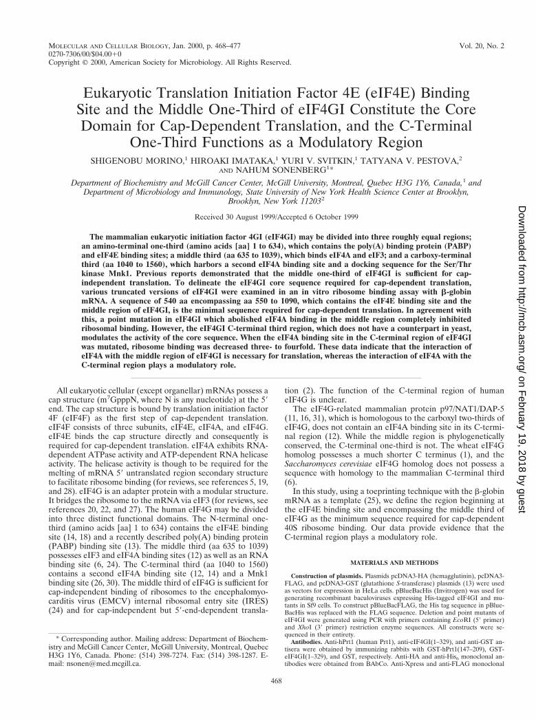
The eIF4E binding site and middle third of eIF4G are nec-essary and sufficient for cap-dependent translation. The to-eprinting analysis has proven extremely useful for the study oftranslation initiation (23, 25). In the presence of b-globinmRNA (a typical cap-dependent mRNA), Met-tRNA, ATP,40S ribosomal subunits, eIF1, eIF1A, eIF2, eIF3, eIF4B,eIF4A, and eIF4F, a 48S ribosomal complex is formed on theinitiation codon of the mRNA. No signal was detected in thepresence of mRNA, Met-tRNA, and ATP alone (Fig. 1, lane1). The ribosomal complex is detected by primer extension asa toeprint 15 to 17 nucleotides downstream from the initiationcodon (25) (lane 2). This toeprint is termed complex II (25).To study the function of eIF4GI in cap-dependent translation,eIF4F was replaced by a combination of recombinanteIF4GI(157–1560), eIF4A, and eIF4E. Complex II was formedwith these three recombinant proteins as efficiently as witheIF4F (compare lane 3 to lane 2). Control experiments inwhich eIF4A or eIF4E were omitted were performed. ComplexII was not detectable in the absence of eIF4A (lane 4), con-firming the importance of eIF4A for 48S ribosomal complexformation. However, the 48S complex was formed at the cor-rect position in the absence of eIF4E (lane 5), albeit with muchlower efficiency (compare lane 5 to lane 3). Complex II was notformed, however, when eIF4GI(157–1560) was omitted (lane6). These results confirm that eIF4G is essential for 48S ribo-somal complex formation, and they validate the use of recom-binant eIF4G in this assay system.
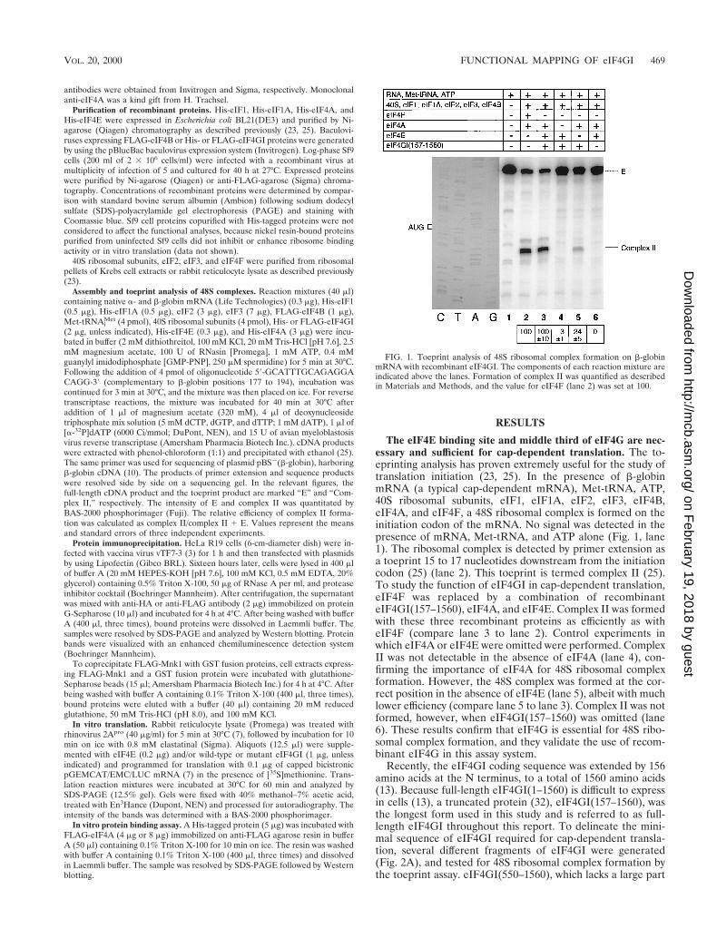
Recently, the eIF4GI coding sequence was extended by 156amino acids at the N terminus, to a total of 1560 amino acids(13). Because full-length eIF4GI(1–1560) is difficult to expressin cells (13), a truncated protein (32), eIF4GI(157–1560), wasthe longest form used in this study and is referred to as full-length eIF4GI throughout this report. To delineate the mini-mal sequence of eIF4GI required for cap-dependent transla-tion, several different fragments of eIF4GI were generated(Fig. 2A), and tested for 48S ribosomal complex formation bythe toeprint assay. eIF4GI(550–1560), which lacks a large part
FIG. 1. Toeprint analysis of 48S ribosomal complex formation on b-globinmRNA with recombinant eIF4GI. The components of each reaction mixture areindicated above the lanes. Formation of complex II was quantified as describedin Materials and Methods, and the value for eIF4F (lane 2) was set at 100.
VOL. 20, 2000 FUNCTIONAL MAPPING OF eIF4GI 469
on February 19, 2018 by guest
http://mcb.asm
.org/D
ownloaded from
of the N-terminal region but retains the eIF4E binding site,functioned as efficiently as control eIF4GI(157–1560) (Fig. 2B,compare lanes 3 and 5). However, deletion of the eIF4E bind-ing site markedly (80%) reduced cap-dependent 40S ribosomalbinding (compare lanes 5 and 6). The residual activity (20%) isconsistent with the background level of binding observed forcontrol eIF4GI in the absence of eIF4E (Fig. 1). The reasonsfor the residual activity will be addressed in Discussion.eIF4GI(157–1090), which lacks the C-terminal third, retained;60% of the activity of control eIF4GI (compare lanes 3 and4). Strikingly, efficient binding of 40S ribosomes (70% of con-trol) was achieved by an eIF4GI protein possessing only the
eIF4E binding site and the middle third (aa 550 to 1090), whichcontains binding sites for eIF3 and eIF4A (lane 7). In contrastto the results obtained for EMCV IRES RNA (24), the middledomain alone failed to support 40S ribosome binding to b-glo-bin mRNA (lane 8). These results were reproducible with awide concentration range (0.5 to 4 mg) of eIF4GI and itsmutants (data not shown).
To further substantiate these conclusions, we extended theexperiments to a rabbit reticulocyte lysate in vitro translationsystem. The lysate was pretreated with rhinovirus 2Apro tocleave the endogenous eIF4G. This treatment results in inhi-bition of cap-dependent translation and stimulation of IRES-
FIG. 2. Functional analysis of eIF4GI deletion mutants. (A) Schematic representation of eIF4GI deletion mutants. PABP, eIF4E, eIF4A, eIF3, and Mnk1 bindingsites are indicated. (B) Toeprint analysis of 48S ribosomal complex formation on b-globin mRNA with eIF4GI deletion mutants. The reaction components are indicatedabove the lanes. The value for eIF4GI(157–1560) (lane 3) was set at 100. (C) Analysis of eIF4GI deletion mutants in a reticulocyte lysate translation system. Translationwas performed as described in Materials and Methods. A rabbit reticulocyte lysate treated with rhinovirus 2Apro was supplemented with recombinant proteins asindicated and programmed for translation with the capped bicistronic mRNA CAT/EMCV IRES/LUC. For quantitation of luciferase (LUC) synthesis, the valueobtained for translation in untreated lysate in the absence of additional proteins (lane 1) was set at 100. For the quantitation of CAT synthesis, the value obtained fortranslation in the treated lysate in the presence of eIF4E alone was subtracted as background, and then the value for treated lysate translated in the presence of eIF4Eand eIF4GI(157–1560) (lane 4) was set at 100. (D) Western blotting of eIF4G deletion mutants. Recombinant protein preparations (;1 mg) containing the sameamount of eIF4GI according to Coomassie blue staining were subjected to SDS-PAGE (10% gel) and analyzed by Western blotting with anti-Xpress antibody (to detectthe epitope located between the His tag and eIF4G coding sequence) or with anti-FLAG antibody.
470 MORINO ET AL. MOL. CELL. BIOL.
on February 19, 2018 by guest
http://mcb.asm
.org/D
ownloaded from
dependent translation (7, 9). The 2Apro-treated lysate was pro-grammed with a bicistronic mRNA in which translation of thefirst cistron (chloramphenicol acetyltransferase [CAT]) is capdependent but translation of the second cistron (luciferase),which is preceded by the EMCV IRES, is cap independent (7).Treatment with 2Apro dramatically (85%) reduced cap-depen-dent translation, as expected (Fig. 2C, compare lane 2 to lane1). While addition of eIF4E alone failed to enhance cap-de-pendent translation of the CAT cistron (lane 3), addition ofeIF4E plus eIF4GI(157–1560) restored cap-dependent trans-lation to 65% of the untreated control level (compare lane 4 tolane 1). The eIF4GI C-terminal third fragment is not criticalfor cap-dependent translation, because eIF4GI(157–1090) re-tained approximately half of the activity of control eIF4GI(compare lanes 4 and 5). In contrast, the eIF4E binding site isimportant for cap-dependent translation, because eIF4GI(613–1560) and eIF4GI(613–1090) were extremely feeble instimulating cap-dependent translation (lanes 7 and 9). Theseresults demonstrate that the minimal region required for cap-dependent translation is the eIF4GI(550–1090) fragment,which contains the eIF4E, eIF3, and one (the middle) eIF4Abinding site. Thus, the eIF4A binding site in the C-terminalregion (aa 1090 to 1560) of eIF4GI (12, 14) does not play acritical role in cap-dependent translation. In this regard, yeasteIF4Gs, which do not possess a region corresponding to theC-terminal third of human eIF4G, bind eIF4A (21). eIF4GIand deletion mutant proteins used in the above experimentswere analyzed by SDS-PAGE followed by Western blotting(Fig. 2D) to indicate that similar amounts of the differentproteins were utilized.
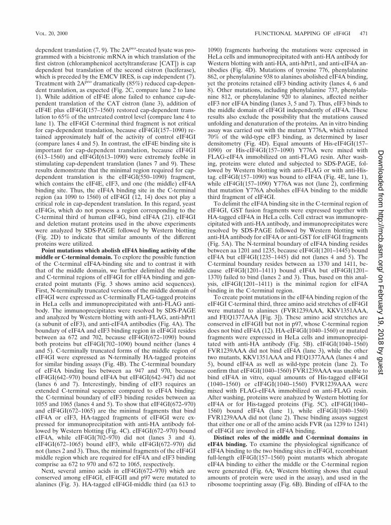
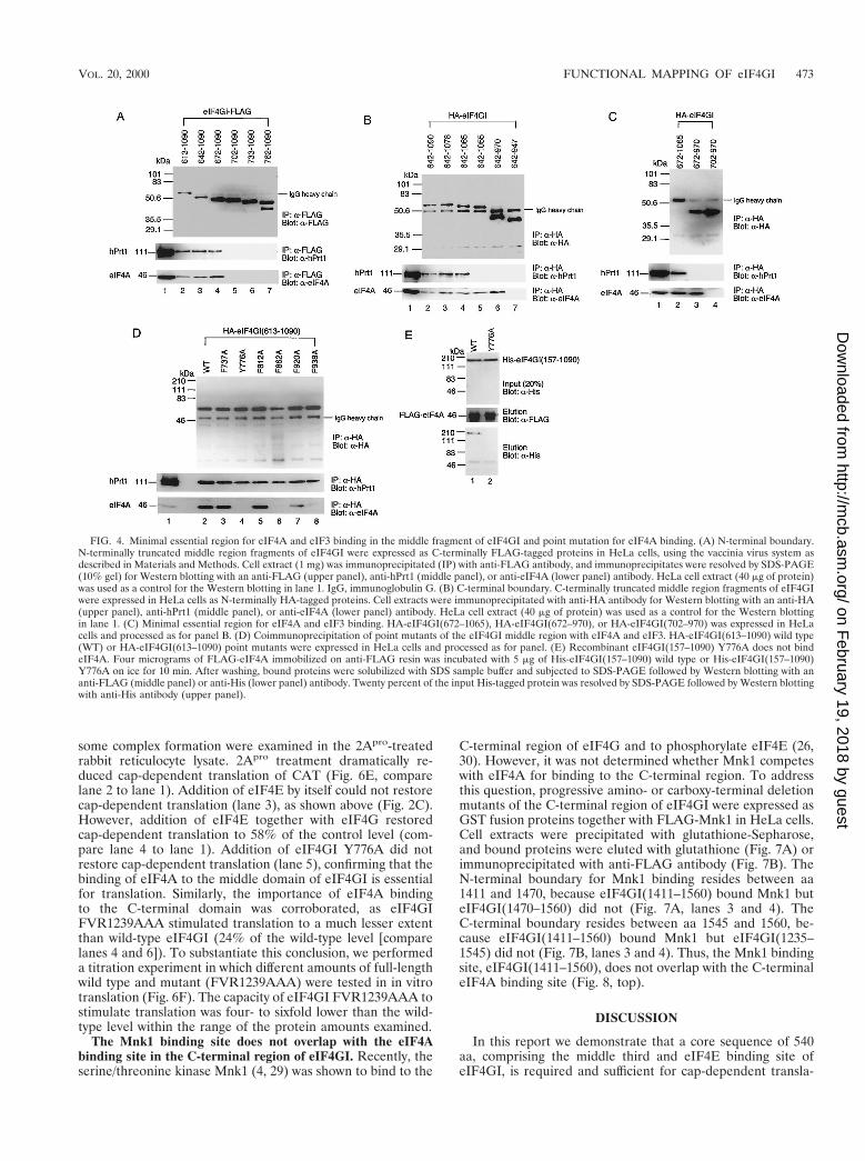
Point mutations which abolish eIF4A binding activity of themiddle or C-terminal domain. To explore the possible functionof the C-terminal eIF4A-binding site and to contrast it withthat of the middle domain, we further delimited the middleand C-terminal regions of eIF4GI for eIF4A binding and gen-erated point mutants (Fig. 3 shows amino acid sequences).First, N-terminally truncated versions of the middle domain ofeIF4GI were expressed as C-terminally FLAG-tagged proteinsin HeLa cells and immunoprecipitated with anti-FLAG anti-body. The immunoprecipitates were resolved by SDS-PAGEand analyzed by Western blotting with anti-FLAG, anti-hPrt1(a subunit of eIF3), and anti-eIF4A antibodies (Fig. 4A). Theboundary of eIF4A and eIF3 binding region in eIF4GI residesbetween aa 672 and 702, because eIF4GI(672–1090) boundboth proteins but eIF4GI(702–1090) bound neither (lanes 4and 5). C-terminally truncated forms of the middle region ofeIF4GI were expressed as N-terminally HA-tagged proteinsfor similar binding assays (Fig. 4B). The C-terminal boundaryof eIF4A binding lies between aa 947 and 970, becauseeIF4GI(642–970) bound eIF4A but eIF4GI(642–947) did not(lanes 6 and 7). Interestingly, binding of eIF3 requires anextended C-terminal sequence compared to eIF4A binding:the C-terminal boundary of eIF3 binding resides between aa1055 and 1065 (lanes 4 and 5). To show that eIF4GI(672–970)and eIF4GI(672–1065) are the minimal fragments that bindeIF4A or eIF3, HA-tagged fragments of eIF4GI were ex-pressed for immunoprecipitation with anti-HA antibody fol-lowed by Western blotting (Fig. 4C). eIF4GI(672–970) boundeIF4A, while eIF4GI(702–970) did not (lanes 3 and 4).eIF4GI(672–1065) bound eIF3, while eIF4GI(672–970) didnot (lanes 2 and 3). Thus, the minimal fragments of the eIF4GImiddle region which are required for eIF4A and eIF3 bindingcomprise aa 672 to 970 and 672 to 1065, respectively.
Next, several amino acids in eIF4GI(672–970) which areconserved among eIF4GI, eIF4GII and p97 were mutated toalanines (Fig. 3). HA-tagged eIF4GI-middle third (aa 613 to
1090) fragments harboring the mutations were expressed inHeLa cells and immunoprecipitated with anti-HA antibody forWestern blotting with anti-HA, anti-hPrt1, and anti-eIF4A an-tibodies (Fig. 4D). Mutations of tyrosine 776, phenylalanine862, or phenylalanine 938 to alanines abolished eIF4A binding,yet the proteins retained eIF3 binding activity (lanes 4, 6 and8). Other mutations, including phenylalanine 737, phenylala-nine 812, or phenylalanine 920 to alanines, affected neithereIF3 nor eIF4A binding (lanes 3, 5 and 7). Thus, eIF3 binds tothe middle domain of eIF4GI independently of eIF4A. Theseresults also exclude the possibility that the mutations causedunfolding and denaturation of the proteins. An in vitro bindingassay was carried out with the mutant Y776A, which retained70% of the wild-type eIF3 binding, as determined by laserdensitometry (Fig. 4D). Equal amounts of His-eIF4GI(157–1090) or His-eIF4GI(157–1090) Y776A were mixed withFLAG-eIF4A immobilized on anti-FLAG resin. After wash-ing, proteins were eluted and subjected to SDS-PAGE, fol-lowed by Western blotting with anti-FLAG or with anti-His-tag. eIF4GI(157–1090) was bound to eIF4A (Fig. 4E, lane 1),while eIF4GI(157–1090) Y776A was not (lane 2), confirmingthat mutation Y776A abolishes eIF4A binding to the middlethird fragment of eIF4GI.
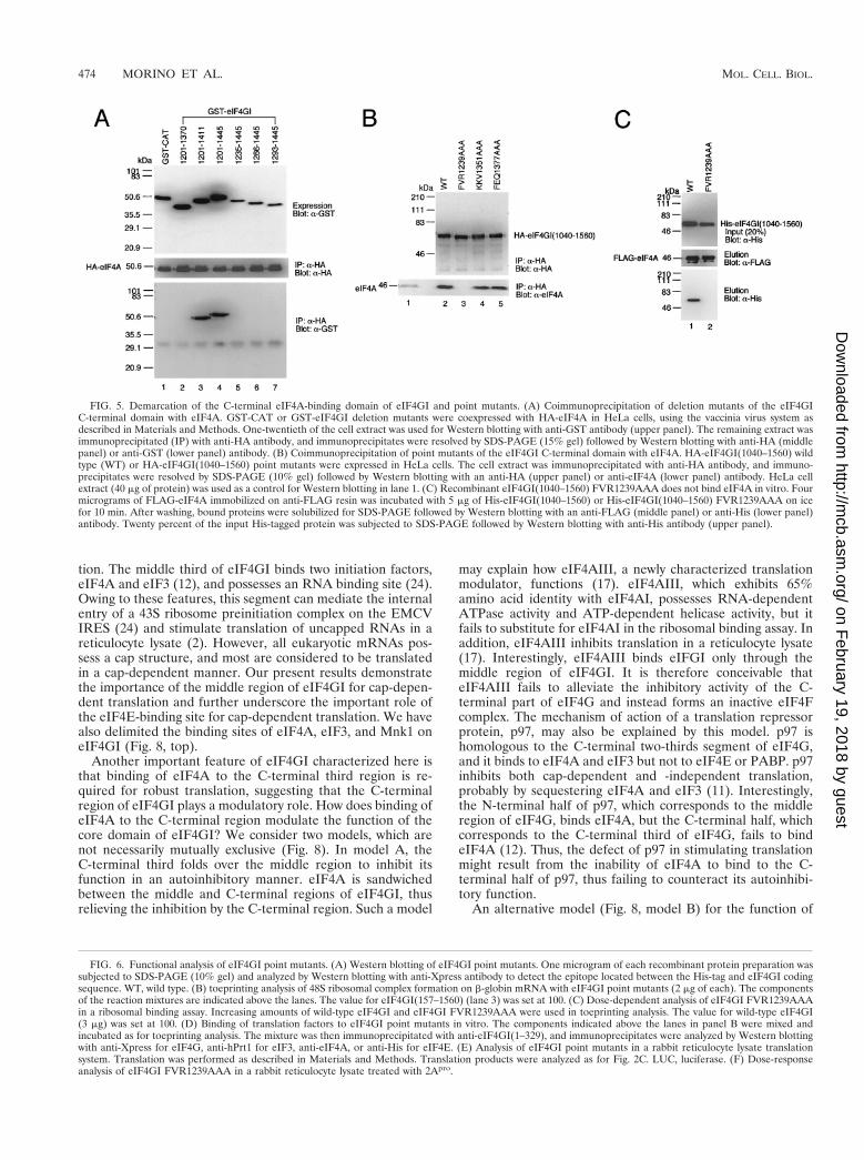
To delimit the eIF4A binding site in the C-terminal region ofeIF4GI, GST fusion fragments were expressed together withHA-tagged eIF4A in HeLa cells. Cell extract was immunopre-cipitated with anti-HA antibody, and immunoprecipitates wereresolved by SDS-PAGE followed by Western blotting withanti-HA antibody for eIF4A or anti-GST for eIF4GI fragments(Fig. 5A). The N-terminal boundary of eIF4A binding residesbetween aa 1201 and 1235, because eIF4GI(1201–1445) boundeIF4A but eIF4GI(1235–1445) did not (lanes 4 and 5). TheC-terminal boundary resides between aa 1370 and 1411, be-cause eIF4GI(1201–1411) bound eIF4A but eIF4GI(1201–1370) failed to bind (lanes 2 and 3). Thus, based on this anal-ysis, eIF4GI(1201–1411) is the minimal region for eIF4Abinding in the C-terminal region.
To create point mutations in the eIF4A binding region of theeIF4GI C-terminal third, three amino acid stretches of eIF4GIwere mutated to alanines (FVR1239AAA, KKV1351AAA,and FEQ1377AAA [Fig. 3]). These amino acid stretches areconserved in eIF4GII but not in p97, whose C-terminal regiondoes not bind eIF4A (12). HA-eIF4GI(1040–1560) or mutatedfragments were expressed in HeLa cells and immunoprecipi-tated with anti-HA antibody (Fig. 5B). eIF4GI(1040–1560)FVR1239AAA did not bind eIF4A (lane 3), while the othertwo mutants, KKV1351AAA and FEQ1377AAA (lanes 4 and5), bound eIF4A as well as wild-type protein (lane 2). Toconfirm that eIF4GI(1040–1560) FVR1239AAA was unable tobind eIF4A in vitro, equal amounts of His-tagged eIF4GI(1040–1560) or eIF4GI(1040–1560) FVR1239AAA weremixed with FLAG-eIF4A immobilized on anti-FLAG resin.After washing, proteins were analyzed by Western blotting foreIF4A or for His-tagged proteins (Fig. 5C). eIF4GI(1040–1560) bound eIF4A (lane 1), while eIF4GI(1040–1560)FVR1239AAA did not (lane 2). These binding assays suggestthat either one or all of the amino acids FVR (aa 1239 to 1241)of eIF4GI are involved in eIF4A binding.
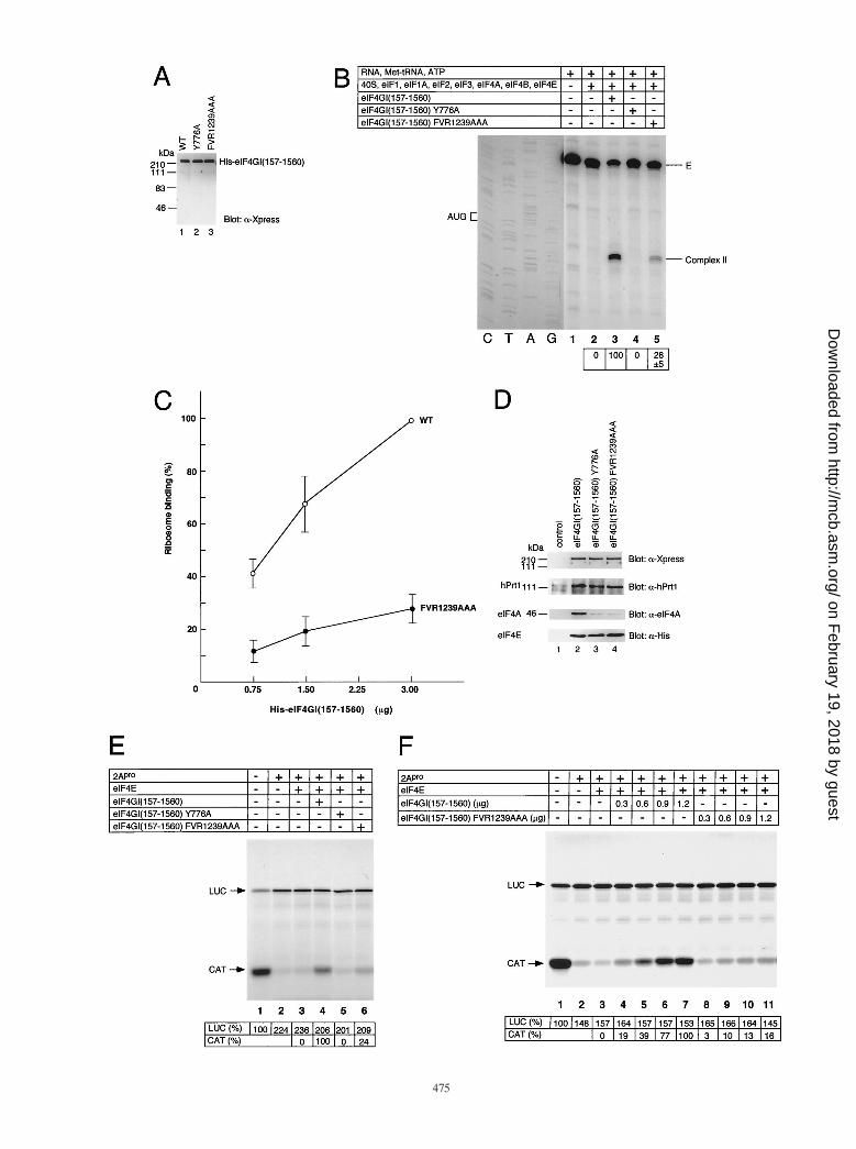
Distinct roles of the middle and C-terminal domains ineIF4A binding. To examine the physiological significance ofeIF4A binding to the two binding sites in eIF4GI, recombinantfull-length eIF4GI(157–1560) point mutants which abrogateeIF4A binding to either the middle or the C-terminal regionwere generated (Fig. 6A; Western blotting shows that equalamounts of protein were used in the assay), and used in theribosome toeprinting assay (Fig. 6B). Binding of eIF4A to the
VOL. 20, 2000 FUNCTIONAL MAPPING OF eIF4GI 471
on February 19, 2018 by guest
http://mcb.asm
.org/D
ownloaded from

middle third of eIF4GI is essential for the 48S ribosomal com-plex formation, because no complex II was formed with full-length eIF4GI Y776A (compare lanes 3 and 4). Surprisingly,binding of eIF4A to the C-terminal region is also important forefficient 48S ribosomal complex formation, because ribosomecomplex II formation was decreased three- to fourfold whenwild-type eIF4GI was replaced with eIF4GI FVR1239AAA(compare lanes 3 and 5). To substantiate these results, weperformed a titration experiment whereby three differentamounts of full-length wild type and mutant (FVR1239AAA)were tested in the toeprinting assay (Fig. 6C). At all concen-trations, eIF4GI FVR1239AAA exhibited only 20 to 30% ac-tivity of wild-type eIF4GI in the ribosomal complex formationassay.
It could be argued that FVR1239AAA mutation causes par-
tial denaturation of eIF4GI, which affects the functions of theother regions of eIF4GI. To address this possibility, the effectsof the mutations in the middle and C-terminal regions in thecontext of full-length eIF4GI on eIF4A, eIF3, and eIF4E bind-ing were determined. Coimmunoprecipitation was performedwith anti-eIF4GI(1–329) antiserum following assembly oftranslation factors on b-globin mRNA (Fig. 6D). Mutation ofeither the middle (lane 3) or C-terminal (lane 4) eIF4A bind-ing site dramatically decreased (10-fold, as determined by laserdensitometry) the association of eIF4A with eIF4GI. In con-trast, binding of eIF4E and eIF3 was not affected by eithermutation. Thus, the defect of the mutant proteins in formationof the ribosomal complex is most probably due to their failurein eIF4A binding.
To further support these results, the mutants used for ribo-
FIG. 3. Protein sequence alignment of human eIF4GI (13, 32), eIF4GII (7), and p97 (11). Conserved amino acids are boxed. Amino acids mutated to alanine arehighlighted. The eIF4E binding site (18) and the rhinovirus 2Apro cleavage site (15) are also indicated.
472 MORINO ET AL. MOL. CELL. BIOL.
on February 19, 2018 by guest
http://mcb.asm
.org/D
ownloaded from
some complex formation were examined in the 2Apro-treatedrabbit reticulocyte lysate. 2Apro treatment dramatically re-duced cap-dependent translation of CAT (Fig. 6E, comparelane 2 to lane 1). Addition of eIF4E by itself could not restorecap-dependent translation (lane 3), as shown above (Fig. 2C).However, addition of eIF4E together with eIF4G restoredcap-dependent translation to 58% of the control level (com-pare lane 4 to lane 1). Addition of eIF4GI Y776A did notrestore cap-dependent translation (lane 5), confirming that thebinding of eIF4A to the middle domain of eIF4GI is essentialfor translation. Similarly, the importance of eIF4A bindingto the C-terminal domain was corroborated, as eIF4GIFVR1239AAA stimulated translation to a much lesser extentthan wild-type eIF4GI (24% of the wild-type level [comparelanes 4 and 6]). To substantiate this conclusion, we performeda titration experiment in which different amounts of full-lengthwild type and mutant (FVR1239AAA) were tested in in vitrotranslation (Fig. 6F). The capacity of eIF4GI FVR1239AAA tostimulate translation was four- to sixfold lower than the wild-type level within the range of the protein amounts examined.
The Mnk1 binding site does not overlap with the eIF4Abinding site in the C-terminal region of eIF4GI. Recently, theserine/threonine kinase Mnk1 (4, 29) was shown to bind to the
C-terminal region of eIF4G and to phosphorylate eIF4E (26,30). However, it was not determined whether Mnk1 competeswith eIF4A for binding to the C-terminal region. To addressthis question, progressive amino- or carboxy-terminal deletionmutants of the C-terminal region of eIF4GI were expressed asGST fusion proteins together with FLAG-Mnk1 in HeLa cells.Cell extracts were precipitated with glutathione-Sepharose,and bound proteins were eluted with glutathione (Fig. 7A) orimmunoprecipitated with anti-FLAG antibody (Fig. 7B). TheN-terminal boundary for Mnk1 binding resides between aa1411 and 1470, because eIF4GI(1411–1560) bound Mnk1 buteIF4GI(1470–1560) did not (Fig. 7A, lanes 3 and 4). TheC-terminal boundary resides between aa 1545 and 1560, be-cause eIF4GI(1411–1560) bound Mnk1 but eIF4GI(1235–1545) did not (Fig. 7B, lanes 3 and 4). Thus, the Mnk1 bindingsite, eIF4GI(1411–1560), does not overlap with the C-terminaleIF4A binding site (Fig. 8, top).
DISCUSSION
In this report we demonstrate that a core sequence of 540aa, comprising the middle third and eIF4E binding site ofeIF4GI, is required and sufficient for cap-dependent transla-
FIG. 4. Minimal essential region for eIF4A and eIF3 binding in the middle fragment of eIF4GI and point mutation for eIF4A binding. (A) N-terminal boundary.N-terminally truncated middle region fragments of eIF4GI were expressed as C-terminally FLAG-tagged proteins in HeLa cells, using the vaccinia virus system asdescribed in Materials and Methods. Cell extract (1 mg) was immunoprecipitated (IP) with anti-FLAG antibody, and immunoprecipitates were resolved by SDS-PAGE(10% gel) for Western blotting with an anti-FLAG (upper panel), anti-hPrt1 (middle panel), or anti-eIF4A (lower panel) antibody. HeLa cell extract (40 mg of protein)was used as a control for the Western blotting in lane 1. IgG, immunoglobulin G. (B) C-terminal boundary. C-terminally truncated middle region fragments of eIF4GIwere expressed in HeLa cells as N-terminally HA-tagged proteins. Cell extracts were immunoprecipitated with anti-HA antibody for Western blotting with an anti-HA(upper panel), anti-hPrt1 (middle panel), or anti-eIF4A (lower panel) antibody. HeLa cell extract (40 mg of protein) was used as a control for the Western blottingin lane 1. (C) Minimal essential region for eIF4A and eIF3 binding. HA-eIF4GI(672–1065), HA-eIF4GI(672–970), or HA-eIF4GI(702–970) was expressed in HeLacells and processed as for panel B. (D) Coimmunoprecipitation of point mutants of the eIF4GI middle region with eIF4A and eIF3. HA-eIF4GI(613–1090) wild type(WT) or HA-eIF4GI(613–1090) point mutants were expressed in HeLa cells and processed as for panel. (E) Recombinant eIF4GI(157–1090) Y776A does not bindeIF4A. Four micrograms of FLAG-eIF4A immobilized on anti-FLAG resin was incubated with 5 mg of His-eIF4GI(157–1090) wild type or His-eIF4GI(157–1090)Y776A on ice for 10 min. After washing, bound proteins were solubilized with SDS sample buffer and subjected to SDS-PAGE followed by Western blotting with ananti-FLAG (middle panel) or anti-His (lower panel) antibody. Twenty percent of the input His-tagged protein was resolved by SDS-PAGE followed by Western blottingwith anti-His antibody (upper panel).
VOL. 20, 2000 FUNCTIONAL MAPPING OF eIF4GI 473
on February 19, 2018 by guest
http://mcb.asm
.org/D
ownloaded from
tion. The middle third of eIF4GI binds two initiation factors,eIF4A and eIF3 (12), and possesses an RNA binding site (24).Owing to these features, this segment can mediate the internalentry of a 43S ribosome preinitiation complex on the EMCVIRES (24) and stimulate translation of uncapped RNAs in areticulocyte lysate (2). However, all eukaryotic mRNAs pos-sess a cap structure, and most are considered to be translatedin a cap-dependent manner. Our present results demonstratethe importance of the middle region of eIF4GI for cap-depen-dent translation and further underscore the important role ofthe eIF4E-binding site for cap-dependent translation. We havealso delimited the binding sites of eIF4A, eIF3, and Mnk1 oneIF4GI (Fig. 8, top).
Another important feature of eIF4GI characterized here isthat binding of eIF4A to the C-terminal third region is re-quired for robust translation, suggesting that the C-terminalregion of eIF4GI plays a modulatory role. How does binding ofeIF4A to the C-terminal region modulate the function of thecore domain of eIF4GI? We consider two models, which arenot necessarily mutually exclusive (Fig. 8). In model A, theC-terminal third folds over the middle region to inhibit itsfunction in an autoinhibitory manner. eIF4A is sandwichedbetween the middle and C-terminal regions of eIF4GI, thusrelieving the inhibition by the C-terminal region. Such a model
may explain how eIF4AIII, a newly characterized translationmodulator, functions (17). eIF4AIII, which exhibits 65%amino acid identity with eIF4AI, possesses RNA-dependentATPase activity and ATP-dependent helicase activity, but itfails to substitute for eIF4AI in the ribosomal binding assay. Inaddition, eIF4AIII inhibits translation in a reticulocyte lysate(17). Interestingly, eIF4AIII binds eIFGI only through themiddle region of eIF4GI. It is therefore conceivable thateIF4AIII fails to alleviate the inhibitory activity of the C-terminal part of eIF4G and instead forms an inactive eIF4Fcomplex. The mechanism of action of a translation repressorprotein, p97, may also be explained by this model. p97 ishomologous to the C-terminal two-thirds segment of eIF4G,and it binds to eIF4A and eIF3 but not to eIF4E or PABP. p97inhibits both cap-dependent and -independent translation,probably by sequestering eIF4A and eIF3 (11). Interestingly,the N-terminal half of p97, which corresponds to the middleregion of eIF4G, binds eIF4A, but the C-terminal half, whichcorresponds to the C-terminal third of eIF4G, fails to bindeIF4A (12). Thus, the defect of p97 in stimulating translationmight result from the inability of eIF4A to bind to the C-terminal half of p97, thus failing to counteract its autoinhibi-tory function.
An alternative model (Fig. 8, model B) for the function of
FIG. 5. Demarcation of the C-terminal eIF4A-binding domain of eIF4GI and point mutants. (A) Coimmunoprecipitation of deletion mutants of the eIF4GIC-terminal domain with eIF4A. GST-CAT or GST-eIF4GI deletion mutants were coexpressed with HA-eIF4A in HeLa cells, using the vaccinia virus system asdescribed in Materials and Methods. One-twentieth of the cell extract was used for Western blotting with anti-GST antibody (upper panel). The remaining extract wasimmunoprecipitated (IP) with anti-HA antibody, and immunoprecipitates were resolved by SDS-PAGE (15% gel) followed by Western blotting with anti-HA (middlepanel) or anti-GST (lower panel) antibody. (B) Coimmunoprecipitation of point mutants of the eIF4GI C-terminal domain with eIF4A. HA-eIF4GI(1040–1560) wildtype (WT) or HA-eIF4GI(1040–1560) point mutants were expressed in HeLa cells. The cell extract was immunoprecipitated with anti-HA antibody, and immuno-precipitates were resolved by SDS-PAGE (10% gel) followed by Western blotting with an anti-HA (upper panel) or anti-eIF4A (lower panel) antibody. HeLa cellextract (40 mg of protein) was used as a control for Western blotting in lane 1. (C) Recombinant eIF4GI(1040–1560) FVR1239AAA does not bind eIF4A in vitro. Fourmicrograms of FLAG-eIF4A immobilized on anti-FLAG resin was incubated with 5 mg of His-eIF4GI(1040–1560) or His-eIF4GI(1040–1560) FVR1239AAA on icefor 10 min. After washing, bound proteins were solubilized for SDS-PAGE followed by Western blotting with an anti-FLAG (middle panel) or anti-His (lower panel)antibody. Twenty percent of the input His-tagged protein was subjected to SDS-PAGE followed by Western blotting with anti-His antibody (upper panel).
FIG. 6. Functional analysis of eIF4GI point mutants. (A) Western blotting of eIF4GI point mutants. One microgram of each recombinant protein preparation wassubjected to SDS-PAGE (10% gel) and analyzed by Western blotting with anti-Xpress antibody to detect the epitope located between the His-tag and eIF4GI codingsequence. WT, wild type. (B) toeprinting analysis of 48S ribosomal complex formation on b-globin mRNA with eIF4GI point mutants (2 mg of each). The componentsof the reaction mixtures are indicated above the lanes. The value for eIF4GI(157–1560) (lane 3) was set at 100. (C) Dose-dependent analysis of eIF4GI FVR1239AAAin a ribosomal binding assay. Increasing amounts of wild-type eIF4GI and eIF4GI FVR1239AAA were used in toeprinting analysis. The value for wild-type eIF4GI(3 mg) was set at 100. (D) Binding of translation factors to eIF4GI point mutants in vitro. The components indicated above the lanes in panel B were mixed andincubated as for toeprinting analysis. The mixture was then immunoprecipitated with anti-eIF4GI(1–329), and immunoprecipitates were analyzed by Western blottingwith anti-Xpress for eIF4G, anti-hPrt1 for eIF3, anti-eIF4A, or anti-His for eIF4E. (E) Analysis of eIF4GI point mutants in a rabbit reticulocyte lysate translationsystem. Translation was performed as described in Materials and Methods. Translation products were analyzed as for Fig. 2C. LUC, luciferase. (F) Dose-responseanalysis of eIF4GI FVR1239AAA in a rabbit reticulocyte lysate treated with 2Apro.
474 MORINO ET AL. MOL. CELL. BIOL.
on February 19, 2018 by guest
http://mcb.asm
.org/D
ownloaded from

the C-terminal region is that the middle region of eIF4GI issterically hidden from free eIF4A by the C-terminal region.eIF4A binds first to the C-terminal region and is subsequentlytransferred to the middle region. This idea is consistent withanother feature of eIF4AIII: while eIF4AIII strongly binds tothe middle third of eIF4GI, it binds the full-length eIF4GI verypoorly (17). To distinguish between these models, dissociationconstants between eIF4A and each region or full-length ofeIF4GI ought to be determined. Also, the number of eIF4Amolecules that bind to eIF4GI at a given time will need to beestablished.
The C-terminal third of eIF4G also plays a role in the phos-phorylation of eIF4E. The distal C-terminal region of eIF4GIcontains a binding site for the serine/threonine kinase Mnk1
(Fig. 7). It has been shown that the C-terminal third of eIF4Grecruits Mnk1 to phosphorylate eIF4E in vivo (26, 30), whichis thought to stimulate cap-dependent translation. Phosphory-lation of eIF4G itself may also affect translation. Interestingly,several serum-responsive phosphorylation sites are localized inthe C-terminal third region of eIF4GI (B. Raught, A.-C. Gin-gras, S. P. Gygi, H. Imataka, S. Morino, A. Gradi, R. Aeber-sold, and N. Sonenberg, unpublished data).
What is the function of the N-terminal third of eIF4G? Thisregion harbors the eIF4E and PABP binding sites and conse-quently engages the mRNA via both its 59 and 39 ends. Whilethe critical role of the eIF4E-binding site for cap-dependenttranslation is confirmed in this study, our experiments did notaddress the importance of the PABP binding site in transla-tion, because the recombinant eIF4GI which we used lackedthis site. It should be very interesting, however, to examine howtranslation is affected when mRNA is circularized through theN terminus of eIF4G in a reconstituted translation system,using full-length eIF4G. However, current models state thatthe PABP interaction with eIF4G is not required for the firstround of translation initiation, but only for subsequent rounds(28). The spacer region between the PABP and eIF4E bindingsites (;400 aa) is the least conserved region between eIF4GIand eIF4GII (7). We do not know the function of this region;no protein has been reported to bind this region, and its de-letion had no effect on ribosomal binding (Fig. 2B) or ontranslation in the reticulocyte lysate (Fig. 2C).
Finally, using the in vitro ribosome binding assay, we dem-onstrated 48S ribosomal complex formation at the correct ini-tiator AUG for the b-globin mRNA in the absence of eIF4E(Fig. 1) or with an eIF4GI mutant lacking the eIF4E bindingsite (Fig. 2B), albeit with low efficiency (20 to 24% of that ofthe complete system). The eIF4E-independent ribosome bind-ing does not seem to represent aberrant ribosomes binding,
FIG. 7. Demarcation of the Mnk1 binding site in the C-terminal region ofeIF4GI. (A) N-terminal boundary. GST, GST-CAT, or GST-eIF4GI deletionmutants were coexpressed with FLAG-Mnk1 in HeLa cells. One-fortieth of thecell extract was subjected to SDS-PAGE for Western blotting with anti-FLAGantibody to confirm the expression of FLAG-Mnk1 (upper panel). The remain-ing extract was mixed with glutathione-Sepharose beads. Bound proteins elutedwith reduced glutathione were subjected to Western blotting with an anti-GST(middle panel) or anti-FLAG (lower panel) antibody. (B) C-terminal boundary.GST, GST-CAT, or GST-eIF4GI deletion mutants were coexpressed withFLAG-Mnk1 in HeLa cells. One-fortieth of the cell extract was subjected toSDS-PAGE for Western blotting with anti-GST antibody to confirm the expres-sion of GST fusion proteins (upper panel). The remaining extract was immuno-precipitated with anti-FLAG antibody, and immunoprecipitates were subjectedto Western blotting with anti-FLAG (middle panel) or anti-GST (lower panel)antibody. IgG, immunoglobulin G.
FIG. 8. Model of eIF4GI functional domains. Previous studies have mappedthe eIF4E (18) and PABP (13) binding sites to the N-terminal third of eIF4G.The middle third region was shown to bind eIF4A and eIF3 (12), while theC-terminal third region was shown to bind eIF4A (12, 14) and Mnk1 (26). SeeDiscussion section for explanations of models.
476 MORINO ET AL. MOL. CELL. BIOL.
on February 19, 2018 by guest
http://mcb.asm
.org/D
ownloaded from

since the ribosomal complex was not formed at the secondAUG of b-globin mRNA (data not shown; the second AUGwas out of the photograph in Fig. 1 and 2B). Thus, the bindingrepresents cap-independent but 59-end-dependent translation,which has been documented in a rabbit reticulocyte lysate (2)and in mammalian cells (8). The first AUG is still predomi-nantly utilized as the translation initiator when the reticulocytelysate is programmed with uncapped mRNA (2) or when un-capped mRNA is transcribed by RNA polymerase III in mam-malian cells (8). These results are consistent with the idea thatthe cap structure dramatically enhances translation, ratherthan being absolutely required for translation in eukaryotes.
In summary, we have defined a minimal core sequence ofeIF4GI which is required for ribosome binding and translation.The core sequence constitutes only one-third of the entireeIF4GI protein. The C-terminal third region, which does nothave a counterpart in yeast, modulates eIF4GI activity. It istherefore of great importance to elucidate the mechanism bywhich the eIF4G C-terminal region controls translation inmetazoan cells.
ACKNOWLEDGMENTS
We thank W. C. Merrick for eIF2, eIF3, and eIF4F proteins used fora preliminary experiment, T. Skern for rhinovirus 2Apro, A. Gradi forthe anti-eIF4GI antibody, and R. Fukunaga for the Mnk1 plasmid. Weare indebted to C. Lister for excellent technical assistance. We thankB. Raught and A.-C. Gingras for sharing unpublished data and criti-cally reading the manuscript.
S.M. was supported by research fellowships of the Japan Society forthe Promotion of Science for Young Scientists. This work was sup-ported by a grant from the Medical Research Council of Canada toN.S. N.S. is a Distinguished Scientist of the Medical Research Councilof Canada and a Howard Hughes Medical Institute InternationalScholar.
ADDENDUM IN PROOF
Since the submission of this paper, a report by De Gregorioet al. (EMBO J. 18:4865–4874, 1999) also defined a conservedcentral domain (aa 642 to 1091) of eIF4G as an autonomousribosome recruitment core in vivo.
REFERENCES
1. Allen, M., A. M. Metz, R. T. Timmer, R. E. Rhoads, and K. S. Browning.1992. Isolation and sequence of the cDNAs encoding the subunits of theisozyme form of wheat protein synthesis initiation factor 4F. J. Biol. Chem.267:23232–23236.
2. De Gregorio, E., T. Preiss, and M. W. Hentze. 1998. Translational activationof uncapped mRNAs by the central part of human eIF4G is 59 end-depen-dent. RNA 4:828–836.
3. Fuerst, T. R., E. G. Niles, F. W. Studier, and B. Moss. 1986. Eukaryotictransient expression system based on recombinant vaccinia virus that syn-thesizes bacteriophage T7 RNA polymerase. Proc. Natl. Acad. Sci. USA83:8122–8126.
4. Fukunaga, R., and T. Hunter. 1997. MNK1, a new MAP kinase-activatedprotein kinase, isolated by a novel expression screening method for identi-fying protein kinase substrates. EMBO J. 16:1921–1933.
5. Gingras, A.-C., B. Raught, and N. Sonenberg. 1999. eIF4 initiation factors:effectors of mRNA recruitment to ribosomes and regulators of translation.Annu. Rev. Biochem. 68:913–963.
6. Goyer, C., M. Altmann, H. S. Lee, A. Blanc, M. Deshmukh, J. L. Woolford,Jr., H. Trachsel, and N. Sonenberg. 1993. TIF4631 and TIF4632: two yeastgenes encoding the high-molecular-weight subunits of the cap-binding pro-tein complex (eukaryotic initiation factor 4F) contain an RNA recognitionmotif-like sequence and carry out an essential function. Mol. Cell. Biol.13:4860–4874.
7. Gradi, A., H. Imataka, Y. V. Svitkin, E. Rom, B. Raught, S. Morino, and N.Sonenberg. 1998. A novel functional human eukaryotic translation initiationfactor 4G. Mol. Cell. Biol. 18:334–342.
8. Gunnery, S., U. Maivali, and M. B. Mathews. 1997. Translation of an un-capped mRNA involves scanning. J. Biol. Chem. 272:21642–21646.
9. Haghighat, A., Y. V. Svitkin, I. Novoa, E. Kuechler, T. Skern, and N. Sonen-berg. 1996. The eIF4G-eIF4E complex is the target for direct cleavage by therhinovirus 2A proteinase. J. Virol. 70:8445–8450.
10. Hellen, C. U. T., G. W. Witherell, M. Schmid, S. H. Shin, T. V. Pestova, A.Gil, and E. Wimmer. 1993. A cytoplasmic 57kDa protein that is required fortranslation of picornavirus RNA by internal ribosome entry is identical to thenuclear pyrimidine tract-binding protein. Proc. Natl. Acad. Sci. USA 90:7642–7646.
11. Imataka, H., H. S. Olsen, and N. Sonenberg. 1997. A new translationalregulator with homology to eukaryotic translation initiation factor 4G.EMBO J. 16:817–825.
12. Imataka, H., and N. Sonenberg. 1997. Human eukaryotic translation initia-tion factor 4G (eIF4G) possesses two separate and independent binding sitesfor eIF4A. Mol. Cell. Biol. 17:6940–6947.
13. Imataka, H., A. Gradi, and N. Sonenberg. 1998. A newly identified N-terminal amino acid sequence of human eIF4G binds poly(A)-binding pro-tein and functions in poly(A)-dependent translation. EMBO J. 17:7480–7489.
14. Lamphear, B. J., R. Kirchweger, T. Skern, and R. E. Rhoads. 1995. Mappingof functional domains in eukaryotic protein synthesis initiation factor 4G(eIF4G) with picornaviral proteases: implications for cap-dependent andcap-independent translational initiation. J. Biol. Chem. 270:21975–21983.
15. Lamphear, B. J., R. Yan, F. Yang, D. Waters, H. D. Liebig, H. Klump, E.Kuechler, T. Skern, and R. E. Rhoads. 1993. Mapping of the cleavage site inprotein synthesis initiation factor eIF-4g of the 2A proteases from humancoxsackievirus and rhinovirus. J. Biol. Chem. 268:19200–19203.
16. Levy-Strumpf, N., L. P. Deiss, H. Berissi, and A. Kimchi. 1997. DAP-5, anovel homolog of eukaryotic translation initiation factor 4G isolated as aputative modulator of gamma interferon-induced programmed cell death.Mol. Cell. Biol. 17:1615–1625.
17. Li, Q., H. Imataka, S. Morino, G. W. Rogers, Jr., N. J. Richter-Cook, W. C.Merrick, and N. Sonenberg. 1999. Eukaryotic translation initiation factor4AIII (eIF4AIII) is functionally distinct from eIF4AI and eIF4AII. Mol.Cell. Biol. 19:7336–7346.
18. Mader, S., H. Lee, A. Pause, and N. Sonenberg. 1995. The translationinitiation factor eIF4E binds to a common motif shared by the translationfactor eIF-4g and the translational repressors 4E-binding proteins. Mol.Cell. Biol. 15:4990–4997.
19. Merrick, W. C., and J. W. B. Hershey. 1996. The pathway and mechanism ofeukaryotic protein synthesis, p. 31–70. In J. Hershey, M. Mathews, and N.Sonenberg (ed.), Translational control. Cold Spring Harbor LaboratoryPress, Cold Spring Harbor, N.Y.
20. Morley, S. J., P. S. Curtis, and V. M. Pain. 1997. eIF4G: translation’s mysteryfactor begins to yield its secrets. RNA 3:1085–1104.
21. Neff, C. L., and A. B. Sachs. 1999. Eukaryotic translation initiation factors 4Gand 4A from Saccharomyces cerevisiae interact physically and functionally.Mol. Cell. Biol. 19:5557–5564.
22. Pain, V. M. 1996. Initiation of protein synthesis in eukaryotic cells. Eur.J. Biochem. 236:747–771.
23. Pestova, T. V., C. U. T. Hellen, and I. N. Shatsky. 1996. Canonical eukaryoticinitiation factors determine initiation of translation by internal ribosomalentry. Mol. Cell. Biol. 16:6859–6869.
24. Pestova, T. V., I. N. Shatsky, and C. U. T. Hellen. 1996. Functional dissectionof eukaryotic initiation factor 4F: the 4A subunit and the central domain ofthe 4G subunit are sufficient to mediate internal entry of 43S preinitiationcomplexes. Mol. Cell. Biol. 16:6870–6878.
25. Pestova, T. V., S. I. Borukhov, and C. U. T. Hellen. 1998. Eukaryotic ribo-somes require initiation factors 1 and 1A to locate initiation codons. Nature394:854–859.
26. Pyronnet, S., H. Imataka, A.-C. Gingras, R. Fukunaga, T. Hunter, and N.Sonenberg. 1999. Human eukaryotic translation initiation factor 4G (eIF4G)recruits Mnk1 to phosphorylate eIF4E. EMBO J. 18:270–279.
27. Sachs, A. B., P. Sarnow, and M. W. Hentze. 1997. Starting at the beginning,middle, and end: translation initiation in eukaryotes. Cell 89:831–838.
28. Sonenberg, N. 1996. mRNA 59 cap-binding protein eIF4E and control of cellgrowth, p. 271–294. In J. Hershey, M. Mathews, and N. Sonenberg (ed.),Translational control. Cold Spring Harbor Laboratory Press, Cold SpringHarbor, N.Y.
29. Waskiewicz, A. J., A. Flynn, C. G. Proud, and J. A. Cooper. 1997. Mitogen-activated protein kinases activate the serine/threonine kinases Mnk1 andMnk2. EMBO J. 16:1909–1920.
30. Waskiewicz, A. J., J. C. Johnson, B. Penn, M. Mahalingam, S. R. Kimball,and J. A. Cooper. 1999. Phosphorylation of the cap-binding protein eIF4E bythe protein kinase Mnk1 in vivo. Mol. Cell. Biol. 19:1871–1880.
31. Yamanaka, S., K. S. Poksay, K. S. Arnold, and T. L. Innerarity. 1997. Anovel translational repressor mRNA is edited extensively in livers containingtumors caused by the transgene expression of the apoB mRNA-editingenzyme. Genes Dev. 11:321–333.
32. Yan, R., W. Rychlik, D. Etchsion, and R. E. Rhoads. 1992. Amino acidsequence of the human protein synthesis initiation factor eIF-4g. J. Biol.Chem. 267:23226–23231.
VOL. 20, 2000 FUNCTIONAL MAPPING OF eIF4GI 477
on February 19, 2018 by guest
http://mcb.asm
.org/D
ownloaded from