Embed Size (px)
Citation preview

Targeted disruption of SHIP leadsto hemopoietic perturbations, lungpathology, and a shortened life spanCheryl D. Helgason,1 Jacqueline E. Damen,1 Patty Rosten,1 Rewa Grewal,1 Poul Sorensen,2
Suzanne M. Chappel,1 Anita Borowski,3 Frank Jirik,3 Gerald Krystal,1 and R. Keith Humphries1,4,5
1Terry Fox Laboratory, British Columbia Cancer Agency, Vancouver, British Columbia V5Z 1L3, Canada; 2Departmentof Pathology and Laboratory Medicine, University of British Columbia, Vancouver, British Columbia V6T 2B5, Canada;3Center for Molecular Medicine and Therapeutics, University of British Columbia, Vancouver, British Columbia V6T 1Z4,Canada; 4Department of Medicine, University of British Columbia, Vancouver, British Columbia V6T 2B5, Canada
SHIP is a 145-kD SH2-containing inositol-5-phosphatase widely expressed in hemopoietic cells. It was firstidentified as a tyrosine phosphoprotein associated with Shc in response to numerous cytokines. SHIP has beenimplicated in FcgRIIB receptor-mediated negative signaling in B cells and mast cells and is postulated todown-regulate cytokine signal transduction in myeloid cells. To define further its role in the proliferation anddifferentiation of hemopoietic progenitors, as well as its function in mature cells, we have generatedembryonic stem cells and mice bearing a targeted disruption of both SHIP alleles. Here we show that althoughSHIP null mice are viable and fertile, they fail to thrive and survival is only 40% by 14 weeks of age.Mortality is associated with extensive consolidation of the lungs resulting from infiltration by myeloid cells.Increased numbers of granulocyte–macrophage progenitors are observed in both the bone marrow and spleenof SHIP−/− mice, perhaps as a consequence of hyper-responsiveness to stimulation by macrophage–colonystimulating factor, granulocyte–macrophage colony stimulating factor, interleukin-3, or Steel factor asobserved in vitro. In contrast, numbers of bone marrow lymphoid and late erythroid progenitors (CFU-E) arereduced. Thus, homozygous disruption of SHIP establishes the crucial role of this molecule in modulatingcytokine signaling within the hemopoietic system and provides a powerful model for further delineating itsfunction.
[Key Words: SHIP; hemopoiesis; embryonic stem cells; knockout; signal transduction]
Received March 18, 1998; revised version accepted April 13, 1998.
SHIP (SH2-containing inositol-5-phosphatase) is a re-cently cloned 145-kD protein that is highly expressed inhemopoietic cells (Damen et al. 1996; Kavanaugh et al.1996; Lioubin et al. 1996). It contains an amino-terminalsrc homology 2 (SH2) domain, a central 58-phosphoino-sitol phosphatase domain, two phosphotyrosine binding(PTB) consensus sequences, and a proline-rich region atthe carboxyl tail. SHIP becomes tyrosine phosphorylatedfollowing activation of the hemopoietic cell-surface re-ceptors for numerous cytokines including erythropoietin(Epo), Steel factor (SF), interleukin-3 (IL-3) (Cutler et al.1993; Damen et al. 1993), IL-2, granulocyte–macrophagecolony-stimulating factor (GM-CSF), and macrophagecolony stimulating factor (M-CSF) (Lioubin et al. 1994).Similarly, cross-linking of the B-cell antigen receptor(Saxton et al. 1994) or T-cell activation (Ravichandran etal. 1993) induce SHIP phosphorylation.
It has been postulated that SHIP may impact on many
signal transduction pathways in hemopoietic cells. Forexample, SHIP selectively hydrolyzes the 58-phosphatefrom inositol 1,3,4,5-tetraphosphate and phosphatidyl-inositol 3,4,5-trisphosphate (PIP3), the latter being aproduct of phosphatidylinositol 38-kinase (PI-3-K) activ-ity. PI-3-K activation in response to growth factor stimu-lation is implicated as a major step in mitogenic signal-ing (Kapeller and Cantley 1994). In addition, both phos-phatidylinositol 3,4-bisphosphate and PIP3 may play arole in regulating the PKB/Akt kinase (Marte and Down-ward 1997). Interestingly, the catalytic activity of SHIPis not altered following cytokine stimulation, suggestingthat its subcellular localization may be an importantregulatory factor (Damen et al. 1996). In this regard,phosphorylated SHIP has been shown to associate withShc (Liu et al. 1997a), Grb2 (Osborne et al. 1996), and thetyrosine phosphatase SHP-2 (Liu et al. 1997b; Sattler etal. 1997), which may change its proximity to the mem-brane and/or various substrates. Furthermore, associa-tion with these molecules suggests that SHIP may func-tion in coupling cytokine receptor activation to the Ras
5Corresponding author.E-MAIL [email protected]; FAX (604) 877-0712.
1610 GENES & DEVELOPMENT 12:1610–1620 © 1998 by Cold Spring Harbor Laboratory Press ISSN 0890-9369/98 $5.00; www.genesdev.org
Cold Spring Harbor Laboratory Press on April 14, 2019 - Published by genesdev.cshlp.orgDownloaded from

signaling pathway in hemopoietic cells (Scharenberg andKinet 1996; Liu et al. 1997c; Tridandapani et al. 1997).Thus, SHIP may regulate the proliferation and differen-tiation of hemopoietic cells by modulating PIP3 levelsand Ras activity following cytokine stimulation.
SHIP has been implicated in the negative signalingpathways that abrogate activation in cells of the immunesystem. Coligation of the low-affinity Fc receptor for im-munoglobulin G (IgG) (FcgRIIB) with the B-cell receptor(BCR) or the high-affinity mast cell IgE receptor (FceRI)blocks the influx of extracellular calcium with minimaleffects on the release of calcium from intracellularstores. This perturbation of calcium signaling may me-diate the negative signals on B-cell growth and mast-celldegranulation. Studies with both mast and B cells de-rived from mice deficient in SHP-1 (me/me) suggest thatSHIP is the primary mediator of inhibitory FcgRIIB sig-naling (Ono et al. 1996; Nadler et al. 1997). It is unclearhowever whether SHIP is an inhibitor of cytokine signaltransduction pathways regulating the proliferation anddifferentiation of other hemopoietic progenitors. Ectopicexpression of SHIP in the FDC-P1 cell line significantlyreduces M-CSF-dependent growth (Lioubin et al. 1996)and increases apoptosis in the DA-ER murine hemopoi-etic cell line (Liu et al. 1997a). These observations sug-gest that SHIP may down-regulate signals required forsurvival and proliferation.
Targeted disruption using homologous recombinationin embryonic stem (ES) cells was carried out to furtherdefine the role of SHIP in the proliferation and differen-tiation of hemopoietic progenitors, as well as its func-tion in mature cells. The data presented suggest thatSHIP plays an important role in down-regulating the mi-togenic signals initiated through stimulation of numer-ous growth factor receptors. Absence of SHIP results in amyeloproliferative-like syndrome and consolidation ofthe lungs by infiltration of macrophages, with a conse-quent decrease in the survival of SHIP−/− mice.
Results
Targeted disruption of SHIP
To examine the in vivo function of SHIP, targeted dis-ruption of the murine SHIP gene was achieved using astrategy in which the entire 254-bp coding sequence ofthe first exon was replaced with the neomycin resistancegene in the antisense orientation using either of the twotargeting vectors described in Figure 1A. Two lines ofSHIP knockout mice, one generated with each targetingvector, were derived using standard techniques. South-ern blot analysis of genomic DNA from both the targetedES cells and representative F2 mice using a 2-kb KpnI–HindIII genomic probe confirmed the expected targetingevent (Fig. 1B). Western blot analysis of hemopoieticcells derived from the in vitro differentiation of SHIPwild type (+/+), heterozygous (+/−), and null (−/−) R1 EScells, as well as bone marrow cells derived from mice ofeach genotype, confirmed the absence of full-length ortruncated protein products in the SHIP−/− cells (Fig. 1C).
SHIP−/− mice fail to thrive and haveprofound splenomegaly
Wild-type, heterozygous, and null F2 progeny were pres-ent at the expected Mendelian ratio of 1:2:1 (n = 360).Although both male and female SHIP−/− mice were vi-able and fertile, they failed to thrive and exhibited a 17%reduction in body weight at 4–5 weeks of age with afurther 5% decrease by 8–10 weeks of age (Table 1).Splenomegaly was also a striking feature in SHIP−/−
mice of both age groups, with nucleated cell counts in-creased two- to threefold above +/+ and +/− littermatecontrols. Total spleen cellularity (red + white cells) andweights were increased five- to sevenfold in the 8–10-week-old knockout animals (not shown). In contrast, thebone marrow of SHIP null mice became progressivelyhypocellular with age (Table 1). The cellularity of thethymus and lymph nodes obtained from SHIP−/− mice inthese two age groups was highly variable and thereforethe differences compared with the +/+ mice were notsignificant. In all cases the phenotype of the +/− micemore closely resembled that of the wild-type mice.
SHIP−/− mice have a shortened life span associatedwith massive myeloid cell infiltration of the lungs
Homozygous knockout mice became moribund as earlyas 4 weeks of age and over 50% died by 10 weeks of age(Fig. 2). Gross anatomical examination of SHIP−/− miceat 8–10 weeks of age revealed, in addition to the pro-nounced splenomegaly, a uniform enlargement andpatchy whitish discoloration of the pleural surfaces ofthe lungs compared to littermate controls (Fig. 3A). His-tologic examination revealed a massive infiltration ofthe air spaces with distended lipid-laden macrophages,many of which were multinucleated (Fig. 3B,C). The his-tiocytic origin of these cells was corroborated by strongchloracetate esterase staining (Fig. 3D). The infiltrateswere located primarily in alveolar spaces in the patternof lipoid pneumonia. In addition, collections of neutro-phils were distributed throughout the consolidated ar-eas, and in some sections neutrophils were present inbronchiolar lumens. Although the degree of lung in-volvement was extensive and became progressively pro-nounced with age, the infiltrates were somewhat patchyin that areas of normal lung could be found adjacent toabnormal areas (Fig. 3B). Stains for bacteria and fungiwere negative, and serological testing showed no evi-dence of viral pathogens or mycoplasma, ruling out ob-vious infectious causes. Similar lung histopathology wasobserved in the second line of SHIP−/− mice generatedand housed in a separate pathogen-free animal facility(not shown).
Numerous hemopoietic abnormalities are observedin SHIP−/− mice
Because SHIP is believed to play an important role in thehemopoietic system, various hematological parametersof these mice were examined. No significant abnormali-
Hemopoietic and lung abnormalities in SHIP null mice
GENES & DEVELOPMENT 1611
Cold Spring Harbor Laboratory Press on April 14, 2019 - Published by genesdev.cshlp.orgDownloaded from

ties were observed in hematocrits, peripheral bloodwhite cell counts, or peripheral blood red cell counts.However, differential counts of peripheral blood smears
revealed a substantial increase in the percentage of cir-culating monocytes and mature neutrophils with a con-comitant decrease in the percentage of circulating lym-
Table 1. Characterization of SHIP F2 mice
ParameterAge
(weeks) +/+a +/−a −/−a
Body weight (grams) 4–5 20.5 ± 1.1 19.8 ± 1.3 17.0 ± 0.6*8–10 26.9 ± 1.6 25.3 ± 1.5 20.9 ± 1.0**
Cellularityb
spleen (×108) 4–5 1.5 ± 0.1 2.5 ± 0.3 3.3 ± 0.4**8–10 2.0 ± 0.2 2.0 ± 0.3 6.0 ± 0.4**
BM (no./femur × 107) 4–5 1.5 ± 0.1 1.4 ± 0.2 1.2 ± 0.28–10 1.5 ± 0.2 1.3 ± 0.2 1.0 ± 0.1*
thymus (×108) 4–5 2.1 ± 0.5 1.9 ± 0.3 1.9 ± 0.18–10 1.3 ± 0.1 1.7 ± 0.3 1.9 ± 0.2
lymph nodesc (×107) 4–5 1.4 ± 0.5 2.0 ± 0.6 4.8 ± 2.78–10 4.7 ± 1.2 N.D. 5.7 ± 0.5
aValues represent the mean ± S.E.M. for at least three animals per determination. (N.D.) Not done. Statistical significance comparedwith the +/+ populations was determined using the Student’s t-test where P ø 0.05 (*) and P ø 0.005 (**).bNucleated cell counts determined using acetic acid for cell dilutions.cIncludes mesenteric, 2 popliteal, and 4 axial nodes.
Figure 1. Targeted disruption of the murineSHIP gene. (A) Partial restriction map of thewild-type 129 SHIP gene (Wild type), the tk-containing targeting vector that replaces thecoding sequence of the first exon with theNeo-resistance gene in the antisense orienta-tion (Vector), and the organization of the tar-geted 129 allele (Targeted). (Dashed lines)Area of homology between the vector andthe endogenous gene. The 2.0-kb genomicprobe used for screening is indicated alongwith expected sizes of the wild-type and tar-geted KpnI fragments. (B) Genomic Southernblot analysis using a 2-kb KpnI–HindIII geno-mic probe. The nontargeted SHIP allele is vi-sualized as a 4.6-kb band in the wild-type EScells and a 2.9-kb C57Bl/6 band in the mice(because of a polymorphism at this locus),whereas the SHIP−/− cells and mice containonly the targeted 5.8-kb band. (C) Westernblot analysis. R1 SHIP wild-type (+/+), het-erozygous (+/−), and null (−/−) ES cells weredifferentiated in vitro as described previously(Helgason et al. 1996). Hemopoietic cellswere derived from suspension cultures of day10 embryoid bodies, and total cell lysateswere probed with an antibody against thecarboxy-terminal region spanning the twoNPxY sequences of SHIP. The predominant145- and 135-kD forms are indicated at left.Total cell lysates were also prepared fromfreshly isolated bone marrow of representa-tive F2 mice and blots were probed with anantibody against the amino terminal SH2 do-main. The predominant 110-kD protein is in-dicated at right.
Helgason et al.
1612 GENES & DEVELOPMENT
Cold Spring Harbor Laboratory Press on April 14, 2019 - Published by genesdev.cshlp.orgDownloaded from

phocytes (Table 2). Cytospins of bone marrow fromSHIP−/− mice showed a similar increase in mature neu-trophils, with a reduction in lymphoid and late erythroidcells (not shown).
Flow cytometric analysis of selected hemopoieticorgans was carried out to further characterize the na-ture of the hemopoietic perturbations in the SHIP−/−
mice. The percentage of cells staining positive for bothMac-1 and Gr-1 (Mac-1+Gr-1+), representing monocytesand granulocyte progenitors, was significantly elevated[1.69 ± 0.16-fold (P = 0.005; n = 5) above +/+ littermates]in the marrow of SHIP−/− mice (Fig. 4A, upper panel). Incontrast, significant reductions in the percentage ofbone marrow cells expressing the B220 antigen werenoted (Fig. 4A, middle panel). In 8–10-week-old SHIP−/−
mice the percentage of B220bright cells was reduced to∼20% of wild-type levels (5.03 ± 0.58% for wild type
vs. 0.95 ± 0.12% for SHIP−/−; P = 0.005; n = 3). Similarly,the percentage of bone marrow Ter119-positive ery-throid cells was decreased to 70.0 ± 5.7% of wild-typelevels in the 4-week-old animals (Fig. 4A, lower panel).The marrow erythroid component was reduced to 30%of normal (31.29 ± 4.94% Ter119+ for +/+ compared to10.16 ± 1.92% for SHIP−/−; n = 3; P = 0.03) by 8–10 weeksof age. Because the bone marrow cellularity (Table 1), aswell as the percentages of B220+ and Ter119+ cells, arereduced in these older SHIP−/− mice, total numbers ofB-lymphoid and late erythroid cells are significantly re-duced in the marrow compartment. In contrast, whenthe increased percentage of Mac-1+Gr-1+ cells is exam-ined in the context of the decreased marrow cellularity,absolute numbers of these cells are not significantly dif-ferent in the SHIP−/− mice compared to their +/+ litter-mates. Results of phenotypic analysis of bone marrowcells derived from +/− mice were similar to those of thewild-type cells (not show).
Representative FACS profiles from the analysis ofsplenic cells isolated from 4–5-week-old littermates arepresented in Figure 4B. At this age there are no signifi-cant differences in the percentages of cells that areMac-1+Gr-1+, B220+, or Ter119+ in the SHIP null mice(n = 5). However, because spleen cellularity of SHIP−/−
mice is increased twofold relative to +/+ mice (Table 1),absolute numbers of cells expressing each of these mark-ers are elevated ∼twofold. In spleens from 8–10-week-oldSHIP−/− mice there are almost 4.5 times as manyMac-1+Gr-1+ cells (6.0 ± 0.2% in +/+ compared with26.2 ± 5.3% for SHIP−/−; n = 3; P = 0.03). This translatesinto a 12-fold increase in absolute numbers of granulo-cyte progenitors and monocytes when the increasedspleen cellularity is considered.
Normal proportions of CD4 and CD8 single-positive,double-positive, and double-negative thymocytes wereseen in SHIP−/− mice at both 4–5 weeks of age (Fig. 4C)and 8–10 weeks of age (not shown). FACS analysis ofperipheral blood cells revealed an increased percentage of
Figure 3. Lung pathology observed inSHIP−/− mice. (A) Representative gross appear-ance of lungs from a 4-week-old wild-type(left) and two 4-week-old SHIP−/− mice (right)showing diffuse enlargement and patchy pleu-ral discoloration of the lungs from the knock-out mice. (B) Low power micrograph of H&Estained sections from a representative 8-week-old SHIP−/− mouse showing patchy cellular in-filtrates in alveolar airspaces adjacent to rela-tively normal appearing lung. (C) High powerphotomicrograph of H&E stained sectionsfrom an 8-week-old knockout mouse demon-strating alveolar infiltration by lipid-ladenmacrophages, including multinucleatedforms, as well as collections of neutrophilsmixed with the macrophages. (D). Chlorac-etate esterase stains for macrophages showingstrong reactivity of alveolar cell infiltrates ofthe SHIP−/− lung.
Figure 2. Decreased survival of SHIP−/− mice. F2 littermates[14 +/+ (diamonds) , 22 +/− (circles), and 14 −/− (squares)] werefollowed over time and the percentage of surviving mice is in-dicated as a function of time.
Hemopoietic and lung abnormalities in SHIP null mice
GENES & DEVELOPMENT 1613
Cold Spring Harbor Laboratory Press on April 14, 2019 - Published by genesdev.cshlp.orgDownloaded from

Mac-1+Gr-1+ cells and a decreased percentage of cellsexpressing the B220 antigen (not shown). Thus, theFACS data are consistent with the changes observed inthe counts of peripheral blood smears (Table 2).
SHIP−/− mice have decreased lymphoid and increasedmyeloid progenitor numbers
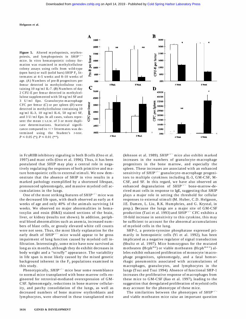
In order to determine if these perturbations in the hemo-poietic organs and peripheral blood arise at the progeni-tor level, bone marrow cells from SHIP+/+, SHIP+/−, andSHIP−/− mice were plated in methylcellulose-based me-dia and various assays performed. Absolute numbers ofclonogenic progenitors observed with +/− cells weresimilar to wild-type levels (data not shown). Numbers ofpre-B lymphoid (Fig. 5A) colony-forming cells were re-duced to 39% of normal in mice 4–5 weeks of age,whereas numbers of day 2 colony-forming unit-erythroid(CFU-E) (Fig. 5B) progenitors were reduced to 47% of thelevels observed in +/+ littermates. In both cases the re-duction in progenitor numbers was more pronounced inanimals 8–10 weeks of age. Neither day 3 burst formingunit (BFU)-erythroid nor day 10 BFU-E progenitor num-bers were significantly reduced (results not shown), sug-gesting a block in the late stages of erythroid maturation.Although slight increases were observed in the numberof granulocyte–macrophage colony forming cells (CFC)in SHIP−/− bone marrow (Fig. 5C), dramatic elevationswere seen in the spleen (Fig. 5D). For example, CFCnumbers were elevated almost 70-fold in the spleens ofSHIP−/− mice 8–10 weeks of age. We observed similarincreases in numbers of CFC-GM, and decreases in CFU-E, using the SHIP−/− ES cells in an in vitro differentiationsystem (not shown) suggesting that these variations indifferentiation potential are intrinsic to the progenitorcells.
Hemopoietic progenitors lacking SHIP arehyper-responsive to multiple cytokines
The generation and maturation of CFC can be achievedboth in vitro and in vivo using a number of differentgrowth factors. We therefore examined the colony-form-ing ability of SHIP−/− bone marrow cells in methylcel-lulose-based media containing various concentrations ofcytokines that play a role in myeloid development. Un-der these conditions, SHIP−/− bone marrow progenitorsexhibited an enhanced sensitivity to all growth factorsexamined (Fig. 6). Both GM-CSF (Fig. 6A) and IL-3 (Fig.6B) yielded 50% maximal colony formation at a 10-foldlower concentration using SHIP−/− cells compared tothose of +/+ and +/− littermates. These observationswere confirmed using bone marrow derived from a sec-ond, independently derived line of SHIP null mice (notshown). In addition, SHIP−/− bone marrow cells weresome two- to threefold more sensitive to SF (Fig. 6C) andM-CSF (Fig. 6D).
In an attempt to determine if the absence of SHIP in-fluenced colony size, suspension cultures were estab-lished with SHIP+/+ or SHIP−/− bone marrow cells in thepresence of 0.01 ng/ml GM-CSF or 1.0 ng/ml IL-3 underconditions similar to those used to assess colony forma-tion. SHIP−/− cultures yielded more cells in the presenceof either GM-CSF or IL-3 than littermate control cul-tures. Moreover, if the cell yields from these suspensioncultures were divided by the number of colonies gener-ated in methylcellulose, it appears that the GM-CSF-stimulated SHIP−/− colonies were approximately doublethe size of those generated by the wild-type cells. In con-trast, no difference in colony size was observed in thepresence of IL-3.
Growth-factor-independent colony formation is oneproperty of certain transformed cell populations. In order
Table 2. Hematological parameters of SHIP F2 mice
ParameterAge
(weeks) +/+a +/−a −/−a
Hematocrit (%) 4–5 47.5 ± 1.3 49.0 ± 1.2 48.9 ± 2.88–10 50.7 ± 1.0 47.2 ± 2.6 42.4 ± 5.7
WBC (×107/ml) 4–5 1.1 ± 0.0 1.1 ± 0.1 1.3 ± 0.38–10 1.0 ± 0.1 0.9 ± 0.1 1.0 ± 0.1
RBC (×109/ml) 4–5 9.9 ± 0.8 10.4 ± 0.3 10.9 ± 1.08–10 10.0 ± 0.3 10.1 ± 0.3 8.4 ± 0.7
Peripheral blooddifferential counts (%)a 4–5 L 68.8 ± 1.8 62.3 ± 2.4 45.3 ± 4.5**
M 15.5 ± 0.8 13.3 ± 0.4 23.3 ± 3.0*N 14.8 ± 1.9 19.0 ± 0.7 28.0 ± 2.1**E 0 5.5 ± 2.4 3.3 ± 0.4
8–10 L 73.3 ± 2.6 67.3 ± 5.6 39.2 ± 3.2**M 10.0 ± 1.4 10.0 ± 3.0 25.0 ± 4.2*N 13.3 ± 1.1 23.3 ± 3.4* 35.5 ± 7.3*E 1.3 ± 0.9 0.8 ± 0.7 0.5 ± 0.3
aValues represent the mean ± S.E.M. for at least three animals per determination. Statistical significance compared with the +/+populations was determined using the Student’s t-test where P ø 0.05 (*) and P ø 0.005 (**).aWright–Giemsa-stained smears of peripheral blood were scored microscopically based on morphology. (L) lymphocyte; (M) monocyte;(N) neutrophil; (E) eosinophil and others.
Helgason et al.
1614 GENES & DEVELOPMENT
Cold Spring Harbor Laboratory Press on April 14, 2019 - Published by genesdev.cshlp.orgDownloaded from

to evaluate the possibility that SHIP−/− progenitors pos-sessed such potential, cells from all three genotypes ofmice were plated in methylcellulose-based media con-taining 15% serum, but no growth factors. No coloniesgreater than 20 cells were observed in cultures from thewild-type, heterozygous, or SHIP null mice. However,51.0 ± 3.9% of the SHIP−/− CFC detectable in cytokine-containing media were capable of giving rise to smallclusters ranging in size from 5–20 cells. No such clusterswere observed using cells from +/+ or +/− mice, suggest-
ing that SHIP−/− progenitors exhibit enhanced survival,but only minimal proliferation, in the absence of exog-enous growth factors.
Discussion
Tyrosine phosphorylation of SHIP is an early response tostimulation of many hemopoietic cell-surface receptors(Cutler et al. 1993; Damen et al. 1993; Lioubin et al.1994). SHIP has been demonstrated to play a major role
Figure 4. Representative FACS profiles of 4–5-week-old SHIP F2 mice. Five mice of each genotypewere analyzed and the average percentages of posi-tive cells determined. A FACS profile representa-tive of each of these values is presented for bonemarrow (A), spleen (B), and thymus (C) of SHIPwild-type (+/+) or null (−/−) mice. Numbers repre-sent the percent of viable (PI−), positively stainedcells within each marked region.
Hemopoietic and lung abnormalities in SHIP null mice
GENES & DEVELOPMENT 1615
Cold Spring Harbor Laboratory Press on April 14, 2019 - Published by genesdev.cshlp.orgDownloaded from
in FcgRIIB inhibitory signaling in both B cells (Ono et al.1997) and mast cells (Ono et al. 1996). Thus, it has beenpostulated that SHIP may play a central role in nega-tively regulating the responses of both primitive and ma-ture hemopoietic cells to external stimuli. We now dem-onstrate that the absence of SHIP in vivo results in amarked pathology exemplified by a shortened lifespan,pronounced splenomegaly, and massive myeloid cell ac-cumulations in the lungs.
One of the most striking features of SHIP−/− mice wasthe decreased life span, with death observed as early as 4weeks of age and only 40% of the animals surviving 14weeks. We observed no major abnormalities in hema-toxylin and eosin (H&E) stained sections of the brain,liver, or kidney (results not shown). In addition, periph-eral blood abnormalities such as anemia, increased num-bers of blast cells, or grossly elevated white cell countswere not seen. Thus, the most likely explanation for theearly death of SHIP−/− mice would appear to be grossimpairment of lung function caused by myeloid cell in-filtration. Interestingly, some mice have now survived aslong as six months, although they do exhibit decreases inbody weight and a ‘‘scruffy’’ appearance. The variabilityin life span is most likely caused by the mixed geneticbackground inherent in the F2 populations examined inthis study.
Phenotypically, SHIP−/− mice bear some resemblanceto normal mice transplanted with bone marrow cells en-gineered for retroviral-mediated overexpression of GM-CSF. Splenomegaly, reductions in bone marrow cellular-ity, and patchy consolidation of the lungs, as well asdecreased numbers of bone marrow erythroblasts andlymphocytes, were observed in these transplanted mice
(Johnson et al. 1989). SHIP−/− mice also exhibit markedincreases in the numbers of granulocyte–macrophageprogenitors in the bone marrow, and especially thespleen. These increases are associated with an enhancedsensitivity of SHIP−/− granulocyte–macrophage progeni-tors to multiple cytokines including IL-3, GM-CSF, M-CSF, and SF. In this regard, we have also observed anenhanced degranulation of SHIP−/− bone-marrow-de-rived mast cells in response to IgE, suggesting that SHIPplays a major role in setting the threshold for cellularresponses to external stimuli (M. Huber, C.D. Helgason,J.E. Damen, L. Liu, R.K. Humphries, and G. Krystal, inprep.). Because the lungs are a major site of GM-CSFproduction (Tazi et al. 1993) and SHIP−/− CFC exhibits a10-fold increase in sensitivity to this cytokine, this maybe sufficient to account for the abnormal accumulationsof myeloid cells in the lung.
SHP-1, a protein-tyrosine phosphatase expressed pri-marily in hemopoietic cells (Yi et al. 1992), has beenimplicated as a negative regulator of signal transduction(Shultz et al. 1997). Mice homozygous for the mutatedmotheaten (Hcphme) or viable motheaten (Hcphme-v) al-leles exhibit enhanced proliferation of monocyte/macro-phage progenitors, splenomegaly, and a fatal hemor-rhagic pneumonitis associated with accumulations ofmacrophages, granulocytes, and lymphocytes in thelungs (Tsui and Tsui 1994). Absence of functional SHP-1increases the proliferative response of macrophages fromthese mice to GM-CSF (Jiao et al. 1997), leading to thesuggestion that deregulated proliferation of myeloid cellsmay account for the phenotype of these mice.
The similarities between the phenotypes of SHIP−/−
and viable motheaten mice raise an important question
Figure 5. Altered myelopoiesis, erythro-poiesis, and lymphopoiesis in SHIP−/−
mice. In vitro hematopoietic colony for-mation was examined in methylcellulosecolony assays using cells from wild-type(open bars) or null (solid bars) SHIP F2 lit-termates at 4–5 weeks and 8–10 weeks ofage. (A) Numbers of pre-B progenitors perfemur detected in methylcellulose con-taining 10 ng/ml IL-7. (B) Numbers of day2 CFU-E per femur detected in methylcel-lulose supplemented with 50 ng/ml SF and3 U/ml Epo. Granulocyte–macrophageCFC per femur (C) or per spleen (D) weredetected in methylcellulose containing 10ng/ml IL-3, 10 ng/ml IL-6, 50 ng/ml SF,and 3 U/ml Epo. In all cases, values repre-sent the mean ± S.E.M. of 3 or more dupli-cate determinations. Statistical signifi-cance compared to +/+ littermates was de-termined using the Student’s t-test.P ø 0.05 (*); P ø 0.01 (**).
Helgason et al.
1616 GENES & DEVELOPMENT
Cold Spring Harbor Laboratory Press on April 14, 2019 - Published by genesdev.cshlp.orgDownloaded from

regarding the relative importance of SHIP and SHP-1 innegatively regulating cytokine signaling. That is, dothese molecules function independently, within thesame pathway, or in an overlapping manner? Studies us-ing homologous recombination in the chicken DT-40cell line suggest that SHP-1 and SHIP function indepen-dently in response to signaling through different recep-tors (Ono et al. 1997). For example, signals initiated bythe killer cell inhibitory receptor are mediated by SHP-1,but not SHIP. Conversely, the proapoptotic signals ini-tiated by FcgRIIB signaling are attenuated by SHIP butnot SHP-1, suggesting that SHIP−/− mice should exhibitB-lymphoid perturbations. In fact, we observe decreasedpercentages of B220+ cells in the bone marrow and spleenof SHIP−/− mice and pre-B colony-forming cell numbersare significantly reduced. Further phenotypic and func-tional analyses of the B-lymphoid populations are cur-rently in progress to evaluate the consequences of loss ofSHIP-mediated negative signaling.
Several signal transduction pathways may be involved
in the enhanced growth factor sensitivity of the SHIP−/−
progenitors. One possible mechanism involves hyperac-tivation of the Ras pathway. SHIP associates with theprotein tyrosine phosphatase SHP-2 in response togrowth factors such as IL-3 and erythropoietin (Liu et al.1997b; Sattler et al. 1997). Because the activity of SHP-2is postulated to contribute to Ras-MAP kinase activa-tion, association of SHIP with SHP-2 could modulateactivity of the Ras pathway. In addition, SHIP may com-pete with Grb2 for Shc, thereby regulating the levels ofShc–Grb2 association within the cell (Liu et al. 1994;Tridandapani et al. 1997). In either case, absence of SHIPcould theoretically lead to enhanced Ras activity. Al-though we have not yet examined the phosphorylation ofShc or activation of the Ras pathway in cytokine-stimu-lated bone marrow progenitors, studies with IgE-stimu-lated SHIP−/− mast cells suggest that Shc phosphoryla-tion is markedly reduced compared to IgE-stimulatedSHIP+/+ mast cells (M. Huber, C.D. Helgason, J.E.Damen, L. Liu, R.K. Humphries, and G. Krystal, in
Figure 6. Altered growth factor responsiveness of SHIP−/− CFC. Bone marrow derived from wild type (– –), heterozygous (– . –), or null(—) littermates was plated in methylcellulose containing the indicated concentrations of (A) GM-CSF, (B) IL-3, (C) Steel factor, or (D)M–CSF. Colonies of >20 cells were scored on day 10 of culture and the numbers of colonies present at each cytokine concentrationwere calculated as the percent of the number formed in the highest concentration of the cytokine indicated. Values represent themean ± S.E.M. for duplicate determinations using at least three mice per group. Statistical significance compared to +/+ littermates wasdetermined using the Student’s t-test. P ø 0.05 (*); P ø 0.008 (***).
Hemopoietic and lung abnormalities in SHIP null mice
GENES & DEVELOPMENT 1617
Cold Spring Harbor Laboratory Press on April 14, 2019 - Published by genesdev.cshlp.orgDownloaded from

prep.). Experiments are now in progress to examine thesebiochemical pathways in more detail.
A second possible mechanism underlying the en-hanced proliferative potential of SHIP−/− granulocyte–macrophage progenitors resides in the phosphatase ac-tivity of SHIP. The PI-3-K pathway plays an importantrole in signaling through cytokine receptors. For ex-ample, PIP3 is capable of stimulating members of theprotein kinase C family (Toker et al. 1994), as well asmodulating the activity of the Akt/PKB kinase (Marteand Downward 1997). In the absence of SHIP, which me-tabolizes this PI-3-K product, uncontrolled activation ofthese pathways could result in enhanced cell survivaland proliferation.
In conclusion, we have provided evidence that sup-ports the hypothesis that SHIP is an important negativeregulator of cytokine signaling in cells of the hemopoi-etic system. SHIP−/− mice provide a powerful model inwhich to further explore SHIP function in hemopoieticstem and progenitor cell compartments, as well as inmature cells. In addition, studies with cells from thesemice should further our understanding of the molecularmechanisms by which signaling thresholds are estab-lished in both mature cells and progenitors.
Materials and methods
SHIP gene targeting
A genomic DNA library derived from the 129 mouse strain wasscreened with a 600-bp fragment derived from the 58 end of theSHIP cDNA. Positive clones were analyzed by restriction map-ping and sequence analysis. The targeting vector was engineeredto contain a 254-bp ApaI–BamHI deletion, thus effectively re-moving the transcriptional start site and most of the first exon.A Tk–neo cassette derived from pMC1TkNeoPolyA was sub-cloned into the deletion site in the opposite transcriptional ori-entation. The targeting vector contains 1.8 kb of SHIP homolo-gous 58 DNA and 6.0 kb of 38 SHIP genomic DNA. A secondtargeting vector was constructed to include the HSV–Tk genefrom pMC1–Tk at the 38 end of the targeting sequence.
Each linearized targeting construct was electroporated intothe R1 ES cell line and colonies were isolated following selec-tion in either G418 alone or G418 plus gancyclovir. GenomicDNA was isolated from pools of 6–8 clones and analyzed byPCR analysis using Elongase (Life Technologies) to generate a2662-bp amplicon derived from an internal Neo primer (58-CAAGATGGATTGCACGCAGG) and a 58 SHIP primer (G3:58-CCAGAAGTGTCTCTATCATGATAGT). Positive colonieswere expanded and purified genomic DNA was digested withKpnI and analyzed by Southern blot analysis using a 2-kb KpnI–HindIII genomic probe. By Southern analysis, the nontargetedSHIP allele and the positively targeted allele were visualized as4.6- and 5.8-kb bands, respectively. One clone out of 300 waspositive using the double-selection protocol, whereas 7 positiveclones were obtained with the single-selection vector. Similarblots were also probed with a Neor fragment to confirm singleintegrations (results not shown).
Germ-line transmission chimeras of clone 4.8B (double-selec-tion vector) and clone 5.1 (single-selection vector) were gener-ated by injection of C57BL/6J blastocysts, followed by breedingonto a C57BL/6J background. Genotype analysis was routinelydone using Southern blot analysis and the probe described
above. Animals were housed in microisolator units and pro-vided with sterilized food and water. Routine testing for viralpathogens and mycoplasma was carried out on both animalcolonies in which the two independent lines were maintained.
Homozygous deletion SHIP ES cells were generated by geneconversion in increasing concentrations of G418 using previ-ously described techniques (Mortensen et al. 1992). Southernblot analysis of genomic DNA confirmed the absence of theSHIP allele. Karyotype analysis was carried out on all clonesused both in vitro and in vivo in this study.
Western blot analysis
Equivalent numbers of nucleated cells, derived from hemopoi-etic suspension cultures of day 10 embryoid bodies (differenti-ated as described previously; Helgason et al. 1996) generatedwith SHIP wild-type, heterozygous, and null R1 ES cells, werewashed once in phosphate-buffered saline, solubilized with1.0% Triton-X 100 at 4°C, and subjected to Western blot analy-sis as described previously (Damen et al. 1993). Bone marrowcells from all three genotypes of mice were treated in a similarmanner. The SHIP antibodies used for Western blot analysiswere generated against the SH2 domain and the region spanningthe two NPxY motifs using GST fusion proteins as describedpreviously (Liu et al. 1997a).
Preparation of sections and slides
Selected organs were fixed in a buffered 4% paraformaldehydesolution, dehydrated in ethanol, and embedded in paraffin forsectioning. Sections were prepared and H&E stained at the Aca-demic Pathology Laboratory, University of British Columbia,Vancouver using standard protocols. Cytospin preparations ofbone marrow and spleen, as well as peripheral blood smears,were routinely stained with a modified Wright-Geimsa stain.
Assays to detect clonogenic progenitors
Nucleated cell counts were performed on bone marrow aspi-rates or cell suspensions of spleen, prepared using a nylon meshscreen. Appropriate cell numbers were plated in a 1.1 ml vol-ume per Petri dish in standard conditions to detect the variousclonogenic progenitors. All cell culture was carried out in ahumidified incubator at 37°C with 5% CO2. Bone marrow pre-Bprogenitors were detected by culture in methylcellulose mediacontaining 10 ng/ml IL7 for 5 to 7 days [StemCell TechnologiesInc. (STI), Vancouver; Methocult M3630]. Methocult M3230(STI) was supplemented with 50 ng/ml Steel factor (supplied asa supernatant from Cos cells engineered to express the protein)and 3 U/ml rhEpo for detection of day 2 CFU-E and day 3 BFU-E.Methocult M3434 (STI) containing 10 ng/ml rmIL-3, 10 ng/mlrhIL-6, 50 ng/ml rmSF, and 3 U/ml rhEpo was used for thedetection of myeloid (CFC), late erythroid, and multipotentialprogenitors in bone marrow and spleen cell preparations. Allcolonies were scored microscopically using standard criteria.
Bone marrow cells derived from mice of all three genotypeswere cultured in Methocult M3234 (STI) in the indicated con-centrations of IL-3, rmGM-CSF (Peprotech), SF (supplied as aCos supernatant), or rhM-CSF (Genetics Institute, Cambridge,MA) for growth factor response curves. Colonies containing 20or more cells were scored. In all cases duplicate determinationswere performed on each sample. Statistical significance com-pared with the +/+ populations was determined using the Stu-dent’s t-test.
Helgason et al.
1618 GENES & DEVELOPMENT
Cold Spring Harbor Laboratory Press on April 14, 2019 - Published by genesdev.cshlp.orgDownloaded from

Flow cytometry
Bone marrow, spleen, thymus, or lymph node cells at a densityof 5–10 × 106 cells/ml were incubated on ice for 30 min with 3µg/ml 2.4G2 (murine anti-IgG Fc receptor antibody) followed byincubation on ice for 40 min with the various FITC-labeled orphycoerythrine-conjugated antibodies. Cells were washed twicein Hank’s balanced salt solution containing 2% fetal bovineserum at 4°C and propidium iodide (Sigma Chemicals, St. Louis,MI) at a concentration of 1 ug/ml was included in the finalwash. Cells were analyzed on a FACStar+ or FACSort (Becton-Dickinson, San Jose, CA).
The monoclonal antibodies used for analysis included: E13–161.7 (anti-Sca-1), RB6–8C5 (anti-Gr-1; granulocytes), M1/70(anti-Mac-1; macrophages), Ter119 (anti-erythroid lineage) (thesources of these antibodies have been described elsewhere;Rebel et al. 1996). Antibodies against CD4, CD8, B220, andCD11b were purchased from Pharmingen (Mississauga, On-tario, Canada).
Acknowledgments
The authors thank James Ihle for helpful discussions regardingthe targeting strategy. In addition, the authors wish to thankGayle Thornbury and Giovanna Cameron for expert technicalassistance on the FACStar+, Gloria Shaw for karyotype analysisof the ES clones, Malin Parmar for assistance with PCR andSouthern blot analysis, and Julie Chow for preparation andstaining of the lung sections, as well as Rosemary Hood, Chris-tian Kalberer, Ling Liu, and Cindy Miller for helpful discus-sions. This work was supported by the National Cancer Insti-tute of Canada (with funds from the Canadian Cancer Societyand the Terry Fox Run) and the Medical Research Council ofCanada.
The publication costs of this article were defrayed in part bypayment of page charges. This article must therefore be herebymarked ‘‘advertisement’’ in accordance with 18 USC section1734 solely to indicate this fact.
References
Cutler, R.L., L. Liu, J.E. Damen, and G. Krystal. 1993. Multiplecytokines induce the tyrosine phosphorylation of Shc and itsassociation with Grb2 in hemopoietic cells. J. Biol. Chem.268: 21463–21465.
Damen, J.E., L. Liu, R.L. Cutler, and G. Krystal. 1993. Erythro-poietin stimulates the tyrosine phosphorylation of Shc andits association with Grb2 and a 145-Kd tyrosine phosphory-lated protein. Blood 82: 2296–2303.
Damen, J.E., L. Liu, P. Rosten, R.K. Humphries, A.B. Jefferson,P.W. Majerus, and G. Krystal. 1996. The 145-kDa proteininduced to associate with Shc by multiple cytokines is aninositol tetraphosphate and phosphatidylinositol 3,4,5-tris-phosphate 5-phosphatase. Proc. Natl. Acad. Sci. 93: 1689–1693.
Helgason, C.D., G. Sauvageau, H.J. Lawrence, C. Largman, andR.K. Humphries. 1996. Overexpression of HOXB4 enhancesthe hematopoietic potential of embryonic stem cells differ-entiated in vitro. Blood 87: 2740–2749.
Jiao, H., W. Yang, K. Berrada, M. Tabrizi, L. Shultz, and T. Yi.1997. Macrophages from motheaten and viable motheatenmutant mice show increased proliferative responses to GM-CSF: Detection of potential HCP substrates in GM-CSF sig-nal transduction. Exp. Hematol. 25: 592–600.
Johnson, G.R., T.J. Gonda, D. Metcalf, I.K. Harihan, and S. Cory.
1989. A lethal myeloproliferative syndrome in mice trans-planted with bone marrow cells infected with a retrovirusexpressing granulocyte-macrophage colony stimulating fac-tor. EMBO J. 8: 441–448.
Kapeller, R. and L.C. Cantley. 1994. Phosphatidylinositol 3-ki-nase. Bioessays 16: 565–576.
Kavanaugh, W.M., D.A. Pot, S.M. Chin, M. Deuterreinhard,A.B. Jefferson, F.A. Norris, F.R. Masiarz, L.S. Cousens, P.W.Majerus, and L.T. Williams. 1996. Multiple forms of an ino-sitol polyphosphate 5-phosphatase form signaling complexeswith SHC and GRB2. Curr. Biol. 6: 438–445.
Lioubin, M.N., G.M. Myles, K. Carlberg, D. Botwell, and L.R.Rohrschneider. 1994. SHC, GRB2, SOS1 and a 150-kilodal-ton tyrosine-phosphorylated protein form complexes withFms in hematopoietic cells. Mol. Cell. Biol. 14: 5682–5691.
Lioubin, M.N., P.A. Algate, S. Tsai, K. Carlberg, R. Aebersold,and L.R. Rohrschneider. 1996. p150SHIP, a signal transduc-tion molecule with inositol polyphosphate-5-phosphataseactivity. Genes & Dev. 10: 1084–1095.
Liu, L., J.E. Damen, R.L. Cutler, and G. Krystal. 1994. Multiplecytokines stimulate the binding of a common 145-kilodaltonprotein to Shc at the Grb2 recognition site of Shc. Mol. Cell.Biol. 14: 6926–6935.
Liu, L., J.E. Damen, M.R. Hughes, I. Babic, F.R. Jirik, and G.Krystal. 1997a. The Src homology 2 (SH2) domain of SH2-containing inositol phosphatase (SHIP) is essential for tyro-sine phosphorylation of SHIP, its association with Shc, andits induction of apoptosis. J. Biol. Chem. 272: 8983–8988.
Liu, L., J.E. Damen, M.D. Ware, and G. Krystal. 1997b. Inter-leukin-3 induces the association of the inositol 5-phospha-tase SHIP with SHP2. J. Biol. Chem. 272: 10998–11001.
Liu, L., J.E. Damen, M. Ware, M. Hughes, and G. Krystal. 1997c.SHIP, a new player in cytokine-induced signaling. Leukemia11: 181–184.
Marte, B.M. and J. Downward. 1997. PKB/Akt: Connectingphosphoinositide 3-kinase to cell survival and beyond.Trends Biol. Sci. 22: 355–358.
Mortensen, R.M., D.A. Conner, S. Chao, A.A.T. Geisterfer-Low-rance, and J.G. Seidman. 1992. Production of homozygousmutant ES cells with a single targeting construct. Mol. Cell.Biol. 12: 2391–2395.
Nadler, M.J.S., B. Chen, J.S. Anderson, H.H. Wortis, and B.G.Neel. 1997. Protein-tyrosine phosphatase SHP-1 is dispens-able for FcgRIIB-mediated inhibition of B cell antigen recep-tor activation. J. Biol. Chem. 272: 20038–20043.
Ono, M., S. Bolland, P. Tempst, and J.V. Ravetch. 1996. Role ofthe inositol phosphatase SHIP in negative regulation of theimmune system by the receptor FcgRIIB. Nature 383: 263–266.
Ono, M., H. Okada, S. Bolland, S. Yanagi, T. Kurosaki, and J.V.Ravetch. 1997. Deletion of SHIP or SHP-1 reveals two dis-tinct pathways for inhibitory signaling. Cell 90: 293–301.
Osborne, M.A., G. Zenner, M. Lubinus, X. Zhang, Z. Songyang,L.C. Cantley, P. Majerus, P. Burn, and J.P. Kochan. 1996. Theinositol 58-phosphatase SHIP binds to immunoreceptor sig-naling motifs and responds to high affinity IgE receptor ag-gregation. J. Biol. Chem. 271: 29271–29278.
Ravichandran, K.S., K.K. Lee, Z. Songyang, L.C. Cantley, P.Burn, and S.J. Burakoff. 1993. Interaction of shc with the zetachain of the T cell receptor upon T cell activation. Science262: 902–905.
Rebel, V.I., C.L. Miller, G.R. Thornbury, W.H. Dragowska, C.J.Eaves, and P.M. Lansdorp. 1996. A comparison of long-termrepopulating hematopoietic cells in fetal liver and adult bonemarrow from the mouse. Expt. Hematol. 24: 638–648.
Sattler, M., R. Salgia, G. Shrikhande, S. Verma, J.-L. Choi, L.R.
Hemopoietic and lung abnormalities in SHIP null mice
GENES & DEVELOPMENT 1619
Cold Spring Harbor Laboratory Press on April 14, 2019 - Published by genesdev.cshlp.orgDownloaded from

Rohrschneider, and J.D. Griffin. 1997. The phosphatidylino-sitol polyphosphate 5-phosphatase SHIP and the protein ty-rosine phosphatase SHP-2 form a complex in hematopoieticcells which can be regulated by BCR/ABL and growth fac-tors. Oncogene 15: 2379–2384.
Saxton, T.M., I. Van Ostveen, D. Botwell, R. Aebersold, andM.R. Gold. 1994. B cell antigen receptor cross-linking in-duces tyrosine phosphorylation of the p21ras oncoproteinactivators SHC and SOS1 as well as assembly of complexescontaining SHC, GRB-2, mSOS1, and a 145-kD tyrosine-phosphorylated protein. J. Immunol. 153: 623–636.
Scharenberg, A.M. and J.-P. Kinet. 1996. The emerging field ofreceptor-mediated inhibitory signaling: SHP or SHIP? Cell87: 961–964.
Shultz, L.D., T.V. Rajan, and D.L. Greiner. 1997. Severe defectsin immunity and hematopoiesis caused by SHP-1 protein-tyrosine-phosphatase deficiency. Trends Biotechnol.15: 302–307.
Tazi, A., F. Bouchonnet, M. Grandsaigne, L. Boumsell, A.J.Hance, and P. Soler. 1993. Evidence that granulocyte mac-rophage-colony-stimulating factor regulated the distributionand differentiated state of dendritic cells/Langerhans cells inhuman lung and lung cancers. J. Clin. Invest. 91: 566–576.
Toker, A.M., M. Meyer, K.K. Reddy, J.R. Falck, R. Aneja, S.Aneja, A. Parra, D.J. Burns, L.M. Ballas, and L.C. Cantley.1994. Activation of protein kinase C family members by thenovel polyphosphoinositides PtdIns-3,4-P2 and PtdIns-3,4,5-P3. J. Biol. Chem. 269: 32358–32367.
Tridandapani, S., T. Kelley, D. Cooney, M. Pradhan, and K.M.Coggeshall. 1997. Negative signaling in B cells: SHIP GrbsShc. Immunol. Today 18: 424–427.
Tsui, F.W.L. and H.W. Tsui. 1994. Molecular basis of the moth-eaten phenotype. Immunol. Rev. 138: 185–206.
Yi, T.L., J.L. Cleveland, and J.N. Ihle. 1992. Protein tyrosinephosphatase containing SH2 domains: Characterization,preferential expression in hematopoietic cells, and localiza-tion to human chromosome 12p12-p13. Mol. Cell. Biol.12: 836–846.
Helgason et al.
1620 GENES & DEVELOPMENT
Cold Spring Harbor Laboratory Press on April 14, 2019 - Published by genesdev.cshlp.orgDownloaded from
10.1101/gad.12.11.1610Access the most recent version at doi: 12:1998, Genes Dev.
Cheryl D. Helgason, Jacqueline E. Damen, Patty Rosten, et al.
span pathology, and a shortened life leads to hemopoietic perturbations, lungSHIPTargeted disruption of
References
http://genesdev.cshlp.org/content/12/11/1610.full.html#ref-list-1
This article cites 29 articles, 16 of which can be accessed free at:
License
ServiceEmail Alerting
click here.right corner of the article or
Receive free email alerts when new articles cite this article - sign up in the box at the top
Cold Spring Harbor Laboratory Press
Cold Spring Harbor Laboratory Press on April 14, 2019 - Published by genesdev.cshlp.orgDownloaded from

![Trends - SciELO · Trends Psychiatry Psychother. 2014;36(2) ... sensorimotor, [2] ... obsessions, or compulsions may help us distinguish](https://img.pdfslide.us/doc/110x75/5ada19d77f8b9a137f8cff51/trends-psychiatry-psychother-2014362-sensorimotor-2-obsessions.jpg)