Embed Size (px)
Citation preview

Ethanol Attenuates Histiotrophic Nutrition Pathways
and Alters the Intracellular Redox Environment and
Thiol Proteome during Rat OrganogenesisJoseph L. Jilek,* Karilyn E. Sant,* Katherine H. Cho,* Matthew S. Reed,†
Jan Pohl,† Jason M. Hansen,‡ and Craig Harris*,1
*Department of Environmental Health Sciences, University of Michigan, Ann Arbor, Michigan 48109;†Biotechnology Core Facility Branch, Centers for Disease Control, Atlanta, Georgia 30333; and ‡Department ofPhysiology and Developmental Biology, College of Life Sciences, Brigham Young University, Provo, Utah 846021To whom correspondence should be addressed at Department of Environmental Health Sciences, University of Michigan, School of Public Health, M6667SPH II, 1415 Washington Heights, Ann Arbor, MI 48109-2029. Fax: 734 936-7283. E-mail: [email protected].
ABSTRACT
Ethanol (EtOH) is a reactive oxygen-generating teratogen involved in the etiology of structural and functionaldevelopmental defects. Embryonic nutrition, redox environment, and changes in the thiol proteome following EtOHexposures (1.56.0 mg/ml) were studied in rat whole embryo culture. Glutathione (GSH) and cysteine (Cys) concentrationswith their respective intracellular redox potentials (Eh) were determined using high-performance liquid chromatography.EtOH reduced GSH and Cys concentrations in embryo (EMB) and visceral yolk sac (VYS) tissues, and also in yolk sac andamniotic fluids. These changes produced greater oxidation as indicated by increasingly positive Eh values. EtOH reducedhistiotrophic nutrition pathway activities as measured by the clearance of fluorescin isothiocyanate (FITC)-albumin fromculture media. A significant decrease in total FITC clearance was observed at all concentrations, reaching approximately50% at the highest dose. EtOH-induced changes to the thiol proteome were measured in EMBs and VYSs using isotope-coded affinity tags. Decreased concentrations for specific proteins from cytoskeletal dynamics and endocytosis pathways(a-actinin, a-tubulin, cubilin, and actin-related protein 2); nuclear translocation (Ran and RanBP1); and maintenance ofreceptor-mediated endocytosis (cubilin) were observed. Kyoto encyclopedia of genes and genomes (KEGG) pathway analysisalso identified a decrease in ribosomal proteins in both EMB and VYS. Results show that EtOH interferes with nutrient uptaketo reduce availability of amino acids and micronutrients required by the conceptus. Intracellular antioxidants such as GSH andCys are depleted following EtOH and Eh values increase. Thiol proteome analysis in the EMB and VYS show selectively alteredactin/cytoskeleton, endocytosis, ribosome biogenesis and function, nuclear transport, and stress-related responses.
Key words: embryo; visceral yolk sac; organogenesis; ethanol; histiotrophic nutrition; glutathione; cysteine; redox potential;redox environment; thiol proteome
Fetal alcohol syndrome (FAS) and fetal alcohol spectrum disor-ders (FASD) are well-characterized consequences of in utero ex-posure to ethanol (EtOH), manifest as a variety of clinicalconditions. These range from behavioral and cognitive defects(Coriale et al. 2013) to craniofacial malformations. However, dueto the complex nature of EtOH exposure and timing during de-velopment, the precise mechanisms governing the teratogenic
activity of EtOH are not well understood. The clinical manifesta-tions of EtOH exposure during organogenesis are believed to in-clude a combination of disrupted cellular and tissue-levelprocesses, including oxidative stress and free radical damage(Dong et al. 2008; Miller et al. 2013; Miller-Pinsler et al. 2015;Smith 1997; Sulik et al. 1988), DNA methylation and histonemodifications (Kaminen-Ahola et al. 2010; Park et al. 2003;
VC The Author 2015. Published by Oxford University Press on behalf of the Society of Toxicology.All rights reserved. For Permissions, please e-mail: [email protected]
475
TOXICOLOGICAL SCIENCES, 147(2), 2015, 475–489
doi: 10.1093/toxsci/kfv145Advance Access Publication Date: July 15, 2015Research Article
Downloaded from https://academic.oup.com/toxsci/article-abstract/147/2/475/1621904by gueston 24 March 2018

Ungerer et al. 2013), and dysregulation of microRNA expression(Balaraman et al. 2013; Wang et al. 2009), among others. Previousstudies have identified the damaging potential of EtOH to be re-lated to its ability to promote excess generation of reactive oxy-gen species (ROS). The specific source of ROS can be traced toproducts of EtOH biotransformation, release from the mito-chondrial respiratory chain or the activation of NADPH oxidases(Dong et al. 2010; Hill et al. 2014). Due to the spatial and temporalcomplexities of mammalian embryogenesis, increased concen-trations of ROS may originate from various cells and tissues de-pendent on developmental stage and related metabolic factors.Examples of cell selectivity for EtOH-induced ROS generation in-clude the populations of neural crest cells that actively migrateduring early embryogenesis to serve as precursors for nerves,glia, glands, muscle, and several other essential structures. Asone of the major targets of EtOH toxicity, perturbation of neuralcrest cell migration and differentiation strongly correlate withFAS and FASD outcomes. Neural crest cells are known to be de-ficient in several cellular antioxidant capacities, and it has beensuggested that this characteristic contributes greatly to in-creased ROS accumulation where alterations in the cellular re-dox environments may be a key step in the general mechanismof the teratogenic effects of EtOH (Davis 1990). The administra-tion of sulforaphane, an inducer of antioxidant responsesthrough Nrf2-mediated pathways, has been shown to provideprotection from ROS in neural crest cells (Chen et al. 2013). In al-most any cellular context, excessive ROS will produce the com-mon consequence of depleting glutathione (GSH) and resultingin the net oxidation of intracellular thiols. Classic definitions of“oxidative stress” would suggest that this overoxidation resultsonly in damaging or deleterious consequences. More contempo-rary views emphasize the roles of cellular oxidation and reduc-tion as critical signaling elements, which are necessary in normaland abnormal cellular regulation and control (Guttmann 2010;Jones 2006). Characterization of the broader cellular redox envi-ronment through the measurement of intracellular redox poten-tials (Eh) provides a means to compare the magnitude of redoxchanges in distinct tissues and fluids (Jones 2002) The introduc-tion of a “systems level” thiol proteomics tool allows for the fur-ther characterization of what happens to the specific proteinsand relevant pathways that are part of the much larger conceptalredox circuitry (Chung et al. 2013; Go et al. 2014).
A central goal in the current work is to determine the extentto which the redox environments of major conceptal tissuesand fluids are altered by EtOH treatment. The numerous re-ports, including those listed earlier, linking increased genera-tion of ROS and developmental EtOH exposure to mechanismsof adverse functional and anatomical birth defects are instruc-tive but still fall short of describing the complete spectrum ofEtOH effects. Perceptions of ROS have changed dramaticallyover the past decade from an understanding that all ROS aredamaging, to the current view that ROS is an essential signalingmolecule and second messenger for normal cell regulation andcontrol (Finkel 2011; Guttmann 2010; Hansen and Harris 2013).Most of what ROS does in this capacity is directly linked to itsability to oxidize critical protein cysteine (Cys) sulfhydryl groupsin enzyme active sites, receptors, transporters, and transcrip-tion factors and thus affect changes in activity and control sig-naling. The scope of changes to these signaling nodes isextensive and complex and can be differentially impacted bythe specific chemical or insult causing the change in ROS status.These are occurring in conjunction with a general reprogrammingof genetic networks resulting from EtOH exposure (Green et al.2007) One, often underappreciated, factor that affects ROS-
mediated outcomes in cases such as EtOH exposure, is the nutri-tional state of the exposed organism, having an impact on thetype and quantity of proteins being synthesized during embryo-genesis and their functional states once formed (Dreosti 1993).
During the sensitive period of organogenesis, both rodentand human conceptuses are undergoing rapid growth and dif-ferentiation in a relatively hypoxic environment prior to the es-tablishment of a fully functional placenta (Harris et al. 2013). Inthe rodent and also likely in the human, nutrients are suppliedthrough the visceral yolk sac (VYS) (Burton et al. 2001), which ac-tively takes up maternal proteins by receptor-mediated endocy-tosis (RME) and degrades them via lysosomal proteolysis (Harriset al. 2013). These 2 processes constitute the histiotrophic nutri-tion pathways (HNP) which provide micronutrients to the em-bryo (EMB) for all of its biosynthesis and metabolism needsduring organogenesis (Beckman et al. 1998; Christensen andBirn 2002). Disruption of HNP by environmental or chemicalagents has been shown to contribute to significant growth retar-dation and dysmorphogenesis (Ambroso and Harris 1993; Brentet al. 1990). Chemical toxicants are known to affect the supply ofnecessary amino acids required for protein and nucleic acid bio-synthesis in the developing EMB. Recent work has also con-firmed that reductions in amino acid supply also significantlyincreases ROS generation (Ambroso et al. 1997; Arriazu et al.2010). Two major consequences of reduced amino acid avail-ability are reduced protein biosynthesis and reduced de novoGSH biosynthesis. These outcomes are linked by changes in thequantity of conceptal proteins and the functional quality of re-maining proteins regarding their cysteine redox status. Systemslevel assessment of qualitative changes to the thiol proteome(Fu et al. 2008) will allow us to identify additional specific path-ways and proteins most affected by EtOH exposure.
To better understand the mechanistic consequences of em-bryonic EtOH exposure, we have, in this report, attempted to in-tegrate information gathered from multiple experimentalplatforms. We provide the backdrop of a concentration depen-dent morphology assessment in whole embryo culture (WEC)and a detailed evaluation of GSH and Cys redox profiles. Undersimilar WEC conditions, we also show EtOH-induced decreasesin the histiotrophic uptake of proteins and correlate these withsystems level changes to the thiol proteome in EMB and VYS.Combined, these data help to integrate toxin-induced changesto proteins and their associated functional pathways throughoxidative posttranslational modifications for an improved un-derstanding of EtOH embryotoxicity that integrates ROS, nutri-tion, and proteomic changes.
MATERIALS AND METHODS
Chemicals and reagents. GSH, glutathione disulphide (GSSG), cys-teine, cysteine (CySS), gamma-glutamyl-glutamate, iodoaceticacid, iodoacetamide, bicinchoninic acid (BCA), fluorescein iso-thiocyanate albumin conjugate (bovine; fluorescin isothiocya-nate [FITC]-albumin), Triton X-100, tri-choloroacetic acid,sodium hydroxide, and sodium dodecylsulfate were purchasedfrom Sigma-Aldrich (St. Louis, Missouri). Dansyl chloride waspurchased from Fluka Chemie/Sigma-Aldrich. EtOH (200 proof)was purchased from Decon Labs, Inc (King of Prussia,Pennsylvania). Chloroform and sodium phosphate werepurchased from Fisher Scientific (Pittsburgh, Pennsylvania).Hanks balanced salt solution (HBSS) and penicillin/streptomy-cin (10 000 units/ml penicillinþ 10 000 lg/ml streptomycin in0.85% saline) were purchased from GIBCO/Life Technologies(Grand Island, New York). Cleavable isotope coded affinity tag
476 | TOXICOLOGICAL SCIENCES, 2015, Vol. 147, No. 2
Downloaded from https://academic.oup.com/toxsci/article-abstract/147/2/475/1621904by gueston 24 March 2018

(ICAT) reagent kits were purchased from AB Sciex/AppliedBiosystems (Framingham, Massachusetts).
Animals. All experiments were conducted in rat WEC (rWEC)using gestational day (GD) 10–11 conceptuses obtained from pri-magravida time-mated and specific pathogen free SpragueDawley rats purchased from Charles River Laboratories(Portage, Michigan). A sperm-positive vaginal smear on themorning following mating was used to confirm pregnancy andwas used to designate GD 0. Pregnant dams were shipped 4–5days prior to use, housed in solid bottom polycarbonate cageswith corn cob bedding, maintained on a 12-h light/dark roomcycle, and were allowed access to standard commercial rodentdiet and water ad libitum.
Culture conditions, exposure, and sampling. Anesthesia, exsangui-nation, and uteri removal were conducted as previouslydescribed, in accordance with approved institutional animalcare and use protocols (Harris et al. 2013). Immediately followingeuthanasia of GD 10 pregnant dams, uteri were removed, placedin HBSS (pH 7.4), and each implantation site was removed usingforceps and irridectomy scissors. Decidual masses were openedusing fine watchmakers forceps under HBSS and completelyintact conceptuses were removed. Reichert’s membranes weretorn away using fine watchmakers forceps and viable concep-tuses, with intact VYS and ectoplacental cones, were trans-ferred into 10 ml of warmed culture medium (not exceeding 1conceptus per milliliter of culture medium). Prior to addition ofconceptuses, culture bottles were saturated with 20% O2, 5%CO2, and 75% N2 and medium was warmed to 37�C. Culturemedia consisted of heat-inactivated rat serum (50%), HBSS (pH7.4, 50%), and 43 ll of penicillin/streptomycin. Culture mediawas resaturated with 95% O2, 5% CO2 after 20 h of culture (GD11) to ensure optimal growth and development. Prior to mor-phology assessment and other functional characterizations inGD 11, conceptal viability was confirmed by the presence of anactive heartbeat and vitelline blood circulation.
Three different EtOH treatment paradigms were used inthese studies. For all concentration response, morphologyassessment, and redox profiling protocols EtOH at concentra-tions of 1.5, 3.0, or 6.0 mg/ml was added directly to the culturemedia containing conceptuses at the onset of the culture period(GD 10) and remained in the media for the entire 26-h periodprior to harvesting and evaluation. HNP evaluations and expo-sures for assessment of the thiol proteome were initiated on GD11 after approximately 20 h in culture as described later.
Morphology assessment. Assessment of overall changes to con-ceptal growth and EMB morphology were made following a 26-hexposure of conceptuses to EtOH (1.5, 3.0, and 6.0 mg/ml) inWEC using a scoring protocol outlined in detail previously(Harris 2012). Exposure ranges were chosen based on previousWEC experiments where it was determined that anatomicallesions were first detected at 6 mg/ml and viability was lost atEtOH concentrations in excess of 12 mg/ml. Concentrations of1.5 mg/ml are believed to be close to concentrations found inhuman fluids at the legal limits of intoxication (0.1–0.8 mg/ml)for many countries (ICAP Blue Book, International Center forAlcohol Policies). [http://www.icap.org/policytools/icapblue-book/ bluebookmodules/16bloodalcoholconcentrationlimits/tabid/176/default.aspx]. Determinations of EMB and VYS proteincontents using the BCA assay procedure (Kirlin et al. 1999) werealso made as a confirmation of EtOH effects on overall conceptalgrowth.
Analysis of histiotrophic nutrition. Whole conceptuses wereexplanted and cultured as indicated earlier for 24 h. Followingthe culture period, histiotrophic clearance of nutrients wasmeasured as demonstrated previously (Ambroso and Harris2012). Briefly, FITC-albumin was added to each culture bottle onGD 11 at a concentration of 100 mg/ml culture media. Culturebottles were incubated for 3 h, and whole conceptuses wereremoved, rinsed in HBSS, and placed in a drop of 250 ml cold50 mM sodium phosphate buffer (pH 6.0) on a plastic culturedish. Entire VYS and EMB samples were collected following agi-tation, rinsed in HBSS, and placed in 250 ml of 0.1% Triton X-100.The remaining fluid was collected (consisting of the extraem-bryonic fluids; EEF). Samples were sonicated, measured for pro-tein concentration by BCA assay (Kirlin et al. 1999) precipitatedby 750 ml of 6% trichloroacetic acid (TCA), and stored for 1 h priorto centrifugation. TCA-soluble supernatants were separatedfrom the pellet and 1 ml of 500nM Tris buffer and 150 ml of 1NNaOH were added to bring the pH to approximately 8.8. TCA-insoluble pellets were solubilized with 150 ml of 1N NaOH, vor-texed, and allowed to incubate at room temperature for 1 h.Following, 1 ml of 500 mM Tris buffer, 150 ml of 1N NaOH, and750 ml of 6% TCA were added to each sample. Aliquots of the cul-ture media were collected, total protein was determined by BCAassay, and diluted in 250 ml of 0.1% Triton X-100, 750 ml of 6%TCA containing 1% SDS, and 150 ml of 1N NaOH. TCA-solubleand TCA-insoluble components were collected as mentionedearlier. Fluorescence for TCA-soluble, TCA-insoluble, and allmedia and “blank” samples was measured in a black polypropy-lene 96-well plate and read using an excitation wavelength of495nm and an emission at 520nm, using a standard curve com-posed of a 1:10 000 dilution of FITC-albumin balanced for pH by500 mM Tris buffer and 6% TCA (Ambroso et al. 1997; Ambrosoand Harris 2012).
Analysis of soluble thiol concentrations and redox potential calculation.Tissue and fluid compartment samples were collected on GD 11from cultured rat embryos as indicated earlier, following the 26-h incubation period. One or two intact conceptuses were col-lected in a 150 ll drop of cold HBSS and the VYS were torn openwithout rupturing the amnion, and gently agitated to disperseyolk sac fluid (YSF) into the drop. The drop was collected andadded to an equal volume of 2�high-performance liquid chro-matography (HPLC) preservation buffer containing 10%perchloric acid, 0.4 M boric acid, and 20 lM c-glutamylglutamate(c-EE). One hundred and fifty microliters of cold HBSS wasadded to reconstitute the drop and the VYS were lifted usingfine watchmakers forceps and placed in 300 ll of 1� HPLC pres-ervation buffer containing 5% perchloric acid, 0.2 m boric acid,and 10 lM (c-EE). The amnion was then removed to disperse theamniotic fluid (AF) into the drop and the EMB was placed in300 ll 1� HPLC preservation buffer. The remaining drop con-taining the AF was collected and combined with an equal vol-ume of 2� HPLC preservation buffer. All samples wereimmediately snap frozen following collection and saved at�74�C prior to HPLC analysis.
Sample derivitization for HPLC was conducted as describedpreviously by Jones (2002) and as modified by Harris andHansen (2012). Samples were then thawed and prepared byultrasonic cell disruption followed by centrifugation and pellet-ing of precipitated protein (14 000� g for 10 min). The superna-tant was then removed and the pellet was solubilized in1 ml 250 mM sodium hydroxide for total protein determinationby (BCA) assay, standardized to a series of known bovine serumalbumin solution (Kirlin et al. 1999). The supernatant was
JILEK ET AL. | 477
Downloaded from https://academic.oup.com/toxsci/article-abstract/147/2/475/1621904by gueston 24 March 2018

reacted with 60 ml iodoacetic acid (14.8 mg/ml) to block allaccessible thiol groups and the pH was adjusted to 9.0 usingsaturated (1 M) potassium tetraborate. Samples were left tostand for 20 min. Amino groups were labeled by the additionof 300 ml dansyl chloride (20 mg/ml in acetone) and sampleswere allowed to react at room temperature overnight inthe dark. Derivitization was completed with the additionof 500 ml chloroform followed by mixing, centrifugation, andremoval of the top sample layer that contains the thiols ofinterest.
GSH, GSSG, cysteine, and cystine were resolved and quanti-fied using reverse-phase HPLC analysis on a Waters 2695Alliance Separations Module fitted with a Supelcosil LC-NH2 col-umn (Sigma-Aldrigh). Mobile phases consisted of (1) 80% metha-nol and 20% ddiH2O and (2) 62.5% methanol, 12.5% glacial aceticacid and 214 mg/ml sodium acetate trihydrate in ddiH2O in agradient at a flow rate of 1 ml/min. Detection of peaks wasmade using a Waters 2474 fluorescence detector (excitation335nm and emission at 518nm).
Calculation of redox potentials from HPLC data using theNernst equation required the calculation of concentrations foreach of the thiols of interest separated by this method, previ-ously explained by Harris et al. (2013). Briefly, volumes of tissuesand fluid compartments for the calculation of concentrationswere estimated using 2 methods. Tissue concentrations for thi-ols in the VYS and EMB were estimated from total tissues pro-tein using the BCA assay, using bovine serum albumin as anauthentic standard. Fluid volumes in the YSF and AF compart-ments were estimated from average digital measurements of aGD 11 conceptus determined using NIH Image software.Approximate volumes using the various spheres (VYS,amnion, and EMB) were calculated using the equation 4pr3
and sequentially subtracting compartments to find thedifference. Final data is, thus, expressed as absolute thiol con-centrations and redox potentials (Eh) calculated from the Nernstequation.
Thiol proteome analysis using cleavable ICAT reagents. Whole GD 10conceptuses were explanted and cultured to GD 11 as describedearlier. All conceptuses were regassed according to standardprotocols (20 h of culture) after which 6.0 mg/ml EtOH wasadded directly to designated bottles for an exposure period of6 h. EMB and VYS samples were obtained at the conclusion andprocessed for ICAT analysis as previously described (Harris et al.2013) and according to the manufacturer’s instructions (ABSciex/Applied Biosystems). Cleavable isotope-coded affinitytags reagents are designed to covalently bond to a specific func-tional group on proteins. The reagents used here employ themaleimide group of the tag to specifically bind all reduced cys-teine residues on proteins following global disulfide reductionwith tris (2-carboxyethyl) phosphine. The samples are thendigested with trypsin and the resulting peptides are enrichedusing the biotin moiety via separation on an avidin column.After enrichment, the biotin tag is removed, by the acid linker,and the remaining peptides remain bound only to maleimideand the stable carbon isotopes. The control samples are labeledwith the (L)ight 12C-isotope and the treated samples are labeledwith the (H)eavy 13C isotope and the 2 peptide samples aremixed prior to analysis. Mass spectroscopy analysis will resolveeach tagged peptide in tandem and allow for the direct calcula-tion of H/L ratios and, following peptide sequence analysis, therelative concentrations of identified proteins. A 100 lg sampleof both untreated control and 6.0 mg/ml EtOH-treated EMB andVYS samples were labeled with the maleimide ICAT reagents.
Untreated control samples were labeled with the 12C (light) iso-tope and 6.0 mg/ml EtOH-treated samples were labeled with the13C (heavy) isotope, to directly compare treatment-relatedchanges in relative protein abundance. Labeled peptide sampleswere prepared for mass spectrometry and analyzed as previ-ously described (Harris et al. 2013) per instructions provided bythe manufacturer (Applied Biosystems). Identification of pro-teins by tryptic peptide fragment analysis and evaluation of H/Lratios were used to compare protein quantities as also describedpreviously (Harris et al. 2013). The principle output from an ICATanalysis following trypsin digestion and resolution by massspectroscopy was the H/L ratio. Only ratios that varied fromunity by more than 15% were considered to be significantly dif-ferent. Proteins meeting these criteria were interrogated usingthe Kyoto encyclopedia of genes and genomes (KEGG) pathwaydatabase (http://www.genome.jp/kegg) to predict functionalpathway associations and links among proteins. Only proteinsfor which pathway data is available in KEGG were used in thisstudy. Further details of the analysis procedure can be found in(Harris et al. 2013)
Statistical analysis. Representative values in this report areexpressed as the mean 6 standard error of the mean (SE). A con-fidence level of 95% (a¼ .05) was used as the threshold for statis-tical significance. Statistical outliers were excluded fromanalysis after meeting the criteria of being 1.5 times the inter-quartile range outside of the first and third quartile measures.All measures were compared using Student’s t tests andANOVA using Tukey’s post hoc test to determine statistical sig-nificance (IBM SPSS).
RESULTS
Morphology AssessmentResults from the morphology assessment showed that EtOHexposure in WEC at concentrations from 1.5 to 3.0 mg/ml hadno significant pairwise effects on overall growth and develop-ment in terms of the scoring of anatomical characteristics ortotal protein content in EMB and VYS (Fig. 1). At EtOH concen-trations of 6.0 mg/ml, morphology scores were significantlyreduced by 15% and EMB protein was decreased by more than45%. No significant changes were seen in VYS protein concen-trations, indicating a selective effect on the EMB proper.Anatomical changes at the higher concentrations were limitedto incomplete axial rotation and some hyperplasia of theprosencephalon.
Histiotrophic Nutrition PathwaysFollowing the 20-h culture period, clearance of fluorescencefrom the culture media into the conceptus was measuredthrough addition of FITC-albumin to the culture media.Following the 3-h incubation period with EtOH and FITC-albumin, clearance was measured by fluorometric analysis ofthe culture media, VYS, EEF, and EMB. Total clearance intothe entire conceptus was found to decrease in aconcentration-dependent manner, expressed as a nearly 50%decrease from control values at the highest concentration(6.0 mg/ml) (Fig. 2). Due to the short incubation period, themajority of FITC was measured in the VYS and EEF (Ambrosoet al. 1997).
Total clearance into each tissue or fluid compartment wasmeasured as a sum of acid-soluble and acid-insoluble FITC,prior to analysis for fluorescence. The fraction of clearance of
478 | TOXICOLOGICAL SCIENCES, 2015, Vol. 147, No. 2
Downloaded from https://academic.oup.com/toxsci/article-abstract/147/2/475/1621904by gueston 24 March 2018

FITC into the VYS was approximately 25% acid-insoluble and75% acid-soluble at all concentrations, suggesting that proteoly-sis (and subsequent cargo release) of the endocytotic vesiclesinto this tissue occurs at the same rate and efficiency
independent of EtOH concentration. Furthermore, this is alsoobserved in the EEF, where the acid-insoluble fraction of FITCconstitutes approximately 3%–6% of the total clearance into thiscompartment.
FIG. 1. Morphology assessment and protein determination as a measure of overall growth. Ethanol (EtOH) was added directly to the culture medium containing intact
gestational day (GD) 10 conceptuses at concentrations of 1.5, 3.0, and 6.0 mg/ml and cultured for 26 h under standard whole embryo culture (WEC) conditions. On GD 11
conceptuses were scored for growth and dysmorphogenesis using our complete morphology assessment protocol (Harris 2012). Total embryo (EMB) and visceral yolk
sac (VYS) protein were measured using the bicinchoninic acid assay as described in Materials and Methods. An * denotes values significantly different from control
( p< .05) with n¼10 conceptuses for each group.
FIG. 2. Histiotrophic nutrition assay, indicating clearance of fluorescin isothiocyanate (FITC)-albumin to VYS, extraembryonic fluid, and EMB from culture media.
Uptake of FITC-albumin from culture media was assayed over a 3-h exposure period on GD 11. Clearance is defined as: ll of media cleared of FITC/h/mg protein as
described in Materials and Methods. All measurements were standardized to total protein. trichloroacetic acid (TCA)-soluble and TCA-insoluble fractions were meas-
ured in each compartment to indicate the amount of degraded or whole protein, respectively. The relative proportions of total cleared FITC are represented by pie
charts along with the total clearance value for both soluble and insoluble fractions. ** Indicates significant decrease in FITC compared with control at p< .01.
JILEK ET AL. | 479
Downloaded from https://academic.oup.com/toxsci/article-abstract/147/2/475/1621904by gueston 24 March 2018

Total GSH and CysAbsolute GSH and Cys concentrations were measured by HPLCat concentrations of 0, 1.5, 3.0, and 6.0 mg/ml EtOH, and totalGSH and Cys were calculated according to the formulaGSHþ 2GSSG or Cysþ 2CySS, respectively, to account for stoi-chiometry of dithiols (Figs. 3 and 4). In the VYS, total Cysremained unaltered with respect to control, except at the high-est concentration, in which a significant decrease in total Cysfrom 0.59 6 0.06 mM to 0.43 6 0.03 mM was observed. Total GSHlevels did not change significantly in the VYS.
Total GSH in the YSF showed a slight decreasing trendover all concentrations, whereas total Cys displayed anincreasing trend toward significance at the intermediate con-centration when compared with control (from 0.58 to 0.83 mM),but then significantly decreased at the highest concentration(0.40 6 0.04 mM). In the AF, total GSH and Cys did not changesignificantly, although an overall decreasing trend of totalCys was observed at the highest concentration, from0.09 6 0.02 mM to 0.07 6 0.01 mM. Generally speaking, these datasuggest that when the complete redox pair was observedsimultaneously, total Cys was impacted to a greater extent thantotal GSH.
Reduced and Oxidized GSHWithin the VYS, GSH decreased nonuniformly from control lev-els at all EtOH concentrations (Fig. 5). GSSG concentrationsincreased very significantly from control, reaching a uniformmaximum of approximately 1.9 mM. This pattern is consistentwith the adjacent fluid compartment of the YSF where GSSGincreases then remains relatively constant at all concentrationscompared with control levels. This may be a consequence ofGSH oxidation in the VYS, and the subsequent induction of GSHshuttling from the YSF into the VYS in an effort to maintainintracellular GSH levels.
Embryonic GSH levels also decrease relative to controls byapproximately 50% in both 1.5 and 6.0 mg/ml EtOH concentra-tion groups, and by a factor of approximately 0.25 in the 3.0 mg/ml EtOH concentration group (Fig. 5). GSSG increased approxi-mately 2-fold at all concentration groups when compared withcontrol levels, with very little variation. Reduced GSH levelsbecome significantly decreased at higher contrentations ofEtOH, however, these levels are in the micromolar range andcontribute less to supporting embryonic GSH homeostasis.Furthermore, when compared with total GSH, GSSG accountsfor more than half of the total at all concentrations of EtOH,
FIG. 3. Total GSH measured by high-performance liquid chromatography (HPLC) analysis (Materials and Methods) and calculated by the sum of reduced GSH (filled
bars, [GSH]) and oxidized glutathione (open bars, 2*[GSSG]). SE bars represent respective GSH and glutathione disulfide (GSSG) concentrations; total glutathione
([GSH]þ[GSSG]) values were compared between each group by ANOVA and Tukey’s post hoc tests. * Indicates significant difference at p< .05 when compared with con-
trol; ** indicates p< .01 when compared with control; *** indicates p< .001 when compared with control.
480 | TOXICOLOGICAL SCIENCES, 2015, Vol. 147, No. 2
Downloaded from https://academic.oup.com/toxsci/article-abstract/147/2/475/1621904by gueston 24 March 2018

while the opposite is true in the control samples; this observa-tion is reflected in the VYS at very similar ratios.
Reduced and Oxidized CysReduced and oxidized Cys in the VYS remains fairly constantacross all concentrations of EtOH, although CySS is significantlydecreased at 6.0 mg/ml EtOH, from a control level of0.29 6 0.03 mM to 0.46 6 0.02 mM (Fig. 6). This is accompanied bya decrease in total Cys in the same group, possibly due to aninhibition of amino acid supply as previously indicated.Additionally, as the primary soluble thiol within fluid compart-ments, CySS levels in the YSF were shown to have increased sig-nificantly at 1.5 and 3.0 mg/ml EtOH concentrations butdecreased significantly from control levels at the 6.0 mg/mlEtOH dose.
Redox PotentialsAbsolute reduced and oxidized GSH and Cys concentrationswere used in the Nernst equation to calculate the respectiveredox potentials (Eh) for tissues and fluids using data from theGSH/GSSG and Cys/CySS redox couples (Fig. 7). Initial Eh valuesin VYS and EMB were �207 and �216 mV, respectively, showing
the EMB to be the more reducing environment. The GSSG/GSHredox potential (Eh) was found to become more positive (moreoxidizing) at all EtOH concentrations and this alteration wasfairly consistent between each fluid compartment and tissue.Additionally, these effects on Eh were most significant at thelowest concentration level (1.5 mg/ml EtOH) and remained atsimilar levels with increasing concentration. A similar trendwas found when examining the CySS/Cys Eh, however, thiseffect was largely confined to YSF and AF and the increase wasnot as robust. CySS/Cys redox potentials in the VYS and EMB atthe 6.0 mg/ml EtOH concentration level were found to haveeither increased significantly or remained the same, respec-tively, from control values. However, this discrepancy is alsoreflected in decreased total Cys, as CySS levels in both caseswere decreased and Cys levels did not change as much fromcontrol levels.
Thiol ProteomeProteins with accessible thiol groups were labeled with isotope-coded affinity tags to quantify the relative change in abun-dance, with respect to control groups, at the 6.0 mg/ml EtOHconcentration group. Paired resolution of identical heavy and
FIG. 4. Total cysteine measured by HPLC analysis (Materials and Methods) and calculated by the sum of reduced cysteine (filled bars, [Cys]) and oxidized cystine (open
bars, 2*[CySS]). SE bars represent respective Cys and CySS concentrations; total cysteine ([Cys]þ[CySS]) values were compared between each group by ANOVA and
Tukey’s post hoc tests. * Indicates significant difference at p< .05 when compared with control; ** indicates p< .01 when compared with control; *** indicates p< .001
when compared with control.
JILEK ET AL. | 481
Downloaded from https://academic.oup.com/toxsci/article-abstract/147/2/475/1621904by gueston 24 March 2018

light isotope tagged peptides by mass spectrometry and thesubsequent identification of their proteins of origin by trypticpeptide analysis made the ratiometric analysis possible. A shiftin the H/L ratio was indicative of an absolute change in protein
abundance within the thiol proteome in both EMB and VYStissues. Proteins with an H/L shift> 15% were included in theanalysis, which shows an overall trend toward decreasedconcentration-related abundance in the VYS and increased
FIG. 5. Changes to reduced and oxidized glutathione (GSH and GSSG, respectively) were compared across untreated and EtOH-exposed conceptuses at various concen-
trations. All rat conceptuses were allowed to grow for 24 h before processing and subsequent derivatization (Materials and Methods). GSSG concentrations in both EMB
and VYS tissues are significantly increased at all concentrations of EtOH. * Indicates significant difference at p< .05; ** indicates p< .01.
FIG. 6. Changes to reduced and oxidized cysteine (Cys and CySS, respectively) were compared across untreated and EtOH-exposed conceptuses at various concentra-
tions. All rat conceptuses were allowed to grow for 24 h before processing and subsequent derivatization (Materials and Methods). * Indicates significant difference at
p< .05; ** indicates p< .01.
482 | TOXICOLOGICAL SCIENCES, 2015, Vol. 147, No. 2
Downloaded from https://academic.oup.com/toxsci/article-abstract/147/2/475/1621904by gueston 24 March 2018

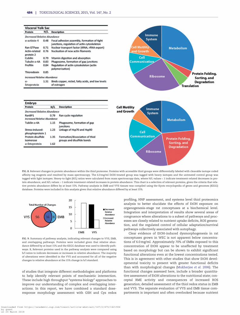
concentration-related abundance in the EMB. Specifically, inthe VYS, proteins involved in actin cytoskeleton dynamicsand RME were found to have reduced abundance. In bothEMB and VYS, proteins associated with nuclear transport(Ran pathway) were reduced and a-fetoprotein was increased(Fig. 8).
ICAT data were also analyzed using a pathway analysis,including proteins that changed in relative abundance by atleast 15% from control (based upon the H/L ratio). Based uponthe total data, a majority of changes in relative abundanceoccurred in the VYS (72 changes), fewer were observed inthe EMB (24 changes), and 16. Of the specific pathwaychanges identified, 16 were common between both VYSand EMB (Fig. 9). Notably, the largest altered pathways were
ribosomal proteins in the VYS, accounting for 14 changedpathways in all (Fig. 8).
DISCUSSION
EtOH is a well-known developmental toxicant and teratogenthat has been shown to affect a broad spectrum of biochemicaland molecular pathways leading to both functional and ana-tomical defects. Animal models of EtOH teratogenicity, includ-ing the rat WEC model used in these studies have revealed anumber of significant endpoints that are altered directly or indi-rectly following exposure. Drawing conclusions regarding themechanistic relatedness of multiple observations across differ-ent species and models is very difficult and has led to the design
FIG. 7. Redox potentials (Eh) for the GSH and Cys redox couples in major tissue (VYS, EMB) and fluid (yolk sac fluid, amniotic fluid) compartments of the intact concep-
tus are shown following 26-h exposure to EtOH at concentrations of 1.5, 3.0, and 6.0 mg/ml in WEC. GSH, GSSG, Cys, and CySS were measured directly by HPLC and Eh
calculated using the Nernst equations described in Materials and Methods. Diagrams show the spatial and anatomical relationships between tissue and fluid compart-
ments along with their respective Eh values expressed in mV units. Color coding provides improved visualization of redox changes and patterns in tissues and fluids
where progression to blue represents more reducing environments and progression to red showing more oxidizing conditions. An * denotes values that are signifi-
cantly different at p< .05 when compared with their appropriate controls. Full color version available online.
JILEK ET AL. | 483
Downloaded from https://academic.oup.com/toxsci/article-abstract/147/2/475/1621904by gueston 24 March 2018
of studies that integrate different methodologies and platformsto help identify relevant points of mechanistic intersection.These include high throughput “systems biology” approaches toimprove our understanding of complex and overlapping inter-actions. In this report, we have combined a standard dose-response morphology assessment with GSH and Cys redox
profiling, HNP assessment, and systems level thiol proteomicsanalysis to better elucidate the effects of EtOH exposure onorganogenesis-stage rat conceptuses at a biochemical level.Integration and interpretation of results show several areas ofcongruence where alterations to a subset of pathways and proc-esses are closely related to nutrient uptake deficits, ROS genera-tion, and the regulated control of cellular adaptation/survivalpathways collectively associated with autophagy.
Clear evidence of EtOH-induced dysmorphogenesis in ratconceptuses grown in WEC is not apparent below concentra-tions of 6.0 mg/ml. Approximately 70% of EMBs exposed to thisconcentration of EtOH appear to be unaffected by treatmentbased on morphology but can be shown to exhibit significantfunctional alterations even at the lowest concentrations tested.This is in agreement with other studies that show EtOH devel-opmental toxicity to present with greater functional deficitsthan with morphological changes (Mukherjee et al. 2006). Thefunctional changes assessed here, include a broader quantita-tive assessment of EtOH alterations to the nutritional state; con-ceptal RME activity and consequences of increased ROSgeneration; detailed assessment of the thiol redox status in EMBand VYS. The separate evaluation of VYS and EMB tissue com-partments is important and often overlooked because nutrient
FIG. 8. Relevant changes in protein abundance within the thiol proteome. Proteins with accessible thiol groups were differentially labeled with cleavable isotope coded
affinity tag reagents and resolved by mass spectroscopy. The 6.0 mg/ml EtOH-treated group was tagged with heavy isotopes and the untreated control group was
tagged with light isotopes. Heavy-to-light (H/L) ratios were calculated from mass spectroscopy data, where H/L values<1 indicate treatment-related decreases in pro-
tein abundance, and H/L values>1 indicate treatment-related increases in protein abundance. This chart is a selection of relevant proteins, given the criteria that rela-
tive protein abundance differs by at least 15%. Pathway analysis in EMB and VYS tissues was compiled using the Kyoto encyclopedia of genes and genomes (KEGG)
database. Proteins were included in this analysis given that relative abundance differed by at least 15%.
FIG. 9. Summary of pathway analysis, indicating relevant changes to VYS, EMB,
and overlapping pathways. Proteins were included given that relative abun-
dance differed by at least 15% and the KEGG database was used to identify path-
ways. B, Relevant proteins used in the pathway analysis were compared using
h/l ratios to indicate decreases or increases in relative abundance. The majority
of alterations were identified in the VYS and accounted for all of the negative
changes to relative abundance at the 15% change in h/l standard.
484 | TOXICOLOGICAL SCIENCES, 2015, Vol. 147, No. 2
Downloaded from https://academic.oup.com/toxsci/article-abstract/147/2/475/1621904by gueston 24 March 2018

uptake, metabolism, and redox control are intimately regulatedbetween these 2 compartments of the developing conceptus.
The major source of amino acids required for fuel and thesupply of anabolic precursors for biosynthesis in the conceptusduring organogenesis comes through HNP via the inverted VYS.Whole proteins and their associated cargoes are taken up byRME at the VYS brush border, incorporated into primary endo-cytotic vesicles, and fused with lysosomes where they are pro-teolytically degraded to constituent amino acids (Ambroso et al.1997; Brent and Fawcett 1998). The reported significantdecreases in the clearance of FITC from media supplied FITC-albumin, as a result of EtOH exposure, corroborate previousstudies that show similar EtOH effects on the VYS (Steventonand Williams 1987; Xu 2005). Based on the lack of a proportionalincrease of the acid-insoluble fraction in the VYS, we concludethat the effect of EtOH on HNP function is due to a direct effecton RME activity and not on the subsequent proteolysis step. TheRME process is initiated by the binding of an exogenous bulk orcarrier protein to the multiligand receptor complex thatincludes the principle proteins megalin and cubilin(Christensen and Birn 2002). Megalin and cubilin have beenshown to coimmunolocalize in the human yolk sac in asimilar manner, suggesting an analogous functional role ofthe VYS in the rodent model, despite obvious differencesbetween human and rodent VYS structure and orientation(Burke et al. 2013).
Protein binding initiates a series of events that integratesactivities of essential membrane proteins and the rearrange-ment of the actin cytoskeleton to initiate endocytosis and for-mation of the primary vesicle. The actin cytoskeleton and itsdynamic regulation is a known target of EtOH toxicity (Loureiroet al. 2011; Oyedele and Kramer 2013; Romero et al. 2010). It isunlikely that EtOH acts directly to affect disruption of actindynamics but the increased generation of ROS is believed toplay an important role (Sakai et al. 2012). The observed oxidationof GSH in the VYS coupled with the significantly more positiveEh values (Fig. 7) are consistent with a role for increased ROSand altered redox regulation. These correlations are also sup-ported by findings from the analysis of ICAT thiol proteomicdata (Fig. 8) where a decreased abundance of several actin-related proteins (Arp2, profilin, and a-actinin) was identified.These proteins are known to play important roles in actinnucleation, branching, and depolymerization. All of these pro-teins must be regulated to facilitate endocytosis and cell migra-tion, as well as other necessary structural functions that areimportant during the preimplantation stage and early embry-onic development (Lassing et al. 2007; Sun et al. 2013). Becausemost of these dynamic changes have been associated withincreased ROS or oxidative stresses, it is reasonable to suggestthat redox-related posttranslational alterations to these pro-teins may contribute significantly to the observed reduction inRME activity. The relative abundance of b-actin is onlydecreased by 8% (Supplemental Fig. 1) following short-termEtOH exposure, which is consistent with changes to signalingmachinery and not just actin availability. Furthermore, thiore-doxin-1 (Trx-1), which has been shown to maintain the redoxstate of actin via reduction of oxidized thiols in cells under con-ditions of oxidative stress (Wang et al. 2010) was also shown tobe significantly depleted in the VYS as a result of EtOH exposure(Fig. 8). Sensitivity to cellular redox disruption via posttransla-tional modification is additionally evidenced by S-glutathiony-lation of b-actin (cytosolic isoform) and profilin, which is a keyprotein in G-actin sequestration and microfilament dynamics(Fratelli et al. 2003).
Redox profile data from this study show the effects ofincreasing EtOH concentrations on GSH and Cys content andredox state in the major tissues (VYS, EMB) and major fluidcompartments (YSF, AF) of the rat conceptus (Figs. 3–6). Noattempts were made to directly quantify ROS, relying on sus-tained changes to the soluble thiol steady states to assess redoxstatus. EtOH, at the concentrations used, did not cause signifi-cant reductions in total GSH (GSHþ 2GSSG) in the VYS and AF,but did produce small decreases in the EMB and YSF.Traditional evidence for “oxidative stress” would interpret theseresults as EtOH having little or no effect on redox status. Furtherexamination shows that significant decreases in GSH occur at1.5 mg/ml EtOH and that corresponding values of GSSG signifi-cantly increase over the entire EtOH concentration range inVYS, EMB, and YSF. There did not appear to be as consistent orextensive pattern of GSH and Cys movement between tissueand fluid compartments as we have reported with other chemi-cal modulators (Harris et al. 2013). In VYS and EMB, GSSG con-centrations increased approximately 2-fold at all dose groupswhen compared with control levels (Fig. 5). This is likely attrib-utable to oxidation of GSH to the disulfide form, as well as con-sistent shuttling of GSSG into the extracellular compartments(YSF and AF) (Ballatori et al. 2009). These results show evidencefor the net oxidation of GSH to GSSG, which is confirmed by Eh
values increasing by 18–41 mV (Fig. 7). The observed magnitudeof redox change in these tissues and fluid compartment is con-sistent with studies reporting significant oxidative posttransla-tional modification of cysteine thiols. Based on these results, itis difficult to draw conclusions regarding the potential deleteri-ous consequences of ROS elevation following EtOH, although,changes in Eh do indicate a permissive environment for redoxsignaling changes. (Jones 2006; Paulsen and Carroll 2009;Schafer and Buettner 2001; Schieber and Chandel 2014).
The increased generation of ROS has been discussed previ-ously in relation to the disruption of HNP functions and cytos-keletal dynamics. Because of the vast number of oxidizableprotein thiols present in cells and tissues, the magnitude ofROS-induced changes is daunting. Disruption of the cellular cys-teine redox circuitry has many important implications in bio-chemical and physiological signaling pathways that alsorequire input from cellular metabolism as with the supply ofreducing equivalents. Functional Trx-1 was previously identi-fied as a critical redox-regulating enzyme that is reduced inabundance following EtOH treatment (Karshikoff et al. 2013).Maintenance of Trx-1 in its reduced and active state is depend-ent on thioredoxin reductase, which receives electrons fromNADPH, its obligate electron donor. Transcriptomic studies inembryonic mouse models have shown that the pentose phos-phate pathway is upregulated in response to EtOH exposure inresistant strains (B6N), indicating a possible adaptive responsein supplying additional NADPH as reducing equivalents (Greenet al. 2007) Additionally, the glutaredoxin system, when bal-anced with physiologically relevant concentrations of GSH,NADPH, and GSH reductase, has been shown to protect againstTrx-1 active site oxidation (Du et al. 2013). Although both sys-tems are distinct redox circuits, the depletion of reduced GSHand reducing equivalents due to EtOH exposure may hinder thecells ability to maintain adequate levels of reduced Trx-1. Thus,this demonstrates that the ROS-generating capacity of EtOHmay target specific cellular signaling pathways, outside of thescope of general oxidative damage to cells and tissues.
Although several correlations were found to exist betweenredox status, HNP activity and potential connections to redoxsignaling and circuitry in the context of EtOH embryotoxicity, it
JILEK ET AL. | 485
Downloaded from https://academic.oup.com/toxsci/article-abstract/147/2/475/1621904by gueston 24 March 2018

is also clear that many other potential pathways may beaffected. A number of studies have found that ROS produced byEtOH metabolism can be linked to cell death via apoptosis (Suliket al. 1981, 1988; Dunty et al. 2001), which is believed to be initi-ated when Eh values increase above �170 mV. Although pro-grammed cell death pathways may be initiated as a result ofincreased ROS production, other responses having the exactopposite outcome are also known to be regulated by ROS.Pathway analysis (KEGG) of EtOH-induced changes to the thiolproteome revealed significant numbers of altered pathway ele-ments mapping to the ribosome, protein folding, sorting, anddegradation pathways (Fig. 8). These pathways are of particularinterest because they are developmentally relevant, they areeasily initiated as a result of nutrient amino acid deficiency(such as through HNP activity inhibition), and they are exten-sively redox regulated processes (Aburto et al. 2012; Cecconi andLevine 2008; Cecconi et al. 2008; Dodson et al. 2013; Zhang 2015).Recent studies on mechanisms of EtOH embryotoxicity haveunderstandably focused on the brain and various aspects ofneurotoxicity. A key element in many of these studies is thedirect association between EtOH and the initiation of autophagyin neural tissues which may be expressed very differently in dif-ferent species and strains of animals (Alimov et al. 2013; Luo2014). This differential sensitivity to EtOH has been shown inmouse models for FASD where B6J dams were dosed twice, 4 hapart, on GD 8 with 2.9 g/kg EtOH, resulting in a significantlyhigher incidence of FASD outcomes when compared with iden-tical exposures in the related B6N strains (Green et al. 2007).Microarray transcript profiling of the embryonic headfold (3-hpost-EtOH exposure) from the resistant B6N strain showeddownregulation of KEGG pathways for ribosomal proteins andproteosome and upregulation of glycolysis and pentose phos-phate pathways. Actin cytoskeleton, focal adhesion, adherensjunction, and other HNP-related genes were expressed andupgregulated in both mouse strains. We have observed similaraffected pathways in our analysis of the thiol proteome suggest-ing central roles for autophagy and its related nutritional andredox-related control mechanisms. A final example from thethiol proteome analysis that shows a high degree of correlationwith the ribosome and more general autophagy pathways ismanifest by the observed decreased abundance of Ran cycle-related nuclear transport proteins, both in the EMB and VYS(Fig. 8). Several proteins involved in the Ran cycle have beenpreviously shown to be redox-sensitive. Specifically, ROS hasbeen shown to alter the GDP dissociation rate from Ran, due toits unique structure (Heo 2008). Ran cycles operate in both EMBand VYS to transport ribonucleoproteins (including 40S and 60Ssubunits) from the site of synthesis in the nucleolus to the cyto-sol (Kodiha et al. 2004; Miyamoto et al. 2004); Failure of this sys-tem results in accumulation of ribosomal and preribosomalmaterial in the nucleus (Lounsbury and Macara 1997). Thus, theabundance of several other downstream proteins may beaffected as a result of an altered Ran-GTPase cycle or decreasedrecycling of Ran proteins; such proteins may lie outside of thethiol proteome or may exist in low abundance, effectively beingmissed by the ICAT procedure. Furthermore, the intracellulardomains of EGFR-family proteins have been found to be proteo-lytically cleaved following ligand binding, then acting as tran-scription factors. Many of these, including EGFR and ErbB2, havebeen shown to be translocated into the nucleus by importin-b,which is dependent on the Ran cycle (Wang and Hung 2009).The resulting collapse of the Ran gradient also results in theaccumulation of importin-a (Kodiha et al. 2004; Miyamoto et al.2004). Additionally, Ran, nucleoporin Nup153, and importin-b
are also proteolytically degraded under conditions of oxidativestress (Kodiha et al. 2004), consistent with our proteomic data inthe VYS. Alternatively, although we did not observe direct alter-ations to Ran in the EMB, our proteomic data (Fig. 8) shows thatRan binding protein-1 (RanBP1) abundance is significantlyreduced in the treatment group. This may, likewise, be indica-tive of a collapse of the Ran cycle, as RanBP1 has been found toreduce inhibition of Ran GTPase-activating protein (RanGAP)(Lounsbury and Macara 1997) and is required for hydrolysis ofRan-bound GTP (Petersen et al. 2000), completing the Ran cycleand maintaining the Ran concentration gradient necessary fornuclear transport. It is also important to note that the Rannuclear translocation activities are integral to the ribosome bio-synthesis pathways that make ribonucleotides in the nucleoliand transport them to the cytosol for ribosome assembly andfunction (Fromont-Racine et al. 2003; Johnson et al. 2002), whichis suggested as a targeted pathway by our data (Fig. 8). Nuclearimport/export pathways are important in development becauseof the significant roles they play in mediating the EtOH-induceddisruption of cell transport, signaling, mitosis, and cell cycleprogression (Dasso 2002).
In conjunction with existing evidence, our data suggestsome novel insights into the mechanism of EtOH teratogenicityduring early organogenesis. We have shown that EtOH altersthe compartmentalized redox environments of the rodent con-ceptus, and have found this to be related to a significant, dose-dependent inhibition of HNP activity. These data demonstratedifferential EtOH-induced effects in the EMB and VYS, and sug-gest that the VYS plays an important role during early organo-genesis and is a distinct target for EtOH toxicity (Fig. 7). Theresulting exacerbated conditions of intracellular oxidation mayalso contribute directly to significant changes in the functionalthiol proteome as well as significant changes in the absoluteamounts of critical proteins. Because our ICAT proteomic analy-sis only focused on the top 2.5% of most abundant proteins, weare certain there is much to yet be learned as we are able tomine deeper into changes involving less abundant proteins.Observed decreases for specific proteins involved in cytoskeletaldynamics and endocytosis (a-actinin, a-tubulin, cubilin, andactin-related protein 2), nuclear translocation (Ran and RanBP1),and maintenance of RME (cubilin) show direct correlation withfunctional changes in redox status and HNP activity. The com-bined alterations of HNP activity, ROS generation, and redox sig-naling and circuitry all impact the broader activation ofautophagy pathways and may contribute significantly to theoverall process of ribosome biogenesis, assembly and functionthat presents an interesting target for future studies in EtOH ter-atogenesis as have been shown in specific studies focused onmechanisms of EtOH neurogenesis (Luo 2014).
SUPPLEMENTARY DATA
Supplementary data are available online at http://toxsci.oxfordjournals.org/.
FUNDING
This work was supported by the Bill and Melinda GatesFoundation, Grand Challenges Explorations, Round 7; theUniversity of Michigan National Institute of EnvironmentalHealth Sciences (NIEHS) Core Center, “Lifestage Exposuresand Adult Disease” (P30 ES017885); and from an
486 | TOXICOLOGICAL SCIENCES, 2015, Vol. 147, No. 2
Downloaded from https://academic.oup.com/toxsci/article-abstract/147/2/475/1621904by gueston 24 March 2018

Institutional Training Grant from the NIEHS to K.E.S. (T32ES007062).
ACKNOWLEDGMENTS
The authors thank Ofra Duchen, Thomas J. Wolfe, andAnthony Su for contributing to the production and analysisof data supporting this work. The findings and conclusionsin this manuscript are those of the authors and do not nec-essarily represent the official views or policies of theCenters for Disease Control and Prevention.
ReferencesAburto, M. R., Hurle, J. M., Varela-Nieto, I., and Magarinos, M.
(2012). Autophagy during vertebrate development. Cells 1,428–448.
Alimov, A., Wang, H., Liu, M., Frank, J., Xu, M., Ou, X., and Luo, J.(2013). Expression of autophagy and UPR genes in the devel-oping brain during ethanol-sensitive and resistant periods.Metab. Brain Dis. 28, 667–676.
Ambroso, J., and Harris, C. (2012). Assessment of histiotrophicnutrition using fluorescent probes. Methods Mol. Biol. 889,407–423.
Ambroso, J. L., and Harris, C. (1993). Chloroquine embryotoxicityin the postimplantation rat conceptus in vitro. Teratology 48,213–226.
Ambroso, J. L., Larsen, S. V., Brabec, R. K., and Harris, C. (1997).Fluorometric analysis of endocytosis and lysosomal proteol-ysis in the rat visceral yolk sac during whole embryo culture.Teratology 56, 201–209.
Arriazu, E., Perez de Obanos, M. P., Lopez-Zabalza, M. J., Herraiz,M. T., and Iraburu, M. J. (2010). Amino acid deprivation de-creases intracellular levels of reactive oxygen species in he-patic stellate cells. Cell. Physiol. Biochem. 26, 281–290.
Balaraman, S., Tingling, J. D., Tsai, P. C., and Miranda, R. C. (2013).Dysregulation of microRNA expression and function contrib-utes to the etiology of fetal alcohol spectrum disorders.Alcohol Res. 35, 18–24.
Ballatori, N., Krance, S. M., Marchan, R., and Hammond, C. L.(2009). Plasma membrane glutathione transporters and theirroles in cell physiology and pathophysiology. Mol. AspectsMed. 30, 13–28.
Beckman, D. A., Lloyd, J. B., and Brent, R. L. (1998). Quantitativestudies on the mechanisms of amino acid supply to rat em-bryos during organogenesis. Reprod. Toxicol. 12, 197–200.
Brent, R. L., Beckman, D. A., Jensen, M., and Koszalka, T. R. (1990).Experimental yolk sac dysfunction as a model for studyingnutritional disturbances in the embryo during early organo-genesis. Teratology 41, 405–413.
Brent, R. L., and Fawcett, L. B. (1998). Nutritional studies of theembryo during early organogenesis with normal embryosand embryos exhibiting yolk sac dysfunction. J. Pediatr. 132,S6–S16.
Burke, K. A., Jauniaux, E., Burton, G. J., and Cindrova-Davies, T.(2013). Expression and immunolocalisation of the endocyticreceptors megalin and cubilin in the human yolk sac and pla-centa across gestation. Placenta 34, 1105–1109.
Burton, G. J., Hempstock, J., and Jauniaux, E. (2001). Nutrition ofthe human fetus during the first trimester–a review. Placenta22(Suppl. A), S70–S77.
Cecconi, F., and Levine, B. (2008). The role of autophagy in mam-malian development: Cell makeover rather than cell death.Dev. Cell 15, 344–357.
Cecconi, F., Piacentini, M., and Fimia, G. M. (2008). The involve-ment of cell death and survival in neural tube defects: A dis-tinct role for apoptosis and autophagy? Cell Death Differ. 15,1170–1177.
Chen, X., Liu, J., and Chen, S. Y. (2013). Sulforaphane protectsagainst ethanol-induced oxidative stress and apoptosis inneural crest cells by the induction of Nrf2-mediated antioxi-dant response. Br. J. Pharmacol. 169, 437–448.
Christensen, E. I., and Birn, H. (2002). Megalin and cubilin:Multifunctional endocytic receptors. Nat. Rev. Mol. Cell Biol. 3,256–266.
Chung, H. S., Wang, S.-B., Venkatraman, V., Murray, C. I., andVan Eyk, J. E. (2013). Cysteine oxidative posttranslationalmodifications: Emerging regulation in the cardiovascularsystem. Circ. Res. 112, 382–392.
Coriale, G., Fiorentino, D., Di Lauro, F., Marchitelli, R., Scalese, B.,Fiore, M., Maviglia, M., and Ceccanti, M. (2013). Fetalalcohol spectrumdisorder (FASD): Neurobehavioral profile,indications for diagnosis and treatment. Riv. Psichiatr. 48,359–369.
Dasso, M. (2002). The Ran GTPase: Theme and variations. Curr.Biol. 12, R502–R508.
Davis, W. L. (1990). Ethanol induces the generation of reactivefree radicals by neural crest cells in vitro. J. Craniofac. Genet.Dev. Biol. 10, 277–293.
Dodson, M., Darley-Usmar, V., and Zhang, J. (2013). Cellularmetabolic and autophagic pathways: Traffic control by redoxsignaling. Free Radic. Biol. Med. 63, 207–221.
Dong, J., Sulik, K. K., and Chen, S. Y. (2008). Nrf2-mediatedtranscriptional induction of antioxidant response in mouseembryos exposed to ethanol in vivo: Implications for the pre-vention of fetal alcohol spectrum disorders. Antioxid. RedoxSignal. 10, 2023–2033.
Dong, J., Sulik, K. K., and Chen, S. Y. (2010). The role of NOX en-zymes in ethanol-induced oxidative stress and apoptosis inmouse embryos. Toxicol. Lett. 193, 94–100.
Dreosti, I. E. (1993). Nutritional factors underlying the expressionof the fetal alcohol syndrome. Ann. N. Y. Acad. Sci. 678,193–204.
Du, Y., Zhang, H., Zhang, X., Lu, J., and Holmgren, A. (2013).Thioredoxin 1 is inactivated due to oxidation induced byperoxiredoxin under oxidative stress and reactivated by theglutaredoxin system. J. Biol. Chem. 288, 32241–32247.
Dunty, W. C., Jr, Chen, S. Y., Zucker, R. M., Dehart, D. B., andSulik, K. K. (2001). Selective vulnerability of embryonic cellpopulations to ethanol-induced apoptosis: Implications foralcohol-related birth defects and neurodevelopmental disor-der. Alcohol. Clin. Exp. Res. 25, 1523–1535.
Finkel, T. (2011). Signal transduction by reactive oxygen species.J. Cell Biol. 194, 7–15.
Fratelli, M., Demol, H., Puype, M., Casagrande, S., Villa, P.,Eberini, I., Vandekerckhove, J., Gianazza, E., and Ghezzi, P.(2003). Identification of proteins undergoing glutathionyla-tion in oxidatively stressed hepatocytes and hepatoma cells.Proteomics 3, 1154–1161.
Fromont-Racine, M., Senger, B., Saveanu, C., and Fasiolo, F.(2003). Ribosome assembly in eukaryotes. Gene 313, 17–42.
Fu, C., Hu, J., Liu, T., Ago, T., Sadoshima, J., and Li, H. (2008).Quantitative analysis of redox-sensitive proteome with DIGEand ICAT. J. Proteome Res. 7, 3789–3802.
Go, Y.-M., Roede, J. R., Orr, M., Liang, Y., and Jones, D. P. (2014).Integrated redox proteomics and metabolomics of mitochon-dria to identify mechanisms of Cd toxicity. Toxicol. Sci. 139,59–73.
JILEK ET AL. | 487
Downloaded from https://academic.oup.com/toxsci/article-abstract/147/2/475/1621904by gueston 24 March 2018

Green, M. L., Singh, A. V., Zhang, Y., Nemeth, K. A., Sulik, K. K.,and Knudsen, T. B. (2007). Reprogramming of genetic net-works during initiation of the fetal alcohol syndrome. Dev.Dyn. 236, 613–631.
Guttmann, R. P. (2010). Redox regulation of cysteine-dependentenzymes. J. Anim. Sci. 88, 1297–1306.
Hansen, J. M., and Harris, C. (2013). Redox control of teratogene-sis. Reprod. Toxicol. 35, 165–179.
Harris, C. (2012). Rodent whole embryo culture. Methods Mol. Biol.889, 215–237.
Harris, C., and Hansen, J. (2012). Oxidative stress, thiols, and re-dox profiles. In: Developmental Toxicology (C. Harris and J. M.Hansen, Eds.), pp. 325–346. Humana Press, NY, New York.
Harris, C., Shuster, D. Z., Roman Gomez, R., Sant, K. E., Reed, M.S., Pohl, J., and Hansen, J. M. (2013) Inhibition of glutathionebiosynthesis alters compartmental redox status and thethiol proteome in organogenesis-stage rat conceptuses. FreeRadic. Biol. Med. 63, 325–337.
Heo, J. (2008). Redox regulation of Ran GTPase. Biochem. Biophys.Res. Commun. 376, 568–572.
Hill, A. J., Drever, N., Yin, H., Tamayo, E., Saade, G., andBytautiene, E. (2014). The role of NADPH oxidase in a mousemodel of fetal alcohol syndrome. Am. J. Obstet. Gynecol. 210,466.e1–466.e5.
Johnson, A. W., Lund, E., and Dahlberg, J. (2002). Nuclear exportof ribosomal subunits. Trends Biochem. Sci. 27, 580–585.
Jones, D. P. (2002). [11] Redox potential of GSH/GSSG couple:Assay and biological significance. In: Methods in Enzymology(S. Helmut and P. Lester, Eds.), pp. 93–112. Academic Press,San Diego, CA.
Jones, D. P. (2006). Redefining oxidative stress. Antioxid. RedoxSignal. 8, 1865–1879.
Kaminen-Ahola, N., Ahola, A., Maga, M., Mallitt, K. A., Fahey, P.,Cox, T. C., Whitelaw, E., and Chong, S. (2010). Maternal etha-nol consumption alters the epigenotype and the phenotypeof offspring in a mouse model. PLoS Genet. 6, e1000811.
Karshikoff, A., Nilsson, L., and Foloppe, N. (2013). Understandingthe -C-X1-X2-C- motif in the active site of the thioredoxin su-perfamily: E. coli DsbA and its mutants as a model system.Biochemistry 52, 5730–5745.
Kirlin, W. G., Cai, J., Thompson, S. A., Diaz, D., Kavanagh, T. J.,and Jones, D. P. (1999). Glutathione redox potential in re-sponse to differentiation and enzyme inducers. Free Radic.Biol. Med. 27, 1208–1218.
Kodiha, M., Chu, A., Matusiewicz, N., and Stochaj, U. (2004).Multiple mechanisms promote the inhibition of classical nu-clear import upon exposure to severe oxidative stress. CellDeath Differ. 11, 862–874.
Lassing, I., Schmitzberger, F., Bjornstedt, M., Holmgren, A.,Nordlund, P., Schutt, C. E., and Lindberg, U. (2007). Molecularand structural basis for redox regulation of beta-actin. J. Mol.Biol. 370, 331–348.
Lounsbury, K. M., and Macara, I. G. (1997). Ran-binding protein1 (RanBP1) forms a ternary complex with Ran and karyo-pherin beta and reduces Ran GTPase-activating protein(RanGAP) inhibition by karyopherin beta. J. Biol. Chem. 272,551–555.
Loureiro, S. O., Heimfarth, L., Reis, K., Wild, L., Andrade, C.,Guma, F. T. C. R., Goncalves, C. A., and Pessoa-Pureur, R.(2011). Acute ethanol exposure disrupts actin cytoskeletonand generates reactive oxygen species in c6 cells.Toxicol.in vitro 25, 28–36.
Luo, J. (2014). Autophagy and ethanol neurotoxicity. Autophagy10, 2099–2108.
Miller, L., Shapiro, A. M., Cheng, J., and Wells, P. G. (2013). Thefree radical spin trapping agent phenylbutylnitrone reducesfetal brain DNA oxidation and postnatal cognitive deficitscaused by in utero exposure to a non-structurally teratogenicdose of ethanol: A role for oxidative stress. Free Radic. Biol.Med. 60, 223–232.
Miller-Pinsler, L., Pinto, D. J., and Wells, P. G. (2015). OxidativeDNA damage in the in utero initiation of postnatal neurode-velopmental deficits by normal fetal and ethanol-enhancedoxidative stress in oxoguanine glycosylase 1 knockout mice.Free Radic. Biol. Med. 78, 23–29.
Miyamoto, Y., Saiwaki, T., Yamashita, J., Yasuda, Y., Kotera, I.,Shibata, S., Shigeta, M., Hiraoka, Y., Haraguchi, T., andYoneda, Y. (2004). Cellular stresses induce the nuclear accu-mulation of importin a and cause a conventional nuclear im-port block. J. Cell Biol. 165, 617–623.
Mukherjee, R. A. S., Hollins, S., and Turk, J. (2006). Fetal alcoholspectrum disorder: An overview. J. R. Soc. Med. 99, 298–302.
Oyedele, O. O., and Kramer, B. (2013). Nuanced but signif-icant: How ethanol perturbs avian cranial neural crestcell actin cytoskeleton, migration and proliferation.Alcohol. 47, 417–426.
Park, P. H., Miller, R., and Shukla, S. D. (2003). Acetylation of his-tone H3 at lysine 9 by ethanol in rat hepatocytes. Biochem.Biophys. Res. Commun. 306, 501–504.
Paulsen, C. E., and Carroll, K. S. (2009). Orchestrating redox sig-naling networks through regulatory cysteine switches. ACSChem. Biol. 5, 47–62.
Petersen, C., Orem, N., Trueheart, J., Thorner, J. W., and Macara,I. G. (2000). Random mutagenesis and functional analysis ofthe Ran-binding protein, RanBP1. J. Biol. Chem. 275,4081–4091.
Romero, A. M., Esteban-Pretel, G., Marın, M. P., Ponsoda, X.,Ballestın, R., Canales, J. J., and Renau-Piqueras, J. (2010).Chronic ethanol exposure alters the levels, assembly, andcellular organization of the actin cytoskeleton and microtu-bules in hippocampal neurons in primary culture. Toxicol. Sci.118, 602–612.
Sakai, J., Li, J., Subramanian Kulandayan, K., Mondal, S., Bajrami,B., Hattori, H., Jia, Y., Dickinson Bryan, C., Zhong, J., Ye, K.,et al. (2012). Reactive oxygen species-induced actin glutathio-nylation controls actin dynamics in neutrophils. Immunity37, 1037–1049.
Schafer, F. Q., and Buettner, G. R. (2001). Redox environment ofthe cell as viewed through the redox state of the glutathionedisulfide/glutathione couple. Free Radic. Biol. Med. 30, 1191–1212.
Schieber, M., and Chandel Navdeep, S. (2014). ROS function in re-dox signaling and oxidative stress. Curr. Biol. 24, R453–R462.
Smith, S. M. (1997). Alcohol-induced cell death in the embryo.Alcohol Health Res. World 21, 287–297.
Steventon, G. B., and Williams, K. E. (1987). Ethanol-induced in-hibition of pinocytosis and proteolysis in rat yolk sac in vitro.Development 99, 247–253.
Sulik, K. K., Cook, C. S., and Webster, W. S. (1988). Teratogens andcraniofacial malformations: Relationships to cell death.Development 103(Suppl.), 213–231.
Sulik, K. K., Johnston, M. C., and Webb, M. A. (1981). Fetal alcoholsyndrome: Embryogenesis in a mouse model. Science 214,936–938.
Sun, S. C., Wang, Q. L., Gao, W. W., Xu, Y. N., Liu, H. L., Cui, X. S.,and Kim, N. H. (2013). Actin nucleator Arp2/3 complex is es-sential for mouse preimplantation embryo development.Reprod. Fertil. Dev. 25, 617–623.
488 | TOXICOLOGICAL SCIENCES, 2015, Vol. 147, No. 2
Downloaded from https://academic.oup.com/toxsci/article-abstract/147/2/475/1621904by gueston 24 March 2018

Ungerer, M., Knezovich, J., and Ramsay, M. (2013). In utero alco-hol exposure, epigenetic changes, and their consequences.Alcohol Res. 35, 37–46.
Wang, L. L., Zhang, Z., Li, Q., Yang, R., Pei, X., Xu, Y.,Wang, J., Zhou, S. F., and Li, Y. (2009). Ethanol expo-sure induces differential microRNA and target gene ex-pression and teratogenic effects which can besuppressed by folic acid supplementation. Hum. Reprod.24, 562–579.
Wang, S. C., and Hung, M. C. (2009). Nuclear translocationof the epidermal growth factor receptor family
membrane tyrosine kinase receptors. Clin. Cancer Res.15, 6484–6489.
Wang, X., Ling, S., Zhao, D., Sun, Q., Li, Q., Wu, F., Nie, J., Qu, L.,Wang, B., Shen, X., et al. (2010). Redox regulation of actin bythioredoxin-1 is mediated by the interaction of the proteinsvia cysteine 62. Antioxid. Redox Signal. 13, 565–573.
Xu, Y. (2005). Effect of ethanol on the development of visceralyolk sac. Hum. Reprod. 20, 2509–2516.
Zhang, J. (2015). Teaching the basics of autophagy and mitoph-agy to redox biologists—Mechanisms and experimentalapproaches. Redox Biol. 4, 242–259.
JILEK ET AL. | 489
Downloaded from https://academic.oup.com/toxsci/article-abstract/147/2/475/1621904by gueston 24 March 2018