Embed Size (px)
Citation preview

Journal of Medical Genetics 1987, 24, 52-59
Prenatal diagnosis of a, antitrypsin deficiency andestimates of fetal risk for diseaseDIANE W COX AND TAMMY MANSFIELDFrom The Research Institute, The Hospital for Sick Children, and the Departments of Paediatrics, MedicalGenetics, and Medical Biophysics, University of Toronto, Toronto, Canada.
SUMMARY Alpha, antitrypsin deficiency is one of the most common metabolic disorders,frequently associated with obstructive lung disease and occasionally with childhood livercirrhosis. Prenatal diagnosis of this deficiency has been accomplished using a DNA polymorph-ism detected by the restriction enzyme A vall. A unique haplotype ofDNA fragments is observedin deficient (PI type ZZ) subjects. Diagnosis is therefore possible directly from fetal tissue, unlikeother prenatal diagnoses using linkage of a DNA polymorphism within a specific family. Thisapproach must be modified for rare deficiency alleles of a, antitrypsin (PI*Mmalton,PI*Mduarte, and PI*QO or null). Knowledge of risk of severe disease in the fetus is importantfor the application of prenatal diagnosis. From the limited data available to date, the risk for a
given PI ZZ fetus to develop severe liver disease has been estimated at 13% where a previous PIZZ sib had no liver disease or liver disease which resolved during early childhood, and a risk of40% where a previous PI ZZ sib had developed severe liver disease.
A deficiency of the plasma protease inhibitor, a,antitrypsin (AAT), occurs in about 1 in 7700 whiteNorth American people' and in some northernEuropean populations it is as frequent as 1 in2000 to 3000. The deficiency is usually associatedwith the genetic type or PI (protease inhibitor) typeZZ, where the Z allele results in production of avariant AAT which is not secreted normally fromthe liver. Persons with this deficiency are almostcertain to develop obstructive lung disease in earlyadult life if they smoke, but may have a near normallife span if smoking is avoided.3 4 Adults have an
increased risk for developing liver disease.5 6 Basedon a prospective study in Sweden,7 about 17% ofsubjects with the deficiency develop clinical signs ofliver disease in infancy. In a portion of this group,liver disease progresses to cirrhosis and early death,as first described in 1969.8 Because of the fatal liverdisease which has occurred in some families, pre-natal diagnosis may be desired.
Prenatal diagnosis was first reported by PI typingof fetal blood samples obtained at fetoscopy.Because of the risk of about 5% for pregnancy lossassociated with this procedure,'0 molecular methodswhich can be carried out on cells obtained from
Received for publication 24 July 1986.Revised version accepted for publication 3 September 1986.
amniotic fluid or from chorionic villus biopsy offeradvantages. Synthetic oligonucleotide probes recog-nise a single base pair mutation1' and are theoreti-cally a reliable approach when the mutation site hasbeen identified. Specific probes for the normal typeM and abnormal type Z proteins have beendeveloped.'2 However, the use of synthetic probesis technically demanding because of the rigid controlof hybridisation conditions which must be appliedfor the detection of a single mismatched base pair.Using the restriction enzyme AvaIl, we have
identified two polymorphisms, one for each of twogenomic probes in the AAT coding and flankingregions. 13 With one of these probes, the polymorph-ism appears to show a pattern of DNA fragmentsunique to persons of PI type ZZ. Reliable prenataldiagnosis is therefore possible, as we have reportedbriefly.'4 In a collaborative study, we have com-pared synthetic and genomic probes for prenataldiagnosis of a, antitrypsin deficiency,15 mostly usingamniocytes. The more simple method using genomicprobes may be preferred in diagnostic laboratoriesand furthermore can be used for rare deficiencyalleles.
In this report, we provide details of the applica-tion of these polymorphisms to prenatal diagnosis ofAAT deficiency, using the DNA haplotype obtainedwith Avall. Unusual features of chorionic villus are
52
copyright. on 22 N
ovember 2018 by guest. P
rotected byhttp://jm
g.bmj.com
/J M
ed Genet: first published as 10.1136/jm
g.24.1.52 on 1 January 1987. Dow
nloaded from

Prenatal diagnosis of a, antitrypsin deficiency
discussed. We also review data available to datefrom our own series and those of other authors toprovide risk figures for genetic counselling forparents at risk of having a child of PI type ZZ. Theserisk figures are crucial for evaluating the appro-priateness of prenatal diagnosis in specific families.
Patients and methods
DNA STUDIESThe analysis of DNA polymorphisms in this studywas carried out on 25 parents and six sibs of 13 PIZZ probands, 32 unrelated subjects of PI type ZZ,including the 13 from the above family units, andthree subjects with rare types of AAT deficiency; PItypes MduarteZ, MmaltonZ, and MnullZ, respec-tively. Families with a PI type ZZ child arerepresentative of those in which prenatal diagnosiscould be carried out. From each of these persons,venous blood samples were collected in EDTA. Infamily 13, the father was of type ZZ and was notincluded in the parental analysis. The 'sib' was afetus for which prenatal testing was carried out onDNA obtained from a 30 mg sample of chorionicvillus at 10 weeks' gestation. Data for controls wereobtained as outlined previously.'3
PI typing was carried out on serum or plasma byisoelectric focusing in acrylamide gels, using ampho-lines of pH 3-6, as described'6 or using PharmolytepH 4-2-4-9 (Pharmacia Fine Chemicals). Sera orplasma from all those with AAT deficiency weretyped by isoelectric focusing followed byimmunofixation'7 to identify rare deficiency variantsif present.
Buffy coats were aspirated from EDTA wholeblood, contaminating red cells were lysed, whitecells were saline washed and lysed with SDS inEDTA containing buffer, and DNA was extractedwith phenol chloroform (method of D Hoar, 1983,personal communication). Digestion with AvaIl wascarried out as recommended by the manufacturer on3 ig DNA. The restriction digest was elec-trophoresed on 0-8 to 1% agarose gel, transferred toBiodyne (Pall®) or more recently Hybond-N®(Amersham), and hybridised to 32p labelled DNAprobes as described.'8 Two DNA genomic probes,provided by S L C Woo, were used: a 4-6 kb EcoRIgenomic fragment, extending from the 5' flankingregion into the first intron of the AAT gene, and a6-5 kb BamHI fragment extending from within exonII to the 3' flanking region of the AAT gene, bothhaving been subcloned from aAT35. "9 Studies withthe 4-6 kb probe have been described.'3
FAMILY DATA FOR ASSESSMENT OF RISKFamilies of probands with AAT deficiency have
been studied at The Hospital for Sick Children since1971, mostly by Drs James Weber and Andrew Sass-Kortsak. During that time, 40 probands and theirfirst degree relatives have been ascertained. PItyping was carried out on all probands, theirparents, and sibs. Twelve sibs were found to be of PItype ZZ. Only those probands and PI ZZ sibs whohave been followed to at least three years of agehave been included. Studies on some of theseprobands have previously indicated that clinicalassessment and tests of liver function by two to threeyears of age usually indicate whether the earlyevidence of liver abnormalities has resolved tonormal, or whether liver deterioration and cirrhosiswill result.20 These early observations have beenborne out by later follow up studies of these samepatients at our hospital; these original patients havebeen followed for up to 10 to 15 years.2' A furthermore detailed follow up study has been initiated.
Published sibships in which there has been aproband affected with liver disease and one or moresibs have been reviewed. Only those series present-ing a number of families have been reviewed, in anattempt to avoid families featured in case reportsand selected because of the occurrence of multipleaffected sibs. Only those studies in which the clinicalstatus of all PI ZZ sibs has been given are included.
Results
STUDIES OF DNA POLYMORPHISMSThe 6-5 kb AAT probe is particularly useful forprenatal diagnosis. Patterns of DNA fragments intypical families, obtained by using the 6&5 kb AATprobe on Avall digested DNA, are shown in fig 1B.DNA fragments observed range in size from 0-48 to2-7 kb. Autoradiograph bands of fragments whichshow polymorphisms have been numbered from 1 to7, as described,13 except that there is probably nopolymorphic band, only a constant band, in position6. Allele frequencies have been calculated from 47non-Z PI types. In summary, there are threeconstant DNA fragments between polymorphicbands 1 and 2. Band 2 (not shown) is rare. Anotherless intense constant band lies in the same positionas polymorphic band 2. Alternate alleles showpresence or absence of band 1; the alternate allele,based on further studies, appears to be band 4.Genotype 14 is shown in fig 1B, lane 7. In normalnon-Z haplotypes, the allele frequency for presenceof band 1 is estimated at 0*70 and presence of band 4is 0*30 for non-Z PI types. Bands 5 and 7 are alleleswith frequencies of 0-70 and 0 30 respectively. Aunique pattern of DNA fragments is observed in all32 subjects of PI type ZZ, representing 64 chromo-somes. This pattern is characterised by an absence
53
copyright. on 22 N
ovember 2018 by guest. P
rotected byhttp://jm
g.bmj.com
/J M
ed Genet: first published as 10.1136/jm
g.24.1.52 on 1 January 1987. Dow
nloaded from
Diane W Cox and Tammy Mansfield
-1
,.... _
I t W _
kb2.2.3
4w 4
2 41113
___Sl,
__
lil P, -.S1$
e .Mb ,?,f- 4 4
2
3 _ _43)4
5..65~::
1 2 3 4
7
5 7 8 9
FIG 1 AutoradiogramsofleucocyteDNA digested withA vailandprobed with theAA T46 kbprobe (A) and6-5kbprobe(Band C). Sizes ofkHindIIImarkerfragmentsareindicatedto therightofeachset. Fragmentnumbers areindicatedon theleft. Anodeisatthebottom. DNA haplotypesareasfollows, PItypesinparentheses: (A) 1, +-;2and4, + +;3, --; (B)5,335714(MIMI);6, 337711 (ZZ); 7,337714 (MlZ);8, 337711 (ZZ); 9, 337714(MIZ); (C) 10,335711 (MmaltonZ); 11,
337711 (ZZ);12, 335711 (MduarteZ). Arrowindicatestracefragmentbetweenfragments4and5observedrarelywithPIZZDNA. DNA Biodyne transfermembrane was used.
of bands 4 and 5 and the presence of bands 1 and 7(fig 1B, lanes 6 and 8). The haplotype, or combina-tion of restriction sites, associated with the PI*Zallele can be indicated as 371. Fragment 1 is placedat the end of the haplotype because we know fromthe sequence of the AAT gene that the band mustlie in the 3' flanking region of the AAT gene. Thispattern is not observed in 32 normal controls or 25MZ parents of PI ZZ probands. The results usingthis probe for normal and PI ZZ AAT deficientsubjects are summarised in table 1. Results for the13 families of PI ZZ probands are shown in table 2,indicating that all PI ZZ children have the uniqueDNA haplotype with an Avall digest and the 6*5 kb
probe. The fetus in family 13 is also predicted to beof PI type ZZ.
Rarely in DNA fragment patterns from PI ZZ
TABLE 1 Frequency of selected polymorphic DNAfragments obtained with restriction enzyme AvaIl.
Avall polymorphisms Controls Parents Probands(n=32) PI MZ P1 ZZ
Probe DNA fragment (n=25) (n=32)pattern
4-6 +* 12 (37-5)t 18 (72-0) 32 (100)65 3 7 1 0 0 32 (100)
*Homozygous for corresponding fragments.tPercentage is given in parentheses.
At B..
4k.
0 11 12
54
-ahr &LA ml
4p
copyright. on 22 N
ovember 2018 by guest. P
rotected byhttp://jm
g.bmj.com
/J M
ed Genet: first published as 10.1136/jm
g.24.1.52 on 1 January 1987. Dow
nloaded from

Prenatal diagnosis of a, antitrypsin deficiency
TABLE 2 Extended haplotype (DNA haplotype* and PI type) in families of PI ZZ probands.
Family no Father Mother Sib Proband
I +374M1 +371Z +374M1-+371Z +371Z-+371Z2 -351M2-+371Z +351S *+371Z +371Z-+371Z3 +374M1-+371Z -351M2-+371Z +371Z *+371Z +371Z-+371Z4 +351M2-+371Z +374M1-+371Z +371Z-+371Z5 +354M1 +371Z +351M1-+371Z +351M1-+371Z +371Z-+371Z6 +374M1 +371Z -351M1-+371Z +371Z +371Z7 -351M2-+371Z +374M1-+371Z -351M2-+371Z +371Z-+371Z8 -351M1-+371Z +354M1-+371Z +371Z-+371Z9 -351M1 +371Z +351S *+371Z +371Z-+371Z10 -351M1 +371Z +374M1 +371Z +371Z *+371Z +371Z +371Z11 +354M3-+371Z +351M1-+371Z +371Z-+371Z12 -351M3-+371Z +351M1-+371Z -351M3-+371Z +371Z-+371Z13t +371M1-+371Z +351M1 +371Z +371Z *+371Z +371Z-+371Z'
*AvaIl digest with 4-6 kb probe results indicated first (+ or -) followed by results with 65 kb probe.fFamily for which prenatal diagnosis was carried out.4Pl type ZZ predicted in fetus, one band was reduced in intensity as is common in chorionic DNA. therefore presence of band 1 in both haplotypes has beeninferred.
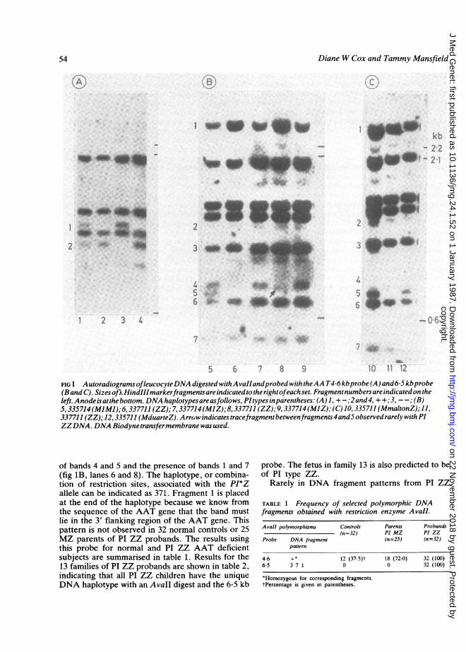
subjects, a weakly hybridising band is observedbetween bands 4 and 5 (fig 1B, lane 8). The markeddifference in intensity (less than 10% intensity ofband 4) and position between bands 4 and 5 makesthis band readily identifiable, when present, inleucocyte or amniocyte DNA.A difference in intensity of certain fragments is
observed between genomic DNA from the chorionicvillus and leucocyte or amniocyte DNA. Thisdifference is noted in the results for family 13, as
shown in fig 2. The chorionic villus DNA (lane 2)has a 'Z specific' band (between 4 and 5), which isalmost equal in intensity to that of band 6. Further-more, band 6 is widened on its leading edge,suggesting the presence of an additional fragmentsomewhat shorter than fragment 6. The adjacentsample (lane 1) has bands 4, 5, and 7 present and isan important control. In this family, the fetus can beonly PI type MZ or ZZ. The fetus has clearly notinherited band 5 from the mother (lane 3). DNAfrom the father (lane 5) is present in excess andshows a visible 'Z specific' band. However, theproband (lane 6) shows only a trace of this band. Atincreased intensity, even the mother shows evidenceof the 'Z specific' band (lane 8). This enhancementof the 'Z band' is consistently noted in chorionicvillus, particularly with Hybond-N transfer mem-
brane. Good separation of the fragments and use ofan adjacent control DNA, having bands 4 and 5, isparticularly important for chorionic DNA. In thisfamily, the diagnosis in the fetus was furtherconfirmed by the absence of a rare TaqI fragment22segregating with the mother's Ml haplotype.The unique DNA haplotype is associated specifi-
cally with the PI*Z deficiency allele. Heterozygotescarrying one rare deficiency allele (PI*Mmalton or
PI*Mduarte) are shown in fig 1C to differ by the
presence of a band 5. This fragment was alsoassociated with the one PI*QO (PI null) alleletested (not shown).Because a unique fragment pattern occurs in PI
ZZ subjects, this probe and AvaII digestion can beused alone for prenatal diagnosis. Although the PIZZ DNA haplotype is unique, DNA from anaffected PI ZZ sib, when available, and fromparents is important to verify that the expected Zhaplotype is present. For chorionic villus DNAparticularly, a confirming polymorphism is useful.The same Biodyne blots of Avall digests werereprobed using the 5' 4-6 kb AAT probe, shown infig 1A. This is one of several polymorphismsdescribed in this DNA region.13 Briefly, one set oftwo fragments shows variability between subjectsdue to two alleles. In 32 normal controls (13 spousesplus 19 random normals)13 and 27 MZ first degreerelatives including all parents in table 2, for whomonly the fragment shown by family studies not tosegregate with the Z allele was included, thefrequency for presence of the restriction site (0.9 kbfragment, band 2) was 0-68 and for absence of site(1-0 kb fragment, band 1) 0-32. Among 32 normalunrelated subjects not carrying the PI Z allele (thatis, PI type M or MS), the 0 9 kb fragment only wasobserved in 12 subjects, while this pattern wasobserved in all of 32 subjects of PI type ZZ,including those in table 1.The extended haplotype for DNA digested with
AvaIl and probed with both 6-5 and 4-6 kb probeswith the PI allele present is shown in table 2. In eightof the 13 families, the AvaIl digest, reprobed withthe 4-6 kb probe, could identify a non-PI ZZ fetus(that is, either PI type MZ or MM) because of theabsence of the restriction site in at least onechromosome of the fetus (+ -) where the PI Z allele
55
copyright. on 22 N
ovember 2018 by guest. P
rotected byhttp://jm
g.bmj.com
/J M
ed Genet: first published as 10.1136/jm
g.24.1.52 on 1 January 1987. Dow
nloaded from

Diane W Cox and Tammy Mansfield
or .
.^-@'~~~~~.040
44__
FIG 2 Autoradiograms of DNA digested with
probed with the 65 kb probe. Band designatioshown in fig lB and C. All lanes contain leuc
with the exception of lane 2. A vaIl DNA haploi
types are as follows: 1, 374-351 (MIMI). 2, 3;(predicted ZZ, DNA from chorionic villus in fa351.371 (MJZ, mother in family 13). 4, 371.3;371 371 (ZZ, father in family 13, 6 Mig DNA).
(ZZ, proband in family 13). 7, 371 *371 (ZZ).
3, 6 ,ig DNA. 9, 351.371 (MJZ). All lanes hay
DNA unless specified. Arrow indicates 'Z spec
Hybond-N transfer membrane was used.
is associated only with presence of th-esite (table 2). This polymorphism w
confirm the diagnosis in these cases. I-
homozygous '+' result in these families
an equal chance of being PI type MZ or Z
DNA haplotypes are associated with
types (D W Cox, unpublished data). PI treliable M subtyping, in combination witi
6-5 results for both parents, can be
predicting which of the other DNA polyiwe have described' could be useful in
family for additional confirmation.
FAMILY STUDIES FOR ASSESSMENT OF RISKOf 38 probands of Pi type ZZ who have beenpatients at The Hospital for Sick Children between
> ^ 1974 and 1984 and who are currently three years ofage or more, five have died of their liver disease andtwo have died of unrelated defects. These otherdefects were severe combined immune deficiency(one patient) and congenital heart disease (onepatient). At least eight patients in the series of Udallet at23 also had additional diseases or abnormalities.This is probably because the deficiency will bepicked up incidentally only in patients in hospital.
PI typing of all sibs of probands was carried outA* for the HSC series. In 10 families where parents are
heterozygotes (PI MZ, SZ) there was more than onePI ZZ child. The clinical status of all PI ZZ childrenborn in the sibships studied is indicated in table 3.
it The first born PI ZZ child has been classified as'normal' if there has been no evidence of liverdisease at any time, 'resolved' if there was neonatalevidence of liver abnormalities with apparent re-solution by two to three years of age and no presentevidence of liver abnormality, or as having 'severe
4 liver disease', that is, deteriorating liver functionfrequently with cirrhosis.
In addition to data from our own series, therehave been four published reports which have de-
* scribed the clinical status of all PI ZZ childrenwithin the sibship. This includes two studies from
- ~ the USA,8 23 one study from Norway,24 and onestudy from Great Britain.25 From the latter study,
A vall and selected cases in their table 3 and case 47 werens are as included. Families never coming to medical atten-
wcyte DNA, tion for liver symptoms were omitted. A group intypes and P1 which patients had 'persisting liver disease' was alsomily 13). 3 omitted: in these cases, there was an increased71 (ZZ). 5, concentration of serum AST (aspartate aminotrans-6, 371 371 ferase) as the only abnormality. However, there was
8, as in lane no indication to what degree the levels of thise about 3 ,mg enzyme were raised in the children involved. In ourcific' band.
TABLE 3 Status of all PI ZZ sibs ofproband (born beforeand after proband).
restriction(ill further[owever, a
would have"Z. Specificspecific PItyping withh the AvaIlhelpful in,morphismsl a specific
No of Proband Liver status in sib Sourcefamilies status* and reference
Normal or Severeresolved
3 S 5 0 Present data7 N,R 7 0 Present data2 S 1 1 USA84 S 5 0 USA231 S 2 0 Norway248 S 3 5 Great Britain257 N,R 5 2 Great Britain25
*S=severe liver disease.N=normal, no liver disease.R=resolved liver disease; normal or slight rise in liver enzymes.
56
copyright. on 22 N
ovember 2018 by guest. P
rotected byhttp://jm
g.bmj.com
/J M
ed Genet: first published as 10.1136/jm
g.24.1.52 on 1 January 1987. Dow
nloaded from

Prenatal diagnosis of a, antitrypsin deficiency
TABLE 4 Risk in PI ZZ sib after proband with resolved orno liver disease.
No of No of sibs Clinical state of sib Referencefamilies (ZZ)
Severe liver Normaldisease
7 7 2 5 258 8 0 8 Present data
15 15 2 (13%) 13 (87%)
TABLE 5 Risk in PI ZZ sib after proband with severe liverdisease.
No of No of sibs Clinical state of sib Referencefamilies (ZZ)
Severe liver Normal*disease
2 2 2 0 84 5 0 5 231 1 0 1 248 9 6 3 252 3 0 3 Present data
17 20 8 (40%) 12 (60%)
*Includes resolved liver disease.
TABLE 6 A priori risk for heterozygous parents having a PIZZ child with severe liver disease (mean estimates).
Previous normal or resolved liver disease (0-25x(-13)= 3%Previous severe liver disease (0-25x0.40)=10%
experience, a modest rise of serum glutamyl oxalo-transaminase (SGOT), about two or three timesnormal concentration, if it is the only abnormal liverfunction test, has been associated with resolved liverdisease. However, a SGOT concentration greaterthan about three times the normal limit has beenassociated with subsequent liver deterioration.211The clinical status of children born after the first
PI ZZ child in a sibship is given in tables 3 and 4,both for our HSC series and for those in publishedreports. Table 4 includes those children born after a
child with normal or resolved liver disease andindicates an average risk of about 13% for having a
PI ZZ child who develops severe liver disease. Table5 includes those children born after a child withsevere liver disease, and indicates an average risk ofabout 40% for a similarly affected child. Table 6shows a priori risks for an affected child beforeprenatal test results are considered.
Discussion
Using the restriction enzyme AvaIl and a genomicprobe for the coding region and 3' flanking region of
the AAT gene, we have shown the presence of aunique series of restriction sites, or haplotype,found only with the PI*Z deficiency allele to date.This unique haplotype should allow reliable prenataldiagnosis. Although this unique haplotype has notbeen found with non-PI*Z alleles to date, thepossibility exists that when larger numbers ofsubjects are studied, this haplotype may occur withnon-Z alleles and, conversely, that other haplotypesmay be associated with the Z allele. Therefore,parents or an affected sib must always be tested. If aPI MZ parent is found to have the Z haplotype withboth M and Z alleles, or if a different haplotype isfound with the Z allele, prenatal diagnosis usingAvaII and the 6-5 probe would not be reliable.However, extending the DNA haplotype by usingthe 4.6 kb probe on the Avall digested DNA, andother DNA polymorphisms we have described,'3might prove useful in distinguishing the M from theZ allele in parent and fetus. In about half of thefamilies we have tested, use of the 4-6 kb probe onAvall digested DNA could discriminate a non-ZZfetus.
This type of prenatal diagnosis with the observa-tion of a specific Z DNA haplotype is appropriateonly where the affected fetus is PI ZZ. We haveobserved a different AvalI 6-5 haplotype with the PIdeficiency alleles Mmalton, Mduarte, and null(QO), each of which is about one hundred times lessfrequent than the Z allele.26 When these raredeficiency alleles are present, identification of anAAT deficient fetus should be possible using theusual approach of identifying the P1/DNA haplo-types of parents and affected proband and grand-parents where necessary. The specific mutations inother rare deficiency alleles have not been identi-fied, so diagnosis using synthetic probes would notbe possible. It should be pointed out that risk forliver disease has not been established for rarealleles.Using genomic probes as in the present study, 2 to
3 [sg DNA is adequate, allowing the direct use ofDNA from uncultured chorionic villus samples.When chorionic villus is cultured, there is a risk ofcontamination by maternal cells, which may lead toan erroneous diagnosis. When the prenatal diagno-sis is carried out by amniocentesis, a single assaymight be possible directly on uncultured amniocytes,although cultured amniocytes are more frequentlyused. Care must be taken with chorionic villus DNAto have excellent separation of fragments in the 0-6kb region and to use appropriate controls. Weprefer to confirm with the 4-6 kb probe or anotherenzyme.
For appropriate genetic counselling and use ofprenatal diagnosis, accurate risk figures are impor-
57
copyright. on 22 N
ovember 2018 by guest. P
rotected byhttp://jm
g.bmj.com
/J M
ed Genet: first published as 10.1136/jm
g.24.1.52 on 1 January 1987. Dow
nloaded from
58
tant. Because of the small number of follow up
studies carried out to date on AAT deficient persons
and their sibs, little information is available. How-ever, we have attempted to summarise our own dataand relevant published reports. While those of PItype ZZ have an increased risk for the developmentof obstructive lung disease in adult life, such lungdisease can be considerably delayed by the avoid-ance of smoking.3 4 Treatment with injected AATis being attempted and there is a rationale forproviding AAT to prevent elastase destruction in
the lung.27 The cause of the liver disease inchildhood is unknown and liver transplant is theonly known therapy. It is therefore probable thatprenatal diagnosis will be considered only becauseof the risk for severe childhood liver disease. Thereis suggestive evidence from the reports summarisedthat the risk for having a child with severe liverdisease may be increased if a previous PI ZZ childhas severe liver disease (40%) compared with therisk when the previous child of PI type ZZ does nothave liver disease (13%). However, the reportedresults vary considerably. For example, the greatestrisk of recurrence of severe liver disease has beenreported in studies from Great Britain, where atleast 67% (six out of nine, excluding 'persisting liverdisease') of subjects have been reported to beaffected after the birth of a PI ZZ child whodevelops severe liver disease. At present, there areinadequate data to determine the precise differencein risk for sibs born after a child with severe liverdisease in comparison with a birth after a child withnormal or resolved liver disease. Furthermore, thedifferences in apparent risk figures between reportsmay be due to biases of ascertainment for variouscountries, or could reflect genetic or environmentalinfluences which vary between geographical loca-tions.The overall risk for heterozygotes to have a child
with severe liver disease can be compared with therisk of 7% for any PI ZZ child to develop severe
liver disease. These figures were obtained from a
Swedish study in which the PI ZZ children ascer-tained at birth7 have been followed to four years ofage. Of the 120 children ascertained, three had de-veloped cirrhosis (two living and one dead at thetime of the report), one had hepatomegaly, andthree had an increase of greater than three times thenormal of their liver enzymes.28 More data will beneeded to determine if the 13% risk after a childwith resolved or no liver disease is really differentfrom the 7% risk for any PI ZZ child of developingsevere liver disease.AAT deficiency has now been recognised for
more than 15 years. Follow up studies of childrenwith liver disease, and of their sibs, is now possible
Diane W Cox and Tammy Mansfield
over a considerable period of time. When suchfollow up studies are carried out, more precise riskfigures should be available. In the meantime, thefigures reported here should help parents to makeinformed decisions.
Note added in proofA polymorphism in MaeIII is useful for prenataldiagnosis (Lancet 1986;ii:741-2). This will be par-ticularly helpful for confirmation with chorionicvillus DNA. We have sucessfully used a combi-nation of the two enzymes for three recent prenataldiagnoses.
We are indebted to Dr James Weber and clinicalfellows for clinical assessment of the PI type ZZprobands; Dr Torben Bech-Hansen for assistancewith DNA probe preparation; and Diane Wills andGail Billingsley (PI typing) and Timothy Durrant(DNA studies, family 13) for technical assistance.Drs J-D Jeppsson, Malmo, and J Gyftadimou,Stockholm, provided chorionic villus and white cellsfrom parents and proband in family 13. We alsogratefully acknowledge the contribution of manyphysicians who have made their patients accessibleto us. This work was supported by grant MA 5426from the Medical Research Council of Canada and agrant from Birth Defects/March of Dimes.
References
'Cox DW, Hoeppner VH, Levison H. Protease inhibitors inpatients with chronic obstructive pulmonary disease: the alphaI-antitrypsin heterozygote controversy. Am Rev Respir Dis1976;113:601-6.
2 Fagerhol MK, Cox DW. The Pi polymorphism: genetic,biochemical and clinical aspects of human a,-antitrypsin. In:Harris H, Hirschhorn K, eds. Advances in human genetics. Vol11. New York, London: Plenum Press, 1981:1-62.
3Larsson C. Natural history and life expectancy in severe alphal-antitrypsin deficiency, PI Z. Acta Med Scand 1983;204:345-51.Janus ED, Phillips NT, Carrell RW. Smoking, lung function,and a,-antitrypsin deficiency. Lancet 1985;i:152-4.
5Cox DW, Smyth S. Risk for liver disease in adults with al-antitrypsin deficiency. Am J Med 1983;74:221-7.
6 Eriksson S, Carlson J, Velez R. Risk of liver cirrhosis andprimary liver cancer in alpha,-antitrypsin deficiency. N Engl JMed 1986;314:736-9.
7Sveger T. Liver disease in alpha,-antitrypsin deficiency detectedby screening of 200,000 infants. N Engl J Med 1976;294:1316-21.
8 Sharp HL, Bridges RA, Krivit W, Freier EF. Cirrhosisassociated with alpha-1-antitrypsin deficiency: a previouslyunrecognized inherited disorder. J Lab Clin Med 1969;73:934-9.
9Jeppsson JO, Cordesius E, Gustavii B, et al. Prenatal diagnosisof alpha-1-antitrypsin deficiency by analysis of fetal bloodobtained at fetoscopy. Pediatr Res 1981;15:254-6.
10 Alter BP. Advances in the prenatal diagnosis of hematologicdiseases. Blood 1984;64:329-40.Conner BJ, Reyes AA, Morin C, Itakura K, Teplitz RL,Wallace RB. Detection of sickle P3-globin allele by hybridizationwith synthetic oligonucleotides. Proc Natl Acad Sci USA1983;80:278-82.
copyright. on 22 N
ovember 2018 by guest. P
rotected byhttp://jm
g.bmj.com
/J M
ed Genet: first published as 10.1136/jm
g.24.1.52 on 1 January 1987. Dow
nloaded from

Prenatal diagnosis of a, antitrypsin deficiency
12 Kidd VJ, Golbus MS, Wallace RB, Itakura K, Woo SLC.Prenatal diagnosis of alpha1-antitrypsin deficiency by directanalysis of the mutation site in the gene. N Engl J Med1984;310:639-42.
13 Cox DW, Woo SLC, Mansfield T. DNA restriction fragmentsassociated with alpha-antitrypsin indicate a single origin fordeficiency allele PI Z. Nature 1985;316:79-81.
14 Cox. DW, Mansfield T. Prenatal diagnosis for alpha1-antitrypsindeficiency. Lancet 1985;i:230.
5 Hejtmancik JF, Sifers RN, Ward P, Harris S, Mansfield T, CoxDW. Prenatal diagnosis of alpha1-antitrypsin deficiency usingrestriction fragment length polymorphism analysis and compari-son with oligonucleotide probe analysis. Lancet 1986;ii:767-70.
16 Cox DW. New variants of a1-antitrypsin: comparison of Pityping techniques. Am J Hum Genet 1981;33:354-65.
17 Arnaud P, Wilson GB, Koistinen J, Fudenberg HH. Immuno-fixation after electrofocusing: improved method for specificdetection of serum proteins with determination of isoelectricpoints. J Immunol Meth 1977;16:221-31.
'8 Linsley PS, Bech-Hansen NT, Siminovitch L, Cox DW. Analysisof a break in chromosome 14 mapping to the region of theimmunoglobulin heavy chain locus. Proc Natl Acad Sci USA1983;80:1997-2001.
19 Leicht M, Long GL, Chandra T, et al. Sequence homology andstructural comparison between the chromosomal human ar-
antitrypsin and chicken ovalbumin genes. Nature 1982;297:655-9.
20 Moroz SP, Cutz E, Cox DW, Sass-Kortsak A. Liver diseaseassociated with alpha,-antitrypsin deficiency in childhood.J Pediatr 1976;88:19-25.
21 Cox DW. Alpha1-antitrypsin deficiency. In: Fisher MM, RoyCC, eds. Pediatric liver disease. Hepatology Research and
Clinical Issues Vol 5. New York, London: Plenum Press,1983:271-82.
22 Matteson KJ, Ostrer H, Chakravarti A, et al. A study ofrestriction fragment length polymorphisms at the human alpha-1-antitrypsin locus. Hum Genet 1985;69:263-7.
23 Udall JN, Dixon M, Newman AP, Wright JA. Liver disease inalpha-antitrypsin deficiency: a retrospective analysis of theinfluence of early breast- vs bottle-feeding. JAMA 1985;253:2679-82.
24 Aagenaes 0, Fagerhol M, Elgjo K,, Munthe E, Hovig T.Pathology and pathogenesis of liver disease in alpha-i-antitrypsin deficiency individuals. Postgrad Med J 1974;50:365-75.
25 Psacharopoulos HT, Mowat AP, Cook PJL, Carlile PA,Portmann B, Rodeck CH. Outcome of liver disease associatedwith alpha1 antitrypsin deficiency (PiZ). Arch Dis Child1983;58:882-7.
26 Cox DW, Billingsley GD, Smyth S. Rare types of al-antitrypsinassociated with deficiency. In: Allen RC, Arnaud P, eds.Electrophoresis 81. Proceedings of the Third InternationalConference on Electrophoresis. New York: de Gruyter, 1981:505-10.
27 Cohen AB. Unravelling the mysteries of alpha1-antitrypsindeficiency. N Engl J Med 1986;314:778-9.
28 Sveger T, Thelin T. Four-year-old children with alphar-antitrypsin deficiency: clinical follow-up and parental attitudestowards neonatal screening. Acta Paediatr Scand 1981;70:171-7.
Correspondence and requests for reprints to DrD W Cox, The Research Institute, The Hospital forSick Children, 555 University Avenue, Toronto,Ontario, Canada M5G 1X8.
59
copyright. on 22 N
ovember 2018 by guest. P
rotected byhttp://jm
g.bmj.com
/J M
ed Genet: first published as 10.1136/jm
g.24.1.52 on 1 January 1987. Dow
nloaded from