Embed Size (px)
Citation preview
DENTAL TECHNIQUE
aMaster StudbCorporate EcVisiting ProfdAdjunct ProeProfessor, D
THE JOURNA
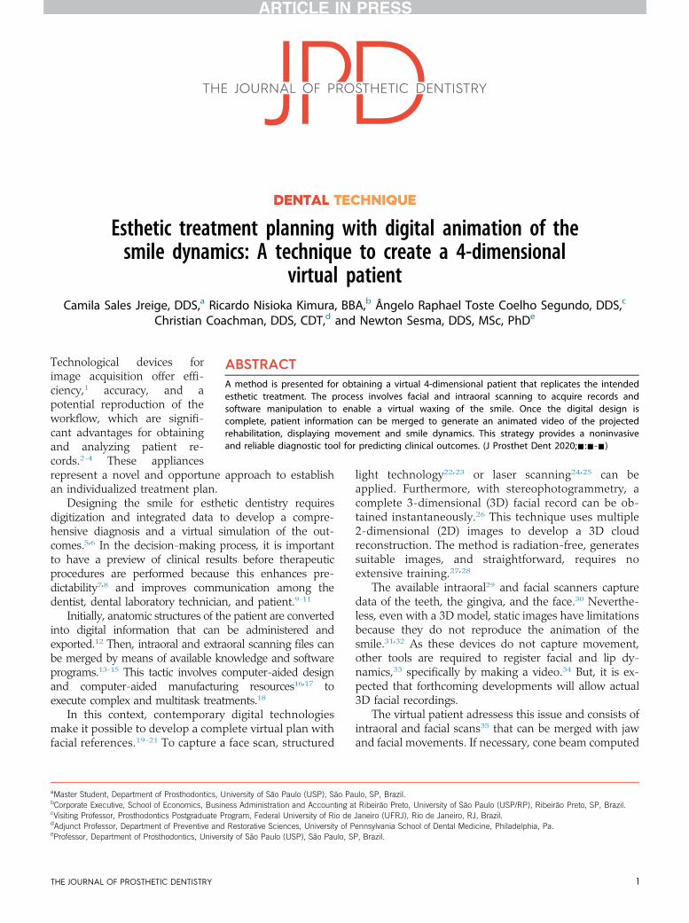
Esthetic treatment planning with digital animation of thesmile dynamics: A technique to create a 4-dimensional
virtual patient
Camila Sales Jreige, DDS,a Ricardo Nisioka Kimura, BBA,b Ângelo Raphael Toste Coelho Segundo, DDS,cChristian Coachman, DDS, CDT,d and Newton Sesma, DDS, MSc, PhDe
ABSTRACTA method is presented for obtaining a virtual 4-dimensional patient that replicates the intendedesthetic treatment. The process involves facial and intraoral scanning to acquire records andsoftware manipulation to enable a virtual waxing of the smile. Once the digital design iscomplete, patient information can be merged to generate an animated video of the projectedrehabilitation, displaying movement and smile dynamics. This strategy provides a noninvasiveand reliable diagnostic tool for predicting clinical outcomes. (J Prosthet Dent 2020;-:---)
Technological devices forimage acquisition offer effi-ciency,1 accuracy, and apotential reproduction of theworkflow, which are signifi-cant advantages for obtainingand analyzing patient re-cords.2-4 These appliances
represent a novel and opportune approach to establishan individualized treatment plan.Designing the smile for esthetic dentistry requiresdigitization and integrated data to develop a compre-hensive diagnosis and a virtual simulation of the out-comes.5,6 In the decision-making process, it is importantto have a preview of clinical results before therapeuticprocedures are performed because this enhances pre-dictability7,8 and improves communication among thedentist, dental laboratory technician, and patient.9-11
Initially, anatomic structures of the patient are convertedinto digital information that can be administered andexported.12 Then, intraoral and extraoral scanning files canbe merged by means of available knowledge and softwareprograms.13-15 This tactic involves computer-aided designand computer-aided manufacturing resources16,17 toexecute complex and multitask treatments.18
In this context, contemporary digital technologiesmake it possible to develop a complete virtual plan withfacial references.19-21 To capture a face scan, structured
ent, Department of Prosthodontics, University of São Paulo (USP), São Paxecutive, School of Economics, Business Administration and Accounting aessor, Prosthodontics Postgraduate Program, Federal University of Rio defessor, Department of Preventive and Restorative Sciences, University of Pepartment of Prosthodontics, University of São Paulo (USP), São Paulo, S
L OF PROSTHETIC DENTISTRY
light technology22,23 or laser scanning24,25 can beapplied. Furthermore, with stereophotogrammetry, acomplete 3-dimensional (3D) facial record can be ob-tained instantaneously.26 This technique uses multiple2-dimensional (2D) images to develop a 3D cloudreconstruction. The method is radiation-free, generatessuitable images, and straightforward, requires noextensive training.27,28
The available intraoral29 and facial scanners capturedata of the teeth, the gingiva, and the face.30 Neverthe-less, even with a 3D model, static images have limitationsbecause they do not reproduce the animation of thesmile.31,32 As these devices do not capture movement,other tools are required to register facial and lip dy-namics,33 specifically by making a video.34 But, it is ex-pected that forthcoming developments will allow actual3D facial recordings.
The virtual patient adressess this issue and consists ofintraoral and facial scans35 that can be merged with jawand facial movements. If necessary, cone beam computed
ulo, SP, Brazil.t Ribeirão Preto, University of São Paulo (USP/RP), Ribeirão Preto, SP, Brazil.Janeiro (UFRJ), Rio de Janeiro, RJ, Brazil.ennsylvania School of Dental Medicine, Philadelphia, Pa.P, Brazil.
1

Figure 1. Pretreatment photographs. A, Smile at rest. B, Animated smile. C, Profile assessment of lips at rest. D, Lateral view of animated smile. E, 12o’clock view.
2 Volume - Issue -
THE JOURNAL OF PROSTHETIC DENTISTRY Jreige et al

Figure 2. Facial scan obtained by postprocessing images in software program (3DF Zephyr; 3DFLOW). A, Stereophotogrammetry cabin (clOner; dOne3D). B, Smile at rest. C, Animated smile. D, Lateral aspect of lips at rest. E, Profile outlook of animated smile. F, 12 o’clock view.
- 2020 3
tomography images36 can also be acquired. All these filescan be overlapped and used together to create a 4-dimensional (4D) digital patient, allowing nonstatic andrealistic treatment planning.37,38
The present article introduces a technique that com-bines facial imaging, intraoral scanning, digital planningsoftware, and virtual animation of smile dynamics toreliably predict esthetic treatment.
TECHNIQUE
1. Initiate patient documentation. Make facial photo-graphs in front, profile, and 12 o’clock views. Aim tocapture the most spontaneous animated smile,especially in patients with a high smile line and/orwith excessive gingival display (Fig. 1).
2. Record a video by using a mobile phone (iPhone;Apple Corp) to save the patient’s dynamic move-ments from the rest position to the animated smile.Upload the video to a cloud storage system.
Jreige et al
3. Acquire a precise facial scan in a stereo-photogrammetry cabin (clOner; dOne 3D) equippedwith 16 cameras (8 MP; 2.8 mm each) programmedto be synchronized and compose the image. Set thepatient’s head in a natural head position as per thelinear references of the laser positioning structure.Capture photographs in a single command in lessthan 0.5 seconds. Perform the procedure in thefollowing smile positions: at rest, half-open, andanimated smile. Export the images by using the jointphotographic group (JPG) format.
4. Import photographs into a photogrammetry and 3Dmodeling software program (3DF Zephyr; 3DFLOW).Apply the point georeferencing tactic to postprocess thedata and transform the 2D images into a 3D coloredmesh (Fig. 2). Save the record in object file (OBJ) format.
5. Scan the maxilla, mandible, and occlusal relationsby using an intraoral scanner (TRIOS; 3Shape A/S).Extract the data in standard tessellation language(STL) file format (Fig. 3).
THE JOURNAL OF PROSTHETIC DENTISTRY

Figure 3. Intraoral scanner 3-dimensional casts. A, Virtual maxillary cast.B, Virtual mandibular cast. C, Occlusion record.
4 Volume - Issue -
TH
6. For complete patient digitization, transfer the 2Dinitial photographs, the intraoral scan, and facialscan to the digital smile design (DSD) app foriPad (Apple Corp). Merge the facial animatedsmile photograph with the intraoral and facialscans. Create an esthetic dental project by usingthe tools available in the DSD system. Choosethe optimal layout of the teeth and define thenew gingival contour, considering the facial ref-erences (Fig. 4).
7. Still using the DSD app software program, followthe 2D proposal to guide a complete 3D maxillarywaxing. Verify the morphology and overallappearance of the planned smile from differentangles, including front, profile, and 12 o’clockviews, to achieve dentofacial harmony (Fig. 5).Save the data in a STL extension.
8. Use the references of the patient’s initial video andoperate an animation software (Maya; Autodesk) todevelop the dynamic path of the smile from the restposition to the animated smile. Adopt the pointcorrelation strategy to associate the 2D video withthe 3D mesh of the intraoral and facial scans.Render data and generate a video of the smileproject to create a 4D patient: a representation of 3dimensions gathered with the patient in movement(Fig. 6, Supplemental Video 1, available online).
E JOURNAL OF PROSTHETIC DENTISTRY
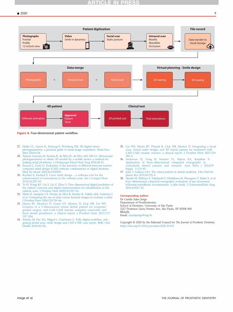
9. Analyze the video and adopt it as a prospectivediagnostic tool to anticipate treatment outcomes.Demonstrate the animated preview to the pa-tient and share data with the professional team.Obtain the patient’s approval and, if necessary,apply changes to the project. Subsequently, usethe 3D digital waxing file to make a resin cast ofthe maxilla (Standard Photopolymer Resin;FlashForge) with a 3D printer (FlashForgeHunter; FlashForge). Fabricate a silicone indexwith impression material (Silagum Putty andHonigum Light; DMG) from the printed cast.Finally, fill the index with bis-acryl resin shadeA1 (Protemp 4; 3M ESPE) and produce trialrestorations to clinically evaluate the smileplanning (Fig. 7). The workflow of the techniqueis visually displayed in Figure 8.
DISCUSSION
The protocol presented in this article characterizes amethod for treatment planning that incorporates themovements of smile dynamics. The visual perception isimproved and clinical outcomes are adequately predicted.
In a conventional workflow, it is expected but notensured that the planned smile will be precisely trans-ferred to the definitive restorations.9 Analog waxing froma 2D photograph of the patient may contain inaccuraciesbecause the results depend mainly on the technician’simpression and expertise.31 The digital process overcomethese concerns by combining imaging devices and new3D reconstruction software.4,8,36
The smile design phase has been previously per-formed by using 2D digital images and has now devel-oped into a 3D process. The intention is essentially thesame, to develop a facially driven smile framework thatwill suggest the optimal 3D position of the teeth andgingiva of the maxillary arch. After data acquisition, thepatient’s digital files can be saved in a compatible formatto be imported to the chosen technological system.Different software programs can be used to develop thenew smile architecture, including Exocad (Align Tech-nology),17,21 3Shape Smile Design (3Shape A/S),29 andCEREC or CAD inLab (Dentsply Sirona).30 These com-puter programs can be effectively applied for planningpurposes.
The DSD app is also a file management system. DSDis a conceptual tool that analyzes the intraoral andextraoral linear references located on the 4 views of thepatient’s face: front, profile, 12 o’clock, andocclusal.5,6,10,11,34 The video assessment is then used toconfirm where these guidelines should be positioned.However, this examination is visual and subjective. Thebenefit of using 3D facial scans with movement is thatthe smile design can be checked, not only from the 4
Jreige et al

Figure 4. Esthetic smile planning with DSD app. A, DSD software program and facial photograph of animated smile. B, Maxillary digital cast mergedwith 2D frontal photograph. C, Intraoral scan digital cast integrated with profile photograph. D, Facial scan imported into software program andsuperimposed on front view photograph. E, Merging intraoral and facial scans. F, Smile design guided by anatomic references. G, Before and afterproject simulation. DSD, digital smile design.
- 2020 5
Jreige et al THE JOURNAL OF PROSTHETIC DENTISTRY

Figure 5. Computerized 3-dimensional waxing. A, 3D waxing cast presented at different angles to evaluate smile. B, Proposed design from frontal view.C, 12 o’clock view. D, 3D waxing incorporated into facial scan in anterior view. E, Smile design and facial scan in profile view. F, 3D waxing in inferiorview. G, Definitive 3-dimensional smile design cast.
6 Volume - Issue -
conventional perspectives but also in an infinite numberof views; moreover, the project can be evaluated withactual facial movements (Supplemental Video 1, availableonline).
The merging of intraoral and facial scans14,15,34 withcomputer manipulation allows complete digitization andthe creation of a virtual patient.19,20 Nevertheless, thecommon reference used to be a static smile position.31,33 Inthe presented technique, an additional software programwas used to convert the data into a realistic 4D patient,capable of showing the smile path. Hence, a completeperiodontal and restorative simulation was conductedconsidering facial anatomy and lip movements.
The virtual plan was assessed in different posi-tions and angles from a 3D perspective with lipdynamics, and the outcome of the additive trialrestorations was a replica of the digital design.When it is not possible to provide trial restorationsbecause of extruded teeth or abnormal tooth an-gulations, the opportunity to virtually preview thesmile is another advantage of 4D simulation. Once
THE JOURNAL OF PROSTHETIC DENTISTRY
the project is approved, the planning files can beused to mill and produce definitive restorationsfrom the same digital library.
Limitations of the proposed strategy include thehigh equipment costs,35 necessitating initial in-vestments or the establishment of partnerships. Inaddition, dental professionals and technicians needtraining to understand and operate computer-aideddesign software.1 The management of digital features isassociated with an inherent learning curve, and sci-entific evidence should guide the educational progress.
The reported method points to a trend towardgraphic simulation. A 4D patient has shown to be anaccurate, realistic, and noninvasive diagnostic plan-ning tool. Clinical studies are needed to validate itsuse in practice.
SUMMARY
The described technique introduces the integration offacial and intraoral scans with a smile design to create a
Jreige et al

Figure 6. Video frames representing 4D virtual patient. 3D smile project incorporated into facial scan. A, Smile at rest. B, Animated smile. C, Lateral viewof lips at rest. D, Profile appearance of animated smile. E, 12 o’clock view.
- 2020 7
dynamic 4D patient. This protocol enables the develop-ment of a comprehensive digital planning that results in afinal animated video of the smile path. The possibility ofevaluating the smile in movement represents a promisingdiagnostic instrument to predict treatment outcomes.
REFERENCES
1. Lewis RC, Harris BT, Sarno R, Morton D, Llop DR, Lin WS. Maxillary andmandibular immediately loaded implant-supported interim complete fixeddental prostheses on immediately placed dental implants with a digitalapproach: a clinical report. J Prosthet Dent 2015;114:315-22.
2. Bukhari S, Goodacre BJ, AlHelal A, Kattadiyil MT, Richardson PM. Three-dimensional printing in contemporary fixed prosthodontics: a techniquearticle. J Prosthet Dent 2018;119:530-4.
3. Codari M, Pucciarelli V, Tommasi DG, Sforza C. Validation of a technique forintegration of a digital dental model into stereophotogrammetric images ofthe face using cone-beam computed tomographic data. Br J Oral MaxillofacSurg 2016;54:584-6.
4. Kim JH, Park YC, Yu HS, Kim MK, Kang SH, Choi YJ. Accuracy of 3-dimensional virtual surgical simulation combined with digital teeth align-ment: a pilot study. J Oral Maxillofac Surg 2017;75:2441.e1-13.
5. Coachman C, Calamita MA. Digital smile design: a tool for treatmentplanning and communication in esthetic dentistry. Quintessence DentTechnol 2012;35:103-11.
6. Coachman C, Georg R, Bohner L, Rigo LC, Sesma N. Chairside 3D digitaldesign and trial restoration workflow. J Prosthet Dent 2020;124:514-20.
Jreige et al
7. Liu S, Srinivasan M, Mörzinger R, Lancelle M, Beeler T, Gross M, et al.Reliability of a three-dimensional facial camera for dental and medical ap-plications: a pilot study. J Prosthet Dent 2019;122:282-7.
8. Guichet DL. Digital workflows in the management of the estheticallydiscriminating patient. Dent Clin North Am 2019;63:331-44.
9. Pimentel W, Teixeira ML, Costa PP, Jorge MZ, Tiossi R. Predictable outcomeswith porcelain laminate veneers: a clinical report. J Prosthodont 2016;25:335-40.
10. Garcia PP, da Costa RG, Calgaro M, Ritter AV, Correr GM, da Cunha LF,et al. Digital smile design and mock-up technique for esthetic treatmentplanning with porcelain laminate veneers. J Conserv Dent 2018;21:455-8.
11. Coachman C, Calamita MA, Sesma S. From 2D to complete digital workflowin interdisciplinary dentistry. J Cosmet Dent 2016;32:62-74.
12. Tapie L, Lebon N, Mawussi B, Fron-Chabouis H, Duret F, Attal JP. Under-standing dental CAD/CAM for restorations e accuracy from a mechanicalengineering viewpoint. Int J Comput Dent 2015;18:343-67.
13. Galantucci LM, Percoco G, Lavecchia F, Di Gioia E. Noninvasive comput-erized scanning method for the correlation between the facial soft and hardtissues for an integrated three-dimensional anthropometry and cephalom-etry. J Craniofac Surg 2013;24:797-804.
14. Lam WYH, Hsung RTC, Choi WWS, Luk HWK, Cheng LYY, Pow EHN.A clinical technique for virtual articulator mounting with natural head posi-tion by using calibrated stereophotogrammetry. J Prosthet Dent 2018;119:902-8.
15. Park JM, Oh KC, Shim JS. Integration of intraoral digital scans with a 3Dfacial scan for anterior tooth rehabilitation. J Prosthet Dent 2019;121:394-7.
16. Alghazzawi TF. Advancements in CAD/CAM technology: options for prac-tical implementation. J Prosthodont Res 2016;60:72-84.
17. Kim JE, Park JH, Moon HS, Shim JS. Complete assessment of occlusal dy-namics and establishment of a digital workflow by using target tracking witha three-dimensional facial scanner. J Prosthodont Res 2019;63:120-4.
THE JOURNAL OF PROSTHETIC DENTISTRY

Figure 7. Result of technique. 3D printed cast and trial restorations. A, Maxillary cast. B, Smile at rest position. C, Animated smile. D, Profileaspect of lips at rest. E, Lateral assessment of animated smile. F, 12 o’clock view. G, Comparison between initial status, 4D virtual plan, andtrial restorations.
8 Volume - Issue -
18. Sailer I, Liu S, Mörzinger R, Lancelle M, Beeler T, Gross M, et al. Comparisonof user satisfaction and image quality of fixed and mobile camera systems for3-dimensional image capture of edentulous patients: a pilot clinical study.J Prosthet Dent 2018;120:520-4.
19. Vandenberghe B. The digital patient e imaging science in dentistry. J Dent2018;74 Suppl 1:S21-6.
20. Li J, Chen Z, Dong B, Wang HL, Joda T, Yu H. Registering maxillomandibularrelation to create a virtual patient integrated with a virtual articulator forcomplex implant rehabilitation: a clinical report. J Prosthodont 19 May 2020.doi: 10.1111/jopr.13204. [Epub ahead of print].
21. Lavorgna L, Cervino G, Fiorillo L, Di Leo G, Troiano G, Ortensi M,et al. Reliability of a virtual prosthodontic project realized through a2D and 3D photographic acquisition: an experimental study on theaccuracy of different digital systems. Int J Environ Res Public Health2019;16:5139.
THE JOURNAL OF PROSTHETIC DENTISTRY
22. Metzler P, Sun Y, Zemann W, Bartella A, Lehner M, Obwegeser JA, et al.Validity of the 3D VECTRA photogrammetric surface imaging system forcranio-maxillofacial anthropometric measurements. Oral Maxillofac Surg2014;18:297-304.
23. Dornelles RFV, Alonso N. New virtual tool for accurate evaluation of facialvolume. Acta Cir Bras 2017;32:1075-86.
24. Zogheib T, Jacobs R, Bornstein MM, Agbaje JO, Anumendem D, Klazen Y,et al. Comparison of 3D scanning versus 2D photography for the identi-fication of facial soft-tissue landmarks. Open Dent J 2018;12:61-71.
25. Bardouille T, Krishnamurthy SV, Hajra SG, D’Arcy RC. Improved localizationaccuracy in magnetic source imaging using a 3-D laser scanner. IEEE TransBiomed Eng 2012;59:3491-7.
26. Hassan B, Greven M, Wismeijer D. Integrating 3D facial scanning in a digitalworkflow to CAD/CAM design and fabricate complete dentures for imme-diate total mouth rehabilitation. J Adv Prosthodont 2017;9:381-6.
Jreige et al
Data transfer tocloud storage
PhotographsFrontalProfile12 o’clock view
VideoSmile in dynamics
Facial scanStatic posture
Intraoral scanMaxillaMandibleOcclusion
Patient digitization File record
3D waxingPhotographs
Data merge Virtual planning - Smile design
Intraoral scan Facial scan++ 2D waxing
Clinical test
Trial restorations3D printed cast
4D patient
Software animationApprovalPatientTeam
Figure 8. Four-dimensional patient workflow.
- 2020 9
27. Heike CL, Upson K, Stuhaug E, Weinberg SM. 3D digital stereo-photogrammetry: a practical guide to facial image acquisition. Head FaceMed 2010;6:18.
28. Salazar-Gamarra R, Seelaus R, da Silva JV, da Silva AM, Dib LL. Monoscopicphotogrammetry to obtain 3D models by a mobile device: a method formaking facial prostheses. J Otolaryngol Head Neck Surg 2016;45:33.
29. Erozan Ç, Ozan O. Evaluation of the precision of different intraoral scanner-computer aided design (CAD) software combinations in digital dentistry.Med Sci Monit 2020;26:e918529.
30. Kurbad A, Kurbad S. Cerec smile design e a software tool for theenhancement of restorations in the esthetic zone. Int J Comput Dent2013;16:255-69.
31. Ye H, Wang KP, Liu Y, Liu Y, Zhou Y. Four-dimensional digital prediction ofthe esthetic outcome and digital implementation for rehabilitation in theesthetic zone. J Prosthet Dent 2020;123:557-63.
32. Mahn E, Sampaio CS, Pereira da Silva B, Stanley K, Valdés AM, Gutierrez J,et al. Comparing the use of static versus dynamic images to evaluate a smile.J Prosthet Dent 2020;123:739-46.
33. Harris BT, Montero D, Grant GT, Morton D, Llop DR, Lin WS.Creation of a 3-dimensional virtual dental patient for computer-guided surgery and CAD-CAM interim complete removable andfixed dental prostheses: a clinical report. J Prosthet Dent 2017;117:197-204.
34. Stanley M, Paz AG, Miguel I, Coachman C. Fully digital workflow, inte-grating dental scan, smile design and CAD-CAM: case report. BMC OralHealth 2018;18:134.
Jreige et al
35. Lin WS, Harris BT, Phasuk K, Llop DR, Morton D. Integrating a facialscan, virtual smile design, and 3D virtual patient for treatment withCAD-CAM ceramic veneers: a clinical report. J Prosthet Dent 2017;119:200-5.
36. Anderson PJ, Yong R, Surman TL, Rajion ZA, Ranjitkar S.Application of three-dimensional computed tomography incraniofacial clinical practice and research. Aust Dent J 2014;59Suppl 1:174-85.
37. Joda T, Gallucci GO. The virtual patient in dental medicine. Clin Oral Im-plants Res 2015;26:725-6.
38. Akashi M, Shibuya Y, Takahashi S, Hashikawa K, Hasegawa T, Kakei Y, et al.Four-dimensional computed tomography evaluation of jaw movementfollowing mandibular reconstruction: a pilot study. J Craniomaxillofac Surg2016;44:637-41.
Corresponding author:Dr Camila Sales JreigeDepartment of ProsthodonticsSchool of Dentistry, University of São Paulo2227 Professor Lineu Prestes Ave, São Paulo, SP 05508-000BRAZILEmail: [email protected]
Copyright © 2020 by the Editorial Council for The Journal of Prosthetic Dentistry.https://doi.org/10.1016/j.prosdent.2020.10.015
THE JOURNAL OF PROSTHETIC DENTISTRY