Embed Size (px)
Citation preview

Esthetic DentistrySampler
Chapter 9: Bonded Cementation of Veneers and Onlays From Supra-Gingival Minimally Invasive Dentistry: A Healthier Approach to Esthetic Restorations, First Edition.
by Jose-Luis Ruiz

163
Part IV
Bonded Cementation of Indirect Restorations


165
Supra-Gingival Minimally Invasive Dentistry: A Healthier Approach to Esthetic Restorations, First Edition. By Jose-Luis Ruiz.© 2017 John Wiley & Sons, Inc. Published 2017 by John Wiley & Sons, Inc.
9
Bonded Cementation of Veneers and Onlays
165
The proper technique during resin cement removal will ensure simplicity and safety, and improve margins.
Cementing Bonded Restorations
Bonded cementation is a crucial step in ensuring correct retention and marginal seal of bonded restorations. It is the bonded cementation of indirect onlays or veneers that tends to greatly concern and sometimes dissuade clinicians from embracing this newer technique. The fact that traditional crowns and bridges can be cemented very successfully with traditional crown and bridge cements such as resin-reinforced glass ionomer cements, which are easy to use and rarely cause sensitivity [1,2,3], can be a reason why many people prefer a full crown proce-dure. They see those familiar procedures as easier. Nev-ertheless, the price paid by the tooth and the gingiva for this apparent simplicity at cementation may be too high (Figure 9.1).
It is easy to understand why some clinicians stay away from bonded restorations. First, postoperative sensitiv-ity continues to irritate dentists and patients after the bonded cementation of indirect bonded restorations [4,5]. Second, the importance and complexity of isola-tion can be challenging. Some educators suggest, with-out good literature support, that unless a rubber dam
Figure 9.1 Crown being cemented with resin-modified glass ionomer cement (Meron Plus AC (VOCO)); note the amount of tooth removed and subgingival margins.
is used, the adhesive procedure will not succeed or will be of inferior quality. Such statements can easily make many clinicians shy away from adhesive procedures. According to Christensen, less than 10% of restorative dentists use a rubber dam routinely [6]. Third, resin cementation is perceived to be a very sensitive and dif-ficult technique, especially the removal of resin cement, because once it acquires its full hardness, removal of any excess can be very difficult. Additionally, previous fail-ures may also be clear in some clinician's minds, which may prevent them from adopting these newer tech-niques. These concerns can easily be overcome with some additional training. It is important to remem-ber that proper preparation will lead to a much easier cementation. Aggressive preparations with a great deal of dentin exposure and subgingival margin placement will make the cementation procedure very difficult. The implementation of a supragingival protocol can greatly simplify this process, as can choosing the appropriate materials correctly (Figure 9.2a–d).

166 9 Bonded Cementation of Veneers and Onlays
Postoperative Sensitivity
Postoperative sensitivity after the cementation of bonded onlays and veneers is well documented [7,8]. One important reason for this sensitivity is the incorrect use of geometric preparation with mechanical retention, which can prevent full seating of the restoration, owing to the film thickness of the adhesive cement. Polymeri-zation and contraction of the cement will continue after maximum seating and will form internal gaps, leading to bite pressure sensitivity if the dentinal tubules are not perfectly sealed (Figure 9.3). Nevertheless, supragingival minimally invasive preparation techniques, combined with correct bonded cementation techniques can virtu-ally eliminate the problem of postoperative pain. Occlusal adjustment is also a contributory factor in postoperative sensitivity and is discussed in this chapter.
Isolation
Excellent isolation during bonded cementation is a must, as any contamination can lead to failure. This may
Figure 9.3 Resin cement film thickness plays an important role when the onlay preparation is geometric and has retention form, as it will prevent full seating and will increase the effects of polymerization shrinkage (courtesy of Dr Raymond L. Bertolotti).
+/– 20 microns
Figure 9.2 (a and b) These images depict how much easier and predictable the isolation is when margins are supragingival compared with bleeding subgingival margins for anterior porcelain veneers. (c and d) These images depict how much easier and predictable the isolation is when margins are supragingival compared with bleeding subgingival margins for bonded onlays.
(a)
(c) (d)
(b)
lead some to suggest that unless a rubber dam is used, failures will occur. This is not supported by the litera-ture or through clinical experience. Experience and the literature attest equal results with or without a rubber dam [9,10,11]. Correct preparation, choice of adhesive

Bonded Cementation has Multiple Layers 167
Figure 9.4 (a) DryShield® isolation system. (b) Mouth prop (Propgard®, Ultradent). (c) OptraGate® (Ivoclar). (d) Cotton roll isolation. (Continued)
[12,13] and technique can play a key role in simplify-ing isolation. The implementation of a supragingival protocol during preparation makes cementation eas-ier. The choice of self-etch bonding systems is of great importance because the acidic monomer of this system is designed to work in multiple levels of moisture on the teeth, making the use of an oral humidity control-ler (rubber dam) unnecessary [14]. A number of simple devices for isolation are available and can be used during bonded cementation, including traditional cotton roll isolation (Figure 9.4a–e). Adhesive cementation requires excellent isolation.
Bonded Cementation has Multiple Layers
It must be remembered that when bonding an indirect restoration there will always be two substrates being bonded together by a resin cement and an adhesive, so there are multiple layers that require great attention (Figure 9.4a–e). The correct choice of adhesive and cement is of great importance. The two substrates or surfaces that are being joined together, the tooth sur-face (enamel and/or dentin) and the restoration surface or intaglio surface must be prepared prior to bonded cementation.
(a) (b)
(c) (d)

168 9 Bonded Cementation of Veneers and Onlays
Choosing the Correct Resin Cement
Resin cements have characteristics which make them the ideal type of cement for bonded indirect restorations, like high tensile strength, high compressive strength, low modulus of elasticity and low solubility [15,16]. The use of these strong adhesive resin cements in fact enhances the strength of composite, feldspathic porcelain and lith-ium disilicate restorations [17], although they have min-imal effect on zirconia and alumina. There are a number of different types of resin cements, each with their own advantages and disadvantages. Depending on the type of activator used for polymerization, the resin cements can be categorized as chemically (self ) cured, light-cured and dual-cured. Most resin cements do not adhere to the tooth without the help of a dental adhesive or bonding system, with the exception of the self-adhesive type cements.
There are many other important differences between resin cements, including filler particle size (which affects the ability to polish the tooth), available colors, opacity, consistency, speed of curing [18]. A crucial characteris-tic of a resin cement required for the implementation of supragingival dentistry is optimal translucency, as rule 5 requires the proper use of translucency to achieve a good supragingival restorative margin blending, and the cement is one of the three components of this rule [19]. Attention must be paid to avoiding the use of opacious cements, which will decrease the translucency of the restorative material of choice, and will force the need to hide the margins subgingivally (Figure 9.5a–c). Finally, choosing a cement with excellent chameleon character-istics, the ability to blend with other colors and absorb-ing them is very desirable, as it simplifies the choice of cement shade.
Figure 9.5 (a) Three drops of opacious, medium and translucent cements, showing different effects covering a line. (b) Without cement, the restoration lacks a visual connection with tooth. (Continued)
Figure 9.4 (Continued)(e) Rubber dam isolation on a second molar, showing the imperfect nature of any system.
(e)
(a) (b)

The Ideal Cement for Bonded Onlays 169
The Ideal Cement for Bonded Onlays
The first important consideration when choosing a resin cement is whether it will be light-curing, self-curing or dual-curing. Full self-curing cements are more hydro-philic, less color stable and harder to use because, after the restoration is fully seated, the clinician has to wait until the cement has set. This may compromise isolation, as the longer that isolation needs to be maintained the more difficult it is. The fact that onlays can vary in thick-ness anywhere from 2 mm to 5 mm (Figure 9.6) clearly makes light-cured cements unpredictable. Research has shown that an insufficient amount of light may reach the cement in thicker and darker restorations and may adversely affect the degree of conversion [20,21]. With light-cured cements it becomes worrisome, as there may be some parts of the cement under the onlay that will not cure correctly or fully. Some clinicians suggest warming the restorative composite regularly. This is impractical in a busy practice, as composite has to be
warmed in advance to make it less thick. Additionally, the rigidity of a traditional resin cement may be undesir-able for cementation, due to polymerization shrinkage.
For all of the above reasons, dual-cure resin cement is preferred (Figure 9.7). It sets on command, making isolation simpler and, in cases of thick porcelain areas where light may not fully penetrate, a chemical activa-tor will polymerize the cement. The literature points to the fact that dual-cure cements that are not light activated will have less desirable mechanical proper-ties [22,23].Attention must be paid to ensuring that the cement is compatible with self-etch bonding systems, as the acidity of self-etch can have a negative effect on some dual-cure cements [24,25]. I have used Clearfil™ Esthetic Cement (Kuraray) extensively with success. Simplicity in practice is a must and for posterior res-torations only one shade is necessary: Universal. The great chameleon ability of this cement allows for an enhanced blending ability of the restoration, provid-ing simple and predictable results. Other cements that
Figure 9.6 The curing light will have a difficult time passing through a 5-mm thick onlay.
Figure 9.7 Dual-cure cement in an automix dispensing syringe, which makes it easy to dispense.
Figure 9.5 (Continued)(c) Translucent cement (Clearfil™ Esthetic Cement, Kuraray) showing improved visual connection.
(c)

170 9 Bonded Cementation of Veneers and Onlays
work well for onlays are Bifix QM (VOCO), Multi-Link (Ivoclar), Starfil 2B (Danville), NX3 Nexus™ Third Gen-eration (Kerr) and Panavia V5 (Kuraray). Panavia F 2.0 also has the capability of direct adhesion. No primer is needed with metal oxides such as zirconia, alumina, and base metal (“non-precious”) alloys. On these sub-strates (metal oxides), only sandblast conditioning is needed [26].
Self-adhesive resin cements also have a history of suc-cessful use, when used as indicated and in appropriate cases, such as mechanically retentive preparations with supragingival margins, where contamination and lower adhesion may not be a problem [27,28].A decrease in bond strength to enamel has been shown, so it may be risky to use self-adhesive resin cements with prepara-tions with no mechanical retention [29]. With these materials, selective enamel etching is contraindicated because if any dentin is etched inadvertently, an impor-tant decrease in dentin bond strength will occur.
The Ideal Cements for Porcelain Veneers
Light-cured cements are considered to be more color stable because they do not include tertiary amine activators [30]. Combined with the fact that anterior porcelain veneers should be thin, light activation is pre-dictable, which makes light-cured cement ideal for por-celain veneers. The most important characteristic of the cement becomes the proper translucency and the ability of the cement to enhance the blending of a thin, translucent restoration. Opacious cement would be contraindicated. The market is replete with veneer cement systems which offer different colors and levels of translucency. The most important determinants of the final shade and opacity of the finished restorations are the tooth (preparation) color and the type, and thickness of the ceramic veneer [31]. The veneer cement will influence the final color [32] to a lesser extent, unless opacious cements are used, but we already know that if supragingival margins are desired, opacious cement is contraindicated. Some techniques suggest using the cement to dramatically alter the color and value of the final restoration, or to mask the color under the res-toration. This technique can be very complicated and can lead to some esthetic failures. Especially problematic are cases where there are variations of shade between the prepared teeth (Figure 9.8), and the different veneer and cement thickness, all which will make the perfect selection of different cements difficult and unpredictable. It is pref-erable to alter the color of the teeth prior to preparation by bleaching, internal bleaching or other means, and to allow a knowledgeable technician to undertake the neces-sary masking on the porcelain, as this will greatly simplify
the results. Occasionally, a very difficult area such as a deep stain can be blocked out with a fully opaque flowable composite, (such as Accolade OP Mask™), which can be placed on the tooth before the impression is taken. The technician should always receive a photograph of the teeth next to a shade tab or tabs, if there is difference in shade between the teeth or parts of a tooth.
Trying-in the veneers before choosing a final cement is very important, as this will allow the clinician to have a preview of the approximate final shade. Most try-in pastes are not accurate. The clinician can perform the try-in procedure using a try-in paste with a good true-to-color characteristic, or with water. An accurate approach is to use Accolade™ PV Cementation System (Danville), which has a very accurate try-in “compos-ite”. This try-in composite has no light-cure initiators and the shades are adjusted to accurately duplicate the shade and translucency of the light-cured composite. By using composite and not a substitute such as glycerine- based pastes, a source of veneer bonding problems is eliminated. The try-in composite need not be fully removed and the residual is polymerized by “diffusion polymerization” when the composite is light-cured [33]. It is important to remember that if Accolade PV try-in paste is being used, the intaglio surface of the veneer must have been prepared with porcelain etch and silane.
My preferred choice is to assess the final shade with water as the medium to visually connect the veneer and the tooth. This will give the clinician a close idea as to how the final restoration will look with a fully clear cement, or without the influence of the cement, and from that point it is easier to select a cement option to either maintain the current value and brightness, or slightly increase or decrease them. Trying to change the shade with a cement is extremely complicated, as the
Figure 9.8 Difference in color on prepared teeth is very difficult and unpredictable to manage using different colored cements.

Bonding Systems 171
possibilities are endless (Figure 9.9). For this reason, it is best to allow the experienced laboratory technician to have most of the control of the final shade and trans-lucency, based on the existing tooth color. Small, final adjustments are done with the cement at the chairside, guided by the try-in procedure.
I use a simple and highly predictable approach of only three translucent veneer cements with slightly different values or brightness (degree of whiteness or blackness), which will be selected at the try-in. A light shade of cement, which is usually used for shade A1 or similar, will virtually maintain the same shade and level of trans-lucency, with an insignificant increase in value achieved with the combined color of the tooth and the veneer. An extra-light cement used for shades B1 or 1M1 (the shades I most often use for porcelain veneers) will very lightly enhance the value and brightness of the final restoration with an insignificant decrease in translucency. A less fre-quently used yellow or universal shade used for shades A2 or darker will maintain the warm effect of darker shades without opacifying them. I also use the Accolade PV shades light, extra-light, and yellow, because of their high degree of translucency. Experience shows that a fully translucent or clear shade is undesirable because if there is an opening on the veneers, as often is the case,
the clear cement will show grey and will adversely affect the esthetic results (Figure 9.10).
Bonding Systems
An absolutely crucial choice for success and longevity is the adhesive system (see Chapter 4). Postoperative sensitivity is not related to the resin cement, but to the adhesive being used, with the exception of self-adhesive cements. To avoid sensitivity, some suggest using an etch and rinse (“total etch”) bonding system for veneers in mostly enamel situations and a self-etch for all other situations (Figure 9.11a,b). While this approach works, in clinical practice it can be very confusing for the den-tist, and especially the clinical team, to change the proto-col with veneers. In reality, there is no need to do this, as extremely predictable results can be achieved with self-etch systems on enamel using a selective etching proto-col. Alternatively, a bonding system which allows either “etch and rinse” or self-etching may be used.
Figure 9.9 Try-in case with cement.
Figure 9.10 Observe, on the mesial margin of onlay cemented with clear cement, an apparent “gap”. In reality, the margin is closed, but the clear cement appears to be missing.
Figure 9.11 Veneer preparation: (a) Mostly in dentin. (b) Mostly in enamel.
(a) (b)

172 9 Bonded Cementation of Veneers and Onlays
Preparing the Tooth for Adhesion
It is of great importance to have a properly cleaned and isolated tooth structure to bond to, as previously dis-cussed. Any contamination from saliva or blood dur-ing cementation could result in deficient adhesion and could lead to eventual failure. Getting cement on the provisional can also be extremely deleterious to good adhesion. After the removal of the temporary resto-ration, any remnants of cement must be removed, as research clearly shows that any remaining cement will decrease the bond strength quite dramatically [34]. An ideal way of cleaning temporary cement from the tooth after the provisional is removed is the use of a simple sandblaster, such as the MicroEtcher IIA (Danville), with 27-micron or 50-micron alumina particles, or Consepsis® Scrub (Ultradent) chlorhexidine antibacterial slurry. The use of hemostatics can have a negative effect on veneers. Ferric sulfate should be avoided, because it has the potential to stain the resin cement over time when the residual ferrous sulfate oxidizes. Aluminum chloride is a better choice when hemostasis is needed. Research has shown that when hemostatics are used and not thoroughly cleaned, the bond strength can be seriously affected. Thorough cleaning of the tooth after the use of hemostatics is a must [35].
Preparing the Buildups for Bonded Cementation
During preparation, enamel margin preservation, margin elevation, immediate dentin sealing or any other type of composite, the buildup must be prepared to achieve ideal adhesion. There is a considerable amount of evidence to suggest that bonding to this composite is very predictable when performed correctly [36]. Proper preparation of the aged composite should include sandblasting with a micro-etcher and 27- or 50-micron alumina powder [37].
Preparation of the Porcelain Intaglio Surface
There is much research into the best protocol for the preparation of the intaglio (bondable) surface of the por-celain restoration. Most researchers agree that porcelain restoration is best treated with hydrofluoric acid, such as Ultradent® Porcelain Etch [38], followed by a good, fresh, silane coupling agent, such as Clearfil® Ceramic Primer (Kuraray) [39,40]. Attention must be paid to the etching
protocol for each type of porcelain; incorrect etching time can decrease bond strength. Leucite-reinforced feldspathic porcelain, whether layered or pressed, should be etched for a full minute, whereas lithium disilicate need only be etched for 20 seconds. After etching, the intaglio surface must be washed very well with an air/water syringe, then dried completely. A ceramic primer should then be applied. Choose one that includes a sol-vent, silane, and MDP monomer, and does not need an additional coating of resin.
Zirconia is not a porcelain based on silicate. It is a metal oxide ceramic that will not etch with hydrofluoric acid, so the protocol for the intaglio surface is also different. The intaglio surface should be sandblasted and then a primer containing the MDP monomer should be applied, after which almost any resin cement (such as Clearfil™ Esthetic Cement) may be used. An alternative is to use a silica-coated aluminum oxide sand, which deposits an adhered silica layer onto the zirconia. The silica layer is then treated with silane to achieve a bond [41]. After sandblasting, a combination primer, containing both silane and MDP (the adhesive monomer in Panavia) [42], has been shown to further enhance adhesion to the silica layer [43]. When using an “adhesive built in” cement such as Panavia, the abrasive can be aluminum oxide, as Panavia bonds directly to metal oxides, and no primer is needed.
Step by Step Onlay Bonded Cementation
The Tooth
1) After dental anesthesia and the removal of the provisional, isolate using your method of choice (Figure 9.12a).
2) The tooth needs to be completely cleaned of rem-nant cement by using MicroEtcher (Danville). This is especially important if composite buildups were done. A scrub or pumice is a second option.
3) The onlay must be adjusted for proper proximal contacts and fit. Do not expect the fit of a porcelain onlay to match the fit of a gold onlay. Some slight opening in the margin is normal with porcelain, and resin cement will be used to seal those slight open-ings permanently (Figure 9.12b).
4) See Intaglio Onlay Preparation below. 5) Selectively etch the enamel for 10 seconds, carefully
avoiding touching the dentin (Figure 9.12c). 6) Wash completely with water and dry. The tooth is
dry when it is not shiny; desiccating the tooth is never desirable (Figure 9.12d).
7) Apply your bonding system of choice (such as Clear-fil SE Protect primer) for 20–25 seconds over the
Step by Step Onlay Bonded Cementation 173
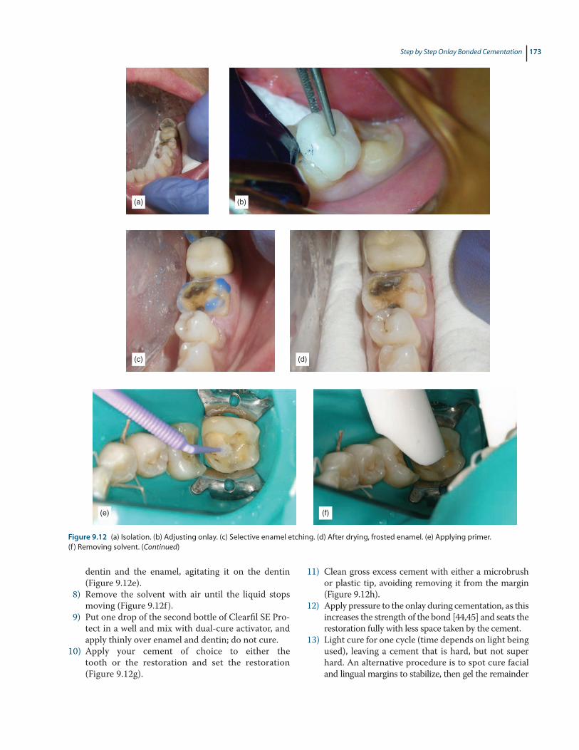
dentin and the enamel, agitating it on the dentin (Figure 9.12e).
8) Remove the solvent with air until the liquid stops moving (Figure 9.12f ).
9) Put one drop of the second bottle of Clearfil SE Pro-tect in a well and mix with dual-cure activator, and apply thinly over enamel and dentin; do not cure.
10) Apply your cement of choice to either the tooth or the restoration and set the restoration (Figure 9.12g).
11) Clean gross excess cement with either a microbrush or plastic tip, avoiding removing it from the margin (Figure 9.12h).
12) Apply pressure to the onlay during cementation, as this increases the strength of the bond [44,45] and seats the restoration fully with less space taken by the cement.
13) Light cure for one cycle (time depends on light being used), leaving a cement that is hard, but not super hard. An alternative procedure is to spot cure facial and lingual margins to stabilize, then gel the remainder
(a)
(c)
(e)
(d)
(f)
(b)
Figure 9.12 (a) Isolation. (b) Adjusting onlay. (c) Selective enamel etching. (d) After drying, frosted enamel. (e) Applying primer. (f ) Removing solvent. (Continued)

174 9 Bonded Cementation of Veneers and Onlays
with brief light cure. Remove the gel. If desired, a metal matrix may be placed through the gel in the contact zones. Then fully light cure (Figure 9.12i).
14) After confirming that the cement is firm, use a no. 12 blade to remove excess cement (Figure 9.12j).
15) Attempt to pass some floss. If this is unsuccessful, use an interproximal finishing tool, such as a Cerisaw™ (DenMat).
16) After the cement is been removed, flood the tooth with an oxygen inhibitor (such as Liquid Lens™, Danville, glycerin-based gel), and do a final cure, one cycle facial, one cycle lingual, one cycle occlusal, using water to keep the tooth cool.a) Full polymerization of dual-cure cements is
indispensable and increases success [46].b) Without this, a layer of resin will always be uncured,
as resin cannot cure in the presence of oxygen. The oxygen-inhibited layer can be 0.25–0.5 mm of uncured resin. Incorrect curing of the cement will lead to the creation of a channel or ditch.
17) Make final bite adjustments:a) Isolate the area with cotton rolls, then use blue
thin articulating paper (such as Artifol®, Bausch, metallic test film).
b) Ask the patient to tap their teeth together hard, twice (it helps to say to the patient “tap, tap”). Marks will usually show on the working cusp and central groove (Figure 9.13a).
c) As soon as there is equal contact throughout the area, use a differently colored paper (red) and ask the patient to grind their teeth (“grind, grind, grind, exaggerate the grind, forward, back, side-ways”) (Figure 9.13b).
d) Whatever is marked in red outside the blue area is an interference and must be removed.
e) Adjustments are complete when the centric con-tacts and the lateral movements are on top of each other.
f ) Having the patient sit up to do a final adjust-ment is an option, although the results will usually be the same. Sometimes you can find an additional adjustment that needs to be made, as there is a slight difference in the bite from lay-ing back to sitting upright.
18) Polish the porcelain using the Sof-Lex™ (3M) or Dialite® (Brasseler) finishing and polishing system.
(g)
(i) (j)
(h)
Figure 9.12 (Continued)(g) Applying cement on onlay. (h) Removing excess using a gum massager. (i) Light-cure onlay for one cycle. (j) Using a no. 12 blade.

Step By Step Veneer Bonded Cementation 175
Intaglio Onlay Preparation
1) Prepare the porcelain for adhesion with hydrochloric acid, and Ultradent Porcelain Etch.a) Pressed feldspathic is etched for 1 minute, lith-
ium disilicate for 20 seconds. Over-etching weakens the ceramic surface so it is undesirable.
b) Note: Onlays may come pre-etched from the laboratory. If so, after making adjustments, only re-etch for 10 seconds or less, to remove con-taminants. Alternatively, cleaning may be done with phosphoric acid.
2) Wash and dry the porcelain thoroughly. Porcelain needs to be bone-dry before applying the ceramic primer (silane).
3) Apply ceramic primer and leave for 1 minute, then air dry.a) Applying resin is not necessary.b) If using some brands of silane, apply for 1 min-
ute, dry, then apply bond.c) The restoration is ready. When using a ceramic
primer, there is no need to protect the onlay from ambient light.
4) If using zirconia, use these intaglio onlay prepara-tion instructions.
Step By Step Veneer Bonded Cementation
The Tooth
1) After dental anesthesia, and the removal of the pro-visional, isolate using OptraGate® (Ivoclar) retractors, cotton rolls and good suction, or your isolation method of choice, including a rubber dam (Figure 9.14a).
2) The tooth needs to be completely cleaned of remnant cement by using a microetcher. This is especially impor-tant if composite buildups have been done. Consepsis scrub or pumice is a second option (Figure 9.14b).
3) Try-in all veneers with water. With teeth very wet, dip the veneer into water and set on the tooth. Repeat with the rest of the veneers. Alternatively, use the Accolade PV try-in system described above. Avoid desiccating adjacent teeth, which will then appear lighter.
4) When the water or try-in paste has created a visual con-nection with the tooth, check that the veneer has the look and color that the patient wishes (Figure 9.14c).
5) Choose an appropriate cement by try-in, as previ-ously described.
6) I prefer to cement only two veneers at a time, to avoid the possibility of any restoration not seating fully, or moving during cementation. However, Dr R Bertolotti prefers to seat all six anterior veneers at once, and has many years of successful results. Read-ers should make the final decision based on their own skill and experience.
7) Carefully place triple-0 cord dipped in water to clearly uncover the margins for cementation. Should hemostasis be required, over the counter Visine works well to soak the cord (Figure 9.14d).
8) Adjust the contact and fit if necessary (Figure 9.14e). 9) It is common for some slight openings to be pres-
ent on the margin with porcelain veneers. A resin cement should be used to permanently seal these slight openings.
10) See Intaglio Veneer Preparation below. 11) Selectively etch the enamel for 10 seconds, being care-
ful to avoid touching the dentin. It is important to use a clear matrix, such as Blue View™ (Garrison), or plumb-er’s tape to separate and isolate the adjacent prepared teeth from being etch or bonded (Figure 9.14f).
12) Wash thoroughly with water and dry. The tooth is dry when it is not shiny; desiccating the tooth is never desirable.
13) Apply your bonding system of choice (such as Clear-fil SE Protect) for 20–25 seconds over the dentin and the enamel, agitating it on the dentin (Figure 9.14g).
(a) (b)
Figure 9.13 (a) After “tap tap,” blue marks are visible. (b) Red marks outside the blue.

176 9 Bonded Cementation of Veneers and Onlays
Figure 9.14 (a) Isolation veneers. (b) Cleaning teeth with Consepsis®. (c) Try-in with water or try-in paste. (d) Placing triple-0 cord. (e) Adjusting contacts. (f ) Etching teeth, placing a matrix to isolate adjacent teeth from etch. (Continued)
(e)
(c) (d)
(f)
(a) (b)
14) Remove the solvent with air until the liquid stops moving.
15) Apply the second component of Clearfil SE Protect, the bond, thinly over the enamel and dentin, but do not cure.
16) Apply veneer cement to both veneer and seat.17) Clean the gross excess cement with a plastic tipped
massage gum (manufactured by GUM) (Figure 9.14h).18) Light cure each veneer for one cycle, leaving a
cement that is hard, but not super hard. Alterna-tively, Dr R. Bertolotti prefers to spot cure in the
center, then gel the margins for easy removal of any excess (Figure 9.14i).
19) Cleanup can be done now, using a no. 12 blade (Figure 9.14j).
20) Try to pass floss through, but if unsuccessful, use a Cerisaw™ (DenMat; Figure 9.14k).
21) Repeat steps 7–20 for all the subsequent veneers, two at a time.
22) After all veneers are cemented, remove the cord and flood the tooth with an oxygen inhibitor liquid. Do the final cure, one cycle lingual, one cycle facial, on all restorations.

Step By Step Veneer Bonded Cementation 177
(g)
(i)
(k)
(j)
(l)
(h)
Figure 9.14 (Continued)(g) Apply primer. (h) Removing cement with a gum stimulator (Buttler). (i) One cycle of curing. (j) Removing cement with no. 12 blade. (k) Using a Cerisaw™ (DenMat). (l) Using a Kincheloe (Hu-Friedy) gingival retractor to check margin integrity.
23) Use a gingival retractor to do a final check for margin quality. If necessary, use a flame diamond to smooth the margins (Figure 9.14l).
24) Polish with the Sof-Lex (3M) or Dialite (Brasseler) system.
25) Do a final bite adjustment (Chapter 12).
Intaglio Veneer Preparation
1) Prepare the porcelain for adhesion with a hydro-chloric acid, Ultradent Porcelain Etch. Pressed feld-spathic is etched for 1 minute, and lithium disilicate for 20 seconds. Over-etching weakens the ceramic surface, so it is undesirable.
Note: Veneers may come pre-etched from the lab-oratory. If so, after making your adjustments, only
re-etch for 10 seconds or less, to remove contami-nants, or clean with phosphoric acid.
2) Wash and dry porcelain thoroughly. Porcelain needs to be bone-dry before applying the silane.
3) Apply ceramic primer and leave for 1 minute, then air dry.a) If using Kuraray ceramic primer, no additional
step of applying resin is needed, since the silane itself contains resin.
b) If using a silane without resin, wait 1 minute, dry, then apply the bond.
c) The restoration is ready. When using Kuraray ceramic primer, there is no need to protect the onlay from ambient light.
4) Go back to step 11 in Step By Step Veneer Bonded Cementation above.

178 9 Bonded Cementation of Veneers and Onlays
References
1 Ehlers V, Kampf G, Stender E, Willershausen B, Ernst CP. Effect of thermocycling with or without 1 year of water storage on retentive strengths of luting cements for zirconia crowns. J Prosthet Dent, 2015; 113(6): 609–615.
2 Lindquist TJ, Connolly J. In vitro microleakage of luting cements and crown foundation material. J Prosthet Dent, 2001; 85(3): 292–298.
3 Ruiz JL, Mitra S. Utilizing RMGI as a liner for direct composite restorations. Compend Contin Educ Dent, 2006; 27: 347–351.
4 Kramer N, Frankenberger R. Clinical performance of bonded leucite reinforced glass ceramic inlays and onlays after 8 years. Dent Mater, 2005; 21: 267–271.
5 Berkowitz G, Horowitz A, Curro FA, Craig RG, Ship JA, Vena D, Thompson VP. Postoperative hypersensitivity in class I resin-based composite restorations in general practice: Interim results. Compend Contin Educ Dent, 2009; 30(6): 356–363.
6 Christensen JG. Real-world moisture control: to dam or not to dam? Clin Rep, 2015; 8(5): 1–3.
7 Clinical Research Associates. Filled polymer crowns: 1- and 2-year status reports. CRA Newsl, 1998; 22(10): 1–3.
8 Christensen GJ. Resin cements and postoperative sensitivity. J Am Dent Assoc, 2000; 131(8): 1197–1199.
9 Thordrup M, Isidor F, Horsted-Bindslev P. A 5-year study of direct and indirect resin composite and ceramic inlays. Quintessence Int, 2001; 32(3): 199–205.
10 Reich S, Wichmann M, Rinne H, Shortall A. Clinical performance of large all ceramic CAD-CAM generated restorations. J Am Dent Assoc, 2004; 135: 605–612.
11 Van Dijken JWV, Horsted P. Effects of rubber dam versus cotton roll on marginal adaptation of composite resin fillings to acid-etch enamel. Acta Odontol Scand, 1987; 45: 303–308.
12 Feigal RJ, Hitt J, Splieth C. Retaining sealant on salivary contaminated enamel. J Am Dent Assoc, 1993; 124(3): 88–97.
13 Hebling J, Feigal RJ. Use of one bottle adhesive as an intermediate bonding layer to reduce sealant microleakage on saliva-contaminated enamel. Am J Dent, 2000; 13; 187–191.
14 Werner JF, Tani C. Effect of relative humidity on bond strength of self-etching adhesives to dentin. J Adhes Dent, 2002; 4(4): 277–282.
15 Stamatacos C, Simon JF. Cementation of indirect restorations: an overview of resin cements. Compend Contin Educ Dent, 2013; 34(1): 42–46.
16 Sakaguchi RL, Powers JM. Materials for adhesion and luting. In Craig's Restorative Dental Materials. 13th ed. Philadelphia, PA: Elsevier; 2012; pp. 327–348.
17 Christensen GJ. Are tooth colored onlays viable alternatives to crowns? Clin Rep, 2012; 5(1): 1, 3.
18 Christensen GJ. Best cement types for indirect restorations. Clin Rep, 2011; 4(10): 1–3.
19 Chaiyabutr, J. Effect of abutment tooth color, cement color, and ceramic thickness on the resulting optical color of a CAD/CAM glass-ceramic lithium disilicate-reinforced crown. J Prosthet Dent, 2011; 105(2): 83–90.
20 Simeone M, Lanza A, Rengo S, Aversa R, Apicella D, Apicella A. Inlay shading effect on the photopolymerization kinetic of a dental composite material used as bonding system in an indirect restoration technique. Dent Mater, 2005; 21(8): 689–694.
21 Runnacles P, Correr GM, Baratto Filho F, Gonzaga CC, Furuse AY. Degree of conversion of a resin cement light-cured through ceramic veneers of different thicknesses and types. Braz Dent J, 2014; 25(1): 38–42.
22 Aguiar TR, de Oliveira M, Arrais CA, Ambrosano GM, Rueggeberg F, Giannini M. The effect of photopolymerization on the degree of conversion, polymerization kinetic, biaxial flexure strength, and modulus of self-adhesive resin cements. J Prosthet Dent, 2015; 113(2): 128–134.
23 Foxton RM, Pereira PN, Nakajima M, Tagami J, Miura H. Durability of dual cure resin cement/ceramic bond with different curing strategies. J Adhes Dent, 2002; 4(1): 49–59.
24 Tay FR, Pashley DH, Yiu CK, Sanares AM, Wei SH. Factors contributing to the incompatibility between simplified-step adhesives and chemically-cured or dual-cured composites. Part I. Single-step self-etching adhesive. J Adhes Dent, 2003; 5(1): 27–40.
25 Pfeifer C, Shih D, Braga RR. Compatibility of dental adhesives and dual-cure cements. Am J Dent, 2003: 16(4): 235–238.
26 Kern M. Bonding to oxide ceramics: laboratory testing versus clinical outcome. Dent Mater, 2015; 31(1): 8–14.
27 Christensen GJ. Self-adhesive resin cements: are they working? Clin Rep, 2015; 8(5): 1–3.
28 Ruiz JL. Simplifying the cementation of porcelain onlays. Dent Today, 2004;. 23(3): 76–79.
29 Burgess JO, Ghuman T, Cakir D. Self-adhesive resin cements. J Esthet Restor Dent, 2010; 22(6): 412–419.
30 Peumans M, Van Meerbeek BV, Lambrechts P, Vanherle G. Porcelain veneers: a review of the literature. J Dent, 2000; 28(3):163177.
31 Barizon KT, Bergeron C, Vargas MA, Qian F, Cobb DS, Gratton DG, Geraldeli S. Ceramic materials for porcelain veneers: part II. Effect of material, shade, and thickness on translucency. J Prosthet Dent, 2014; 112(4): 864–870.
32 Chang J, Da Silva JD, Sakai M, Kristiansen J, Ishikawa-Nagai S. The optical effect of composite luting cement on all ceramic crowns. J Dent, 2009; 37(12): 937–943.
33 Dorsman GJ, Bertolotti RL. Shade accurate composite modified for long preview during veneer try-in.

References 179
International Association for Dental Research, General Session & Exhibition, June 28 – July 1, 2006, Brisbane, Australia. Abstract No. 1140.
34 Fonseca RB, Martins LR, Quagliatto PS, Soares CJ. Influence of provisional cements on ultimate bond strength of indirect composite restorations to dentin. J Adhes Dent, 2005; 7(3): 225–230.
35 O’Keefe KL, Pinzon LM, Rivera B, Powers JM. Bond strength of composites to astringent-contaminated dentin using self etch adhesives. Am J Dent, 2005; 18(3): 168–172.
36 Sadighpour L, Geramipanah F, Allahyari S, Fallahi Sichani B, Kharazi Fard MJ. In vitro evaluation of the fracture resistance and microleakage of porcelain laminate veneers bonded to teeth with composite fillings after cyclic loading. J Adv Prosthodont, 2014; 6(4): 278–284.
37 Gresnigt MM, Ozcan M, Kalk W, Galhano G. Effect of static and cyclic loading on ceramic laminate veneers adhered to teeth with and without aged composite restorations. J Adhes Dent, 2011; 13(6): 569–577.
38 Peumans M, Hikita K, De Munck J, Van Landuyt K, Poitevin A, Lambrechts P, Van Meerbeek B. Effects of ceramic surface treatments on the bond strength of an adhesive luting agent to CAD-CAM ceramic. J Dent, 2007; 35(4): 282–288.
39 Graiff L, Piovan C, Vigolo P, Mason PN. Shear bond strength between feldspathic CAD/CAM ceramic and
human dentine for two adhesive cements. J Prosthodont, 2008; 17(4): 29–49.
40 Della Bona A, Anusavice KJ, Hood JA. Effect of ceramic surface treatment on tensile bond strength to a resin cement. Int J Prosthodont, 2002; 15(3): 248–253.
41 Wolfart M, Lehmann F, Wolfart S, Kern M. Durability of the resin bond strength to zirconia ceramic after using different surface conditioning methods. Dent Mater, 2006; 23: 45–50.
42 Bertolotti RL, Lacy AM, Watanabe LG. Adhesive monomers for porcelain repair. Int J Prosthodont, 1989; 2: 483–489.
43 Kern M. Bonding to oxide ceramics: Laboratory testing versus clinical outcome. Dent Mater, 2015; 31(1): 8–14.
44 Goracci C, Cury AH, Cantoro A, Papacchini F, Tay FR, Ferrari M. Microtensile bond strength and interfacial properties of self-etching and self-adhesive resin cements used to lute composite onlays under different seating forces. J Adhes Dent, 2006; 8(5): 327–335.
45 Chieffi N, Chersoni S, Papacchini F, Vano M, Goracci C, Davidson CL, Tay FR, Ferrari M. Effect of the seating pressure on the adhesive bonding of indirect restorations Am J Dent, 2006; 19(6): 333–336.
46 Foxton RM, Pereira PN, Nakajima M, Tagami J, Miura H. Durability of the dual-cure resin cement/ceramic bond with different curing strategies. J Adhes Dent, 2002; 4(1): 49–59.