Embed Size (px)
Citation preview
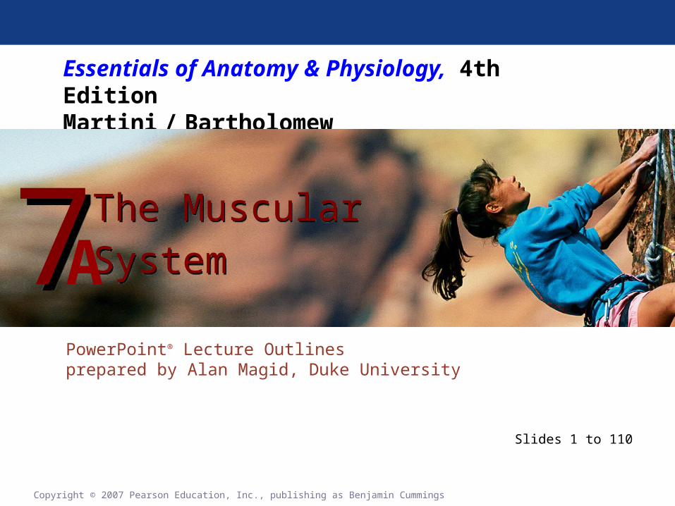
Essentials of Anatomy & Physiology, 4th EditionMartini / Bartholomew
PowerPoint® Lecture Outlines prepared by Alan Magid, Duke University
The Muscular
System
The Muscular
System77
Copyright © 2007 Pearson Education, Inc., publishing as Benjamin Cummings
Slides 1 to 110
A

Overview of Muscular System
Types of Muscle Tissue•Under voluntary control
• Skeletal muscles•Attach to the skeleton•The muscular system
•Under involuntary control• Cardiac muscle
•Heart wall• Smooth muscle
•Visceral organs
Copyright © 2007 Pearson Education, Inc., publishing as Benjamin Cummings

Overview of Muscular System
Skeletal muscles•Perform four functions
• Produce movement of skeleton (“dynamic”)• Maintain posture, balance & body position
(“static”)• Guard “entrances” (mouth) and “exits” (anus)
•oral sphincter & anal sphincter• Maintain body temperature (ex: shivering)
Copyright © 2007 Pearson Education, Inc., publishing as Benjamin Cummings
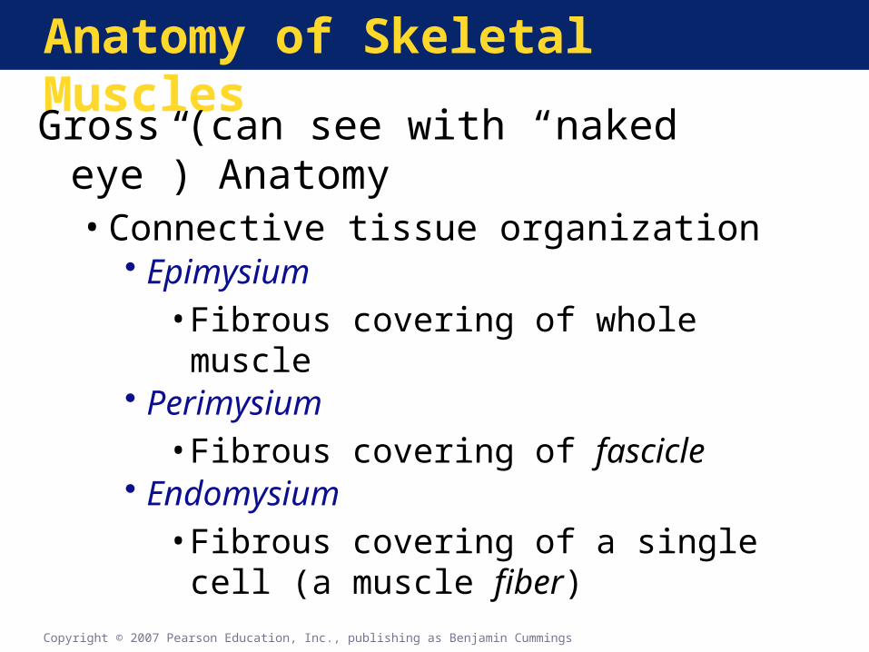
Anatomy of Skeletal Muscles
Gross (can see with “naked eye”) Anatomy•Connective tissue organization
• Epimysium•Fibrous covering of whole muscle
• Perimysium•Fibrous covering of fascicle
• Endomysium•Fibrous covering of a single cell (a muscle
fiber)
Copyright © 2007 Pearson Education, Inc., publishing as Benjamin Cummings

Anatomy of Skeletal Muscles
The Organization of a Skeletal Muscle
Figure 7-1
Quiz on all of these!

•Skeletal muscles attach to bones…• directly
•by way of tendons (thick dense regular connective tissue “ropes”
• indirectly•by way of aponeuroses (thin connective
tissue (sheets”)

Stop Here!
Your first quiz will cover only the material on the first 5 slides that precede this one.
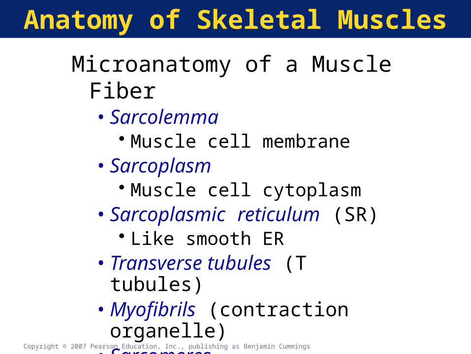
Anatomy of Skeletal Muscles
Microanatomy of a Muscle Fiber•Sarcolemma
• Muscle cell membrane•Sarcoplasm
• Muscle cell cytoplasm•Sarcoplasmic reticulum (SR)
• Like smooth ER•Transverse tubules (T tubules)•Myofibrils (contraction organelle)•Sarcomeres
Copyright © 2007 Pearson Education, Inc., publishing as Benjamin Cummings
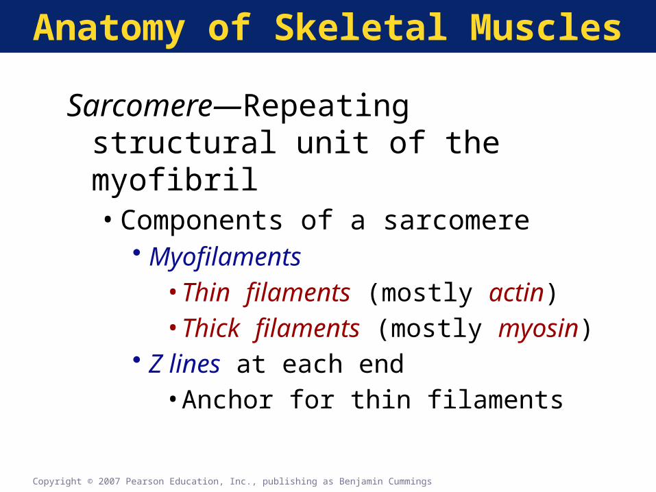
Anatomy of Skeletal Muscles
Sarcomere—Repeating structural unit of the myofibril•Components of a sarcomere
• Myofilaments•Thin filaments (mostly actin)•Thick filaments (mostly myosin)
• Z lines at each end•Anchor for thin filaments
Copyright © 2007 Pearson Education, Inc., publishing as Benjamin Cummings

Anatomy of Skeletal MusclesThe Organization of a Single Muscle Fiber
Figure 7-2(a)

Anatomy of Skeletal Muscles
The Organization of a
Single Muscle Fiber
Figure 7-2(b)

Anatomy of Skeletal Muscles
The Organization of a Single Muscle Fiber
Figure 7-2(cde)Anatomy of Skeletal MusclesPLAY

Anatomy of Skeletal Muscles
Changes in the Appearance of a Sarcomere During Contraction of a Skeletal Muscle Fiber
Figure 7-3 (1 of 2)

Anatomy of Skeletal Muscles
Changes in the Appearance of a Sarcomere During Contraction of a Skeletal Muscle Fiber
Figure 7-3 (2 of 2)

Control of Muscle Contraction
Steps in Neuromuscular Transmission
•Motor neuron action potential
•Acetylcholine release and binding
•Action potential in sarcolemma
•T tubule action potential
•Calcium release from SR
Copyright © 2007 Pearson Education, Inc., publishing as Benjamin Cummings

Control of Muscle Contraction
The Neuromuscular Junction•Synaptic terminal
• Acetylcholine release
•Synaptic cleft•Motor end plate
• Acetylcholine receptors• Acetylcholine binding• Acetylcholinesterase
•Acetylcholine removal
Copyright © 2007 Pearson Education, Inc., publishing as Benjamin Cummings

Control of Muscle Contraction
The Structure and Function of the Neuromuscular Junction
Figure 7-4(a)
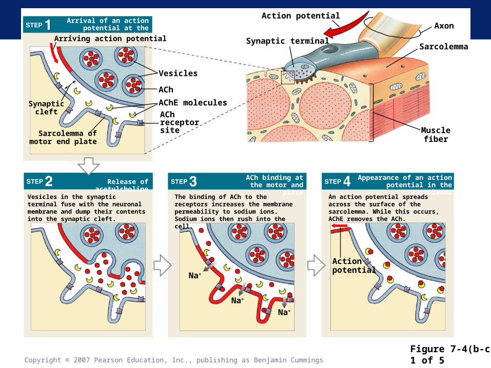
Figure 7-4(b-c)1 of 5Copyright © 2007 Pearson Education, Inc., publishing as Benjamin Cummings
Synapticcleft
Vesicles in the synaptic terminal fuse with the neuronal membrane and dump their contents into the synaptic cleft.
The binding of ACh to the receptors increases the membrane permeability to sodium ions. Sodium ions then rush into the cell.
An action potential spreads across the surface of the sarcolemma. While this occurs, AChE removes the ACh.
Appearance of an action potential in the sarcolemma
ACh binding at the motor and plateRelease of acetylcholine
Arrival of an action potential at the synaptic terminal
Sarcolemma ofmotor end plate
Arriving action potential
Vesicles
ACh
AChE molecules
AChreceptorsite
Action potential
Synaptic terminal
Axon
Sarcolemma
Musclefiber
Actionpotential
Na+
Na+
Na+
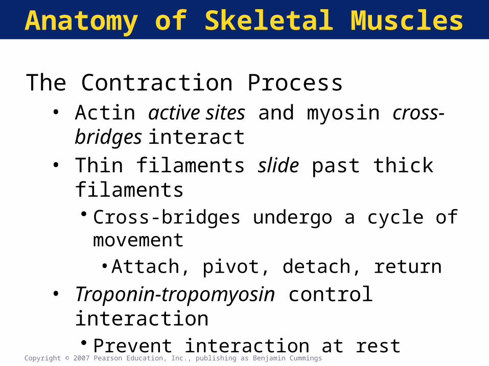
Anatomy of Skeletal Muscles
The Contraction Process• Actin active sites and myosin cross-bridges
interact• Thin filaments slide past thick filaments
• Cross-bridges undergo a cycle of movement•Attach, pivot, detach, return
• Troponin-tropomyosin control interaction• Prevent interaction at rest
Copyright © 2007 Pearson Education, Inc., publishing as Benjamin Cummings

Copyright © 2007 Pearson Education, Inc., publishing as Benjamin Cummings
Figure 7-51 of 7
Resting sarcomere
Myosin head
Myosin reactivation
Active-site exposure
Cross bridge detachment
Cross-bridge formation
Pivoting of myosin head
Troponin
ActinTropomyosin
ADP
P+
ADP
P +
ADP
P+
Active site
Sarcoplasm
Ca2+
Ca2+
ADP
P +
ADP
+ P
Ca2+
ADP+P
Ca2+
Ca2+
ADP + P
Ca2+
ADP + P
Ca2+
ATP
ATP
Ca2+
Ca2+
Ca2+
ADP
P +
+ P
ADP

Control of Muscle Contraction
Table 7-1
Summary of Contraction Process

Control of Muscle Contraction
Key Note
Skeletal muscle fibers shorten as thin filaments interact with thick filaments and sliding occurs. The trigger for contraction is the calcium ions released by the SR when the muscle fiber is stimulated by its motor neuron. Contraction is an active process; relaxation and the return to resting length is entirely passive.
Copyright © 2007 Pearson Education, Inc., publishing as Benjamin Cummings


![[Aryeh Cohen, Shaul Magid] Beginning Again Toward(BookFi.org)](https://img.pdfslide.us/doc/110x75/553013364a7959a42c8b464b/aryeh-cohen-shaul-magid-beginning-again-towardbookfiorg.jpg)