Embed Size (px)
Citation preview

EssentialsOncology Practise Essentials
Oncology Basics
Tutorial 2 Cancer Chemotherapy

EssentialsOncology Practise Essentials
Oncology Basics
16
This tutorial introduces you to the history, goals of therapy, classification, and clinical uses of chemotherapy. It also reviews some of the barriers to successful therapy.
Goals and Objectives
Upon completion of this program you will be able to:
• Define the goals of therapy in the treatment of cancer.• Classify the tumour response criteria using Response Evaluation Criteria in Solid Tumors
(RECIST).• Define the categories of cancer drug therapy.• Describe the treatment intent of chemotherapy.• Describe the clinical uses of chemotherapy.• Explain the method of calculating dosing of antineoplastic therapy.• Describe the mechanisms of chemotherapy resistance and methods to overcome resistance.
Tutorial 2 Cancer Chemotherapy
17 acpho.orgcapho.org
Tutorial 2 Cancer Chemotherapy
Oncology Basics
EssentialsOncology Practise Essentials
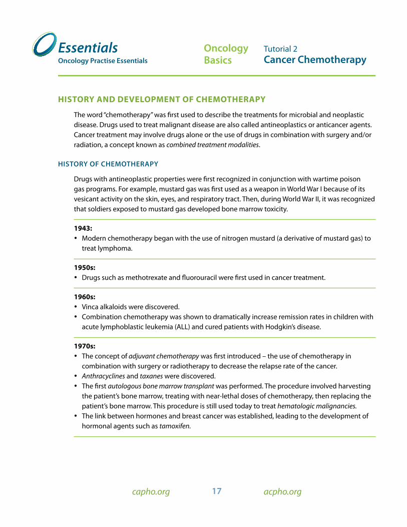
hisTory AnD DeveloPMenT of CheMoTherAPy
The word “chemotherapy” was first used to describe the treatments for microbial and neoplastic disease. Drugs used to treat malignant disease are also called antineoplastics or anticancer agents. Cancer treatment may involve drugs alone or the use of drugs in combination with surgery and/or radiation, a concept known as combined treatment modalities.
HISTOry Of CHEMOTHEraPy
Drugs with antineoplastic properties were first recognized in conjunction with wartime poison gas programs. For example, mustard gas was first used as a weapon in World War I because of its vesicant activity on the skin, eyes, and respiratory tract. Then, during World War II, it was recognized that soldiers exposed to mustard gas developed bone marrow toxicity.
1943: • Modern chemotherapy began with the use of nitrogen mustard (a derivative of mustard gas) to
treat lymphoma.
1950s: • Drugs such as methotrexate and fluorouracil were first used in cancer treatment.
1960s: • Vinca alkaloids were discovered. • Combination chemotherapy was shown to dramatically increase remission rates in children with
acute lymphoblastic leukemia (ALL) and cured patients with Hodgkin’s disease.
1970s: • The concept of adjuvant chemotherapy was first introduced – the use of chemotherapy in
combination with surgery or radiotherapy to decrease the relapse rate of the cancer. • Anthracyclines and taxanes were discovered. • The first autologous bone marrow transplant was performed. The procedure involved harvesting
the patient’s bone marrow, treating with near-lethal doses of chemotherapy, then replacing the patient’s bone marrow. This procedure is still used today to treat hematologic malignancies.
• The link between hormones and breast cancer was established, leading to the development of hormonal agents such as tamoxifen.

18 acpho.orgcapho.org
Tutorial 2 Cancer Chemotherapy
Oncology Basics
EssentialsOncology Practise Essentials
1990s: • Targeted therapy began based on research advances elucidating the signaling pathways involved
in both normal and cancerous cell proliferation. By “targeting” certain pathways, cancer cell growth could be controlled.
• Monoclonal antibodies such as rituximab for non- Hodgkin’s lymphoma were first used. Kinase inhibitors, such as imatinib for chronic myelogenous leukemia (CML) were also developed. As our knowledge of molecular biology in cancer grows, the discovery of novel targeted therapies continues to increase.
Figure 4 illustrates a detailed timeline of the history of chemotherapy.
1942 1948 1955 1958 1959 1965 1970 1972 1975 1978 1989 1992 2001 2004
Louis Goodman and Alfred Gilman use nitrogen mustard to treat a patient with non-Hodgkin’s lymphoma and demonstrate for the first time that chemotherapy can induce tumour regression.
The National Chemotherapy Program begins at the National Cancer institute (NCI); a systematic programme for drug screening commences.
The Food and Drug Administration (FDA) approves the alkylating agent cyclophosphamide.
Vincent DeVita and colleagues cure lymphomas with combination chemotherapy.
A combination of cyclophosphamide, methotrexate and fluorouracil (CMF) was shown to be effective as adjuvant treatment for node-positive breast cancer.
The NCI introduces ‘disease oriented’ screening using 60 cell lines derived from different types of human tumour.
Studies by Brian Druker lead to FDA approval of imatinib mesylate (Glivec) for chronic myelogenous leukaemia, a new paradigm for targeted therapy in oncology.
The FDA approves bevacizumab (Avastin), the first clinically proven antiangiogenic agent, for the treatment of colon cancer.
Syndey Farber uses antifolates to successfully induce remissions in children with lymphoblastic leukaemia (ALL).
George Hitchings and Gertrude Elion synthesize the purine analogue 6-mercaptopurine.
Roy Hertz and Min Chiu Li demonstrate that methotrexate as a single agent can cure choriocarcinoma, the first solid tumour to be cured by chemotherapy.
Combination chemotherapy (POMP regimen) is able to induce long-term remissions in children with ALL.
Emil Frei and colleagues demonstrate that chemotherapy given after surgical removal of osteosarcoma can improve cure rates (adjuvant chemotherapy).
The FDA approves cisplatin for the treatment of ovarian cancer, a drug that would prove to have activity across a broad range of solid tumours.
The FDA approves paclitaxel (Taxol), which becomes the first ‘blockbuster’ oncology drug.
Researchers at Harvard University define mutations in the epidermal growth factor receptor that confer selective responsiveness to the targeted agent gefitinib, indicating that molecular testing might be able to prospectively identify subsets of patients that will respond to targeted agents.
Figure 4 The history of Chemotherapy
Reprinted by permission from Macmillan Publishers Ltd: Nature Reviews Cancer 5, 65-72 (January 2005)Chemotherapy and the war on Cancer, Bruce A. Chabner & Thomas G. Roberts, Jr.

19 acpho.orgcapho.org
Tutorial 2 Cancer Chemotherapy
Oncology Basics
EssentialsOncology Practise Essentials
goAls of TherAPy in MAlignAnT DiseAse
The goals of therapy in the management of malignant disease depend on a number of factors – type of cancer, stage of disease, patient-specific factors, and patient perspectives. The goal may be to: (1) cure the cancer, (2) prolong survival without curing the disease, or (3) provide palliation and improve the quality of life of patients with incurable malignancies. Chemotherapy may be used with or without other treatment modalities, depending on the goals of the treatment.
CUrInG THE CanCEr
“Cured” means the patient is cancer-free and has a life expectancy equivalent to that of the general population. In general, it is impossible to know with certainty that a patient is completely cured, as we are unable to detect microscopic cancer cells. In most cancers, patients are considered cured if they remain alive five years from the time of diagnosis with no signs of disease recurrence. This does not apply to cancers known to have significant risk of relapse even after five years (e.g. breast cancer and melanoma). Today many cancers are considered curable, including early stage Hodgkin’s disease, Ewing’s sarcoma, testicular cancer and acute lymphocytic leukemia (ALL).
PrOlOnGInG SUrvIval
When a cure is not achievable, chemotherapy and other cancer treatments can be used to increase survival time. The goal is to induce remission and/or prevent the cancer from spreading. For example, chemotherapy may be used to prolong survival in patients with advanced breast cancer, multiple myeloma and hormone-refractory prostate cancer.
DefiniTions of resPonse CriTeriA
The World Health Organization and the National Cancer Institute developed reCisT (response evaluation Criteria in solid Tumours) to standardize the assessment of changes in tumour size for use in clinical practice and the medical literature. RECIST was first developed in 2000 (version 1.0) and recently revised in 2009 (version 1.1).
RECIST 1.1:
• Complete response (Cr) Disappearance of all target lesions.
• Partial response (Pr) At least a 30% decrease in tumour size with no evidence of new disease.
• Progressive disease (PD) At least 20% increase in tumour size AND an absolute increase of at least 5 mm, or the development of new lesions.

20 acpho.orgcapho.org
Tutorial 2 Cancer Chemotherapy
Oncology Basics
EssentialsOncology Practise Essentials
• stable disease (sD) Neither sufficient shrinkage to qualify for PR nor sufficient increase to qualify for PD.
For many malignancies, especially hematologic malignancies, response involves the elimination of tumour cells and the return of tumour “markers” to normal levels.
CATegories of CAnCer Drug TherAPy
Cancer drug therapy is generally classified into four main categories: cytotoxic chemotherapy, hormonal agents, biologics/immune therapy and targeted therapy.
CyTOTOxIC CHEMOTHEraPy
Cytotoxic agents interfere with the synthesis of DNA or RNA by disrupting cell replication and the production of proteins that are vital to cell function. The agents (which are most often associated with the treatment of cancer) can cause significant toxicity, and also require careful attention to safe handling and disposal.
HOrMOnal aGEnTS (Or anTIHOrMOnal aGEnTS) / EndOCrInE aGEnTS
Hormonal agents are used when cancer cells have hormone receptors needed for the growth process. These agents interfere with the production or action of hormones, thereby slowing or halting cancer cell growth. These drugs often have less severe side effects than cytotoxic chemotherapy drugs. For example, bicalutamide, is a common hormonal agent used in prostate cancer.
IMMUnOTHEraPy
Immunotherapy can modify the host’s response to the tumour or stimulate the immune system to fight the cancer. Two examples are retinoids (vitamin A and its metabolites) and interferon.
Retinoids affect the growth and differentiation of cells, and are used in the treatment of acute promyelocytic leukemia (APL). The exact mechanism of action is not known, but tretinoin induces maturation of leukemic cells and the appearance of normal hematopoietic cells.
Interferon has many mechanisms of action, one of which is to increase the activity of the host’s lymphocytes and macrophages, thus, augmenting the immune system to control the malignancy.

21 acpho.orgcapho.org
Tutorial 2 Cancer Chemotherapy
Oncology Basics
EssentialsOncology Practise Essentials
TarGETEd THEraPIES
Cytotoxic chemotherapy lacks selectivity and is toxic to both tumour and normal cells. Advances in molecular biology have led to a better understanding of the targets and pathways involved in the growth of cancer cells. Consequently, novel agents that specifically target the biochemical processes of malignant cells have resulted in improved patient outcomes with fewer adverse effects. These agents include:
• Monoclonal antibodies are immunoglobulins that bind to specific cell surface proteins or circulating ligands. rituximab is an example of a monoclonal antibody used for non-Hodgkin’s lymphoma. Rituximab binds to the CD20 antigen found on the surface of normal and malignant B cells, and mediates B cell lysis. Monoclonal antibodies can bind to growth factors (such as the vascular endothelial growth factor) and growth factor receptors (such as the epidermal growth factor receptor).
• Other targeted therapies include molecules that work inside the cell to block signal transduction pathways, resulting in interference with the proliferation and growth of cancer cells. erlotinib, a tyrosine kinase inhibitor, works this way.
TreATMenT inTenT of CheMoTherAPy (with and without other modalities)
The goal of chemotherapy, in the presence or absence of other treatment modalities, is dependent on the type of malignancy, prognostic factors, patient-specific factors, and the goals of the patient. The treatment intent can be categorized into the following:
• Induction Chemotherapy • Consolidation Chemotherapy• Maintenance Chemotherapy• Neoadjuvant Chemotherapy • Adjuvant Chemotherapy • Palliative Chemotherapy• Local Chemotherapy• Systemic Chemotherapy
IndUCTIOn CHEMOTHEraPy
Induction chemotherapy is the use of drug therapy in the primary treatment of advanced or disseminated cancers such as leukemias and lymphomas. No alternative treatment exists for these cancers. The goal is to induce a complete response.

22 acpho.orgcapho.org
Tutorial 2 Cancer Chemotherapy
Oncology Basics
EssentialsOncology Practise Essentials
COnSOlIdaTIOn CHEMOTHEraPy
Consolidation chemotherapy (also known as intensification therapy) is given once a remission is achieved with induction therapy. The goal is to sustain a remission.
MaInTEnanCE CHEMOTHEraPy
Maintenance chemotherapy is often given in lower doses than induction or consolidation therapy to prolong disease remission. It is commonly indicated in the treatment of acute lymphocytic leukemias and acute promyelocytic leukemias.
nEOadjUvanT CHEMOTHEraPy
Neoadjuvant chemotherapy is the use of chemotherapy prior to local treatment (e.g. surgery or radiation therapy), or when local therapy is deemed to be unsafe (e.g. if the tumour can not be safely resected due to proximity to a major blood vessel or organ). The goal is to enhance the effectiveness of local treatment by reducing the tumour burden. This is done when local therapy is deemed to be only partially effective (e.g. large tumours). A good response occurs when the tumour reduces in size, warranting additional cycles of treatment, while a poor response prompts the need for other treatment as the turmor is unresponsive to chemotherapy.
In head and neck cancers, neoadjuvant chemotherapy can shrink large tumours, facilitating subsequent surgical resection. This improves cosmetic outcomes and requires less extensive surgeries. However, there is some concern that neoadjuvant treatment may delay the administration of definitive local therapy (e.g. surgery or radiotherapy).
adjUvanT CHEMOTHEraPy
Given following local treatment such as surgery or radiation, adjuvant chemotherapy is routinely used to treat many cancers (e.g. breast and colorectal) with the intent to cure the disease. The objective is to destroy undetectable tumour cells or any malignant cells left behind after surgery or radiation. This approach is based on the rationale that:
• Residual or micrometastatic disease will be more susceptible to chemotherapy. Smaller tumours have a better vascular supply that enable better drug penetration, and have a higher growth fraction. Also, small tumours are not as genetically diverse as they have not undergone enough cell divisions to develop biologically significant mutations. Therefore, there is a lower likelihood of developing resistant cells.
The indicator of tumour response is lost in the adjuvant setting because the primary tumour has been removed. Thus disease-free survival (i.e. time after treatment during which there is no re-growth of cancer cells to a clinically detectable level) is the primary endpoint following adjuvant treatment.

23 acpho.orgcapho.org
Tutorial 2 Cancer Chemotherapy
Oncology Basics
EssentialsOncology Practise Essentials
PallIaTIvE CHEMOTHEraPy
Palliation means treatment is given to alleviate symptoms, stabilize disease and improve/maintain the patient’s quality of life without curing or significantly halting the disease. The aim is to provide relief for the patient by decreasing tumour size or retarding its growth. Palliation may involve the use of chemotherapy or even targeted therapy, in addition to best supportive care.
lOCal CHEMOTHEraPy TrEaTMEnT
Select cancers can be treated by local chemotherapy. Chemotherapy can be instilled directly into confined body spaces (e.g. into the cerebrospinal fluid [CSF]) or by site-directed perfusion into specific regions that are most affected by the cancer [e.g. hepatic artery infusion]).
SySTEMIC CHEMOTHEraPy
Unlike local chemotherapy, the intent of systemic chemotherapy is to not only eliminate cancer cells at the tumour site, but also cancer cells that have potentially spread to other areas of the body. In the adjuvant setting, systemic chemotherapy is given after definitive treatment (e.g. surgery) with the goal of eliminating any micrometastases that have escaped localized treatment with the intent of a clinical cure. The survival benefit from systemic chemotherapy following definitive treatment depends on the stage of the disease. For example, patients with small, localized disease with no nodal involvement may not gain much additional benefit from systemic chemotherapy if the tumour has been completely excised with clear margins (margins of surgical excision are clear of cancer cells). In advanced disease where the tumour cannot be safely excised, or if the cancer has already metastasized, systemic chemotherapy may help reduce the tumour bulk and decrease cancer-related symptoms (palliation). Although uncommon, a small percentage of patients with dessiminated disease may achieve a clinical cure with systemic chemotherapy.
CliniCAl use of CheMoTherAPy
COMBInEd CHEMOTHEraPy
To optimize the killing of tumour cells, chemotherapy regimens often use a combination of clinically active drugs rather than a single agent. Commonly used combination chemotherapy regimens are often referred to using acronyms. Examples include:
• ABvD (ADriamycin®-Doxorubicin, Bleomycin, vinblastine and Dacarbazine)• ChoP (Cyclophosphamide, hydroxyl daunorubicin (doxorubicin), oNCOVIN® [vincristine] and
Prednisone)

24 acpho.orgcapho.org
Tutorial 2 Cancer Chemotherapy
Oncology Basics
EssentialsOncology Practise Essentials
• feC (5-fluorouracil, epirubicin and Cyclophosphamide)• gDP (gemcitabine, Dexamethasone and Prednisone)
Combination chemotherapy has three important objectives: • Provides maximal cell kill within the range of toxicity tolerated by the patient for each drug • Provides a broader range of coverage of resistant cells in a heterogeneous tumour population• Prevents or slows development of new resistant cells
DESIRED FEATURES of antineoplastic drugs used in combination regimens include:
a. Demonstrated clinical activity against the tumour cell when used alone. Drugs producing a complete response are preferred to those producing partial responses.
b. Exhibits different toxicities or at least minimally overlapping toxicities. This minimizes multiple insults to the same organ system when used in combination, which allows administration at full doses. When overlapping toxicities are unavoidable (e.g. bone marrow suppressive drugs), administration of partial doses or longer intervals between treatments may be necessary.
c. Undergoes different mechanisms of action. Drugs can attack cancer cells at different stages of the cell cycle to achieve better cell kill. Less resistance will develop since attacking with various mechanisms overwhelms the cancer cell’s capacity to mutate and acquire resistance.
d. Demonstrates synergy when used together. Clinical trials have shown significantly improved response rates when some combination therapies are used when compared to the use of single agents. An example of biochemical synergy is the combined use of 5-fluorouracil and leucovorin (folinic acid). 5-fluorouracil binds to thymidylate synthetase, an enzyme responsible for synthesizing the nucleic acid, thymidine. At high concentrations, folinic acid stabilizes the combination of 5-fluorouracil and thymidylate synthetase thus increasing the potency of 5-fluorouracil.
e. The combination of anticancer drugs with rescue drugs allows the use of higher dosages while rescuing normal cells from toxicity. • Use of granulocyte-colony-stimulating factors (G-CSF or filgrastim) with high dose
chemotherapy helps accelerate marrow recovery and prevents severe nadirs (lowest counts). • Use of leucovorin with methotrexate to selectively “rescue” normal cells from the adverse
effects of methotrexate. Methotrexate inhibits the production of reduced folates. Administration of leucovorin provides normal cells with the reduced folates needed for nucleic acid and protein synthesis.
• Use of mesna with ifosfamide to prevent hemorrhagic cystitis. Ifosfamide produces toxic metabolites that can cause severe inflammation to the bladder lining, potentially resulting in hemorrhagic cystitis. Mesna combines with ifosfamide’s metabolites in the bladder to render a non-toxic product.

25 acpho.orgcapho.org
Tutorial 2 Cancer Chemotherapy
Oncology Basics
EssentialsOncology Practise Essentials
dOSaGE Of anTInEOPlaSTIC drUGS
Chemotherapy doses are generally calculated according to body surface area (BSA), which is based on the patient’s height and weight. Doses are expressed in units of mg/m².
• There are a number of formulas used to calculate the BSA. Some protocols and clinical trials dictate a certain method be used to calculate doses of chemotherapy. Clinicians should confirm with the prescriber which formula was used. As illustrated in Figure 5, the Mosteller formula is a common method due to its accuracy and simplicity.
BsA is used for dosage calculations rather than weight because: • BSA translates better between species (from animal to
human) for comparison of activity and toxicity.
• BSA is more stable and less variable than weight. Body weight can change considerably in a very short period, and this can affect dosage. Physiologic functions – cardiac output, blood circulation to the liver and kidneys, liver metabolism and excretion of drugs by the kidneys – do not change to the same extent as body weight. These functions are better correlated with BSA and are more important than weight with respect to drug dosage.
Dosing with BsA, actual and ideal body weight, and obesity While dosing of chemotherapy drugs such as docetaxel and gemcitabine correlates well with BSA, dosing of other cytotoxic drugs such as carboplatin does not. Carboplatin dosing correlates better with renal function.
Use of actual body weight versus ideal body weight remains controversial in oncology. While actual body weight is more commonly used, ideal body weight is generally used for acute leukemia protocols and peripheral blood stem cell or bone marrow transplant-supported high dose protocols.
Figure 5 Mosteller formula
• Average BSA for an adult male: 1.9 m²
• Average BSA for an adult female: 1.6 m²
http://www.cancernorth.nhs.uk/hpSite/groups/networkcrosscuttinggroups/chemotherapy/documents/prescribingadvice
Wt(kg)×Ht(cm)3600
BSA(m2) =

26 acpho.orgcapho.org
Tutorial 2 Cancer Chemotherapy
Oncology Basics
EssentialsOncology Practise Essentials
There is no single accepted method for accurately calculating chemotherapy doses in obese patients. Some physicians set an upper limit, or a “cap” for the BSA prior to calculating the chemotherapy dose. A commonly used “cap” is 2.2 m2. It is important to consider patient-specific factors such as age, performance status, comorbidities and tolerability of chemotherapy regimen when assessing chemotherapy dosing in obese patients. Although “capping” the BSA may reduce the risk of significant adverse effects, it may potentially increase the risk for under-dosing the patient. Therefore, it is important to also consider the treatment intent (e.g. you areless likely to “cap” the BSA if treating with a curative intent).
Maintaining Dosage Dosage is based on the balance of the drug’s pharmacologic activity against its toxicity (usually hematologic toxicity). The goal is to use the maximum dosage associated with tolerable adverse effects. Modification to the initial dosage may be required based on certain patient characteristics, most notably:
• Impairment in renal and hepatic function (required for drug elimination) • Extensive prior treatment with chemotherapy or radiation therapy
These clinical features frequently require dosage reduction to avoid unacceptable toxicity. However, this may compromise the efficacy of the chemotherapy regimen. Failing to maintain adequate and frequent doses at the maximum tolerated level is one of the most common reasons for treatment failure in cancer. This is particularly critical when treating a patient in the adjuvant setting.
Chemotherapy is now used at higher doses than ever (e.g. aggressive therapy, dose intensification and bone marrow/stem cell transplants) as the dose is a main determinant of outcome with drug-sensitive tumours. Aggressive dosing of highly myelotoxic drugs has been made possible by the development of granulocyte colony stimulating factors – genetically engineered stimulators of stem cells that make white blood cells.
SCHEdUlE Of CHEMOTHEraPy adMInISTraTIOn
The treatment schedule or interval between treatments is determined by two key factors:
• recovery of host from toxicity: Chemotherapy is generally given intermittently rather than daily. Hematologic toxicity is usually the rate limiting factor and often prevents frequent administration of chemotherapy. Therefore, scheduling of chemotherapy is based mainly on the time required for the patient to recover from the toxic effects of chemotherapy, which generally range from three to four weeks. An ANC of ≥ 1.5 x 10⁹cells/L and a platelet count of ≥ 100 x 109cells/L are typically required before the next cycle of chemotherapy can be administered. Toxicity is highly dependent on the drug dosage schedule.

27 acpho.orgcapho.org
Tutorial 2 Cancer Chemotherapy
Oncology Basics
EssentialsOncology Practise Essentials
Figure 6 The size of Cancer Cells
1.0 cm, actual size 1,000,000,000 cells
1.0 mm, actual size 1,000,000 cells
0.1 mm, actual size 1,000 cells
www.cancerguide.org
• The drug’s mechanism of action: Scheduling is also dependent on whether or not the chemotherapy agent is phase-specific. The effect of cell-cycle non-specific drugs is dose-dependent. These agents are administered at large dosages with a period between cycles to allow recovery from toxicity. Phase-specific agents are more effective when delivered by low dose continuous exposure.
BArriers To suCCessful CheMoTherAPy
TOxICITy TO nOrMal CEllS
For traditional cytotoxic chemotherapy, toxicity to normal cells is dose-dependent. Chemotherapy cannot be given at doses that cause irreversible destruction of normal cells. The doses necessary to kill 100% of malignant cells can be fatal to the patient.
COMPlETE CanCEr CEll KIll rEqUIrEd
Cure requires elimination of all viable malignant cells from the body. Once the cancer cell population is reduced to less than1 million cells, it becomes virtually undetectable. However, there is no objective means to determine when to stop treatment once cancer cells are no longer detectable.
laTE dETECTIOn
Most cancers are well advanced by the time they are large enough for discovery. As illustrated in Figure 6, a tumour 1 cm in diameter already has one billion cells. Later detection compromises treatment. Larger tumours may have metastasized and are less responsive to drugs due to their slower growth. Also, the patient may already be debilitated by the disease and less able to tolerate treatment.
POSITIOn In THE CEll CyClE
In general, solid tumours have a lower growth fraction and thus, are less responsive to anticancer drugs. Most of their cells are in the G0 phase (not actively participating in the cell cycle) and have time to repair damage caused by drugs.
28 acpho.orgcapho.org
Tutorial 2 Cancer Chemotherapy
Oncology Basics
EssentialsOncology Practise Essentials
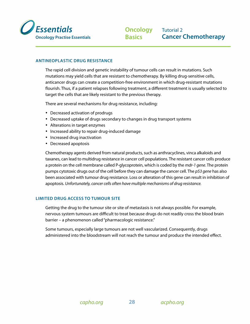
anTInEOPlaSTIC drUG rESISTanCE
The rapid cell division and genetic instability of tumour cells can result in mutations. Such mutations may yield cells that are resistant to chemotherapy. By killing drug-sensitive cells, anticancer drugs can create a competition-free environment in which drug-resistant mutations flourish. Thus, if a patient relapses following treatment, a different treatment is usually selected to target the cells that are likely resistant to the previous therapy.
There are several mechanisms for drug resistance, including:
• Decreased activation of prodrugs • Decreased uptake of drugs secondary to changes in drug transport systems • Alterations in target enzymes • Increased ability to repair drug-induced damage• Increased drug inactivation • Decreased apoptosis
Chemotherapy agents derived from natural products, such as anthracyclines, vinca alkaloids and taxanes, can lead to multidrug resistance in cancer cell populations. The resistant cancer cells produce a protein on the cell membrane called P -glycoprotein, which is coded by the mdr-1 gene. The protein pumps cytotoxic drugs out of the cell before they can damage the cancer cell. The p53 gene has also been associated with tumour drug resistance. Loss or alteration of this gene can result in inhibition of apoptosis. Unfortunately, cancer cells often have multiple mechanisms of drug resistance.
lIMITEd drUG aCCESS TO TUMOUr SITE
Getting the drug to the tumour site or site of metastasis is not always possible. For example, nervous system tumours are difficult to treat because drugs do not readily cross the blood brain barrier – a phenomenon called “pharmacologic resistance.”
Some tumours, especially large tumours are not well vascularized. Consequently, drugs administered into the bloodstream will not reach the tumour and produce the intended effect.