Embed Size (px)
Citation preview

Essential role of mda-5 in type I IFN responsesto polyriboinosinic:polyribocytidylic acidand encephalomyocarditis picornavirusLeonid Gitlin*†, Winfried Barchet*†, Susan Gilfillan*, Marina Cella*, Bruce Beutler‡, Richard A. Flavell§,Michael S. Diamond¶, and Marco Colonna*�
¶Division of Infectious Diseases, Department of Medicine, and *Department of Pathology and Immunology, Washington University School of Medicine,Campus Box 8118, 660 South Euclid Avenue, St. Louis, MO 63110; ‡Department of Immunology, Scripps Research Institute, La Jolla, CA 92037; and§Section of Immunobiology, Howard Hughes Medical Institute, Yale University School of Medicine, New Haven, CT 06520
Communicated by Emil R. Unanue, Washington University School of Medicine, St. Louis, MO, April 17, 2006 (received for review April 6, 2006)
The innate immune system recognizes viral dsRNA through twodistinct pathways; the Toll-like receptor 3 (TLR3) pathway detectsdsRNA phagocytosed in endosomes; the helicases retinoic acid-induced protein I (RIG-I) and melanoma differentiation-associatedgene-5 (mda-5) detect cytoplasmic dsRNA generated during viralreplication. Both RIG-I and mda-5 can bind polyriboinosinic:polyri-bocytidylic acid (polyI:C), the synthetic analog of viral dsRNA, andmediate type I IFN responses to polyI:C and multiple RNA viruses invitro. We generated mda-5-deficient mice and showed that mda-5is the dominant receptor mediating type I IFN secretion in responseto polyI:C in vitro and in vivo. Moreover, mda-5��� mice exhibiteda selectively impaired antiviral response to encephalomyocarditispicornavirus, indicating functional specialization of mda-5 in vivo.
innate immunity � virus
To detect RNA viruses, the innate immune system must be ableto sense conserved viral components (1). During infection of
host cells, these viruses generate RNA–RNA strand pairs in theprocess of RNA-dependent RNA synthesis. Some DNA virusesalso produce dsRNA during their life cycle. Thus, dsRNA canfunction as a pathogen-associated molecular pattern (PAMP)signaling viral infection (1). Indeed, this PAMP is recognized by theinnate immune system, eliciting a prompt antiviral response. Thesynthetic analog of viral dsRNA, polyriboinosinic:polyribocytidylicacid (polyI:C), triggers the innate immune system to secrete theantiviral cytokines IFN-� and IFN-� as well as cytokines thatinduce an inflammatory response.
The innate immune system has developed two pathways for therecognition of dsRNA (2). One pathway is mediated by Toll-likereceptor 3 (TLR3) (3). Because of its endosomal location (4), TLR3allows cells to detect dsRNA that is phagocytosed from theextracellular space where it is released by virally infected cells thatundergo lysis or necrosis (5). TLR3 may also allow detection ofdsRNA viruses that are internalized from the extracellular spacethrough receptor-mediated endocytosis. TLR3 signals through theTIR domain-containing adaptor TRIF (6, 7), which activatesTANK-binding kinase-1 through TRAF3 (8, 9) and the inducibleI-�B kinase IKK-�. These kinases phosphorylate and activate IFNregulatory factors 3 and 7 (10, 11), which mediate transcriptionalactivation of IFN-� and IFN-� genes as well as IFN-inducible genes(12, 13). TRIF also triggers a signaling cascade that activates NF-�Band the transcription of proinflammatory cytokine genes (2). TheTLR3 pathway has been implicated in the host responses torespiratory syncytial virus and influenza A virus infections in vitro(14, 15) and West Nile virus and murine cytomegalovirus (MCMV)infections in vivo (7, 16). However, additional in vivo studies withlymphocytic choriomeningitis virus, reovirus, and MCMV haveindicated that antiviral responses are also mediated by pathwaysindependent of TLR3 (17, 18).
A second pathway for detection of dsRNA is mediated bycytosolic sensors of dsRNA, which allow all cells to directly detect
intracellular viral infection (2, 19). The prototypic cytosolic sensoris the dsRNA-dependent protein kinase (PKR). PKR is a serine–threonine kinase that binds dsRNA in its N-terminal regulatoryregion and induces phosphorylation of the � subunit of the eu-karyotic protein synthesis initiation factor 2 (eIF2�), blockingcellular protein synthesis (20). PKR-deficient mouse embryonicfibroblasts have defective type I IFN responses to polyI:C and someRNA viruses, such as the encephalomyocarditis virus (EMCV)(21). However, this defect is completely corrected by pretreatmentof cells with type I IFN, suggesting the existence of type I IFN-inducible mechanisms for the recognition of dsRNA. Moreover,PKR is not essential for in vivo responses to RNA viruses (21, 22).Recently, the IFN-inducible helicase retinoic acid-induced proteinI (RIG-I) has been shown to bind polyI:C and mediate type I IFNresponses to polyI:C in transfected cells (23, 24). RIG-I contains aDExD�H box RNA helicase domain, which unwinds dsRNAthrough its ATPase activity, and a caspase recruitment domain(CARD). Overexpression of RIG-I in transfected cells enhancedcellular secretion of type I IFN in response to Newcastle diseasevirus (NDV), vesicular stomatitis virus (VSV), and EMCV. RIG-I-deficient primary cells revealed reduced type I IFN responses toNDV, VSV, Sendai virus, hepatitis C virus, and West Nile virusinfections (25–28). Because RIG-I��� mice are embryonic lethalor die within a few weeks of birth (25), the function of RIG-I inantiviral responses in vivo is unclear.
The melanoma differentiation-associated gene-5 (mda-5), alsoknown as Helicard, is another cytoplasmic sensor of dsRNA thatcontains a helicase domain and a CARD (29–32). mda-5 isubiquitously expressed in low abundance and is induced by IFN-�and TNF-�. In transfected cells, mda-5 triggers type I IFN re-sponses to NDV; inhibits EMCV, vesicular stomatitis virus, andNDV replication; and induces type I IFN-mediated inhibition oftumor cell growth (24, 30, 31). An essential role for mda-5 inantiviral responses is suggested by the existence of paramyxovirusproteins that antagonize mda-5 function, most likely to neutralizehost responses (24, 30). Additionally, it has been shown that mda-5is cleaved by apoptotic cells, and the processed protein significantlysensitizes cells to DNA degradation (32). Nonetheless, the functionof mda-5 in antiviral responses in primary cells and in vivo remainsuncertain.
Through the CARD, both RIG-I and mda-5 recruit an adaptorprotein containing an N-terminal CARD, designated IPS-1,
Conflict of interest statement: No conflicts declared.
Freely available online through the PNAS open access option.
Abbreviations: polyI:C, polyriboinosinic:polyribocytidylic acid; TLR3, Toll-like receptor 3;MCMV, murine cytomegalovirus; EMCV, encephalomyocarditis virus; CARD, caspase re-cruitment domain; DC, dendritic cell.
†L.G. and W.B. contributed equally to this work.
�To whom correspondence should be addressed. E-mail: [email protected].
© 2006 by The National Academy of Sciences of the USA
www.pnas.org�cgi�doi�10.1073�pnas.0603082103 PNAS � May 30, 2006 � vol. 103 � no. 22 � 8459–8464
IMM
UN
OLO
GY
Dow
nloa
ded
by g
uest
on
Janu
ary
16, 2
022
MAVS, VISA, or Cardif (24, 33–36). This molecule is presentin the outer mitochondrial membrane and mediates sequentialrecruitment and activation of TANK-binding kinase-1, induc-ible I-�B kinase (IKK-�), and IFN regulatory factor 3, ulti-mately leading to type I IFN secretion. The RIG-I�mda-5�IPS-1 pathway is targeted by viral and endogenous inhibitors.As a means of immune evasion, hepatitis C virus targets IPS-1
through the protease NS3-4a and attenuates type I IFNresponses (24, 26, 36–38). The endogenous protein LGP2,which contains a helicase domain but lacks a CARD, has beenproposed as a negative regulator of RIG-I and mda-5 (24, 39).
Because both mda-5 and RIG-I can detect dsRNA in thecytosol, induce type I IFN responses through the same signalingpathway, and are targeted by common inhibitors (24), it isunclear whether RIG-I and mda-5 serve redundant functions orspecialize in the recognition of different viruses. To address therole of mda-5 in vivo, particularly in mediating type I IFNresponses to dsRNA and viruses, we generated mda-5��� mice.Challenge of mda-5��� dendritic cells (DC) and macrophagesin vitro and mda-5��� mice in vivo with polyI:C demonstratedthat mda-5 is the main cytosolic receptor for polyI:C. mda-5���DC and macrophages exhibited a selective impairment of type IIFN and proinflammatory cytokine secretion in response to thepicornavirus EMCV, whereas responses to other RNA viruseswere slightly impaired, if at all. Moreover, mda-5��� micesuccumbed earlier than WT mice after EMCV infection in vivo.These results reveal a unique role of mda-5 in the recognition ofpolyI:C and an unexpected viral specificity within the cytosolicsensors of dsRNA.
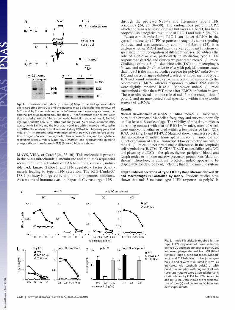
ResultsNormal Development of mda-5��� Mice. mda-5��� mice wereborn at the expected Mendelian frequency and survived normallyuntil at least 6–8 weeks of age. The viability of mda-5��� mice isin striking contrast with that of RIG-I��� mice, most of whichwere embryonic lethal or died within a few weeks of birth (25).RNA blot (Fig. 1) and RT-PCR (data not shown) analyses revealedthat abrogation of mda-5 transcript in mda-5��� mice did notaffect expression of RIG-I transcript. Flow cytometric analysis ofmda-5��� mice did not reveal major differences in the lymphoidcell populations (B, CD4� T, CD8� T, �� T, natural killer cells, DC,and plasmacytoid DC) in the spleen, thymus, peripheral blood, andlymph nodes or in bone marrow precursor populations (data notshown). Therefore, in contrast to RIG-I, mda-5 appears to bedispensable for development, including that of the immune system.
PolyI:C-Induced Secretion of Type I IFN by Bone Marrow-Derived DCand Macrophages Is Controlled by mda-5. Previous studies haveshown that mda-5 mediates type I IFN responses to polyI:C in
Fig. 1. Generation of mda-5���mice. (a) Map of the endogenous mda-5allele, targeting construct, and the mutated mda-5 allele after the removal ofMC1-neoR by Cre recombination. mda-5 exons are shown as gray boxes, theexternal probe as an open box, and the MC1-neoR construct as an arrow. LoxPsites are designated by filled arrowheads. Restriction enzyme sites: B, BamHI;Bgl, BglII; and RV, EcoRV. (b) DNA blot analysis of ES cell DNA. Genomic DNAwas cut with BamHI, and the blot was hybridized with the probe indicated ina. (c) RNA blot analysis of total liver and kidney RNA of WT, heterozygous, andmda-5��� littermates. Mice were injected with polyI:C 3 days before collec-tion of organs. For each mouse, the left lane represents liver, and the right lanerepresents kidney. mda-5 (Top), RIG-I (Middle), and hypoxanthine-guaninephosphoribosyl transferase (HPRT) (Bottom) blots are shown.
Fig. 2. mda-5 is critically required for thetype I IFN response of bone marrow-derived DC and macrophages to polyI:C. DCand macrophages derived from WT (filledsymbols), mda-5-deficient (open symbols,a–c), and TLR3-deficient mice (gray sym-bols, b and c) were stimulated in vitro, asindicated, with synthetic polyI:C or withpolyI:C in complex with Fugene. Cell cul-ture supernatants were assessed after 24 hof stimulation by ELISA for IFN-� (a and b)and IFN-� (c). Data shown are representa-tive of four (a) and two (b and c) indepen-dent experiments.
8460 � www.pnas.org�cgi�doi�10.1073�pnas.0603082103 Gitlin et al.
Dow
nloa
ded
by g
uest
on
Janu
ary
16, 2
022

transfected cells (24, 30). However, because primary cells expressadditional sensors of polyI:C, such as TLR3 and RIG-I, it remainsunknown whether mda-5 is essential for polyI:C recognition or isredundant. To address the contribution of mda-5 to polyI:C re-sponses in primary immune cells, DC and macrophages weregenerated from bone marrow cells of mda-5��� and WT mice andwere stimulated with polyI:C to induce type I IFN secretion. Weused high concentrations of naked polyI:C (100–3 �g�ml) as wellas low concentrations of polyI:C (1.5–0.05 �g�ml) complexed witha transfection reagent that optimizes polyI:C effects by facilitatingintracellular delivery. Strikingly, secretion of IFN-� in response topolyI:C was completely abrogated in mda-5��� cells as comparedwith WT cells (Fig. 2a). mda-5 deficiency also abrogated type I IFNresponses to dsRNA generated by in vitro transcription (Fig. 7,which is published as supporting information on the PNAS website). Because a prior study demonstrated that dsRNA inducesup-regulation of costimulatory molecules through TLR3-dependent and independent mechanisms (40), we investigated thecontribution of mda-5 to this up-regulation. Interestingly, mda-5��� and WT bone marrow-derived DC exhibited similar cellsurface expression profiles of B7.1 (CD80), B7.2 (CD86), and CD40(data not shown).
To compare the relative contributions of mda-5 and TLR3�TRIF to polyI:C-induced secretion of type I IFN, we stimulatedmda-5��� and TLR3��� bone marrow-derived DC with polyI:Cand measured the levels of IFN-� and IFN-� by ELISA. mda-5deficiency abrogated secretion of IFN-� and IFN-�, whereas TLR3deficiency had little or no effect (Fig. 2 b and c). Similar results wereobtained with bone marrow-derived macrophages (data notshown). We also compared the contributions of mda-5 and TLR3to polyI:C-induced secretion of proinflammatory cytokines andchemokines in DC. We observed only a limited reduction of IL-6secretion in mda-5��� DC (Fig. 8, which is published as supportinginformation on the PNAS web site). Our data suggest that mda-5is absolutely required for type I IFN responses of bone marrow-derived DC and macrophages to polyI:C in vitro, whereas it has amodest effect on proinflammatory cytokines and no effect on cellsurface expression of costimulatory molecules. We conclude that,at least in bone marrow-derived DC and macrophages, mda-5 isfunctionally dominant over TLR3 for type I IFN responses topolyI:C.
We also analyzed the response of thioglycollate-induced perito-neal macrophages to polyI:C administered either in solution ortransfected as a complex with cationic lipid. To address the con-tributions of TLR3-TRIF and mda-5 to polyI:C-induced cytokineresponses, we compared peritoneal macrophages from WT andTrifLPS2 mice (7), which carry a single base-pair deletion in theTRIF gene that causes instability or inactivation of the mutantprotein. In contrast to bone marrow-derived DC and macrophages,thioglycollate-induced macrophages produced low amounts ofIFN-� when stimulated with naked polyI:C. However, we observedconsiderable induction of inflammatory cytokines that was severelyimpaired in TrifLPS2 cells (5- to 10-fold reduction), indicating asignificant contribution of the TLR3-TRIF pathway to polyI:Crecognition in these cells (Fig. 3a). PolyI:C delivered with atransfection reagent induced higher amounts of IFN-�, which werenot affected by the TrifLPS2 mutation; in fact, TRIF-deficientmacrophages secreted higher amounts of IFN-� than WT controlsin several of these assays. Other inflammatory cytokines were alsoindependent of TRIF signaling when polyI:C was delivered in thismanner.
In contrast, mda-5��� peritoneal macrophages respondedpoorly to both naked and transfected polyI:C; in comparisonwith WT controls, cytokine secretion was reduced 2- to 3-fold inboth cases, and IFN-� secretion strikingly diminished in re-sponse to transfected polyI:C (Fig. 3b). This result suggests thatthe endosomal and cytoplasmic pathways may directly or indi-rectly, through type I IFN secretion, synergize in this particular
case. Together, these data imply that, depending on the specificcell type analyzed, mda-5 may either exercise a dominant role orcooperate with the TLR system for antiviral responses todsRNA.
mda-5 Deficiency Attenuates PolyI:C-Induced Type I IFN Secretion inVivo. To determine whether mda-5 plays also a major role inresponses to polyI:C in vivo, mda-5��� and WT mice werei.v.-injected with polyI:C, either naked or in complex with atransfection reagent. Serum concentrations of IFN-�, IFN-�,and proinflammatory cytokines and chemokines were measuredat different time points after polyI:C injection. IFN-� and IFN-�responses to naked polyI:C were abrogated in mda-5��� mice(Fig. 4a). When polyI:C was complexed with transfection re-agent, the IFN-� response was abrogated, but IFN-� was par-tially mda-5-independent (Fig. 4b). Additionally, a deficiency ofmda-5 significantly decreased serum levels of the proinflamma-tory cytokines IL-6 and monocyte chemoattractant protein-1(MCP-1) (Fig. 4c).
To address the relative contribution of TLR3-TRIF andmda-5 to polyI:C-induced cytokine responses in vivo, we i.v.injected polyI:C in TLR3-deficent and TrifLPS2 mice. In bothcases, we observed no significant reduction of serum levels ofIFN-�, IFN-�, IL-6, or MCP-1 in response to polyI:C, eithernaked (Fig. 4 d–f ) or in association with a transfection reagent(data not shown). These results demonstrate that mda-5 isfunctionally dominant over TLR3 for type I IFN responses topolyI:C in vivo.
mda-5 Is Required for Recognition of EMCV in Vitro and Host Re-sponses in Vivo. The essential role of mda-5 in response to polyI:Cin primary cells and in vivo suggested that mda-5 may be essential
Fig. 3. Distinct and complementary contributions of mda-5 and TLR3-TRIF topolyI:C-induced responses in thioglycollate-induced peritoneal macrophages.Macrophages (2 � 105 per well) from WT and TRIFLPS2 mice (a) or WT andmda-5��� mice (b) were collected 48 h after i.p. thioglycollate injection andstimulated with polyI:C either naked (100 �g�ml) or in complex with Fugene(1.5 �g�ml). After 24 h, cell culture supernatants were assessed for IFN-� byELISA and for TNF-�, MCP-1, and IL-6 by cytokine bead array. Data shown arerepresentative of two independent experiments.
Gitlin et al. PNAS � May 30, 2006 � vol. 103 � no. 22 � 8461
IMM
UN
OLO
GY
Dow
nloa
ded
by g
uest
on
Janu
ary
16, 2
022

for responses to specific RNA viruses that produce dsRNAintermediates during their life cycles. However, dsRNA pro-duced by RNA viruses could be alternatively and dominantlysensed by RIG-I. To address the question of whether mda-5 andRIG-I are redundant or specialize in the recognition of differentviruses, we assessed type I IFN secretion of mda-5��� and WTbone marrow-derived DC and macrophages after in vitro stim-ulation with an array of different RNA and DNA viruses,including West Nile virus, reovirus, Sindbis virus, influenza A,MCMV, herpes simplex virus-1, and EMCV. The viral panel alsoincluded a recombinant influenza A virus expressing an RNA-binding-defective NS1 protein (R38A NS1) (22, 41). NS1 bindsdsRNA and sequesters it from intracellular sensors, therebypromoting viral evasion from host responses (42). Thus, therecombinant influenza A virus expressing R38A NS1 protein isattenuated and induces high levels of type I IFN (41).
Remarkably, mda-5 deficiency completely abrogated DC andmacrophage secretion of IFN-� only in response to EMCV (Fig.5a). mda-5 deficiency also compromised IL-6 and MCP-1 se-cretion in response to EMCV (Fig. 5b). These results wereconfirmed in thioglycollate-induced peritoneal macrophages(Fig. 5c). In contrast, we observed only limited (�2-fold)reduction of IFN-� secretion in response to recombinant influ-enza A with the R38A NS1 mutation, West Nile virus, Sindbisvirus, and herpes simplex virus-1 (data not shown) and noreduction at all with MCMV (Fig. 5d) and reovirus (data notshown). These results demonstrate a remarkable specificity ofmda-5 for the detection of EMCV, although we cannot excludea contribution of mda-5 to the recognition of dsRNA from otherviruses.
To assess the involvement of mda-5 in the control of EMCVinfection in vivo, mda-5��� and WT littermate control micewere injected i.v. with a lethal dose of EMCV and monitored for
survival. After i.v. injection, EMCV infects the central nervoussystem, causing lethal encephalitis within 72–96 h (43). mda-5��� developed symptoms of hind-limb paralysis �24–72 hearlier than WT mice and succumbed sooner to the infection(Fig. 6), consistent with an essential role of mda-5 in hostresistance to EMCV infection in vivo.
DiscussionOur study demonstrates that mda-5 is remarkably important fortype I IFN responses to polyI:C by DC and macrophages in vitroand in mice in vivo. This result is surprising, given previousdemonstrations that both the TLR3�TRIF pathway and RIG-Ican mediate type IFN responses to polyI:C. Why does mda-5predominate over TLR3�TRIF in the type I IFN response topolyI:C? One important difference between mda-5 and TLR3 isthat mda-5 is located in the cytosol, whereas TLR3 is found inendosomal compartments (4). Thus, naked polyI:C or polyI:C incomplex with a transfection reagent may predominantly reachthe cytosol rather than endosomes, resulting in preferentialactivation of mda-5. Cytosolic entry of polyI:C may be facilitatedby a transmembrane transporter or channel that may be mim-icked by the transfection reagent used in our experiments.
TLR3�TRIF may play a relevant role in vivo when polyI:C orviral dsRNA reach appropriate endosomal compartments. Thisevent could occur when cells phagocytose dsRNA released intothe extracellular space by virally infected cells undergoing lysisor necrosis or when cells internalize viruses through receptor-mediated endocytosis. Consistent with this, phagocytosis ofvirus-infected cells or cells containing synthetic dsRNA en-hances antigen presentation via stimulation of TLR3 in antigen-presenting cells (5). In our study, mda-5 was essential for bonemarrow-derived DC and macrophage type I IFN responses topolyI:C. In contrast, TLR3-TRIF induced responses to polyI:C
Fig. 4. mda-5 deficiency strongly impairs the type I IFN response to polyI:C in vivo. WT, mda-5-deficient, TLR3-deficient, or TRIFLPS-2 (LPS-2 mutant) mice wereinjected i.v., as indicated, with either 100 �g of polyI:C or 10 �g of polyI:C complexed with Fugene. Serum samples were taken 3, 6, and 9 h after stimulationand were analyzed by ELISA for IFN-� and IFN-� (a, b, d, and e). Lines show cytokine kinetics in individual mice. IL-6 and MCP-1 were assessed by cytokine beadarray (c and f ). Error bars indicate SEM.
8462 � www.pnas.org�cgi�doi�10.1073�pnas.0603082103 Gitlin et al.
Dow
nloa
ded
by g
uest
on
Janu
ary
16, 2
022

in peritoneal macrophages, consistent with previous studies (3,6, 7, 44). In vivo, in the spleen, TLR3 is found primarily in CD8�
DC but not in marginal-zone DC (45). Because the marginal
zone of the spleen is the first to encounter the contents of theblood, it may be a major target of polyI:C sensing after i.v.administration. Thus, the dominant role of mda-5 over TLR3 insystemic responses to polyI:C could reflect the preferentialinvolvement of tissues that express relatively low levels of TLR3.
mda-5 appeared to predominate over RIG-I in type I IFNresponses to polyI:C. One possible explanation is that responsesto polyI:C require the concerted activation of RIG-I and mda-5.In this case, mda-5 deficiency would inhibit this signaling cas-cade, preempting RIG-I function. However, it is also possiblethat mda-5 has a higher affinity for polyI:C than RIG-I orrecognizes a specific feature of this particular dsRNA mimic.Therefore, even if RIG-I can bind polyI:C and trigger IFN-�secretion in transfected cells in vitro (23), it may not be required,and mda-5 in fact may be the dominant cytosolic receptor forpolyI:C in vitro and in vivo.
Our observation that mda-5 is selectively required for cytokineresponses to EMCV but not other viruses supports a model inwhich mda-5 and RIG-I have different specificities for dsRNAmolecules. mda-5 deficiency completely abolished type I IFNand cytokine responses to EMCV in DC and macrophages invitro, and mda-5��� mice were highly susceptible to EMCVinfection. In contrast, cytokine responses to other viruses wereonly marginally affected, if at all. Because the amino acidsequences of the helicase domains of mda-5 and RIG-I are only�35% identical, it is possible that this genetic diversity results indifferent specificity for distinct dsRNA conformations, resultingin preferential recognition of different viruses. EMCV andEMC-like viruses share structural characteristics in theirdsRNA, such as the presence of a homopolymeric polyC acidtract within the 5� untranslated sequence (46). These polyCtracts are retained during viral replication in vitro and in vivo andare associated with virulence. Thus, it will be important todetermine whether mda-5 is required for the recognition of
Fig. 6. mda-5-deficient mice show increased susceptibility to lethal infectionswith EMCV. (a) mda-5��� mice (n � 4, open symbols) and WT littermate controls(n � 6, filled symbols) on a pure 129 SvJ background were injected i.v. with 1,000plaque-forming units (pfu) of EMCV and then monitored for survival. mda-5���micediedafterameansurvival timeof69h,whereasWTmicesurvivedforameanof 87 h (P � 0.07). (b) mda-5��� mice (n � 5, open symbols) and WT littermatecontrols (n � 5, filled symbols) on a B6x129 SvJ background were injected i.v. with300 pfu of EMCV and monitored for survival. mda-5��� mice died after a meansurvival time of 82 h, whereas WT mice survived for a mean of 166 h (P � 0.008).
Fig. 5. mda-5 is critically required for type I IFN and cytokine response to EMCV. Bone marrow-derived DC (a, b, and d), bone marrow-derived macrophages (a, b,and d), and thioglycollate-induced peritoneal macrophages (c and d) from WT (filled symbols) and mda-5-deficient (open symbols) mice were stimulated in vitro withEMCV or MCMV at indicated multiplicities of infection. Production of IFN-� (a and c) and IL-6 and MCP-1 (b and c) in response to EMCV. (d) IFN-� response to MCMV.Cell culture supernatants were examined for IFN-� by ELISA and for IL-6 and MCP-1 by cytokine bead array. Data shown are representative of two independentexperiments.
Gitlin et al. PNAS � May 30, 2006 � vol. 103 � no. 22 � 8463
IMM
UN
OLO
GY
Dow
nloa
ded
by g
uest
on
Janu
ary
16, 2
022
other ECM-like viruses and polyC-less EMCV. In addition, itwill be crucial to assess the mda-5 contribution to innateresponses to other members of the picornavirus family, whichincludes important human and agricultural pathogens (43).
The lack of a dramatic effect of mda-5 deficiency on type I IFNsecretion by other viruses we tested could be due to the presenceof RIG-I or additional yet-uncharacterized sensors that contrib-ute to type I IFN secretion. Additionally, some of these virusescould encode novel immune evasion molecules that specificallyantagonize mda-5 function. Indeed, recent studies have shownthat human and murine paramyxoviruses encode such a protein,which inhibits mda-5 function and interferes with type I IFNresponses (24, 30). A more detailed study of the RNA virusesthat are not affected by mda-5 deficiency may identify additionalvirally encoded mda-5 inhibitors. Such investigation will helpelucidate the pathogenesis of these viruses and facilitate thedevelopment of specific antagonists that enhance innate immu-nity against these viruses and possibly improve clinical outcome.
Materials and MethodsGeneration of mda-5��� Mice. The targeting construct was de-signed to replace the first exon and nucleotides �400 bpupstream of mda-5 with an MC1-neor expression cassette (Fig.1). Correctly targeted ES cells (129x1�SvJ) were injected intoC57BL�6 (B6) blastocysts. Chimeras were bred to B6 transgenicmice expressing Cre recombinase under the cytomegaloviruspromoter (47) to delete the MC1-neor cassette. The resultingmda-5��� heterozygotes were intercrossed to obtain mda-5��� mice. Chimeras were also bred to 129x1�SvJ mice andmda-5��� heterozygotes to obtain mda-5��� mice on a pure129x1�SvJ background.
Mice. TLR3��� (3) and TRIFLPS (7) mice were on a C57BL�6background. Age-matched WT control C57BL�6 mice wereobtained from Taconic Farms.
Cell Cultures and Stimulation in Vitro. Mouse bone marrow-derivedDC and macrophages were generated as described (22). Toelicit primary macrophages in mice, 1.5 ml of 2% thioglycollatemedia was injected i.p., and cells were isolated by peritoneallavage. All cells were stimulated for 18–24 h, as indicated in thefigure legends. Synthetic polyI:C (Amersham Pharmacia) wasused as such or complexed with Fugene (3 �l��g nucleic acid)(Roche Applied Science, Indianapolis). dsRNA was generatedby in vitro transcription of a 500-bp template within the firef lyluciferase ORF by using the MEGAscript RNA transcriptionkit (Ambion, Austin, TX). EMCV (EMCV-k) was obtainedfrom R. Silverman (Cleveland Clinic, Cleveland) and passagedin L929 cells.
Stimulations and Infections in Vivo. Mice were injected with 100 �gof polyI:C or 10 �g of polyI:C complexed with 30 �l of Fugene.For in vivo infections with EMCV, 1,000 or 300 plaque-formingunits were injected i.v. in mda-5��� mice on either a mixed or129x1�SvJ background with appropriate controls.
Cytokine Analysis and Flow Cytometry. IFN-� and IFN-� weredetected in cell-free supernatants and mice sera by ELISA (PBLBiomedical Laboratories, New Brunswick, NJ). TNF-�, MCP-1,and IL-6 were measured by cytometric bead array (BDBiosciences).
We thank Julia Klesney-Tait, Isaiah Turnbull, and Rachel Presti forhelpful critiques; and Andrew Pekosz, Skip Virgin, David Leib, andWayne Yokoyama (Washington University School of Medicine, St.Louis); Robert Silverman (Cleveland State University, Cleveland); andTerence Dermody (Vanderbilt University School of Medicine, Nash-ville, TN) for viruses. L.G. is a postdoctoral fellow of the CancerResearch Institute, and B.B. is supported by National Institutes of HealthGrant GM060031.
1. Janeway, C. A., Jr., & Medzhitov, R. (2002) Annu. Rev. Immunol. 20, 197–216.2. Akira, S., Uematsu, S. & Takeuchi, O. (2006) Cell 124, 783–801.3. Alexopoulou, L., Holt, A. C., Medzhitov, R. & Flavell, R. A. (2001) Nature 413, 732–738.4. Matsumoto, M., Funami, K., Tanabe, M., Oshiumi, H., Shingai, M., Seto, Y., Yamamoto,
A. & Seya, T. (2003) J. Immunol. 171, 3154–3162.5. Schulz, O., Diebold, S. S., Chen, M., Naslund, T. I., Nolte, M. A., Alexopoulou, L., Azuma,
Y. T., Flavell, R. A., Liljestrom, P. & Reis e Sousa, C. (2005) Nature 433, 887–892.6. Yamamoto, M., Sato, S., Hemmi, H., Hoshino, K., Kaisho, T., Sanjo, H., Takeuchi, O.,
Sugiyama, M., Okabe, M., Takeda, K. & Akira, S. (2003) Science 301, 640–643.7. Hoebe, K., Du, X., Georgel, P., Janssen, E., Tabeta, K., Kim, S. O., Goode, J., Lin, P., Mann,
N., Mudd, S., et al. (2003) Nature 424, 743–748.8. Hacker, H., Redecke, V., Blagoev, B., Kratchmarova, I., Hsu, L. C., Wang, G. G., Kamps,
M. P., Raz, E., Wagner, H., Hacker, G., et al. (2006) Nature 439, 204–207.9. Oganesyan, G., Saha, S. K., Guo, B., He, J. Q., Shahangian, A., Zarnegar, B., Perry, A. &
Cheng, G. (2006) Nature 439, 208–211.10. Fitzgerald, K. A., McWhirter, S. M., Faia, K. L., Rowe, D. C., Latz, E., Golenbock, D. T.,
Coyle, A. J., Liao, S. M. & Maniatis, T. (2003) Nat. Immunol. 4, 491–496.11. Sharma, S., tenOever, B. R., Grandvaux, N., Zhou, G. P., Lin, R. & Hiscott, J. (2003) Science
300, 1148–1151.12. Honda, K., Yanai, H., Takaoka, A. & Taniguchi, T. (2005) Int. Immunol. 17, 1367–1378.13. Sato, M., Suemori, H., Hata, N., Asagiri, M., Ogasawara, K., Nakao, K., Nakaya, T., Katsuki,
M., Noguchi, S., Tanaka, N. & Taniguchi, T. (2000) Immunity 13, 539–548.14. Rudd, B. D., Burstein, E., Duckett, C. S., Li, X. & Lukacs, N. W. (2005) J. Virol. 79, 3350–3357.15. Guillot, L., Le Goffic, R., Bloch, S., Escriou, N., Akira, S., Chignard, M. & Si-Tahar, M.
(2005) J. Biol. Chem. 280, 5571–5580.16. Wang, T., Town, T., Alexopoulou, L., Anderson, J. F., Fikrig, E. & Flavell, R. A. (2004) Nat.
Med. 10, 1366–1373.17. Edelmann, K. H., Richardson-Burns, S., Alexopoulou, L., Tyler, K. L., Flavell, R. A. &
Oldstone, M. B. (2004) Virology 322, 231–238.18. Schroder, M. & Bowie, A. G. (2005) Trends Immunol. 26, 462–468.19. Lopez, C. B., Moltedo, B., Alexopoulou, L., Bonifaz, L., Flavell, R. A. & Moran, T. M. (2004)
J. Immunol. 173, 6882–6889.20. Saunders, L. R. & Barber, G. N. (2003) FASEB J. 17, 961–983.21. Yang, Y. L., Reis, L. F., Pavlovic, J., Aguzzi, A., Schafer, R., Kumar, A., Williams, B. R.,
Aguet, M. & Weissmann, C. (1995) EMBO J. 14, 6095–6106.22. Barchet, W., Krug, A., Cella, M., Newby, C., Fischer, J. A., Dzionek, A., Pekosz, A. &
Colonna, M. (2005) Eur. J. Immunol. 35, 236–242.23. Yoneyama, M., Kikuchi, M., Natsukawa, T., Shinobu, N., Imaizumi, T., Miyagishi, M., Taira,
K., Akira, S. & Fujita, T. (2004) Nat. Immunol. 5, 730–737.24. Yoneyama, M., Kikuchi, M., Matsumoto, K., Imaizumi, T., Miyagishi, M., Taira, K., Foy, E.,
Loo, Y. M., Gale, M., Jr., Akira, S., et al. (2005) J. Immunol. 175, 2851–2858.25. Kato, H., Sato, S., Yoneyama, M., Yamamoto, M., Uematsu, S., Matsui, K., Tsujimura, T.,
Takeda, K., Fujita, T., Takeuchi, O. & Akira, S. (2005) Immunity 23, 19–28.
26. Foy, E., Li, K., Sumpter, R., Jr., Loo, Y. M., Johnson, C. L., Wang, C., Fish, P. M., Yoneyama,M., Fujita, T., Lemon, S. M. & Gale, M., Jr. (2005) Proc. Natl. Acad. Sci. USA 102,2986–2991.
27. Sumpter, R., Jr., Loo, Y. M., Foy, E., Li, K., Yoneyama, M., Fujita, T., Lemon, S. M. & Gale,M., Jr. (2005) J. Virol. 79, 2689–2699.
28. Fredericksen, B. L. & Gale, M., Jr. (2006) J. Virol. 80, 2913–2923.29. Kang, D. C., Gopalkrishnan, R. V., Lin, L., Randolph, A., Valerie, K., Pestka, S. & Fisher,
P. B. (2004) Oncogene 23, 1789–1800.30. Andrejeva, J., Childs, K. S., Young, D. F., Carlos, T. S., Stock, N., Goodbourn, S. & Randall,
R. E. (2004) Proc. Natl. Acad. Sci. USA 101, 17264–17269.31. Kang, D. C., Gopalkrishnan, R. V., Wu, Q., Jankowsky, E., Pyle, A. M. & Fisher, P. B. (2002)
Proc. Natl. Acad. Sci. USA 99, 637–642.32. Kovacsovics, M., Martinon, F., Micheau, O., Bodmer, J. L., Hofmann, K. & Tschopp, J.
(2002) Curr. Biol. 12, 838–843.33. Kawai, T., Takahashi, K., Sato, S., Coban, C., Kumar, H., Kato, H., Ishii, K. J., Takeuchi,
O. & Akira, S. (2005) Nat. Immunol. 6, 981–988.34. Seth, R. B., Sun, L., Ea, C. K. & Chen, Z. J. (2005) Cell 122, 669–682.35. Xu, L. G., Wang, Y. Y., Han, K. J., Li, L. Y., Zhai, Z. & Shu, H. B. (2005) Mol. Cell 19,
727–740.36. Meylan, E., Curran, J., Hofmann, K., Moradpour, D., Binder, M., Bartenschlager, R. &
Tschopp, J. (2005) Nature 437, 1167–1172.37. Breiman, A., Grandvaux, N., Lin, R., Ottone, C., Akira, S., Yoneyama, M., Fujita, T.,
Hiscott, J. & Meurs, E. F. (2005) J. Virol. 79, 3969–3978.38. Li, X. D., Sun, L., Seth, R. B., Pineda, G. & Chen, Z. J. (2005) Proc. Natl. Acad. Sci. USA
102, 17717–17722.39. Rothenfusser, S., Goutagny, N., DiPerna, G., Gong, M., Monks, B. G., Schoenemeyer, A.,
Yamamoto, M., Akira, S. & Fitzgerald, K. A. (2005) J. Immunol. 175, 5260–5268.
40. Hoebe, K., Janssen, E. M., Kim, S. O., Alexopoulou, L., Flavell, R. A., Han, J. & Beutler,B. (2003) Nat. Immunol. 4, 1223–1229.
41. Donelan, N. R., Basler, C. F. & Garcia-Sastre, A. (2003) J. Virol. 77, 13257–13266.42. Wang, W., Riedel, K., Lynch, P., Chien, C. Y., Montelione, G. T. & Krug, R. M. (1999) RNA
5, 195–205.43. Rueckert, R. R. (1996) in Fields Virology, eds. Fields, B. N., Knipe, D. M. & Howley, P. M.
(Lippincott–Raven, Philadelphia), Vol. 1, pp. 609–654.44. Hemmi, H., Takeuchi, O., Sato, S., Yamamoto, M., Kaisho, T., Sanjo, H., Kawai, T.,
Hoshino, K., Takeda, K. & Akira, S. (2004) J. Exp. Med. 199, 1641–1650.45. Edwards, A. D., Diebold, S. S., Slack, E. M., Tomizawa, H., Hemmi, H., Kaisho, T., Akira,
S. & Reis e Sousa, C. (2003) Eur. J. Immunol. 33, 827–833.46. Osorio, J. E., Martin, L. R. & Palmenberg, A. C. (1996) Virology 223, 344–350.47. Schwenk, F., Baron, U. & Rajewsky, K. (1995) Nucleic Acids Res. 23, 5080–5081.
8464 � www.pnas.org�cgi�doi�10.1073�pnas.0603082103 Gitlin et al.
Dow
nloa
ded
by g
uest
on
Janu
ary
16, 2
022