Embed Size (px)
Citation preview
Essential Oils Loaded in Nanosystems: A Developing Strategy for a Successful Therapeutic Approach http://www.ncbi.nlm.nih.gov/pmc/articles/PMC4058161/
Evid Based Complement Alternat Med. 2014; 2014: 651593.
Published online 2014 May 29. doi: 10.1155/2014/651593
PMCID: PMC4058161
Essential Oils Loaded in Nanosystems: A Developing Strategy for a Successful Therapeutic Approach
Anna Rita Bilia, 1 ,* Clizia Guccione, 1 Benedetta Isacchi, 1 Chiara Righeschi, 1 Fabio Firenzuoli, 2and Maria Camilla Bergonzi 1
Author information ► Article notes ► Copyright and License information ►
This article has been cited by other articles in PMC.
Abstract
Go to:
1. Introduction
Spices have been used since antiquity for their perfume, medicinal and preservative properties
and to impart aroma and flavour to food. Hippocrates, the “father of medicine,” prescribed
perfume fumigations and massages with aromatic oils. Turpentine was known by the Greeks and
Romans for its properties against lung diseases and biliary lithiasis. Dioscorides saying the best
was the white, clear variety. Pliny, Hippocrates, and Galen favoured its properties too. Venice
turpentine was known during the Middle Ages, and the city became one of the principal markets
for this medicinal drug [1]. The first distillation of essential oils appeared in the East (India and
Persia) [1] more than 2000 years ago and was improved in the 9th century by the Arabs [2].
Nevertheless, the first authentic written account of distillation of essential oil is ascribed to
Villanova (ca. 1235–1311), a Catalan physician [1], and only by the 13th century, the essential
oils (EOs) were being made by pharmacies and their pharmacological effects were described in
pharmacopoeias [2]. By contrast, their use does not appear to have been widespread in Europe
until the 16th century; turpentine, juniper wood, rosemary, spike (lavender), clove, mace,
nutmeg, anise, and cinnamon became common essential oils. In this century the term “essential
oil” was used for the first time by Paracelsus von Hohenheim, who named the effective
component of a drug, “Quinta essential” [1]. By the middle of the 20th century, the role of
essential oils had been reduced almost entirely to be used in perfumes, cosmetics, and food
flavourings: rather in pharmaceutical preparations they still represent an important part of the
traditional medicine and several monographs are reported in the official pharmacopoeias. At
present ca. 3000 essential oils (EOs) are known, and 10% of them have commercial importance
[3] for the pharmaceutical, agronomic, food, sanitary, cosmetic, and perfume industries.
2. EOs Chemical Composition
EOs are volatile, limpid, and rarely coloured liquids, lipid soluble and soluble in organic solvents
with a generally lower density than that of water. They can be synthesized by all plant organs,
that is, buds, flowers, leaves, stems, twigs, seeds, fruits, roots, wood, or bark and are stored in
secretory cells, cavities, canals, epidermic cells, or glandular trichomes. Constituents are
lipophilic and highly volatile secondary plant metabolites, reaching a mass below a molecular
weight of 300, that can be physically separated from other plant components or membranous
tissue [4].
Nowadays there are several methods for extracting essential oils. These may include use of
liquid carbon dioxide or microwaves, low or high pressure distillation employing boiling water
or hot steam. As defined by the International Organization for Standardization (ISO), the term
“essential oil” is reserved for a “product obtained from vegetable raw material, either by
distillation with water or steam, or from the epicarp of citrus fruits by a mechanical process, or
by dry distillation” (ISO 9235, 1997), that is, by physical means only. Furthermore, essential oils
for medical purposes need to comply with national or international pharmacopoeias.
The chemical profile of the essential oil products differs not only in the number and type of
molecules but also in their stereochemical structures, and can be very different according to the
selected method of extraction. The extraction product can fluctuate in quality, quantity, and
composition according to climate, soil composition, plant organ, age, and vegetative cycle stage
[5]. Most of the commercialized essential oils are chemotyped by gas chromatography and mass
spectrometry analysis. Analytical monographs have been published (European Pharmacopoeia,
ISO, WHO, Council of Europe) to ensure good quality of essential oils. The EOs are generally
complex mixtures of volatile organic compounds produced as secondary metabolites in plants;
they include hydrocarbons (terpenes and sesquiterpenes) and oxygenated compounds (alcohols,
esters, ethers, aldehydes, ketones, lactones, phenols, and phenol ethers) [1].
Generally EOs contain about 20–60 components up to more than 100 single substances, at quite
different concentrations; two or three are major components at fairly high concentrations (20–
70%) compared to others components present in trace amounts. For example, carvacrol (30%)
and thymol (27%) are the major components of the Origanum species essential oil.
Generally, these major components determine the biological properties of the essential oils. The
components include different groups of distinct biosynthetical origin. The main group is
composed of terpenoids, phenylpropanoids, and short-chain aliphatic hydrocarbon derivatives,
which are all characterized by low molecular weight. Representative structures are depicted
in Figure 1.
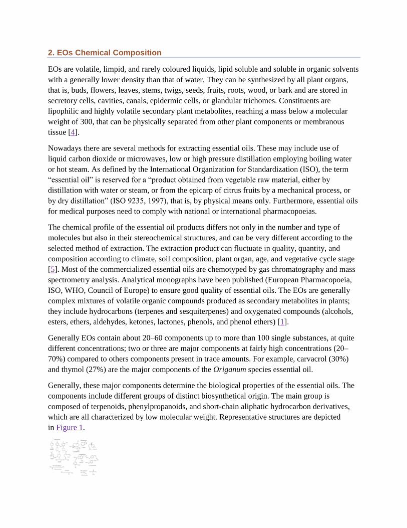
Figure 1
Representative structures typical of essential oils.
Terpenes are made from combinations of several 5-carbon-base (C5) units called isoprene and
form structurally and functionally different classes. The biosynthesis of the terpenes consists of
synthesis of the isopentenyl diphosphate (IPP) precursor, repetitive addition of IPPs to form the
prenyldiphosphate precursor of the various classes of terpenes, modification of the allylic
prenyldiphosphate by terpene specific synthetases to form the terpene skeleton, and, finally,
secondary enzymatic modification (redox reaction) of the skeleton to attribute functional
properties to the different terpenes. Terpenoids derive from the C5-building blocks isopentenyl
diphosphate (IPP) and its isomer dimethylallyl diphosphate (DMAPP) and are generally
represented by monoterpenes (C10) and sesquiterpenes (C15), while hemiterpenes (C5) are quite
rare [6]. Terpenes containing oxygen in the form of hydroxyl, ether, aldehyde, ketone, or
carboxylic moieties are called terpenoids.
The monoterpenes (Figure 1) are formed from the coupling of two isoprene units (C10). They are
the most representative molecules constituting 90% of the essential oils and allow a great variety
of structures. They consist of several functions including acyclic hydrocarbons (myrcene and
ocimene); monocyclic hydrocarbons (limonene, terpinenes, p-cymene, and phellandrenes);
bicyclic hydrocarbons (pinenes, camphene, and sabinene); acyclic alcohols (geraniol, linalool,
citronellol, lavandulol, and nerol); monocyclic alcohols (menthol, α-terpineol, and carveol);
bicyclic alcohols (borneol, fenchol, chrysanthenol, and thuyan-3-ol); acyclic aldehydes (geranial,
neral, and citronellal); acyclic ketone (tegetone), monocyclic ketone (menthones, carvone,
pulegone, and piperitone); bicyclic ketone (camphor, fenchone, thuyone, and pinocarvone);
acyclic esters (linalyl acetate or propionate and citronellyl acetate); monocyclic esters (menthyl
or α-terpinyl acetate); bicyclic esters (isobornyl acetate); ethers (1,8-cineole and menthofuran);
peroxides (ascaridole); and phenols (thymol, carvacrol).
The sesquiterpenes are formed from the assembly of three isoprene units (C15). The extension of
the chain increases the number of cyclisations which allows a great variety of structures (Figure
1). Also sesquiterpenes include hydrocarbons (azulene, β-bisabolene, cadinenes, β-
caryophyllene, farnesenes, and zingiberene); alcohols (bisabolol, β-nerolidol, farnesol, β-
santalol, and patchoulol); ketones (germacrone, β-vetinone, and turmerones); and epoxide
(caryophyllene oxide and humulene epoxides).
Other aromatic molecules are phenylpropanoids formed via the shikimic acid pathway leading to
phenylalanine [6] and occurring less frequently than the terpenes.
Aromatic compounds originated from the shikimate pathway (phenylpropanoids, Figure 1)
comprise aldehydes (cinnamaldehyde); alcohols (cinnamic alcohol); phenols (chavicol and
eugenol); methoxy derivatives (anethole, estragole, and methyleugenols); methylenedioxy
compounds (apiole, myristicin, and safrole).
Nitrogenous or sulphured components such as glucosinolates or isothiocyanate derivatives
(garlic and mustard oils) are also characteristic secondary metabolites of diverse aromatic plants
or of processed, grilled, or roasted products. In addition, some essential oils contain photoactive
molecules like coumarins and furocoumarins (Citrus aurantium ssp. bergamiaessential oil
contains psoralens) and short-chain aliphatic substances such as 3-octanone and methyl nonyl
ketone (Figure 1).
Go to:
3. Limits and Challenges for the Rational Clinical Use of Essential Oils
The most recent applications of EOs include being as antioxidants and preservatives in food [7],
incorporated into foodstuff packaging material [8], and application as plant and crop protectants
[9]. Traditionally, essential oils have been used for many biological properties including
bactericidal, virucidal, fungicidal, antiparasitical, insecticidal, and other medicinal properties
such as analgesic, sedative, anti-inflammatory, spasmolytic, and locally anesthetic remedies [9–
11].
At present, promising approaches have been reported using essential oils or components thereof
in medicinal products for human or veterinary use [12]. The most effective way to use most EOs
is by external application, as gargles and mouthwashes or inhalation; rarely they are used orally
even if generally regarded as safe (GRAS) to ingest. In this case of oral administration they are
generally diluted with milk, soy milk, or olive oil. Topical application is generally safe; the oil is
diluted in a formulation but sometimes can give skin reactions and in particular some oils
(specifically citrus oils) are UV sensitive and may cause irritation or darkening of skin upon
exposure to sunlight up to 4 days after application.
In case of inhalation when using strong oils, limit time in immediate vicinity of an essential oil
diffuser as the concentrated vapours may cause eye irritation, some of them are not
recommended for diffusing or direct inhalation.
There is adequate evidence suggesting that although essential oils are metabolized quickly, their
distribution throughout the body is considered to be relatively high.
Most essential oil components are metabolized and either eliminated by the kidneys in the form
of polar compounds following limited phase I enzyme metabolism by conjugation with
glucuronate or sulfate or exhaled via the lungs as CO2. For example, after oral administration of
(−)-menthol, 35% of the original menthol content was excreted renally as menthol glucuronide
[13, 14]. The same happens with thymol, carvacrol, limonene, and eugenol. After their oral
administration, sulphate and glucuronide forms have been detected in urine and in plasma,
respectively [15, 16]. The fast metabolism and short half-life of active compounds have led to
the belief that there is a minimum risk of accumulation in body tissues [17].
EO compounds are small, fat soluble molecules, able to permeate the membranes including the
skin before being captured by the microcirculation and drained into the systemic circulation,
which reaches all targets organs [9, 18]. In general, the respiratory tract offers the most rapid
way of entry followed by the dermal pathway [19]. Topically, aromatherapy EOs can sometimes
cause irritation of the skin, especially if the oils are not diluted. Some oils, such as bergamot oil,
can also cause photosensitization and induce malignant change. Applying excessive amounts of
highly concentrated oils to a large surface of the skin or on broken skin can result in significant
systemic absorption and increase the chance of serious side effects, such as convulsions because
EOs are permeation enhancers.
Besides the high volatility, EOs can easily decompose, owing to direct exposure to heat,
humidity, light, or oxygen. A recent manuscript has reviewed the factors influencing essential oil
stability; specific knowledge on the chemical composition and properties of essential oil is
fundamental for an adequate use [20].
Degradation of EOs constituents is due to oxidation, isomerization, cyclization, or
dehydrogenation reactions, triggered either enzymatically or chemically [21], strongly influenced
by the conditions during processing and storage of the plant material, upon distillation, and in the
course of subsequent handling of the oil itself [22]. Furthermore, besides organoleptic alterations
and viscosity changes, some aged essential oils as well as oxidized terpenoids have revealed
skin-sensitizing capacities [23] leading to a hypersensitivity reaction synonymous to allergic
contact dermatitis [24].
Go to:
4. Nanoencapsulation Technology
Encapsulation of bioactive compounds represents a feasible and efficient approach to modulate
drug release, increase the physical stability of the active substances, protect them from the
interactions with the environment, decrease their volatility, enhance their bioactivity, reduce
toxicity, and improve patient compliance and convenience [25].
A significantly large part of current literature on the encapsulation of EOs deals with
micrometric size capsules, which are used for the protection of the active compounds against
environmental factors (e.g., oxygen, light, moisture, and pH), to decrease oil volatility and to
transform the oil into a powder. Encapsulation in nanometric particles is an alternative for
overcoming these problems but additionally, due to the subcellular size, may increase the cellular
absorption mechanisms and increasing bioefficacy.
Nanosystems applied to the skin are used to facilitate local therapies even if it is still under
discussion of the mechanisms of penetration trough skin. It is accepted that topical drug delivery
with nanoparticles targets the nanoparticles into the deeper layers of skin and generally they do
not reach the viable epidermis. Only where the cheratine barrier is compromised, however, such
as in aged or diseased skin, an enhanced particle penetration occurs. The use of nanoparticles
provides a sustained and slow release of the active constituents; nanoparticles represent a
reservoir. In addition, nanoparticles can interact with skin at a cellular level as adjuvants to
enhance immune reactivity for topical vaccine applications.
Hair follicles and furrows were regarded as insignificant as potential routes for drug delivery,
covering less than 1% of the human skin surface area, but their complex vascularisation and deep
invagination with a thinning stratum corneum have led to a reappraisal of this view. It has been
demonstrated that in particular hair follicles are an efficient reservoir for nanoparticle-based drug
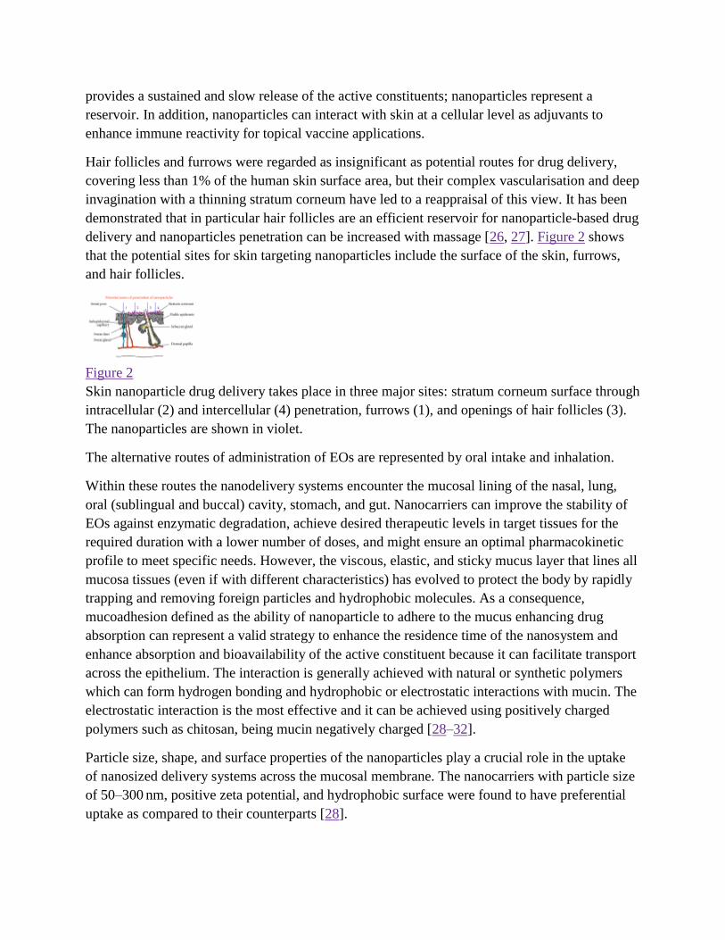
delivery and nanoparticles penetration can be increased with massage [26, 27]. Figure 2 shows
that the potential sites for skin targeting nanoparticles include the surface of the skin, furrows,
and hair follicles.
Figure 2
Skin nanoparticle drug delivery takes place in three major sites: stratum corneum surface through
intracellular (2) and intercellular (4) penetration, furrows (1), and openings of hair follicles (3).
The nanoparticles are shown in violet.
The alternative routes of administration of EOs are represented by oral intake and inhalation.
Within these routes the nanodelivery systems encounter the mucosal lining of the nasal, lung,
oral (sublingual and buccal) cavity, stomach, and gut. Nanocarriers can improve the stability of
EOs against enzymatic degradation, achieve desired therapeutic levels in target tissues for the
required duration with a lower number of doses, and might ensure an optimal pharmacokinetic
profile to meet specific needs. However, the viscous, elastic, and sticky mucus layer that lines all
mucosa tissues (even if with different characteristics) has evolved to protect the body by rapidly
trapping and removing foreign particles and hydrophobic molecules. As a consequence,
mucoadhesion defined as the ability of nanoparticle to adhere to the mucus enhancing drug
absorption can represent a valid strategy to enhance the residence time of the nanosystem and
enhance absorption and bioavailability of the active constituent because it can facilitate transport
across the epithelium. The interaction is generally achieved with natural or synthetic polymers
which can form hydrogen bonding and hydrophobic or electrostatic interactions with mucin. The
electrostatic interaction is the most effective and it can be achieved using positively charged
polymers such as chitosan, being mucin negatively charged [28–32].
Particle size, shape, and surface properties of the nanoparticles play a crucial role in the uptake
of nanosized delivery systems across the mucosal membrane. The nanocarriers with particle size
of 50–300 nm, positive zeta potential, and hydrophobic surface were found to have preferential
uptake as compared to their counterparts [28].
Diverse absorption mechanisms have been established and two have been predominantly used:
the paracellular route that is slow and passive and the transport through a lipoidal route and it is
also known as the transcellular process which is responsible for the transport of lipophilic drugs
that show a rate dependency on their lipophilicity. Drug also crosses cell membranes by an active
transport route via carrier-mediated means or transports through the opening of tight junctions
interacting with the tight junction proteins [28–32].
For instance the increase in the absorption of nanocarriers by enterocytes is due to tight junction
modulation, receptor-mediated endocytosis and transcytosis, phagocytosis via specialized
microfold cells (M cells) of the Peyer's patches, and other mucosa associated lymphoid tissues
(MALT) and lymphatic absorption via chylomicron uptake mechanism from the enterocytes
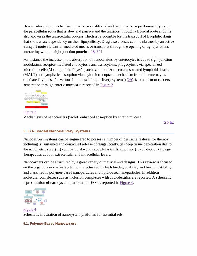
(mediated by lipase for various lipid-based drug delivery systems) [29]. Mechanism of carriers
penetration through enteric mucosa is reported in Figure 3.
Figure 3
Mechanisms of nanocarriers (violet) enhanced absorption by enteric mucosa.
Go to:
5. EO-Loaded Nanodelivery Systems
Nanodelivery systems can be engineered to possess a number of desirable features for therapy,
including (i) sustained and controlled release of drugs locally, (ii) deep tissue penetration due to
the nanometric size, (iii) cellular uptake and subcellular trafficking, and (iv) protection of cargo
therapeutics at both extracellular and intracellular levels.
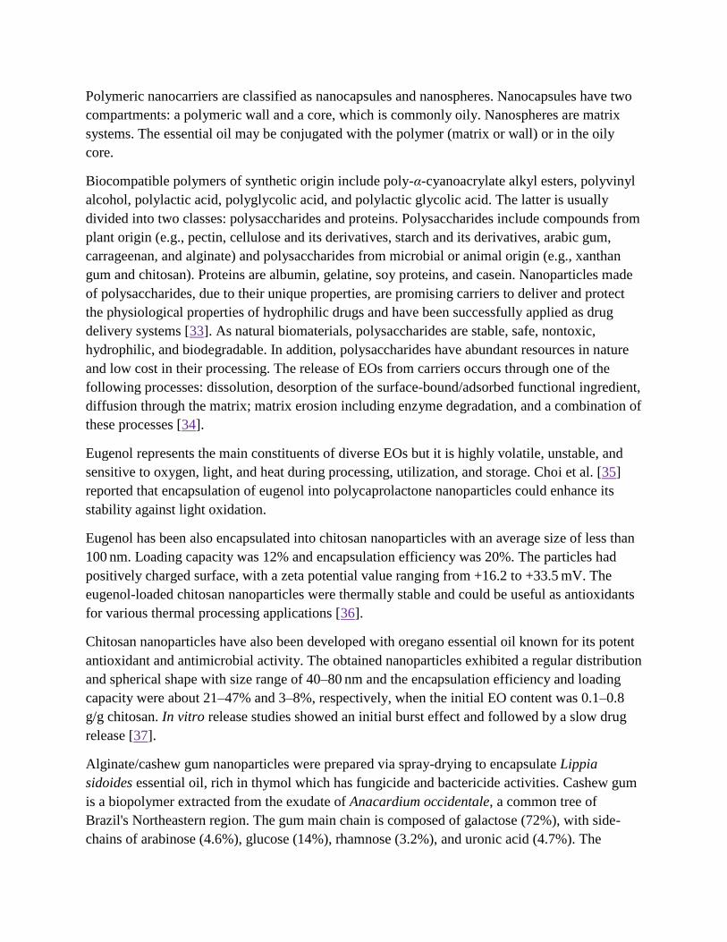
Nanocarriers can be structured by a great variety of material and designs. This review is focused
on the organic nanocarrier systems, characterised by high biodegradability and biocompatibility,
and classified in polymer-based nanoparticles and lipid-based nanoparticles. In addition
molecular complexes such as inclusion complexes with cyclodextrins are reported. A schematic
representation of nanosystem platforms for EOs is reported in Figure 4.
Figure 4
Schematic illustration of nanosystem platforms for essential oils.
5.1. Polymer-Based Nanocarriers
Polymeric nanocarriers are classified as nanocapsules and nanospheres. Nanocapsules have two
compartments: a polymeric wall and a core, which is commonly oily. Nanospheres are matrix
systems. The essential oil may be conjugated with the polymer (matrix or wall) or in the oily
core.
Biocompatible polymers of synthetic origin include poly-α-cyanoacrylate alkyl esters, polyvinyl
alcohol, polylactic acid, polyglycolic acid, and polylactic glycolic acid. The latter is usually
divided into two classes: polysaccharides and proteins. Polysaccharides include compounds from
plant origin (e.g., pectin, cellulose and its derivatives, starch and its derivatives, arabic gum,
carrageenan, and alginate) and polysaccharides from microbial or animal origin (e.g., xanthan
gum and chitosan). Proteins are albumin, gelatine, soy proteins, and casein. Nanoparticles made
of polysaccharides, due to their unique properties, are promising carriers to deliver and protect
the physiological properties of hydrophilic drugs and have been successfully applied as drug
delivery systems [33]. As natural biomaterials, polysaccharides are stable, safe, nontoxic,
hydrophilic, and biodegradable. In addition, polysaccharides have abundant resources in nature
and low cost in their processing. The release of EOs from carriers occurs through one of the
following processes: dissolution, desorption of the surface-bound/adsorbed functional ingredient,
diffusion through the matrix; matrix erosion including enzyme degradation, and a combination of
these processes [34].
Eugenol represents the main constituents of diverse EOs but it is highly volatile, unstable, and
sensitive to oxygen, light, and heat during processing, utilization, and storage. Choi et al. [35]
reported that encapsulation of eugenol into polycaprolactone nanoparticles could enhance its
stability against light oxidation.
Eugenol has been also encapsulated into chitosan nanoparticles with an average size of less than
100 nm. Loading capacity was 12% and encapsulation efficiency was 20%. The particles had
positively charged surface, with a zeta potential value ranging from +16.2 to +33.5 mV. The
eugenol-loaded chitosan nanoparticles were thermally stable and could be useful as antioxidants
for various thermal processing applications [36].
Chitosan nanoparticles have also been developed with oregano essential oil known for its potent
antioxidant and antimicrobial activity. The obtained nanoparticles exhibited a regular distribution
and spherical shape with size range of 40–80 nm and the encapsulation efficiency and loading
capacity were about 21–47% and 3–8%, respectively, when the initial EO content was 0.1–0.8
g/g chitosan. In vitro release studies showed an initial burst effect and followed by a slow drug
release [37].
Alginate/cashew gum nanoparticles were prepared via spray-drying to encapsulate Lippia
sidoides essential oil, rich in thymol which has fungicide and bactericide activities. Cashew gum
is a biopolymer extracted from the exudate of Anacardium occidentale, a common tree of
Brazil's Northeastern region. The gum main chain is composed of galactose (72%), with side-
chains of arabinose (4.6%), glucose (14%), rhamnose (3.2%), and uronic acid (4.7%). The
averaged sizes of the nanoparticles were in the range 223–399 nm, and zeta potential values
ranging from −30 to −36 mV. Encapsulated oil levels varied from 1.9 to 4.4% with an
encapsulation efficiency of up to 55%. The in vitro release profile showed that between 45 and
95% of oil was released within 30–50 h. The addition of cashew gum to alginate has proven to be
able to maximize the hydrophilic character of the polymer matrices, allowing a quicker release at
a satisfactory oil loading. Moreover, the oil release profile revealed that the use of alginate in
synergy with cashew gum for EO encapsulation presents itself as a potential delivery system
with tailored release rate, loading, and encapsulation efficacy [38].
Using the same EO from Lippia sidoides nanoparticles made of chitosan (a deacetylated form of
chitin, chemically D-glucosamine and N-acetyl-D-glucosamine linked by beta (1–4) linkages)
and cashew gum aimed to improve essential oil loading and release profiles. Samples designed
using relative ratios, matrix: oil, 10 : 2; gum : chitosan, 1 : 1; and 5% gum concentration, showed
high loading (11.8%) and encapsulation efficiency (70%), with average sizes in the range 335–
558 nm. In vitro release profiles showed that nanoparticles presented slower and sustained
release. The nanocarriers presented efficacy against St. aegypti larvae, where the mortality rate
was related to the loading values and gum : chitosan ratios. In particular, samples gum : chitosan
1 : 1 and gum : chitosan 1 : 10 showed, respectively, 87% and 75% of mortality after 48 h,
reaching over 90% of mortality at 72 h. These results showed that the gum-chitosan nanoparticles
were designed and present sustained release features [39].
The formation of heat-resistant flavour nanocapsules of jasmine essential oil was achieved by
gelatin and arabic gum. Their heat-resistance capability against 80°C was evaluated by both
structural characteristics (size, polydispersity index, and zeta potential) and flavour analysis. The
results showed that the nanocapsules were stable at 80°C for 7 h, even if the GC-MS revealed
that jasmine essential oil began to destroy above 5 h [40].
Thymol loaded in zein (a corn prolamine protein) nanoparticles stabilized with sodium caseinate
and chitosan hydrochloride were prepared and characterized. In the absence of sodium caseinate,
the particle size and zeta potential of zein nanoparticles were 118.30 nm and +28.10 mV,
respectively. The zeta potential of rein nanoparticles after coating with sodium caseinate
reversed from positive to negative (in the range of −33.60 to −38.95 mV), while size was around
200 nm. Due to the presence of sodium caseinate, the stabilized zein nanoparticles showed a shift
of isoelectric point from 6.18 to 5.05 and had a desirable redispersibility in water at neutral pH
after lyophilization. Encapsulated thymol was more effective in suppressing Gram-positive
bacterium than unencapsulated thymol for a longer time period. Zein nanoparticles presented a
two-phase release profile of the EO. The authors believe that the rapid first phase represents the
portion of thymol that was in the external phase of the film; the slower second phase represents
thymol that was contained in the zein particles [41].
Polylactic glycolic acid nanocapsules containing eugenol or transcinnamaldehyde both presented
a two-phase EO release. The first phase was rapid (under 30 minutes) and approximately 20% of
the EO load was detected; the second release phase was prolonged and after 72 hours 64% of
eugenol and 87% of transcinnamaldehyde were detected. Considering that PLGA has a low
degradation rate, the release was governed mostly by diffusion with a possible influence of
polymer swelling and bulk erosion. The first release phase may be attributed to the molecules
that are adsorbed to the polymeric wall, while the second release phase represents the EO present
in the core of the nanocapsules which diffuses through the polymeric wall [42].
Polylactic glycolic acid nanocapsules containing carvacrol have also been produced. Size was
about 209.8 nm, polydispersity was 0.260, zeta potenzial was −18.99, drug loading was 21%, and
encapsulation efficiency was 26%. In vitro release profile occurred with an initial “burst” release
followed by a slower release due to the concentration gradient. Nanoparticles showed a 60%
release after 3 h and approach to completeness after 24 h with approximately 95% of carvacrol
released. The effect of carvacrol EO antimicrobial activity was enhanced because the
nanoparticles significantly altered rheological characteristic of bacterial biofilms potentially
facilitating the action of carvacrol [43].
Methyl cellulose/ethyl cellulose polymeric nanoparticles containing thymol attaining the
relatively high thymol loading level of 43.53% (weight of encapsulated thymol to weight of the
thymol-loaded spheres) were able to reduce and preserve levels of E. coli in an oil/water lotion
and in a hydrophilic gel, of P. aeruginosa in an oil/water lotion and of S. aureus in an oil/water
lotion and in a water/oil cream. Interestingly, free thymol was also capable of reducing
microbiologic levels in these formulations, but the preservation period was shorter except for
the S. aureus in an oil/water lotion where free thymol maintained low microbial levels for the
same period as the nanocapsules. Effective bacterial suppression by encapsulated thymol was
also observed when used in cream and aqueous gel formulations [44].
5.2. Lipid-Based Nanocarriers
Lipid-based nanocarriers include micro- and nanometric-scaled emulsions and lipid
nanoparticles, roughly divided in liposomes, micelles, niosomes, solid lipid nanoparticles (SLN),
and nanostructured lipid carriers (NLC). Liposomes and niosomes are colloidal association of
amphiphilic lipids that organize themselves spontaneously in bilayer vesicles and that are
suitable with hydrophilic and hydrophobic compounds. SLN and NLC are solid particles at room
and human body temperatures that present lipid core, which makes these carriers a proper
medium for entrapment of lipophilic compounds, as EO.
As these nanoparticles are composed of lipids and/or phospholipids, they have the ability to
interact with several cell types. So, these carriers can been seen as alternatives for treatment of
microbial infections, due to their capacity of interaction with infected cells. Furthermore, the
association of EO with lipid nanoparticles has different objectives, but the main aims are the
enhancement of the stability and the solubility in aqueous media of EO, maintenance or even
enhancement of their biological activity, and drug targeting.
5.2.1. Micro- and Nanoemulsions
Microemulsions can be defined as homogeneous thermodynamically stable transparent
dispersions of two immiscibleliquids stabilized by an interfacial film of surfactants. They have
droplet size above 500 nm and require very low energyto formulate emulsion, since they form
spontaneously when aqueous, oily, and amphiphilic components are brought intocontact, besides
having a lower production cost compared to nanoemulsions. One major drawback to
microemulsions isthat formation requires high surfactant concentration, which can cause toxicity
when used in pharmaceutical applications.
In contrast nanoemulsions can be prepared using lower surfactant concentrations.
Nanoemulsions are fine oil-in-water dispersions, nonequilibrium systems with a spontaneous
tendency to separate into the constituent phases. Nevertheless, nanoemulsions may possess a
relatively high kinetic stability even for several years, due to their very small size, essentially the
consequence of significant steric stabilization between droplets. They have droplet covering the
size range of 10–500 nm and also referred to as miniemulsions, ultrafine emulsions, and
submicrometer emulsions.
Antimicrobial properties of micro- and nanoemulsion are believed to result from the small size of
oil particles that have a high surface tension which can fuse with and subsequently disrupt the
membrane of isolated prokaryotic cells, viruses, and eukaryotic cells of fungi but they do not
affect eukaryotic cells of higher organisms. A synergistic antimicrobial effect could be afforded
by including some substances which possess strong antimicrobial activity into the formula,
reducing the amounts of active substances and detergents used for killing microorganisms by the
conventional method and the cost of raw materials. Furthermore, irritation caused from
detergents in the formula is not likely to happen when they are used in low concentrations.
Encapsulation in nanoemulsion-based delivery systems of two antimicrobial compounds, a
terpenes mixture extracted from Melaleuca alternifolia and D-limonene, deals with the issues of
formulation and fabrication in order to retain and possibly enhance the antimicrobial activity of
the encapsulated compounds.
The nanoemulsions based on food-grade ingredients were investigated by determining the
minimum inhibitory concentration (MIC) and minimum bactericidal concentration (MBC) for
three different classes of microorganisms (Lactobacillus delbrueckii, Saccharomyces cerevisiae,
and Escherichia coli). The increase of the antimicrobial activity resulted in dependance on the
formulation andmean diameter of the delivery systems as well as on the microorganisms class.
Additionally, GC-MS analysis revealed that high intensity processing for nanoemulsion
production may affect the chemicalstability of several active compounds.
The results of the accelerated shelf life studies show that for both fruit juices after 2 days, the
total inactivation of the initial microbial load of 103 CFU/mL was already reached for the
terpenes concentrations of 5.0 g/L and 10 g/L. At a terpenes concentration of 1.0 g/L,
microorganism growth is delayed by 5 days in orange juice and 2 days in pear juice in
comparison to the control [45].
Another study reported the preparation of a self-nanoemulsifying drug delivery system for the
oral delivery of zedoary turmeric oil, an essential oil extracted from the dry rhizome ofCurcuma
zedoaria. The optimized formulation consisting of EO, ethyl oleate, Tween 80, Transcutol P
(30.8 : 7.7 : 40.5 : 21, w/w), and loaded with 30% drug was prepared. Upon mixing with water, the
formulation was rapidly dispersed into fine droplets with a mean size of 68.3 ± 1.6 nm and zeta
potential of −41.2 ± 1.3 mV. The active components remained stable in the optimized
formulation stored at 25°C for at least 12 months. Following oral administration in rats, both
AUC and Cmax of germacrone, a representative bioactive marker of zedoary turmeric oil,
increased by 1.7-fold and 2.5-fold, respectively, compared with the unformulated zedoary
turmeric oil [46].
5.2.2. Liposomes
Liposomes are one of the most studied colloidal delivery systems; in fact, they were first
developed for drug delivery purposes as early as 1970s [47, 48].
Liposomes consist of vesicular self-assembled system comprising of one or more bilayers,
usually formed using a phospholipid, surrounding an aqueous core. Liposomes can contain (i)
one bilayer forming unilamellar vesicles (ULV), (ii) several concentric bilayers forming
multilamellar vesicles, or (iii) nonconcentric bilayers forming multivesicular vesicles (MVV).
The size of these structures can be rather small (in the range of 20 nm) or rather large (exceeding
1 μm). Owing to the presence of the hydrophilic compartment and lipophilic palisade, they can
be used as carriers for both lipophilic and hydrophilic molecules [48].
Bioactive compounds compartmentalised in liposomes can be protected against degradation and
in case of lipophilic compounds, liposomal encapsulation can also lead to increased
solubilisation [48].
The effect of liposomal inclusion on the stability and in vitro antiherpetic activity of Santolina
insularis EO was investigated. Vesicles were obtained from hydrogenated soya
phosphatydilcholine and cholesterol. Formulations were examined for their stability for over one
year monitoring the drug leakage from vesicles and the average size distribution. The stability of
the incorporated oil was verified by studying its quali-quantitative composition. The antiviral
activity was studied against herpes simplex virus type 1 (HSV-1) by plaque reduction and yield
reduction assays. Results showed that Santolina insularis EO can be incorporated in high
amounts in the prepared liposomes, which successfully prevented its degradation. Moreover,
stability studies pointed out that vesicle dispersions were stable for at least one year and neither
oil leakage nor vesicle size alteration occurred during this period. Antiviral activity assays
demonstrated that Santolina insularis essential oil is effective in inactivating HSV-1 and that the
activity is principally due to direct virucidal effects. Free EO proved to be more effective than
liposomal oil and a different activity was discovered which related to the vesicular structure. The
ED(50) values, significantly lower when cells were preincubated with the EO before the virus
adsorption, indicate an intracellular mechanism in the antiviral activity ofSantolina
insularis [49].
The effect of liposomal inclusion on the in vitro antiherpetic activity of Artemisia arborescensL.
EO was investigated. In order to study the influence of vesicle structure and composition on the
antiviral activity of the vesicle-incorporated oil, multilamellar (MLV) and unilamellar (SUV)
positively charged liposomes were prepared. Liposomes were obtained from hydrogenated
(P90H) and nonhydrogenated (P90) soy phosphatidylcholine. Formulations were examined for
their stability for over one year, monitoring the oil leakage from vesicles and the average size
distribution. The antiviral activity was studied against herpes simplex virus type 1 (HSV-1) by a
quantitative tetrazolium-based colorimetric method. Results showed that Artemisia EO can be
incorporated in good amounts in the prepared vesicular dispersions. Stability studies pointed out
that vesicle dispersions were very stable for at least six months and neither oil leakage nor
vesicle size alteration occurred during this period. After one year of storage oil retention was still
good, but vesicle fusion was present. Antiviral assays demonstrated that the liposomal
incorporation of A. arborescens EO enhanced its in vitro antiherpetic activity especially when
vesicles were made with P90H. On the contrary, no significant difference in antiviral activity
was observed between the free and SUV-incorporated oil. P90H MLV showed a higher activity
than P90 MLV (EC50 values of 18.3 and 43.6 μg/mL for P90H MLV and P90MLV, resp.), while
no significant differences of the antiviral activity were observed between the free essential oil
and SUV vesicles. Incorporation of A. arborescens essential oil in multilamellar liposomes
greatly improved its activity against intracellular HSV-1 [50].
A modified technique of rapid expansion of supercritical solutions (RESS) was applied to
incorporate EO extracted from Atractylodes macrocephala Koidz into liposomes. The optimised
entrapment efficiency, drug loading, and average particle size of liposomes were found to be
82.18%, 5.18%, and 173 nm, respectively. The physicochemical properties including the
entrapment efficiency, dissolution rate, and stability met the characteristic for a pharmaceutical
use of the developed formulation [51].
Carvacrol, thymol, p-cymene, and c-terpinene were identified as major constituents and isolated
from the EOs from Origanum dictamnus L. The above components were successfully
encapsulated in phosphatidyl choline-based liposomes and the possible improvement of their
antioxidant and antimicrobial activities was tested against four Gram-positive and four Gram-
negative bacteria and three human pathogenic fungi, as well as the food-borne pathogen, Listeria
monocytogenes. In order to investigate any possible synergistic or antagonistic effect between
carvacrol/thymol and carvacrol/c-terpinene, the antimicrobial activities of the mixtures were also
determined before and after encapsulation in liposomes. All tested compounds presented
enhanced antimicrobial activities after the encapsulation [52].
A study examined carvacrol (derivatives) and thymol encapsulated in liposomes to increase their
bioavailability and stability. Similarly, the endurance to humidity and UV light was enhanced
[53].
5.2.3. SLN Solid Lipid Nanoparticles
Solid lipid nanoparticles (SLN) refer to nanoscale size particles prepared using lipids that remain
solid at room temperature (or/and body temperature). The lipid component may comprise of a
broad range of lipid and lipid-like molecules such as triacylglycerols or waxes [54]. The
diameter of such lipid particles can be also quite small, that is, in the range between 50 nm and 1
μm. Active ingredients can be solubilised homogeneously either in the core of the SLNs or in the
outside part [55]. The advantage of SLNs as delivery system for lipophilic active components is
reported to lie in the immobilisation of active elements by the solid particle structure leading to
an increased chemical protection, less leakage, and sustained release [56].
This physical property allows a better control of both the physical (against recrystallisation) and
chemical (against degradation) stability of the delivered constituents.
The effect of SLN incorporation on transdermal delivery and in vitro antiherpetic activity
ofArtemisia arborescens EO has been investigated. Two different SLN formulations were
prepared using the hot-pressure homogenization technique, Compritol 888 ATO as lipid, and
Poloxamer 188 (SLN 1) and Miranol Ultra C32 (SLN 2) as surfactants.
One day after production, the SLN 1 had a size of 223 nm (0.243 polydispersion index) while the
particle size of SLN 2 prepared using Miranol Ultra C32 as surfactant was 219 nm (0.301
polydispersion index, PI). The mean particle size of the formulations increased only slightly after
two years of storage, indicating a high physical stability of both SLN 1 and SLN 2 formulations.
In particular, 2 years after production, SLN 1 and SLN 2 formulations showed a mean size of
242 nm (0.285 PI) and 239 nm (0.321 PI). The PI values were always smaller than 0.350
indicating a fairly narrow size distribution of the particles. Formulations were examined for their
stability for two years by monitoring average size distribution and zeta potential values. The
antiviral activity of free and SLN incorporated EO was tested in vitroagainst Herpes Simplex
Virus-1 (HSV-1), while the effects of essential oil incorporation into SLN on both the
permeation through and the accumulation into the skin strata were investigated by using in
vitro diffusion experiments through newborn pig skin and an almond oilArtemisia essential oil
solution as a control. Results showed that both SLN formulations were able to entrap the EO in
high yields and that the mean particle size increased only slightly after two years of storage,
indicating a high physical stability. In vitro antiviral assays showed that SLN incorporation did
not affect the EO antiherpetic activity. The in vitro skin permeation experiments demonstrated
the capability of SLN of greatly improving the oil accumulation into the skin, while oil
permeation occurred only when the oil was delivered from the control solution [57].
Alhaj and coworkers developed a formulation based on Nigella sativa essential oil into solid
lipid nanoparticles SLN. SLN formulations were prepared using hydrogenated palm oil Softisan
154 and N. sativa essential oil as lipid matrix, sorbitol, and water. Data showed a high physical
stability for formulations at various storage temperatures during 3 months of storage. In
particular, average diameter of N. sativa essential oil loaded SLN did not vary during storage and
increased slightly after freeze-drying the SLN dispersions. Therefore, obtained results showed
that the studied SLN formulations are suitable carriers in pharmaceutical and cosmetic fields
[58].
Frankincense and myrrh are gum resins obtained from the genera Boswellia andCommiphora,
respectively. Both genera belong to the family Burseraceae, which is native to Northeast Africa
and the Middle East. Frankincense and myrrh have been used for medical purposes in China and
India for thousands of years. Modern pharmacological research has revealed that essential oils
are the primary effective components in frankincense and myrrh oil (FMO) that exhibit a broad
spectrum of biological activities such as antimicrobial, anti-inflammatory, and antitumor
activities. As with other essential oils, the instability and poor water solubility of FMO result in
poor oral bioavailability, which limits its clinical application. The components of FMO are
sensitive to light, air, and high temperature, and FMO stimulates the gastrointestinal tract,
making it unsuitable for oral administration. A study has reported the preparation of solid lipid
nanoparticles for the oral delivery of frankincense and myrrh essential oils (FMO). Aqueous
dispersions of SLNs were successfully prepared by a high-pressure homogenization method
using Compritol 888 ATO as the solid lipid and soybean lecithin and Tween 80 as the
surfactants. Round SLNs were with a mean size of 113.3 nm, a zeta potenzial of −16.8 mV, and
an encapsulation efficiency of 80.60%. SLN formulation increased the antitumor efficacy of
FMO in H22-bearing Kunming mice. Compritol 888 ATO showed reasonable FMO
solubilization capacity. The poorly water-soluble drug FMO was efficiently encapsulated into the
nanoparticles. Particles prepared under proper formulation conditions were spherical with
diameters of 220 nm [59]. Solid lipid nanoparticles (SLNs) of essential oil of Zataria
multiflora have been developed. The results showed that the encapsulation efficiency was
38.66%. Results of particle size determination showed a mean size of 650 nm and SLNs were
spherical as shown by TEM. The DSC curve of sodium dodecyl sulfate, polyethylene glycol,
cetyl alcohol, and EO was different from EO containing SLNs, which indicated that the EO can
interact with the matrix of lipid during the preparation of the SLNs. 93.2% of the essential oil
was released after 24 h. The results of characterization of the SLNs indicated the potential
application of essential oil of Z. multiflora loaded SLN as carrier system [60].
5.3. Molecular Complexes
A simple strategy to deliver active ingredients is by physically complexing them with other
molecules in order to have a better solubility profile and/or an increase in the chemical stability
of the complexed system. In this context a molecular complex is referring to the physical
association between a host and a guest (active ingredient) molecule and in the case of EOs the
complexes are reported with cyclodextrins (CDs).
Cyclodextrins are natural macrocyclic oligosaccharides well known for having toroid-shaped
structures with rigid lipophilic cavities and a hydrophilic outer surface insuring good dissolution
of the complex in an aqueous environment. They are able to enclose highly hydrophobic
molecules inside their hydrophobic cavity, constituting a true molecular encapsulation [61]. The
major advantages of the use of CD-complexation in pharmaceutical applications, foods,
cosmetics, and toiletries are protection of the active ingredients against oxidation, light induced
reactions, decomposition and thermal decomposition, loss by evaporation and sublimation, and
elimination or reduction of undesired tastes/odours, to reduce or prevent gastric-intestinal
irritation (mainly due to anti-inflammatory drugs) or ocular disturbances, prevent drug-drug or
drug-additive interactions, or even to convert oils and liquid drugs into microcrystalline or
amorphous powders and to reduce microbiological contamination, fibres, or the elimination of
other undesired components and hygroscopicity [62]. Moreover, formation of inclusion complex
(IC) increases the guest's in vivo stability against hydrolysis, oxidation, decomposition, and
dehydration, consequently increasing bioavailability and bioefficacy. There are three main types
of CDs: α-, β-, and γ-cyclodextrins, corresponding to 6, 7, and 8 glucopyranose units linked by α-
(1,4) bonds, respectively. The dimensions of the internal cavity are 0.5–0.8 nm and are crucial
for the “encapsulation” of guest molecules [63].
In the last years, physicochemical properties and, consequently, the inclusion capacity of the
natives' CD have been improved by chemical modification of their hydroxyl groups [64].
Besides natural cyclodextrins, a growing number of semisynthetic derivatives and copolymers
have been prepared and are already commercially available. Many of them found use as
structural and chiral selectors, with new properties given by the type and number of substituents.
The semisynthetic derivatives of cyclodextrins show better solubility in water, can decrease and
modulate the release rate of water soluble molecules, are able to enhance the dissolution rate and
the inclusion capacity, and also decrease the side effects of some molecules.
The majority of the publications is concerning the encapsulation of essential oils with β-CD and
its derivatives: randomly methylated-β-cyclodextrin, hydroxypropyl-β-cyclodextrin, and low
methylated-β-cyclodextrin.
The IC of thymol and cinnamaldehyde and β-CD was investigated [65] in order to study the
influence of water adsorption by CDs and their complexes on the release of encapsulated
compounds. The results showed that β-CD encapsulates efficiently both of them, in a 1 : 1 molar
ratio. The ICs were obtained upon mixing the components in aqueous media and subsequent
freeze-drying, as confirmed by differential scanning calorimetry. The samples were stored at
constant relative humidity, from 22% to 97%, at 25°C. The release of encapsulated compounds
was determined following the melting enthalpy of each guest. Water sorption isotherms for β-CD
and the complexes showed constant and low water sorption at RH < 80%; then the uptake of
water increased abruptly. The amount of sorbed water at each RH was smaller for the complexes
than for β-CD. The guest molecules displaced water molecules from inside the cavity of β-CD.
No thymol or cinnamaldehyde release was detected at RH < 84%, and it increased abruptly from
84% RH, coincidentally with the abrupt increase of absorbed water. Water sorption significantly
affects β-CD complexes stability, which is thus governed by the shape of the water sorption
isotherm. The stability studies showed that the inclusion complexes thymol-β-CD and
cinnamaldehyde-β-CD remain stable up to 75% RH during long storage times. In fact, the guests
released from the β-CD complexes were detectable in the region of the water adsorption isotherm
at which a sharp increase of water content occurred (84% RH). These results show the relevance
of selecting appropriated storage conditions for hydrophobic flavours encapsulated in β-CD or
for predicting the shelf life of functional products formulated with nanoencapsulated compounds
[65].
β-Caryophyllene (BCP), a natural sesquiterpene existing in the essential oil of many plants, has
exhibited a wide range of biological activities such as antimicrobial, anticarcinogenic, anti-
inflammatory, antioxidant, anxiolytic-like, and local anaesthetic effects. However, its volatility
and poor water solubility limit its application in pharmaceutical field. Liu and coworkers
investigated and compared the oral bioavailability and the pharmacokinetics of free BCP and
BCP/β-CD IC after a single oral dose of 50 mg/kg on rats [66]. BCP was rapidly released from
inclusion complex and the in vivo data showed that BCP/β-CD IC displayed earlier Tmax, higher
Cmax and the AUC0-12 h showed approximately 2.6 times higher increase than those of free
BCP. The β-CD has significantly increased the oral bioavailability of the drug in rats than free
BCP [66].
The essential oil of Chamomilla recutita (L.) Rauschert, syn. Matricaria recutita L., contains up
to 50% (−)-α-bisabolol which contributes to the anti-inflammatory properties of camomile oil.
Bisabolol is a very lipophilic substance, with a tendency to oxidise decreasing anti-inflammatory
activity ca. 50%. (−)-α-Bisabolol was found to form an inclusion complex with β-CD in solution
as well as in the solid state. To investigate molecular associations of β-CD with pure (−)-α-
bisabolol or (−)-α-bisabolol as a component of camomile EO, Waleczek et al. undertook phase
solubility studies [67]. The complex constant was 273 M−1 for the pure (−)-α-bisabolol and 304
M−1 for (−)-α-bisabolol as a constituent of the EO. The intrinsic solubility of pure (−)-α-bisabolol
(4.85 × 10⁻⁴ M) and (−)-α-bisabolol as a component of the EO (1.82 × 10⁻⁴ M) differ
significantly. Computer simulation proved an inclusion complex having a stoichiometric
composition of 2 : 1 (β-CD : drug) [67].
Thymol is a monoterpene present in Lamiaceae plants, specially oreganos and thymes.
Cinnamaldehyde (3-phenyl-2-propenal) represents 65–75% of the cinnamon EO. Thymol and
cinnamaldehyde are frequently used as flavours, but they are also becoming increasingly
important as naturally occurring antimicrobial, antioxidant, and antiseptic agents. As natural and
artificial flavours they are very sensitive to the effects of light, oxygen, humidity, and high
temperatures. The study of Hill et al. [68] aimed to elucidate the physicochemical characteristics
of essential oils and β-Cyclodextrin (EO-β-CD) inclusion complexes and their resulting
antimicrobial activity. Cinnamon bark extract, transcinnamaldehyde, clove bud extract, eugenol,
and a 2 : 1 (transcinnamaldehyde : eugenol) mixture were microencapsulated by the freeze-drying
method. EO-β-CD complexes were characterized for particle size, morphology, polydispersity
index (PI), entrapment efficiency, and phase solubility. All particles showed a spherical shape,
smooth surface, no significant differences in size distribution, and strong tendency to
agglomerate. The entrapment efficiencies ranged from 41.7 to 84.7%, where pure compounds
were higher (P < 0.05) than extracts.
The oils and their β-CD complexes were analyzed for their antimicrobial activity
againstSalmonella enterica serovar Typhimurium LT2 and Listeria innocua. All the samples
effectively inhibited bacterial growth within the concentration range tested, except free eugenol.
The EO-β-CD complexes were able to inhibit both bacterial strains at lower concentrations than
free oils, likely due to their increased water solubility which determined an increased contact
between pathogens and essential oils. The cinnamon bark and clove bud olis/β-CD complexes
were the most powerful antimicrobials, despite showing the lowest entrapment efficiencies
amongst the oils. The results indicate that such EO inclusion complexes could be useful
antimicrobial delivery systems with a broad spectrum of application in food systems where
Gram-positive and -negative bacteria could present a risk [68].
Garlic (Allium sativum L.) is a widely distributed plant and is used throughout the world not only
as a spice and a food, but also as a folk medicine, and many of the beneficial health-related
biological effects have been attributed to its characteristic organosulphur compounds [69]. Steam
distillation is widely used to extract and condense the volatile organosulphur compounds in
garlic, and the final oily product is called garlic oil (GO). The compounds of GO mainly are
diallyl disulphide, diallyl trisulphide, allyl propyl disulphide, a small quantity of disulphide, and
probably diallyl polysulphide [70]. GO is recognised to be more potent than aqueous extracts of
garlic and exhibits a wide range of pharmacological properties including antimicrobial,
antidiabetic, antimutagenic, and anticarcinogenic effects [71]. However, the application of GO is
limited due to its volatility, strong odour, insolubility in water, and low physicochemical
stability.
The characterisation of ICs of GO/β-CD was investigated [72]. The calculated apparent stability
constant of IC was 1141 M−1, and the water solubility of GO was significantly improved.
Furthermore, the release rate of GO from the inclusion complex was controlled. The results of
this study clearly demonstrated that GO could be efficiently complexed with β-CD to form an
inclusion complex by the coprecipitation method in a molar ratio of 1 : 1. The aqueous solubility
and stability of GO were significantly increased by inclusion in β-CD [72].
Isothiocyanates (ITCs) are hydrolysis products of sulphur-containing compounds called
glucosinolates, which occur naturally in cruciferous vegetables, such as broccoli and cabbage.
Mechanical disruption of cruciferous plant tissues releases ITCs, due to the hydrolysis reaction
catalysed by myrosinase bound to the cell wall and possesses antimicrobial activities [73]. ITCs,
in particular allyl isothiocyanate (AITC), have been extensively studied for their antibacterial
effect for food. Inclusion complexation reactions between isothiocyanates (ITCs), namely, allyl
isothiocyanate (AITC) and phenyl isothiocyanate (PITC), and randomly methylated β-
cyclodextrin (RM-β-CD) were reported [74].
The apparent activation energy of IC dissociation suggested a reduction of volatility and physical
stabilisation by inclusion complexation.
RM-β-CD demonstrated a strong solubilising effect on the poorly water-soluble AITC and PITC
in the aqueous phase. RM-β-CD was more effective in solubilising PITC though the AITC/RM-
β-CD complex had higher apparent stability constants. Despite the fact that a greater amount of
AITC was solubilised in the aqueous phase at any given concentration of RM-β-CD, the solid
state AITC/RM-β-CD complex showed an inclusion ratio remarkably lower than that of the
PITC/RM-β-CD complex. Both of the ITCs may form inclusion complexes with RM-β-CD at
guest to host ratios of 1 : 1 and 1 : 2 in the aqueous phase [74].
The inclusion interactions of cyclodextrins (CDs) and β-cyclodextrin polymers with linalool and
camphor in Lavandula angustifolia EO were investigated in order to prepare novel controlled
release systems for the delivery of essential oil used as ambient odours [75].
The complexation behaviour and the retention capacity of α-CD, β-CD, γ-CD, hydroxypropyl-β-
cyclodextrin (HPBCD), randomly methylated-β-cyclodextrin (RAMEB), a low methylated-β-
cyclodextrin (CRYSMEB), and cross-linked β-CD polymers for linalool and camphor, two major
components of Lavandula angustifolia EO, were studied. The complexation and the retention
capacity of CDs and CD polymers were investigated under solid support or in aqueous media by
static headspace gas chromatography. The release profile of aroma from solid support was
studied by multiple headspace extraction (MHE). The retention capacity of the CD derivatives
was measured in static experiments.
The stability constants with monomeric CD derivatives were determined for standard compounds
and for the compounds in essential oil. The RAMEB showed the higher formation constant both
for the standard compounds (833 M−1 for linalool and 1194 M−1 for camphor) and for the
compounds in the in Lavandula angustifolia essential oil (1074 M−1 for linalool and 2963 M−1 for
camphor). All studied CDs and CD polymers reduce the volatility of the aroma compounds and
stable 1 : 1 inclusion complexes are formed. β-CD is the most versatile CDs for the two guests,
leading to greater formation constant and retention ability in aqueous phase [75].
Go to:
6. Concluding Remarks
EOs have promising potentials for maintaining and promoting health, as well as preventing and
potentially treating some diseases. However, the generally low water solubility and stability,
together with the high volatility and side effects associated with their use have limited their
application in medicine. Nanotechnology is an innovative approach that has potential
applications in medicinal and health research. Indeed, nanoparticles are a very attractive tool and
are able to solve the major inconvenience of EOs use increasing the chemical stability in the
presence of air, light, moisture and, high temperatures, factors which can lead to the rapid
evaporation and to the degradation of the active components. In addition, nanocarriers ensure the
easier and safer handling of the liquid substances by changing them in solid powders,
determining retention of volatile ingredients and taste masking, setting up controlled release
and/or consecutive delivery of multiple active ingredients, reducing toxic side effects, improving
water solubility of hydrophobic ingredients, and enhancing bioavailability and efficacy.
Nanoencapsulation of EOs in liposomes, solid lipid nanoparticles, nano- and microemulsions,
and polymeric nanoparticles represent a promising strategy for overcoming EOs limitations,
lowering their dose and increasing long-term safety of these constituents.
Go to:
Conflict of Interests
The authors declare that there is no conflict of interests regarding the publication of this paper.
Go to:
References
1. Guenther E. The Essential Oils. Malabar, Fla, USA: Krieger Publishing Company; 1972.
2. Bauer K, Garbe D, Surburg H. Common Fragrance and Flavor Materials: Preparation,
Properties and Uses. Weinheim, Germany: Wiley-VCH; 2001.
3. FAO. Flavours and Fragrances of Plant Origin. Rome, Italy: FAO; 1995.
4. Sell C. Chemistry of essential oils. In: Başer KH, Buchbauer G, editors. Handbook of
Essential Oils. Science, Technology, and Applications. Boca Raton, Fla, USA: CRC Press; 2010.
pp. 121–150.
5. Angioni A, Barra A, Coroneo V, Dessi S, Cabras P. Chemical composition, seasonal
variability, and antifungal activity of Lavandula stoechas L. ssp. stoechas essential oils from
stem/leaves and Flowers. Journal of Agricultural and Food Chemistry. 2006;54(12):4364–
4370. [PubMed]
6. Pichersky E, Noel JP, Dudareva N. Biosynthesis of plant volatiles: nature’s diversity and
ingenuity. Science. 2006;311(5762):808–811. [PMC free article] [PubMed]
7. Tiwari BK, Valdramidis VP, O’Donnell CP, Muthukumarappan K, Bourke P, Cullen PJ.
Application of natural antimicrobials for food preservation. Journal of Agricultural and Food
Chemistry. 2009;57(14):5987–6000. [PubMed]
8. Kuorwel KK, Cran MJ, Sonneveld K, Miltz J, Bigger SW. Essential oils and their principal
constituents as antimicrobial agents for synthetic packaging films. Journal of Food
Science.2011;76(9):R164–R177. [PubMed]
9. Adorjan B, Buchbauer G. Biological properties of essential oils: an updated review.Flavour
and Fragrance Journal. 2010;25(6):407–426.
10. Bakkali F, Averbeck S, Averbeck D, Idaomar M. Biological effects of essential oils—a
review. Food and Chemical Toxicology. 2008;46(2):446–475. [PubMed]
11. Buchbauer G, Jirovetz L, Jager W, Plank C, Dietrich H. Fragrance compounds and essential
oils with sedative effects upon inhalation. Journal of Pharmaceutical Sciences.1993;82(6):660–
664. [PubMed]
12. Franz CM. Essential oil research: past, present and future. Flavour and Fragrance
Journal. 2010;25(3):112–113.
13. Bronaugh RL, Wester RC, Bucks D, Maibach HI, Sarason R. In vivo percutaneous
absorption of fragrance ingredients in rhesus monkeys and humans. Food and Chemical
Toxicology. 1990;28(5):369–373. [PubMed]
14. Kohlert C, van Rensen I, Marz R, Schindler G, Graefe EU, Veit M. Bioavailability and
pharmacokinetics of natural volatile terpenes in animals and humans. Planta
Medica.2000;66(6):495–505. [PubMed]
15. Guénette SA, Ross A, Marier J-F, Beaudry F, Vachon P. Pharmacokinetics of eugenol and its
effects on thermal hypersensitivity in rats. European Journal of Pharmacology.2007;562(1-
2):60–67. [PubMed]
16. Michiels J, Missotten J, Dierick N, Fremaut D, Maene P, de Smet S. In vitro degradation and
in vivo passage kinetics of carvacrol, thymol, eugenol and trans-cinnamaldehyde along the
gastrointestinal tract of piglets. Journal of the Science of Food and
Agriculture.2008;88(13):2371–2381.
17. Kohlert C, Schindler G, März RW, et al. Systemic availability and pharmacokinetics of
thymol in humans. Journal of Clinical Pharmacology. 2002;42(7):731–737. [PubMed]
18. Baser KHC, Buchbauer G. Handbook of Essential Oils: Science, Technology, and
Applications. New York, NY, USA: CRC Press; 2010.
19. Moss M, Cook J, Wesnes K, Duckett P. Aromas of rosemary and lavender essential oils
differentially affect cognition and mood in healthy adults. International Journal of
Neuroscience. 2003;113(1):15–38. [PubMed]
20. Turek C, Stintzing FC. Stability of essential oils: a review. Comprehensive Reviews in Food
Science and Food Safety. 2013;12(1):40–53.
21. Scott RPW. Essential oils. In: Worsfold P, Townshend A, Poole C, editors. Encyclopedia of
Analytical Science. 2nd edition. London, UK: Elsevier; 2005. pp. 554–561.
22. Schweiggert U, Carle R, Schieber A. Conventional and alternative processes for spice
production—a review. Trends in Food Science and Technology. 2007;18(5):260–268.
23. Christensson JB, Forsström P, Wennberg A-M, Karlberg A-T, Matura M. Air oxidation
increases skin irritation from fragrance terpenes. Contact Dermatitis. 2009;60(1):32–
40.[PubMed]
24. Divkovic M, Pease CK, Gerberick GF, Basketter DA. Hapten-protein binding: from theory to
practical application in the in vitro prediction of skin sensitization. Contact
Dermatitis. 2005;53(4):189–200. [PubMed]
25. Ravi Kumar MN. Nano and microparticles as controlled drug delivery devices. Journal of
Pharmacy & Pharmaceutical Sciences. 2000;3(2):234–258. [PubMed]
26. Schneider M, Stracke F, Hansen S, Schaefer UF. Nanoparticles and their interactions with
the dermal barrier. Dermato-Endocrinology. 2009;1(4):197–206. [PMC free article][PubMed]
27. Prow TW, Grice JE, Lin LL, et al. Nanoparticles and microparticles for skin drug
delivery.Advanced Drug Delivery Reviews. 2011;63(6):470–491. [PubMed]
28. Roger E, Lagarce F, Garcion E, Benoit J-P. Biopharmaceutical parameters to consider in
order to alter the fate of nanocarriers after oral delivery. Nanomedicine. 2010;5(2):287–
306.[PubMed]
29. Thanki K, Gangwal RP, Sangamwar AT, Jain S. Oral delivery of anticancer drugs:
challenges and opportunities. Journal of Controlled Release. 2013;170:15–40. [PubMed]
30. Lai SK, Wang Y-Y, Hanes J. Mucus-penetrating nanoparticles for drug and gene delivery to
mucosal tissues. Advanced Drug Delivery Reviews. 2009;61(2):158–171.[PMC free
article] [PubMed]
31. Kushwaha SKS, Keshari RK, Rai AK. Advances in nasal trans-mucosal drug
delivery.Journal of Applied Pharmaceutical Science. 2011;1(7):21–28.
32. Singh SG, Singh RP, Gupta SK, Kalyanwat R, Yadav S. Buccal mucosa as a route for drug
delivery: mechanism, design and evaluation. Research Journal of Pharmaceutical, Biological
and Chemical Sciences. 2011;2(3):358–372.
33. Liu Z, Jiao Y, Wang Y, Zhou C, Zhang Z. Polysaccharides-based nanoparticles as drug
delivery systems. Advanced Drug Delivery Reviews. 2008;60(15):1650–1662. [PubMed]
34. Soppimath KS, Aminabhavi TM, Kulkarni AR, Rudzinski WE. Biodegradable polymeric
nanoparticles as drug delivery devices. Journal of Controlled Release. 2001;70(1-2):1–
20.[PubMed]
35. Choi M-J, Soottitantawat A, Nuchuchua O, Min S-G, Ruktanonchai U. Physical and light
oxidative properties of eugenol encapsulated by molecular inclusion and emulsion-diffusion
method. Food Research International. 2009;42(1):148–156.
36. Woranuch S, Yoksan R. Eugenol-loaded chitosan nanoparticles: I. Thermal stability
improvement of eugenol through encapsulation. Carbohydrate Polymers. 2013;96:578–
585.[PubMed]
37. Hosseini SF, Zandi M, Rezaei M, Farahmandghavi F. Two-step method for encapsulation of
oregano essential oil in chitosan nanoparticles: preparation, characterization and in vitro release
study. Carbohydrate Polymers. 2013;95(1):50–56. [PubMed]
38. de Oliveira EF, Paula HCB, de Paula RCM. Alginate/cashew gum nanoparticles for essential
oil encapsulation. Colloids and Surfaces B: Biointerfaces. 2014;113:146–151.[PubMed]
39. Abreu FOMS, Oliveira EF, Paula HCB, De Paula RCM. Chitosan/cashew gum nanogels for
essential oil encapsulation. Carbohydrate Polymers. 2012;89(4):1277–1282. [PubMed]
40. Lv Y, Yang F, Li X, Zhang X, Abbas S. Formation of heat-resistant nanocapsules of jasmine
essential oil via gelatin/gum arabic based complex coacervation. Food
Hydrocolloids.2014;35:305–314.
41. Zhang Y, Niu Y, Luo Y, et al. Fabrication, characterization and antimicrobial activities of
thymolloaded zein nanoparticles stabilized by sodium caseinate-chitosan hydrochloride double
layers. Food Chemistry. 2014;142:269–275. [PubMed]
42. Gomes C, Moreira RG, Castell-Perez E. Poly (DL-lactide-co-glycolide) (PLGA)
Nanoparticles with Entrapped trans-Cinnamaldehyde and Eugenol for Antimicrobial Delivery
Applications. Journal of Food Science. 2011;76(2):N16–N24. [PubMed]
43. Iannitelli A, Grande R, di Stefano A, et al. Potential antibacterial activity of carvacrol-loaded
poly(DL-lactide-co-glycolide) (PLGA) nanoparticles against microbial biofilm.International
Journal of Molecular Sciences. 2011;12(8):5039–5051. [PMC free article][PubMed]
44. Wattanasatcha A, Rengpipat S, Wanichwecharungruang S. Thymol nanospheres as an
effective anti-bacterial agent. International Journal of Pharmaceutics. 2012;434(1-2):360–
365. [PubMed]
45. Donsì F, Annunziata M, Sessa M, Ferrari G. Nanoencapsulation of essential oils to enhance
their antimicrobial activity in foods. Food Science and Technology.2011;44(9):1908–1914.
46. Zhao Y, Wang C, Chow AHL, et al. Self-nanoemulsifying drug delivery system (SNEDDS)
for oral delivery of Zedoary essential oil: formulation and bioavailability studies.International
Journal of Pharmaceutics. 2010;383(1-2):170–177. [PubMed]
47. Gregoriadis G. Liposome Technology: Interactions of Liposomes with the Biological
Milieu. CRC Press; 2006.
48. Musthaba SM, Baboota S, Ahmed S, Ahuja A, Ali J. Status of novel drug delivery
technology for phytotherapeutics. Expert Opinion on Drug Delivery. 2009;6(6):625–
637.[PubMed]
49. Valenti D, de Logu A, Loy G, et al. Liposome-incorporated Santolina insularis essential oil:
preparation, characterization and in vitro antiviral activity. Journal of Liposome
Research.2001;11(1):73–90. [PubMed]
50. Sinico C, de Logu A, Lai F, et al. Liposomal incorporation of Artemisia arborescens L.
essential oil and in vitro antiviral activity. European Journal of Pharmaceutics and
Biopharmaceutics. 2005;59(1):161–168. [PubMed]
51. Wen Z, Liu B, Zheng Z, You X, Pu Y, Li Q. Preparation of liposomes entrapping essential
oil from Atractylodes macrocephala Koidz by modified RESS technique. Chemical Engineering
Research and Design. 2010;88(8):1102–1107.
52. Liolios CC, Gortzi O, Lalas S, Tsaknis J, Chinou I. Liposomal incorporation of carvacrol and
thymol isolated from the essential oil of Origanum dictamnus L. and in vitro antimicrobial
activity. Food Chemistry. 2009;112(1):77–83.
53. Coimbra M, Isacchi B, Van Bloois L, et al. Improving solubility and chemical stability of
natural compounds for medicinal use by incorporation into liposomes. International Journal of
Pharmaceutics. 2011;416(2):433–442. [PubMed]
54. Mehnert W, Mäder K. Solid lipid nanoparticles: production, characterization and
applications. Advanced Drug Delivery Reviews. 2001;47(2-3):165–196. [PubMed]
55. McClements DJ, Decker EA, Weiss J. Emulsion-based delivery systems for lipophilic
bioactive components. Journal of Food Science. 2007;72(8):R109–R124. [PubMed]
56. Weiss J, Decker EA, McClements DJ, Kristbergsson K, Helgason T, Awad T. Solid lipid
nanoparticles as delivery systems for bioactive food components. Food
Biophysics.2008;3(2):146–154.
57. Lai F, Sinico C, de Logu A, Zaru M, Müller RH, Fadda AM. SLN as a topical delivery
system for Artemisia arborescens essential oil: in vitro antiviral activity and skin permeation
study. International Journal of Nanomedicine. 2007;2(3):419–425. [PMC free article][PubMed]
58. Alhaj NA, Shamsudin MN, Alipiah NM, et al. Characterization of Nigella sativa L. essential
oil-loaded solid lipid nanoparticles. American Journal of Pharmacology and
Toxicology. 2010;5(1):52–57.
59. Shi F, Zhao J-H, Liu Y, Wang Z, Zhang Y-T, Feng N-P. Preparation and characterization of
solid lipid nanoparticles loaded with frankincense and myrrh oil. International Journal of
Nanomedicine. 2012;7:2033–2043. [PMC free article] [PubMed]
60. Moghimipour E, Ramezani Z, Handali S. Solid lipid nanoparticles as a delivery system for
Zataria multiflora essential oil: formulation and characterization. Current Drug
Delivery.2013;10(2):151–157. [PubMed]
61. Dodziuk H. Cyclodextrins and Their Complexes. Weinheim, Germany: Wiley-VCH, GmbH
& KGaA; 2006.
62. Rubistein MH. Pharmaceutical Technology, Drug Stability. chapter 1. Chichester, UK: Ellis
Horwood; 1989.
63. Duchêne D, Wouessidjewe D, Ponchel G. Cyclodextrins and carrier systems. Journal of
Controlled Release. 1999;62(1-2):263–268. [PubMed]
64. Matsuda H, Arima H. Cyclodextrins in transdermal and rectal delivery. Advanced Drug
Delivery Reviews. 1999;36(1):81–99. [PubMed]
65. Ponce Cevallos PA, Buera MP, Elizalde BE. Encapsulation of cinnamon and thyme essential
oils components (cinnamaldehyde and thymol) in β-cyclodextrin: effect of interactions with
water on complex stability. Journal of Food Engineering. 2010;99(1):70–75.
66. Liu H, Yanga G, Tanga Y, et al. Physicochemical characterization and pharmacokinetics
evaluation of β-caryophyllene/β-cyclodextrin inclusion complex. International Journal of
Pharmaceutics. 2013;450:304–310. [PubMed]
67. Waleczek KJ, Cabral Marques HM, Hempel B, Schmidt PC. Phase solubility studies of pure
(-)-α-bisabolol and camomile essential oil with β-cyclodextrin. European Journal of
Pharmaceutics and Biopharmaceutics. 2003;55(2):247–251. [PubMed]
68. Hill LE, Gomes C, Taylor TM. Characterization of beta-cyclodextrin inclusion complexes
containing essential oils (trans-cinnamaldehyde, eugenol, cinnamon bark, and clove bud extracts)
for antimicrobial delivery applications. Food Science and Technology.2013;51(1):86–93.
69. Rybak ME, Calvey EM, Harnly JM. Quantitative determination of allicin in garlic:
supercritical fluid extraction and standard addition of alliin. Journal of Agricultural and Food
Chemistry. 2004;52(4):682–687. [PubMed]
70. Pranoto Y, Salokhe VM, Rakshit SK. Physical and antibacterial properties of alginate-based
edible film incorporated with garlic oil. Food Research International. 2005;38(3):267–272.
71. Agarwal KC. Therapeutic actions of garlic constituents. Medicinal Research
Reviews.1996;16:111–124. [PubMed]
72. Wang J, Cao Y, Sun B, Wang C. Physicochemical and release characterisation of garlic oil-
β- cyclodextrin inclusion complexes. Food Chemistry. 2011;127(4):1680–1685.
73. Delaquis PJ, Mazza G. Antimicrobial properties of isothiocyanates in food preservation.Food
Technology. 1995;49(11):73–84.
74. Neoh TL, Yamamoto C, Ikefuji S, Furuta T, Yoshii H. Heat stability of allyl isothiocyanate
and phenyl isothiocyanate complexed with randomly methylated β-cyclodextrin. Food
Chemistry. 2012;131(4):1123–1131.
75. Ciobanu A, Mallard I, Landy D, Brabie G, Nistor D, Fourmentin S. Inclusion interactions of
cyclodextrins and crosslinked cyclodextrin polymers with linalool and camphor in Lavandula
angustifolia essential oil. Carbohydrate Polymers. 2012;87(3):1963–1970.