Embed Size (px)
Citation preview

Essential idea: The lungs are actively
ventilated to ensure that gas exchange
can occur passively.
Understandings:
• Ventilation maintains concentration gradients of oxygen and carbon dioxide between air in
alveoli and blood flowing in adjacent capillaries.
• Type I pneumocytes are extremely thin alveolar cells that are adapted to carry out gas
exchange.
• Type II pneumocytes secrete a solution containing surfactant that creates a moist surface
inside the alveoli to prevent the sides of the alveolus adhering to each other by reducing
surface tension.
• Air is carried to the lungs in the trachea and bronchi and then to the alveoli in bronchioles.
• Muscle contractions cause the pressure changes inside the thorax that force air in and out of
the lungs to ventilate them.
• Different muscles are required for inspiration and expiration because muscles only do work
when they contract.
Applications and skills:
• Application: Causes and consequences of lung cancer.
• Application: Causes and consequences of emphysema.
• Application: External and internal intercostal muscles, and diaphragm and
abdominal muscles as examples of antagonistic muscle action.
• Skill: Monitoring of ventilation in humans at rest and after mild and vigorous
exercise. (Practical 6)

The entire process of gas exchange
between the atmosphere and body
cells is called respiration.
Events of respiration include:
Movement of air into and out of the lungs-commonly
called breathing or ventilation
Gas exchange between air and blood in the lungs
Gas transport in blood between the lungs and body
cells
Gas exchange between blood and body cells.
Oxygen utilization and carbon dioxide production at
the cellular level is called cellular respiration.


The flow of air in and out of the alveoli is called
ventilation and has two stages:
Inspiration (or inhalation) and Expiration (or
exhalation).
Lungs are not muscular and cannot ventilate
themselves, but instead the whole thorax moves and
changes size, due to the action of two sets of
muscles
• the intercostal muscles
• the diaphragm.
Ventilation


This is the diffusion of gases (oxygen and carbon
dioxide)
There are two sites for gas exchange
(a)Alveoli: Oxygen diffuses into the blood from the
alveoli and carbon dioxide diffuses from the blood
into the alveoli
(b)Tissues: Oxygen diffuses from blood into the cells
and carbon dioxide diffuses from cells to the blood
Gas Exchange
Aerobic respiration uses oxygen in the
mitochondria and produces carbon dioxide
Anaerobic respiration does not use oxygen
but still produces carbon dioxide
Cell Respiration
Functions of the
Respiratory System
1. Provides extensive gas exchange surface area between air and circulating blood
2. Moves air to and from exchange surfaces of lungs
3. Protects respiratory surfaces from outside environment
4. Produces sounds-speaking, singing, olfactory sense
A ventilation system is needed to maintain
concentration gradients in the alveoli
Oxygen can always diffuse down its
concentration gradient from the air to the blood
Carbon dioxide can diffuse down its
concentration gradient from the blood to the
air.
The need for a ventilation
system

Ventilation
• Ventilation – process of bringing in fresh air and removing stale air
• Intercostal muscles in the rib cage and diaphragm increase and decrease the size of the lung cavity, causing you to inhale and exhale
• Remember gas laws
– Increased volume = decreased pressure
– Decreased volume = increased pressure


Inhalation (Inspiration)
• External intercostal muscles between ribs
contract, pushing ribcage out and up.
• Diaphragm contracts, pushes downward, and
enlarges the thoracic cavity.
• Pressure inside thoracic cavity drops below
atmospheric pressure
• Air flows into lungs until lung pressure rises
above atmospheric pressure.
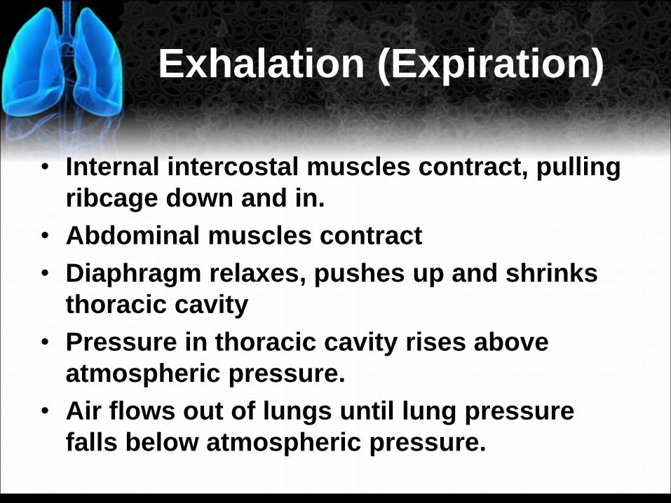
Exhalation (Expiration)
• Internal intercostal muscles contract, pulling
ribcage down and in.
• Abdominal muscles contract
• Diaphragm relaxes, pushes up and shrinks
thoracic cavity
• Pressure in thoracic cavity rises above
atmospheric pressure.
• Air flows out of lungs until lung pressure
falls below atmospheric pressure.

Quick Review – Inhalation
and Exhalation
• Air moves from HIGH to LOW pressure
Inhalation:
• Increasing lung volume = drop in pressure
– therefore air rushes into the lungs
Exhalation:
• Decreasing lung volume = increase in pressure
– Therefore air is pushed out of the lungs
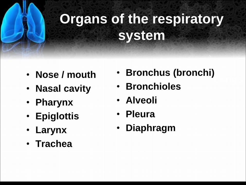
Organs of the respiratory
system
• Nose / mouth
• Nasal cavity
• Pharynx
• Epiglottis
• Larynx
• Trachea
• Bronchus (bronchi)
• Bronchioles
• Alveoli
• Pleura
• Diaphragm
Lungs


Lungs
Lungs are chambers containing moist
respiratory surfaces that are protected within
the body, where water loss is minimized and
the body wall provides support

How Does the Human
Respiratory System Work?
The human respiratory system can
be divided into two parts
– The conducting portion, a series of
passageways that carry air into and
out of the gas-exchange portion of the
respiratory system
– The gas-exchange portion, where
gases are exchanged with the blood in
tiny sacs within the lungs
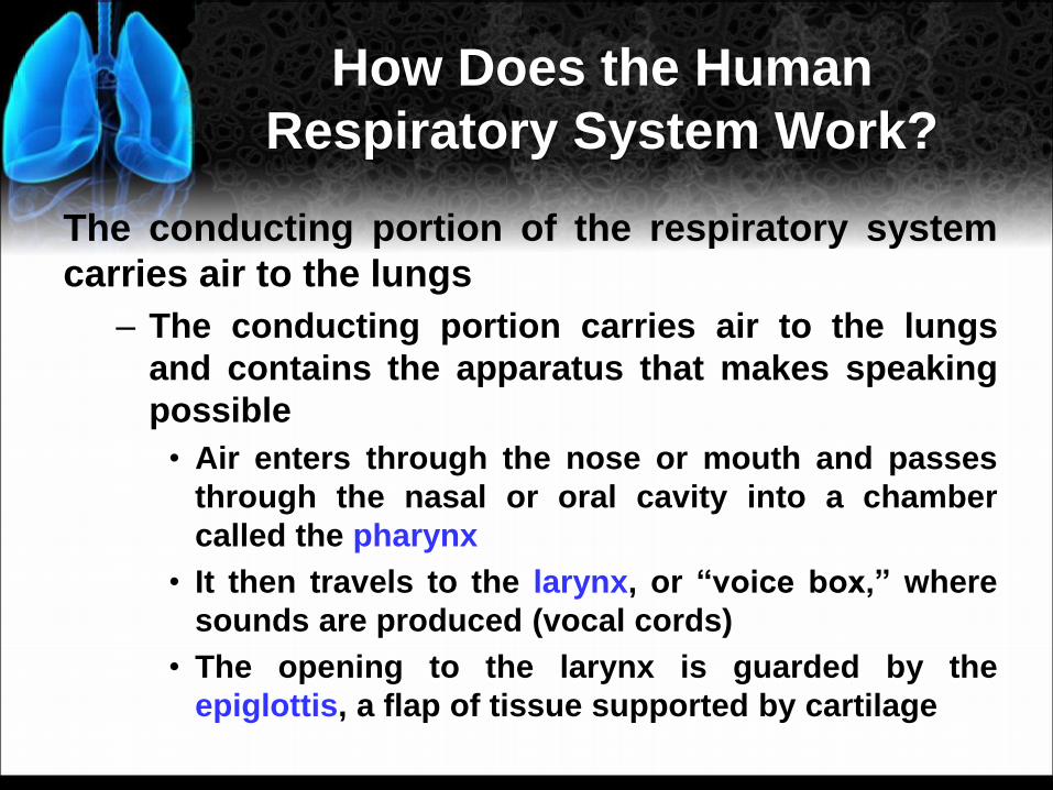
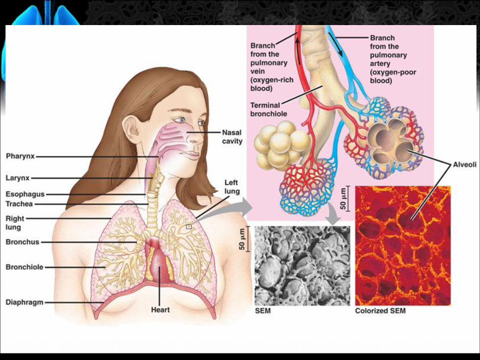
The conducting portion of the respiratory system
carries air to the lungs
– The conducting portion carries air to the lungs
and contains the apparatus that makes speaking
possible
• Air enters through the nose or mouth and passes
through the nasal or oral cavity into a chamber
called the pharynx
• It then travels to the larynx, or “voice box,” where
sounds are produced (vocal cords)
• The opening to the larynx is guarded by the
epiglottis, a flap of tissue supported by cartilage
How Does the Human
Respiratory System Work?

– Inhaled air travels past the larynx into the
trachea, a flexible tube whose walls are
reinforced with semicircular bands of stiff
cartilage
– The trachea splits into two bronchi, one leading
to each lung
– Inside the lung, each bronchus branches
repeatedly into ever small tubes called
bronchioles
– Bronchioles lead to microscopic alveoli, the tiny
air sacs where gas exchange occurs
How Does the Human
Respiratory System Work?

Alveoli – cup shaped structures at the end of the
bronchioles that resemble bunches of grapes; are in
direct contact with capillaries (gas exchange);
covered with SURFACTANT that keep them from
collapsing


(air in
alveolus)
(extracellular
fluid)
alveolar
wall
surfactant
fluid
red
blood
cells
hemoglobin
O2
O2
O2 capillary
walls
(plasma) cells of
body tissues
respiratory
membrane
Oxygen Transport

CO2
CO2
CO2
CO2
CO2
CO2 CO2
CO2
CO2
CO2
+ H2O
H2O
+
H+
H+ HCO3–
1
2
3
4
5 HCO3–
HCO3–
Carbon Dioxide
Transport
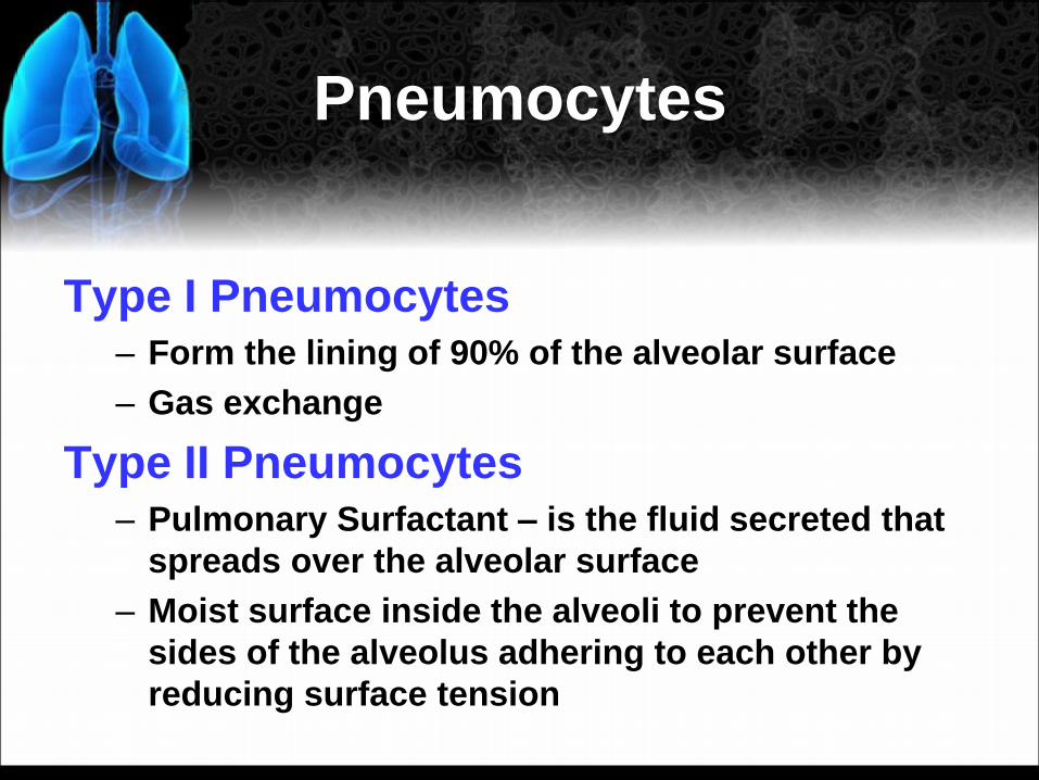
Pneumocytes
Type I Pneumocytes – Form the lining of 90% of the alveolar surface
– Gas exchange
Type II Pneumocytes – Pulmonary Surfactant – is the fluid secreted that
spreads over the alveolar surface
– Moist surface inside the alveoli to prevent the
sides of the alveolus adhering to each other by
reducing surface tension

Alveolar Adaptations
Alveoli are structurally adapted for gas
exchange:
1. Covered by dense network of capillaries
with high CO2 and low O2 concentrations
– Creates the concentration gradient necessary for
diffusion
2. Thin walls (one cell thick) ensure gases
only diffuse a short distance into capillaries

3. Moist inner surface allows gases to
dissolve and prevents alveoli from
sticking together.
4. Millions of alveoli provide adequate
surface area for exchange.
Alveolar Adaptations
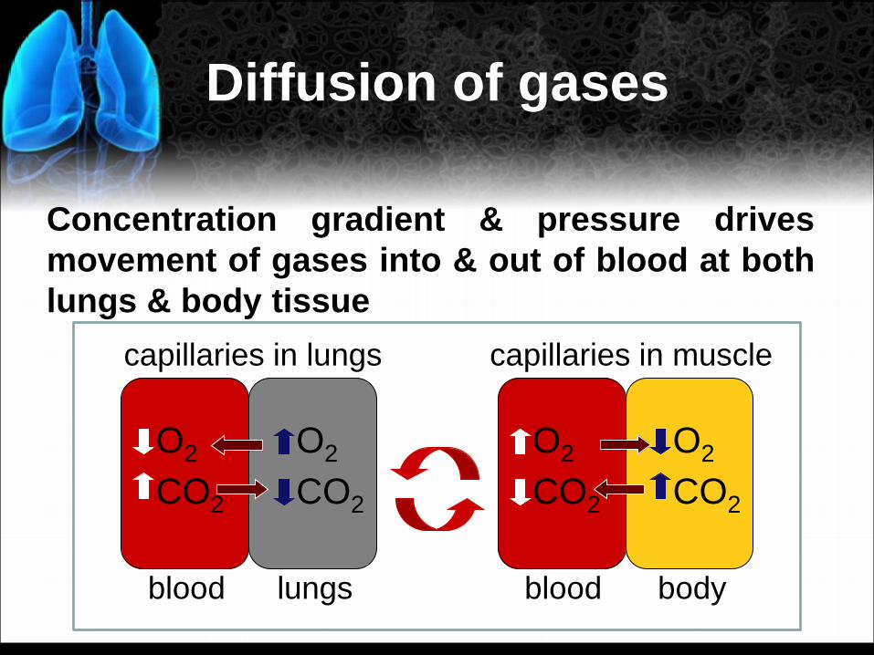
Diffusion of gases
Concentration gradient & pressure drives
movement of gases into & out of blood at both
lungs & body tissue
blood lungs
CO2
O2
CO2
O2
blood body
CO2
O2
CO2
O2
capillaries in lungs capillaries in muscle

Gas Exchange and Partial
Pressure
• Diffusion of gas
always occurs down
the partial pressure
gradient
• O2 tends to move
toward tissues and
CO2 tends to move
toward air.
PO2
(mm Hg)
PCO2
(mm Hg)
Air 152 3
Lungs 100 40
Blood 40 46
Tissues <40 >46
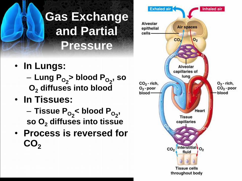
Gas Exchange
and Partial
Pressure
• In Lungs: – Lung PO2
> blood PO2, so
O2 diffuses into blood
• In Tissues: – Tissue PO2
< blood PO2,
so O2 diffuses into tissue
• Process is reversed for CO2

Breathing rate is controlled by the respiratory center of
the brain
– Breathing rate can be primarily modified by CO2 receptors located in the medulla that adjust the breathing rate to maintain a constant low level of CO2 in the blood, while also ensuring that O2 levels remain adequate
– As a backup system, there are also O2 receptors in the aorta and carotid arteries that stimulate the respiratory center to increase the rate and depth of breathing if O2 levels in the blood drop