Embed Size (px)
Citation preview

Essential Dynamics Sampling Study of Adenylate Kinase:Comparison to Citrate Synthase and Implication for theHinge and Shear Mechanisms of Domain Motions
Catherine Snow,1 Guoying Qi,1 and Steven Hayward1,2*1School of Computing Sciences, University of East Anglia, Norwich NR4 7TJ, United Kingdom2School of Biological Sciences, University of East Anglia Norwich NR4 7TJ, United Kingdom
ABSTRACT Essential dynamics sampling simu-lations of the domain conformations of unligandedEscherichia coli adenylate kinase have been per-formed to determine whether the ligand-inducedclosed-domain conformation is accessible to the openunliganded enzyme. Adenylate kinase is a three-domain protein with a central CORE domain andtwo flanking domains, the LID and the NMPbinddomains. The sampling simulations were applied tothe CORE and NMPbind domain pair and the COREand LID domain pair separately. One aim is to com-pare the results to those of a similar study on theenzyme citrate synthase to determinewhether a simi-lar domain-lockingmechanism operates in adenylatekinase. Although for adenylate kinase the simulationssuggest that the closed-domain conformation of theunliganded enzyme is at a slightly higher free energythan the open for both domain pairs, the results areradically different to those found for citrate synthase.In adenylate kinase the targeted domain conforma-tions could always be achieved, whereas this was notthe case in citrate synthase due to an apparent free-energy barrier between the open and closed confor-mations. Adenylate kinase has been classified as aprotein thatundergoes closure throughahingemech-anism, whereas citrate synthase has been assigned tothe shear mechanism. This was quantified here interms of the change in the number of interdomaincontacting atoms upon closure which showed a con-siderable increase in adenylate kinase. For citratesynthase this number remained largely the same, sug-gesting that the domain faces slide over each otherduring closure. This suggests that shear and hingemechanisms of domain closure may relate to the exis-tence or absence of an appreciable barrier to closurefor the unliganded protein, as the latter can hingecomparatively freely, whereas the formermust followamore constrainedpath. In general though it appearsa bias toward keeping the unliganded enzyme in theopen-domain conformationmay be a common featureof domain enzymes. Proteins 2007;67:325–337.VVC 2007Wiley-Liss, Inc.
Key words: molecular dynamics simulation; hingebending; principal component analy-sis; DynDom
INTRODUCTION
A sizeable proportion of enzymes have domain struc-tures with their active sites located in the interdomaincleft. For these enzymes the domains must be open toreceive the functional ligand which upon binding indu-ces the closed conformation. The ligand is then con-tained in the highly specific environment required forcatalysis. These domain movements are relatively easyto characterize in comparison to other conformationalchanges and therefore domain enzymes form an idealtarget for the study of the induced fit mechanism.
One enzyme whose domain movement has been stud-ied in some detail is citrate synthase.1 The binding ofthe small oxaloacetate molecule to citrate synthase indu-ces domain closure and molecular dynamics simulations(MD)2 together with essential dynamics sampling simu-lations (EDS)3 and the available crystallographic datashow that in the absence of oxaloacetate the domainsare unable to close fully. The barrier to closing seems tobe associated with the interaction of two parallel a-heli-ces at the domain interface.3 The interaction of oxaloace-tate with these helices is consistent with the movementrequired to achieve the fully closed structure, suggestingthat this interaction helps it overcome the energy bar-rier. Combined NMR and fluorescence work on maltose-binding protein has revealed a large energy barrier todomain closure for that protein,4 and a recent simulationstudy has also provided some support for this.5 In addi-tion, a recent MD simulation study on liver alcohol de-hydrogenase has shown that its well-known flexible loopis a NAD-sensitive switch that blocks domain closure inthe absence of NAD.6 Thus an emerging theme is thatfor some enzymes there exist ligand-sensitive switcheskeeping domains open in the absence of their functionalligand. For enzymes this would keep the binding site ac-cessible to the ligand resulting in a more efficient
Grant sponsor: Wellcome Trust JIF and BBSRC.
*Correspondence to: Steven Hayward, School of Computing Scien-ces, University of East Anglia, Norwich NR4 7TJ, UK. E-mail:[email protected]
Received 21 June 2006; Revised 14 September 2006; Accepted 21September 2006
Published online 13 February 2007 in Wiley InterScience (www.interscience.wiley.com). DOI: 10.1002/prot.21280
VVC 2007 WILEY-LISS, INC.
PROTEINS: Structure, Function, and Bioinformatics 67:325–337 (2007)

enzyme than one that is opening and closing resulting inthe binding site being inaccessible part of the time.The DynDom program7,8 is able to determine domains
and interdomain bending regions from conformationalchange as represented, for example, by two X-ray struc-tures. Interdomain bending regions are of particular in-terest as they control the relative movement of domainsand have been found to be intimately involved in ligand-induced domain closure.9 Some proteins, such as lacto-ferrin have two interdomain bending regions acting astwo separated mechanical hinges that control the do-main closure much like the two hinges of a door.10 It hasbeen suggested that lactoferrin closes via rotational dif-fusion of the domains.11 Other proteins, such as citratesynthase have a number of interdomain bending regionsand a complex interface between the domains. Relatedto this is the concept of hinge and shear mechanisms ofdomain closure.11 In the hinge mechanism two domaininterfaces come together during domain closure througha rotation about a localized hinge region of the backbonelinking the two domains. In the case of shear, the facesof the domains are in contact and remain in contact asthe protein opens and closes. Adenylate kinase (ADK)has been categorized as a protein that undergoes a hingemotion, whereas citrate synthase has been categorizedas a protein that undergoes a shear motion.11 Althoughthe DynDom analysis of the domain movement in citratesynthase gives a well defined hinge axis, these twoviews can easily be reconciled, as the shear conceptcomes from an atomic-scale analysis at the domain inter-face, which when viewed at the scale of the domainsthemselves, can be regarded as a rotation of the twodomains about some hinge axis. These findings indicatethat there may be a relationship between the complexity
of the interdomain interface and the existence or other-wise of a barrier to domain closure. Thus a similar EDSstudy to that carried out on citrate synthase on a pro-tein that has a much simpler domain interface andundergoes domain closure via the hinge mechanismwould be of value. ADK is just such a protein.
ADK, is a monomeric ubiquitous enzyme, which has akey role in energy maintenance within the cell, as itmaintains cellular adenylate levels by catalyzing thereaction Mg2þ.ATP þ AMP $ Mg2þ ADP þ ADP. ADKsand the family of monophosphate kinases have beenextensively studied both kinetically and structurally.Structurally the enzyme consists of three domains: alarge central domain normally referred to as the‘‘CORE,’’ an AMP binding domain normally referred toas the ‘‘NMPbind,’’ and a ATP-binding domain, normallyreferred to as the ‘‘LID’’ which covers the phosphates atthe active centre12 (Fig. 1). The LID and NMPbinddomains are not covalently linked directly to each otherand there are no contacts between the NMPbind andLID domains in the open-domain conformation. A Dyn-Dom analysis determined two domain pairs, which cor-respond to the CORE and the LID domain, and theCORE and the NMPbind domain.9 There are two inter-domain bending regions for each domain pair. The do-main interface is not complex like citrate synthase andis more reminiscent of that of lactoferrin.
This domain structure is conserved in all ADKs, how-ever, there are a number of different isoforms of theenzyme. To date six ADK isoforms have been character-ized in mammalian tissue with different subcellularlocalization and substrate specificity.13 The highly con-served region found in nearly all nucleoside monopho-phate kinases is named the P-loop which interacts and
Fig. 1. Backbone trace of ADK the open form (4AKE) on the left, and the closed form (1ANK) with theligands AMP and AMPPNP on the right in spacefilling model. The core domain is colored blue, the AMPdomain red and the ATP domain yellow. Green backbone indicates the interdomain bending regions.
326 C. SNOW ET AL.
PROTEINS: Structure, Function, and Bioinformatics DOI 10.1002/prot
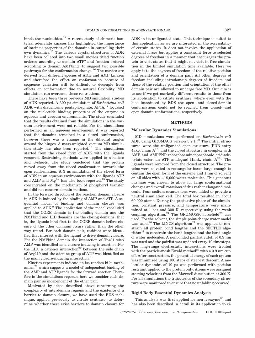
binds the nucleotides.14 A recent study of chimeric bac-terial adenylate kinases has highlighted the importanceof intrinsic properties of the domains in controlling theirown dynamics.15 The various crystal structures of ADKhave been collated into two small movies titled ‘‘motionordered according to domain ATP’’ and ‘‘motion orderedaccording to domain AMPbind’’ to suggest two possiblepathways for the conformation change.16 The movies arederived from different species of ADK and AMP kinasesand therefore the effect on conformation because ofsequence variation will be difficult to decouple fromeffects on conformation due to natural flexibility. MDsimulation can overcome these restrictions.There have been three previous MD simulation studies
of ADK reported. A 300 ps simulation of Escherichia coliADK with diadenosine pentaphosphate, AP5A,17 focussedon the nucleotide binding properties of the enzyme inaqueous and vacuum environments. The study concludedthat the results obtained from the simulations in the vac-uum environment were not reliable. For the simulationsperformed in an aqueous environment it was reportedthat the domains remained in a closed conformation,however there was a change in the dihedral anglesaround the hinges. A mass-weighted vacuum MD simula-tion study has also been reported.18 The simulationsstarted from the closed form of ADK with the ligandsremoved. Restraining methods were applied to a-helicesand b-sheets. The study concluded that the proteinmoved away from the closed conformation towards theopen conformation. A 3 ns simulation of the closed formof ADK in an aqueous environment with the ligands ATPand AMP and Mg2þ has also been reported.19 The studyconcentrated on the mechanism of phosphoryl transferand did not concern domain motions.In the forward direction of the reaction domain closure
in ADK is induced by the binding of AMP and ATP. A se-quential model of binding and domain closure wasapplied to ADK.9 The application of the model concludedthat the CORE domain is the binding domain and theNMPbind and LID domains are the closing domains, thatis, the ligands bind first to the CORE domain before clo-sure of the other domains occurs rather than the otherway round. For each domain pair, residues were identi-fied that interact with the ligand to drive domain closure.For the NMPbind domain the interaction of Thr31 withAMP was identified as a closure-inducing interaction. Forthe LID, a cation-p interaction20 between the side chainof Arg119 and the adenine group of ATP was identified asthe main closure-inducing interaction.9
Kinetics experiments indicate an iso random bi bi mech-anism21 which suggests a model of independent binding ofthe AMP and ATP ligands for the forward reaction There-fore in the simulations reported here we consider each do-main pair as independent of the other pair.Motivated by ideas described above concerning the
complexity of interdomain regions and the existence of abarrier to domain closure, we have used the EDS tech-nique, applied previously to citrate synthase, to deter-mine whether there exist barriers to domain closure for
ADK in its unliganded state. This technique is suited tothis application as we are interested in the accessibilityof certain states. It does not involve the application ofexternal forces but applies a constraint force to selecteddegrees of freedom in a manner that encourages the pro-tein to visit states that it might not visit in free simula-tion in the limited simulation time available. Here weapply it to the degrees of freedom of the relative positionand orientation of a domain pair. All other degrees offreedom including intradomain degrees of freedom andthose of the relative position and orientation of the otherdomain pair are allowed to undergo free MD. Our aim isto see if we get markedly different results to those fromits application to citrate synthase, where even with thebias introduced by EDS the open- and closed-domainconformations could not be reached from closed- andopen-domain conformations, respectively.
METHODS
Molecular Dynamics Simulations
MD simulations were performed on Escherichia coliADK using GROMACS version 3.2.1.22 The initial struc-tures were the unliganded open structure (PDB entry4ake, chain A12) and the closed structure in complex withAMP and AMPPNP (phosphoaminophosphonic acid-ade-nylate ester, an ATP analogue) (1ank, chain A23). Theligands were removed from the closed structure. The pro-teins were solvated in rectangular boxes large enough tocontain the open form of the enzyme and 1 nm of solventon all sides with �18,000 water molecules. This generousbox size was chosen to allow for large conformationalchanges and overall rotations of this rather elongated mol-ecule. Four sodium counter ions were added to provide aneutral simulation cell. The total box resulted in about60,000 atoms. During the productive phase of the simula-tion, constant pressure, and temperature were main-tained at 1 bar and 300 K, respectively, using the weakcoupling algorithm.24 The GROMOS96 forcefield25 wasused. For the solvent, the simple point charge water modelwas used.26 The LINCS algorithm27 was applied to con-strain all protein bond lengths and the SETTLE algo-rithm28 to constrain the bond lengths and the bond angleof water molecules. A nonbonded pairlist cutoff of 0.9 nmwas used and the pairlist was updated every 10 timesteps.The long-range electrostatic interactions were treatedwith the particle-mesh Ewald method29 with a 0.9 nm cut-off. After construction, the potential energy of each systemwas minimized using 100 steps of steepest descent. A mo-lecular dynamics of 10 ps was performed with positionrestraint applied to the protein only. Atoms were assignedstarting velocities from the Maxwell distribution at 300 K.For all simulations the trajectories of the secondary struc-ture were monitored to ensure that no unfolding occurred.
Rigid Body Essential Dynamics Analysis
This analysis was first applied for hen lysozyme30 andhas also been described in detail in its application to ci-
327DOMAIN CONFORMATIONS OF ADENYLATE KINASE
PROTEINS: Structure, Function, and Bioinformatics DOI 10.1002/prot
trate synthase.2 It involves removing intradomain con-formational change from the trajectories of the free MDsimulations using least-squares best-fitting techniquesto get a trajectory of the relative motion of the domains.The usual principal component analysis is then appliedto this trajectory to give six nonzero eigenvalues. As theprotein here comprised three domains, the analysis wasapplied separately to the trajectory of the CORE andNMPbind domain, and the trajectory of the CORE andLID domain.
Essential Dynamics Sampling
The EDS technique31 is used to increase or decreasethe distance from a reference conformation in a definedsubspace. The subspace was that defined by the sixeigenvectors corresponding to the six nonzero eigenval-ues. This technique has previously been applied to ci-trate synthase and the paper reporting that work3 has adetailed description of its usage in the application to do-main motions. Here, as previously, sampling is per-formed in two distinct modes: targeting and exploring.In targeting, contraction is performed to a specified tar-get conformation. In exploring, initial expansion occursfrom a specified reference conformation (e.g., the crystal-lographic open conformation), but when expansion ishalted according to two parameters, the final conforma-tion becomes the new reference conformation from whicha new expansion is started. The two parameters are themaximum number of sampling cycles before changingthe origin of expansion, and the slope, which sets a min-imum on the rate of expansion. These parameters werefixed to 5000 steps and 0.0004 nm/step, respectively, thesame as used for the citrate synthase simulations. Thetargeting simulations were stopped when the radiusfailed to decrease further. In all the simulationsreported, EDS was applied to the specified residues ofthe domains of interest, the other domain being allowedto undergo free MD.In each of the EDS simulations, the sampling space
was the six eigenvectors from the rigid-body essentialdynamics analysis of the domain pair concerned, appliedto the free MD trajectories with the same starting con-formation. For example, the sampling space for the EDSfor the LID-CORE-domain motion that started from theclosed conformation was derived from the rigid-bodyessential dynamics analysis of the LID-CORE-domaintrajectory of the trajectories from the free MD simula-tions that started from the closed conformation.
Depiction of Relative Motions of the Domains
The trajectories are displayed by projecting onto the2D space specified by the first two eigenvectors of therigid-body essential dynamics analysis of the combinedopen trajectories for the domain pair concerned. Thisallows one to see the trajectories of the domain confor-mations.
Domain Contact Analysis for Hingeand Shear Mechanism
In the shear mechanism of domain closure there arepreserved contacts over an extended domain interfacewhereas in the hinge mechanism the two domain inter-faces come together upon closure. Obviously, around thehinge axis itself one would expect to see preserved con-tacts in both mechanisms. The simple proceduredescribed below was used to distinguish between a shearand hinge mechanism taking this observation intoaccount. The molecular graphics program RasMol32 wasused to analyze interdomain atomic contacts from theopen and closed structures in the following way. Consid-ering a domain pair, a ‘‘domain set’’ was defined for eachdomain that comprised all the atoms from that domain.Then atoms in the domain set within 5.5 A10 of thehinge axis were removed to give a ‘‘non-axis-domainset.’’ Finally a subset of atoms in each non-axis-domainset was determined to be those within 4 A of any atomfrom the non-axis-domain set of the other domain. Thesesets, the ‘‘domain-contact sets,’’ were determined forboth open and closed structures.
RESULTS
A DynDom analysis of the functional movement inEscherichia coli ADK between the free enzyme (4ake_chainA12) and the AMP and AMPPNP liganded enzyme(1ank_A23), gave a three domain protein, with domain 1comprising residues 1–29, 76–116, and 160–214, domaintwo comprising residues 30–75, and domain three com-prising residues 117–159. The dynamical domains identi-fied by DynDom are almost identical to the visuallyidentified structural domains, where domain 1 is theCORE, domain 2 is the NMPbind domain and domain 3is the LID domain. The interdomain bending residuesidentified by the DynDom program are 29–30 and 75–76for the CORE-NMPbind domain pair, and 116–118 and157–172 for the CORE-LID domain pair. For conven-ience, we will refer to the CORE, NMPbind, and LIDdomains, as the core, AMP, and ATP domains, respec-tively, from here onwards.
Analysis of All Available X-Ray Structures
There are 29 X-ray files containing structures of ADKin the current protein data bank (PDB) containing 56structures of the individual chains. These come fromexperiments using a variety of ligand, species, isoforms,and mutants. Variation also comes from different crystalpacking.
The nonredundant form of the DynDom database,groups PDB chains into families based on sequence simi-larity.33 ADK sequences were classified into 10 distinctfamilies containing more than one member. Familieswere defined by a representative protein. All memberswithin a family had at least 90% sequence identity withthe representative protein.
328 C. SNOW ET AL.
PROTEINS: Structure, Function, and Bioinformatics DOI 10.1002/prot

All sequences from the 10 families and the sequenceof any single member family were aligned using multiplesequence alignment. The alignment showed large gapsbecause of the different forms of ADK. The short ADKsrepresented by bacterial and mitochondrial enzymes donot contain the 20–30 residue insertion inside the LIDdomain; this is also true of the ADKs from archaea. Itwas therefore decided to remove these sequences.The remaining 29 sequences were realigned and a con-
formational clustering algorithm was applied to thesestructures.33 The algorithm clusters structures based ontheir root mean-square deviations after a window aver-aging process. The structures fell into 10 conformationalclusters, 9 containing more than one structure and 1containing just a single structure. These 10 clusters,indicated as eight circles (three are conformationally
close and are represented by a large circle) in Figure 2,represent six different movements as determined by a‘‘dimensional clustering’’ process,33 and indicated by thestraight lines in Figure 2. The unbroken lines in Figure2 are transitions that are domain movements accordingto the DynDom program and those with broken lines arethose that are not. The lengths of these lines indicateroughly the extent of the transition. One can see thatthere are three main independent domain movements(represented by the long unbroken lines in Fig. 2)between four main states at 4ake_B,12 2ak3_A,35 1ak2,12
and 2aky.36 The four states are: a free enzyme with bothAMP and ATP domains open (4ake_B); a free enzymewith the ATP domain predominantly closed and theAMP domain open (1ak2); an AMP liganded enzymewith the AMP domain closed and the ATP domain open
Fig. 2. This figure is based on a conformational and dimensional clustering process33 of 29 structuresof ADK. The circles represent sets of structures with similar domain conformations. The lines representunique movements between these conformations. Unbroken lines are domain movements and broken onesare not domain movements according to the DynDom program. The process yielded 10 clusters, 9 tight,and 1 extended as defined in Qi et al.,33 but it was found that three of them, the extended (3aky, 1aky,2aky,1p3jA, 1s3gA) and two tight (1e4vA, 1e4vB, 1akeA, 2eckA, 2eckB, 1akeB, 1ankA, 1ankB), (1e4yA,1e4yB) were conformationally close enough to each other to be considered as a single cluster, indicatedas the large circle labelled by, 2aky, one of the structures it contains. All these structures are fully ligandedhaving both domains closed upon a ligand. 1zakB34 (representing 1zakB, 1zakA) is also a fully ligandedstructure but there is no domain movement between it and the other closed-domain structures in the largecluster. 4akeB, (representing 4akeB, 4akeA) is the free enzyme structure with both domains open. 2ak3Ais bound to AMP and has its AMP domain closed. Close to this is 1zd8A (representing 1zd8A, 2ak3B)which is unliganded. 1ak2 (representing 1ak2, 2ak2) has its ATP domain nearly closed but is a freeenzyme. Close to this is 1dvrB (representing 1dvrA, 1dvrB) which is liganded to ATP only and has its ATPdomain closed. 1zin (representing 1zin, 1zip, 1zio, all fully liganded) has no line as it doesn’t have a uniquedomain movement but combines a large part of the movement between 4akeB and 2aky (in the large clus-ter) and a small part of the movement between 4akeB and 2ak3A. Thus this analysis yields three uniquedomain movements between four main states.
329DOMAIN CONFORMATIONS OF ADENYLATE KINASE
PROTEINS: Structure, Function, and Bioinformatics DOI 10.1002/prot

(2ak3_A); a AP5A liganded enzyme with both AMP andATP domains closed (2aky). 1dvr_B12 is from yeast andhas ATP bound with its ATP domain closed but its AMPdomain open. It is close to 1ak2 from Escherichia coli,which is unliganded but its ATP domain is nearly closedapparently due to crystal contacts. 1zd8_A which a humanadenylate kinase 3 is unliganded but its AMP domain isin a closed conformation with its ATP domain open. It isclose to 2ak3_A which has AMP bound. In summary, theX-ray structures yield four main domain states: both AMPand ATP domains open; both AMP and ATP domainsclosed; ATP domain closed, AMP domain open; AMP do-main closed, ATP domain open. The existence of a closedATP domain when the AMP domain is open and vice-versa, both in the presence and absence of ligands, sup-ports our approach of applying EDS to each domain pairseparately.
Free MD Simulation Study
Two independent 5 ns MD simulations were performedstarting from the open conformation and a further twosimulations, again each of 5 ns, starting from the closedconformation. These simulations provided us with thetrajectories to define the EDS sampling spaces asdescribed at the end of the Methods section.
AMP-Core-Domain Motion
We report first the results of the analysis of themotion of the AMP domain relative to the core. Table Igives details of the EDS simulations of the AMP-core-domain motion for both sets of simulations: those thatstart from the open conformation and those that startfrom the closed.
Simulations from open-domain conformation
Free MD simulations. The two 5 ns MD simulationsthat started from the crystallographic open conformationwere combined into a single trajectory and a rigid-bodyessential dynamics analysis performed on the trajectoryof the core-AMP domains. This trajectory was projectedonto the space defined by the first two eigenvectors fromthis analysis. Approximately 91% of the domain fluctua-tion occurred in this 2D space. The trajectory is shown
in all four panels as orange dots in Figure 3. The crys-tallographic open- and closed-domain conformations arealso indicated in Figure 3.
Essential dynamics sampling: exploring mode.Three exploring mode simulations were performed;details of these simulations are shown in Table I. Run 1started from the open conformation that was used in theoriginal free MD simulations. The second simulation,Run 2, began from the final conformation of Run 1. Thethird exploring simulation, Run 3, began from the finalconformation of Run 2. Figure 3(a) shows the AMP-core-domain trajectories of these exploring simulations pro-jected onto the first two eigenvectors described in theprevious section. The trajectories of the exploring modesimulations show that domain conformations are visitedthat were not visited in the original free simulations. InRun 3 the protein probes the space between the crystal-lographic open and closed domain conformation but theclosed domain conformation is never achieved. This mayindicate the presence of an energy barrier or gradient.
Essential dynamics sampling: to-closed target-ing mode. In these simulations the crystallographicclosed-domain conformation is the target. The trajecto-ries that resulted from these runs can be seen in Figure3(b). Run 4 started from the open conformation and thetrajectory moved towards the target and remained there.The starting structure for Run 5 was the conformationthat is furthest from the original open conformation thatoccurred during Run 1. Run 6 was the conformationthat is furthest from the original open conformation thatoccurred during Run 2. Both Run 5 and 6 went straighttowards their target conformation. This shows that theability to reach the closed target conformation is inde-pendent of the starting conformation.
Essential dynamics sampling: back-targeting.Figure 3(b) also shows further simulations, which targetback to original crystallographic open-domain conforma-tion. These back-targeting simulations were performed tocheck that nothing had occurred to prevent the proteinachieving its original domain conformation. Two targetingsimulations were performed, Run 7, for which the start-ing structure was the same as Run 5, and Run 8, forwhich the starting structure was the same as Run 6. Tra-jectories for these simulations are displayed in Figure
TABLE I. Runs for AMP and Core Domains for Both Open to Closed and Closed to Open
Run Mode Starting structure Length of simulation (ps)
1 Exploring Relaxed crystal 15002 Exploring Final structure which resulted from run 1 15003 Exploring Final structure which resulted from run 2 15004 Target Relaxed crystal (targeting from open crystal to closed crystal and vice versa) 5005 Target Furthest structure from original starting structure which occurred in run 1 5006 Target Furthest structure from original starting structure which occurred in run 2 5007 Targetback Same as run 6 5008 Targetback Same as run 7 500
330 C. SNOW ET AL.
PROTEINS: Structure, Function, and Bioinformatics DOI 10.1002/prot
3(b). It shows that they go directly back to the crystallo-graphic open-domain conformation. This demonstratesthat in the targeting mode there is no essential differencein terms of accessibility between the open- and closed-do-main conformations for the AMP-core-domain motion.
Simulations from closed-domain conformation
Free MD simulations. The trajectories of the AMP-core domains from the two 5 ns free MD simulationsstarting from the crystallographic closed conformationwere projected onto the space defined by the first twoeigenvectors from the rigid-body essential dynamic anal-ysis of the combined trajectories from the simulationsstarting from the open-domain conformation. These areshown in all four panels as brown dots in Figure 3.
Essential dynamics sampling: exploring mode.Domain conformations are visited that are not in theoriginal free simulations. In contrast to the runs fromthe crystallographic open conformation, which failed toreach the crystallographic closed-domain conformation,Run 3 reached the crystallographic open-domain confor-mation from the closed conformation.
Essential dynamics sampling: to-open targetingmode. In these simulations the crystallographic open-domain conformation is the target. Figure 3(d) showsthe domain trajectories from Runs 4 to 6. All the trajec-tories go directly from their starting conformations tothe targeted conformation. This shows that the ability toreach the target open-domain conformation is independ-ent of the starting conformation.
Essential dynamics sampling: back-targeting.Two further targeting simulations were performed aschecking simulations. Run 7 began with the same start-ing structure for Run 5, and Run 8 began with the samestarting structure as Run 6. Both the trajectories aredisplayed in Figure 3(d). Both go directly back to thecrystallographic closed-domain conformation.
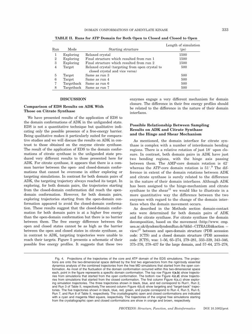
ATP-Core-Domain Motion
We report below the results of the analysis of themotion of the ATP domain relative to the core. Table IIgives details of the EDS simulations of the ATP-core-do-main motion for both sets of simulations: those thatstart from the open conformation, and those that startfrom the closed.
Simulations from open-domain conformation
Free MD simulations. The two 5 ns simulations,which started from the crystallographic open conforma-tion, were combined into a single trajectory and rigid-body essential dynamics analysis performed on the tra-jectory of the ATP-core domain. The trajectory of theseparts of the protein was projected onto the space definedby the first two eigenvectors. Approximately 83% of thedomain fluctuation occurred in this space. The trajectoryis shown as orange dots in Figure 4. The crystallo-graphic open- and closed-domain conformations are alsoindicated in Figure 4.
Essential dynamics sampling: exploring mode.The results in Figure 4(a) are very similar to those seenfor the exploring simulations of the AMP-core domain.In all runs the protein explores a larger space than thatseen in the original free simulations. The trajectoryfrom Run 1 showed that the protein moved towards theclosed-domain conformation. Run 2 which began fromthe final conformation of Run1 shows that the proteinmoves back towards the open-domain conformation andthis continues with Run 3, which explores even moreopen-domain conformations.
Essential dynamics sampling: to-closed target-ing mode. The crystallographic closed-domain confor-mation is successfully targeted in all runs. Run 4 startsfrom the open conformation. Run 5, which starts fromthe final conformation of Run 2, is more open than thecrystallographic open-domain conformation. These areshown in Figure 4(b).
Essential dynamics sampling: back-targeting.Two further targeting runs were performed to checkthat conformations achieved in the initial exploring runswere able to target back to the original starting confor-mation. As in previous cases targeting back to the origi-nal conformation was successful. These runs are alsoshown in Figure 4(b).
Simulations from closed-domain conformation
Free MD simulations. The trajectories of the ATP-core domains from the two 5 ns free MD simulationsstarting from the crystallographic closed conformationwere projected onto the space defined by the first twoeigenvectors from the rigid-body essential dynamic anal-ysis of the combined trajectories from the simulationsstarting from the open-domain conformation. These areshown as brown dots in Figure 4.
Essential dynamics sampling: exploring mode.The results in Figure 4(c) show the exploring Runs 1–3.The protein visits the open-domain conformation andlike the exploring sampling runs starting from the open-domain conformation, the space around and beyond theopen-domain conformation is widely explored. Thus incontrast to the runs from the crystallographic open con-formation, which failed to reach the crystallographicclosed-domain conformation, the crystallographic open-domain conformation was reached from the closed.
Essential dynamics sampling: to-open targetingmode. Figure 4(d) shows Runs 4–6 targeted to the open-do-main conformation. As previously seen for the AMP domain,the trajectory goes directly from its starting domain confor-mations to the target closed-domain conformation and it isnot dependent on the starting domain conformation.
Essential dynamics sampling: back-targeting.Two further targeting simulations, Runs 7 and 8, wereperformed to target back to the original starting crystal-lographic closed conformation. As in previous cases tar-geting back to the original conformation was successful.These runs are also shown in Figure 4(d).
331DOMAIN CONFORMATIONS OF ADENYLATE KINASE

Fig. 3. Projections of the trajectories of the core and AMP domain of the EDS simulations. The projec-tions are onto the two-dimensional space defined by the first two eigenvectors from the rigid-body essentialdynamics analysis of the combined trajectories from the free MD simulations that started from the open con-formation. As most of the fluctuation of the domain conformation occurred within this two dimensional space,each point in the figure represents a specific domain conformation. The top row Figure 3(a,b) show trajecto-ries from simulations that started from the open conformation. The bottom row Figure 3(c,d) show trajectoriesfrom simulations that started from the closed conformation. The first column, Figure 3(a,c) show exploringsimulation trajectories. The three simulations shown in black, blue and red correspond to Run1, Run 2, andRun 3 of Table I respectively. The second column, Figure 3(b,d), show targeting and ‘‘target-back’’ trajector-ies.The five trajectories shown in black, blue, red, green, and purple correspond to Run 4, Run 5, Run 6,Run 7, and Run 8 respectively. The crystallographic open and closed conformations are indicated with acyan and magenta filled square, respectively. The trajectories of the original free simulations starting from thecrystallographic open and closed conformations are show in orange and brown, respectively.
Figure 4.
332 C. SNOW ET AL.
PROTEINS: Structure, Function, and Bioinformatics DOI 10.1002/prot
DISCUSSION
Comparison of EDS Results on ADK WithThose on Citrate Synthase
We have presented results of the application of EDS tothe domain conformations of ADK in the unliganded state.EDS is not a quantitative technique but qualitative indi-cating only the possible presence of a free-energy barrier.Being qualitative makes it particularly suited for compara-tive studies and we will discuss the results on ADK in con-trast to those obtained on the enzyme citrate synthase.The result of the application of EDS to the domain confor-mations of citrate synthase in the unliganded state pro-duced very different results to those presented here forADK. For citrate synthase, it appears that there is a com-mon barrier between the open- and closed-domain confor-mations that cannot be overcome in either exploring ortargeting simulations. In contrast for both domain pairs ofADK, the targeting trajectory always reached its target. Inexploring, for both domain pairs, the trajectories startingfrom the closed-domain conformation did reach the open-domain conformation. However, for both domain pairs,exploring trajectories starting from the open-domain con-formation appeared to avoid the closed-domain conforma-tion. These results suggest that the closed-domain confor-mation for both domain pairs is at a higher free energythan the open-domain conformation but there is no barrierbetween them. The free energy difference between theopen and closed states cannot be as high as the barrierbetween the open and closed states in citrate synthase, asin contrast to ADK, targeting trajectories were unable toreach their targets. Figure 5 presents a schematic of theirpossible free energy profiles. It suggests that these two
enzymes engage a very different mechanism for domainclosure. The difference in their free energy profiles shouldbe related to the difference in the nature of their domaininterfaces.
Possible Relationship Between SamplingResults on ADK and Citrate Synthaseand the Hinge and Shear Mechanism
As mentioned, the domain interface for citrate syn-thase is complex with a number of interdomain bendingregions. There is a relative rotation of just 188 upon clo-sure. In contrast, both domain pairs in ADK have justtwo bending regions, with the hinge axis passingbetween them. The AMP-core domain rotation is 428whereas the ATP-core domain rotation is 518.9 The dif-ference in extent of the domain rotations between ADKand citrate synthase is surely related to the differencein the nature of their domain interfaces. Although ADKhas been assigned to the hinge-mechanism and citratesynthase to the shear11 we would like to illustrate in amore quantitative way the difference between the twoenzymes with regard to the change of the domain inter-faces when the domain movement occurs.
As described in the Methods section domain-contactsets were determined for both domain pairs of ADKand for citrate synthase. For citrate synthase the domaindecomposition, based on the movement (http://www. cmp.uea.ac.uk/dyndom/dyndomRun.do?ddid¼CITRA1R4&action ¼view)37 between and open-domain structure (PDB accessioncode: 1CTS) and a closed domain structure (PDB accessioncode: 2CTS), was: 1–56, 65–274, 279–281, 333–339, 343–346,375–376, 378–437 for the large domain, and 57–64, 275–278,
TABLE II. Runs for ATP Domain for Both Open to Closed and Closed to Open
Run Mode Starting structureLength of simulation
(ps)
1 Exploring Relaxed crystal 15002 Exploring Final structure which resulted from run 1 15003 Exploring Final structure which resulted from run 2 15004 Target Relaxed crystal (targeting from open crystal to
closed crystal and vice versa)500
5 Target Same as run 3 5006 Target Same as run 4 5007 Targetback Same as run 6 5008 Targetback Same as run 7 500
Fig. 4. Projections of the trajectories of the core and ATP domain of the EDS simulations. The projec-tions are onto the two-dimensional space defined by the first two eigenvectors from the rigid-body essentialdynamics analysis of the combined trajectories from the free MD simulations that started from the open con-formation. As most of the fluctuation of the domain conformation occurred within this two-dimensional spaceeach, point in the figure represents a specific domain conformation. The top row Figure 4(a,b) show trajecto-ries from simulations that started from the open conformation. The bottom row Figure 4(c,d) show trajecto-ries from simulations that started from the closed conformation. The first column Figure 4(a,c) show explor-ing simulation trajectories. The three trajectories shown in black, blue, and red correspond to Run1, Run 2,and Run 3 of Table II, respectively.The second column Figure 4(b,d) show targeting and ‘‘target-back’’ trajec-tories. The five trajectories shown in black, blue, red, green, and purple correspond to Run 4, Run 5, Run 6,Run 7, and Run 8 of Table II, respectively. The crystallographic open and closed conformations are indicatedwith a cyan and magenta filled square, respectively. The trajectories of the original free simulations startingfrom the crystallographic open and closed conformations are show in orange and brown, respectively.
333DOMAIN CONFORMATIONS OF ADENYLATE KINASE
PROTEINS: Structure, Function, and Bioinformatics DOI 10.1002/prot

282–332, 340–342, 347–374, 377–377 for the small domain.Table III gives the relevant numbers. There is a striking dif-ference between the domain pairs of ADK and citrate syn-thase. For citrate synthase in the open conformation thereare 258 atoms from both domains that are in the domain-contact sets. This increases to only 284 for the closed struc-ture. These atoms come from 61 residues in the open whichincreases to 66 in the closed. Of these, 57 are common toboth open and closed conformations. There is some ambiguityas to whether the helix 328–341 belongs to the large or smalldomain. Assuming it belongs wholly to the small domainresults in little change in the numbers presented above. For
both domain pairs of ADK, there are 77 atoms in the domain-contact sets in the open conformation, which increases to 170in the closed. These atoms are from 24 residues in the open,which increases to 51 in the closed. Of these 20 are commonto both open and closed conformations. So in citrate synthasethere is only a 10% increase in the number of contactingatoms, whereas for ADK there is a 121% increase. Figure 6shows the difference structurally. These results support theidea of different mechanisms acting in these two enzymes. Itcertainly supports the idea of a preserved interface in thecase of citrate synthase, and the formation of a rather newinterface in ADK. It therefore supports the idea of a shear-type mechanism acting for the former and a hinge-type mech-anism for the latter. Taking our results from the EDS simula-tions into consideration it seems reasonable to suggest thatthis difference relates to the nature of the energy barrierbetween the open- and closed-domain conformations. This issupported by the fact that parts of the a-helices 328–341 and222–235 whose interaction was determined to be the origin ofthe energy barrier3 are part of the shear interface.
Nature of Energy Profiles and InteractionWith Substrates
For ADK it seems likely that the source of the increasein energy upon closure resides at the interdomain bendingregions themselves and involves a strain being induced asthe domains close. A recent application of a so-called plas-tic network model to ADK has shown a gentle rise instrain energy from the open- to closed- domain conforma-tion.38 The model allows one to identify the strain energyat particular residues and peaks in the strain energy arefound at or very close to the bending residues identified bythe DynDom program.38 Given that for the unligandedstate, the closed-domain conformation is at a higher freeenergy than the open, it must be the interaction with thesubstrate that enables the enzyme to adopt the closed-do-main conformation. For the ATP-core domain pair, thisinteraction appears to come from a cation-p20 interactionbetween Arg119 and the adenine moiety of ATP.9 For theAMP-core domain pair it appears to be between Thr31and the adenine moiety of AMP.9
In the case of citrate synthase the probable site thatgave rise to the free energy barrier could be found.3 It islocated at a pair of parallel a-helices at the domain inter-face, one located in the large domain, the other in thesmall domain. The substrate, oxaloacetate binds near thesetwo helices and it is thought that the interaction of oxalo-acetate, once bound to the large domain, with Arg329 iscrucial for converting to the closed- domain conformation.3
Our results on ADK suggest that there is a biastowards the open-domain conformation for both domainpairs but no appreciable barrier. It is possible that theclosed-domain conformation is accessible to equilibriumfluctuations but we cannot conclude that from our simula-tions. The existence of a nearly closed ATP domain (1ak2in Fig. 2) and a closed AMP domain (1zd8_A in Fig. 2) inthe absence of any ligand suggests that weak crystalpacking forces can induce a near closed-domain conforma-
Fig. 5. A schematic representation of the free energy profiles for (a)citrate synthase and (b) ADK that would explain both sets of resultsfrom the EDS studies of both enzymes. For an unliganded citrate syn-thase an energy barrier cannot be overcome in going from the open-domain conformation to the closed-domain conformation and viceversa, in both exploring and targeting studies. Although for both domainpairs of an unliganded ADK the open-domain conformation could bereached from the closed-domain conformation, the closed-domain con-formation could not be reached from the open-domain conformation inexploring simulations. This suggests that the closed-domain conforma-tion for both domains pairs of ADK is at a higher free energy than theopen-domain conformation. However, the free energy difference shouldnot be as high as the barrier for citrate synthase as in targeting; theclosed-domain conformation could be successfully targeted from theopen-domain conformation.
334 C. SNOW ET AL.
PROTEINS: Structure, Function, and Bioinformatics DOI 10.1002/prot

tion for both domain pairs. In contrast, evidence suggeststhat for citrate synthase the unliganded open-domain con-formation is never able to access the closed-domain confor-mation in equilibrium and no closed crystal structures arefound in the absence of its functional ligand.The EDS technique is rather qualitative in that it
only indicates the presence of energy barriers or gra-dients. Potential of mean force calculations or umbrellasampling techniques, although more computationallydemanding, would overcome this by yielding quantita-tively accurate free energy profiles.
Comparison to Other Domain Proteins
Like ADK, near closed forms of bacteriophage T4 lyso-
zyme in its unliganded state have been induced by crystal
contacts39 even though in solution spin-labelling experi-
ments suggest a predominance of the open-domain confor-
mation.40 Also like ADK, and in contrast to citrate synthase
it has two interdomain bending regions through which the
hinge axis passes.7 Two hinges would appear to control the
direction of rotation as in the door-closing model10 but
would not necessarily restrict rotational freedom about the
TABLE III. Atom Contacts Between Domains in Open and Closed Structures for Citrate Synthase and ADK
Protein/domain pair DomainNumber of domain-contactatoms in open structure
Number of domain-contactatoms in closed structure
Citrate synthase Large 126 143Small 132 141
Adenylate kinase/core-AMP Core 21 38AMP 21 32
Adenylate kinase/core-ATP Core 16 42ATP 19 58
Fig. 6. (a) Open structure of citrate synthase. Large domain is colored blue, small red. Arrow indi-cates the hinge axis. In cyan and spacefilling model is the domain contact set of atoms from the largedomain, and in orange the domain contact set from the small domain. (b) The closed structure of ci-trate synthase. (c) The open structure of ADK. Blue is the core domain, yellow is the ATP domainand red the AMP domain. The cyan spacefilling model is the domain contact set of atoms from thecore domain, and orange the domain contact set from the AMP or ATP domains. (d) The closed struc-ture of ADK.
335DOMAIN CONFORMATIONS OF ADENYLATE KINASE
PROTEINS: Structure, Function, and Bioinformatics DOI 10.1002/prot
hinge axis. It is also classified as a hinge mechanism pro-tein.11 A recent computational study on ribose-bindingprotein has shown for that protein the open-domain con-formation is about 1–2 kcal/mol below that of the closed-domain conformation and the closed- domain conforma-tion is populated albeit less than the open-domain confor-mation in equilibrium.41 Again this protein has basicallytwo mechanical hinges, which determine the hinge axisand is classified as a protein belonging to the hinge mech-anism rather than the shear (http://www.molmovdb.org/).42 In liver alcohol dehydrogenase domain closure isinduced by the binding of NADþ. The block to closurehas been shown to be a loop that changes conformation toallow domain closure upon the binding of NADþ.6 Thusliver alcohol dehydrogenase like citrate synthase has ablock to closure in the absence of its functional ligandand like citrate synthase it has been categorized as a pro-tein belonging to the shear mechanism.
CONCLUSIONS
These results suggest that for some proteins a weakbias towards the open-domain conformation is presentfor the unliganded protein whereas for others more so-phisticated mechanisms keep the domains open in theabsence of a ligand. We suggest that this difference isrelated to the complexity of the domain interface andthat those proteins with two simple hinges are able tohinge reasonably freely between states albeit with a biastowards an open domain conformation. The two domainpairs in ADK are examples of this. It seems reasonablethat if an enzyme is to employ a sophisticated blockingmechanism to keep the domains open in the absence ofthe ligand then this blocking mechanism must belocated at the domain interface and may require a moreextensive set of interactions than provided for by a sim-ple hinge. It is plausible therefore to suggest that thehinge and shear mechanisms of domain closure relate tothe absence or presence, respectively, of a significantenergy barrier between the open- and closed-domainconformations for the unliganded enzyme.
ACKNOWLEDGMENTS
C.S. is supported by an EPSRC studentship. We thankI. Daidone for her support.
REFERENCES
1. Wiegand G, Remington SJ. Citrate synthase, structure, control,and mechanism. Annu Rev Biophys Biophys Chem 1986;15:97–117.
2. Roccatano D, Mark AE, Hayward S. Investigation of the mecha-nism of domain closure in citrate synthase by molecular dynam-ics simulation. J Mol Biol 2001;310:1039–1053.
3. Daidone I, Roccatano D, Hayward S. Investigating the accessibil-ity of the closed domain conformation of citrate synthase usingessential dynamics sampling. J Mol Biol 2004;339:515–325.
4. Millet O, Hudson RP, Kay LE. The energetic cost of domainreorientation in maltose-binding protein as studied by NMR andfluorescence spectroscopy. Proc Natl Acad Sci USA 2003;100:12700–12705.
5. Stockner T, Vogel HJ, Tieleman DP. A salt-bridge motif involvedin ligand binding and large-scale domain motions of the malt-ose-binding protein. Biophys J 2005;89:3362–3371.
6. Hayward S, Kitao A. Molecular dynamics simulations of NADþ-induced domain closure in horse liver alcohol dehydrogenase.Biophys J 2006;91:1823–1831.
7. Hayward S, Berendsen HJC. Systematic analysis of domainmotions in proteins from conformational change: new resultson citrate synthase and T4 lysozyme. Proteins 1998;30:144–154.
8. Hayward S, Lee RA. Improvements in the analysis of domainmotions in proteins from conformational change: DynDom ver-sion 1.50. J Mol Graph Model 2002;21:181–183.
9. Hayward S. Identification of specific interactions that driveligand-induced closure in five enzymes with classic domainmovements. J Mol Biol 2004;339:1001–1021.
10. Hayward S. Structural principles governing domain motions inproteins. Proteins 1999;36:425–435.
11. Gerstein M, Lesk AM, Chothia C. Structural mechanisms fordomain movements in proteins. Biochemistry 1994;33:6739–6749.
12. Muller CW, Schlauderer GJ, Reinstein J, Schulz GE. Adenylatekinase motions during catalysis: an energetic counterweight bal-ancing substrate binding. Structure 1996;4:147–156.
13. Ren H, Wang LY, Bennett M, Liang YH, Zheng XF, Lu F, Li LF,Nan J, Luo M, Eriksson S, Zhang CM, Su XD. The crystalstructure of human adenylate kinase 6: an adenylate kinaselocalized to the cell nucleus. Proc Natl Acad Sci USA 2005;102:303–308.
14. Byeon IJL, Shi ZT, Tsai MD. Mechanism of adenylate kinase––the essential lysine helps to orient the phosphates and the active-site residues to proper conformations. Biochemistry 1995;34:3172–3182.
15. Bae E, Phillips GN. Roles of static and dynamic domains in sta-bility and catalysis of adenylate kinase. Proc Natl Acad Sci USA2006;103:2132–2137.
16. Vonrhein C, Schlauderer GJ, Schulz GE. Movie of the structuralchanges during a catalytic cycle of nucleoside monophosphatekinases. Structure 1995;3:483–490.
17. Kern P, Brunne RM, Folkers G. Nucleotide-binding properties ofadenylate kinase from Escherichia coli––a molecular-dynamicsstudy in aqueous and vacuum environments. J Comput-AidedMol Des 1994;8:367–388.
18. Elamrani S, Berry MB, Phillips GN, McCammon JA. Studyof global motions in proteins by weighted masses moleculardynamics: adenylate kinase as a test case. Proteins 1996;25:79–88.
19. Krishnamurthy H, Lou HF, Kimple A, Vieille C, Cukier RI.Associative mechanism for phosphoryl transfer: a moleculardynamics simulation of Escherichia coli adenylate kinase com-plexed with its substrates. Proteins 2005;58:88–100.
20. Gallivan JP, Dougherty DA. Cation-p interactions in structuralbiology. Proc Natl Acad Sci USA 1999;96:9459–9464.
21. Sheng XR, Li X, Pan XM. An iso-random BiBi mechanism foradenylate kinase. J Biol Chem 1999;274:22238–22242.
22. Berendsen HJC, van der Spoel D, van Drunen R. GROMACS: amessage-passing parallel molecular dynamics implementation.Comput Phys Commun 1995;91:43–56.
23. Berry MB, Meador B, Bilderback T, Liang P, Glaser M, PhillipsGN. The closed conformation of a highly flexible protein––thestructure of Escherichia coli adenylate kinase with bound ampand amppnp. Proteins 1994;19:183–198.
24. Berendsen HJC, Postma JPM, Gunsteren WFv, Nola AD. Molec-ular dynamics with coupling to an external bath. J Chem Phys1984;81:3684–3690.
25. van Gunsteren WF, Billeter SR, Eising AA, Hunenberger PH,Kruger P, Mark AE, Scott WRP, Tironi IG. Biomolecular simula-tion: the GROMOS96 manual and user guide. ETH Zurich,Switzerland: vdf Hochschlverlang; 1996.
26. Berendsen HJC, Postma JPM, van Gunsteren WF, Hermans HJ.Interaction models for water in relation to protein hydration.In: Pullman B, editor. Intermolecular forces. Dordrecht, Holland:Reidel; 1981. pp 331–342.
27. Hess B, Bekker H, Berendsen HJC, Fraije JGEM. LINCS: a lin-ear constraint solver for molecular simulations. J Comput Chem1997;18:1463–1472.
336 C. SNOW ET AL.
PROTEINS: Structure, Function, and Bioinformatics DOI 10.1002/prot
28. Miyamoto S, Kollman PA. SETTLE: an analytical version of theSHAKE and RATTLE algorithms for rigid water models. J Com-put Chem 1992;13:952–962.
29. Darden T, York D, Pedersen L. Particle mesh Ewald––an N.Log(N) method for Ewald sums in large systems. J Chem Phys1993;98:10089–10092.
30. Hayward S, Kitao A, Berendsen HJC. Model free methods to an-alyze domain motions in proteins from simulation. A compari-son of a normal mode analysis and a molecular dynamics simu-lation of lysozyme. Proteins 1997;27:425–437.
31. Amadei A, Linssen ABM, de Groot BL, van Aalten DM, Berend-sen HJC. An efficient method for sampling the essential sub-space of proteins. J Biomol Struct Dyn 1996;13:615–625.
32. Sayle R, Milner-White EJ. Rasmol: biomolecular graphics forall. Trends Biochem Sci 1995;20:374–375.
33. Qi G, Lee RA, Hayward S. A comprehensive and non-redundant data-base of protein domainmovements.Bioinformatics 2005;21: 2832–2838.
34. Wild K, Grafmuller R, Wagner E, Schulz GE. Structure, cataly-sis and supramolecular assembly of adenylate kinase frommaize. Eur J Biochem 1997;250:326–331.
35. Diederichs K, Schulz GE. The refined structure of the complexbetween adenylate kinase from beef-heart mitochondrial matrix
and its substrate Amp at 1.85 A resolution. J Mol Biol 1991;217:541–549.
36. Abele U, Schulz GE. High-resolution structures of adenylatekinase from yeast ligated with inhibitor Ap(5)a, showing thepathway of phosphoryl transfer. Protein Sci 1995;4:1262–1271.
37. Lee RA, Razaz M, Hayward S. The DynDom database of proteindomain motions. Bioinformatics 2003;19:1290–1291.
38. Maragakis P, Karplus M. Large amplitude conformationalchange in proteins explored with a plastic network model: ade-nylate kinase. J Mol Biol 2005;352:807–822.
39. Faber HR, Matthews BW. A mutant T4 lysozyme displays fivedifferent crystal conformations. Nature 1990;348:263–266.
40. McHaourab HS, Oh KJ, Fang CJ, Hubbell WL. Conformation ofT4 lysozyme in solution. Hinge-bending motion and the sub-strate-induced conformational transition studied by site-directedspin labeling. Biochemistry 1997;36:307–316.
41. Ravindranathan KP, Gallicchio E, Levy RM. Conformationalequilibria and free energy profiles for the allosteric transition ofthe ribose-binding protein. J Mol Biol 2005;353:196–210.
42. Gerstein M, Krebs W. A database of macromolecular move-ments. Nucleic Acids Res 1998;26:4280–4290.
337DOMAIN CONFORMATIONS OF ADENYLATE KINASE
PROTEINS: Structure, Function, and Bioinformatics DOI 10.1002/prot