1
AGA Abstracts devoid of morbidity and mortality. The aim of the present study was to investigate risk factors for esophageal perforation following PD and to evaluate safety and outcome of conservative management of esophageal tears after PD in achalasia patients. Materials and Methods: All medical records of achalasia patients treated with PD in our unit between January 1992 and September 2010 were analyzed for complications. In November 2010 all patients that were complicated by an esophageal perforation were contacted by telephone for evaluation of long-term outcome. Clinical outcome was assessed by the Vantrappen criteria for achalasia. Data are presented as mean±SEM. Results: From a total of 833 dilations in 373 patients (57±1.0 years, 52% male) with manometry-confirmed achalasia, 17 (4.6% of patients, 2.0% of dilations) were complicated by a transmural esophageal perforation diagnosed by an esophagogram. A multi-disciplinary team of gastroenterologists, thoracic surgeons and radiologists was in charge of post-perforation management. Perforation occurred at a balloon diameter of 30 mm in 4 (0.8% of PD at this diameter), 35 mm in 9 (3.1% of PD at this diameter) and 40 mm in 4 (4% of PD at this diameter) patients. Age was the only significant difference associated with perforation (69±3.3 vs. 56±1.0 years in uncomplicated patients; p<0.01), with age above 65 years being a risk factor for perforation (OR 3.64, 95% CI 1.16-11.37). All patients were treated conservatively with broad-spectrum antibiotics (21±2.2 days) and nil per os (15±1.9 days). The majority of the patients (70.5%) received total parenteral nutrition for 11±1.6 days. In six patients (35%) the clinical course was further complicated by a pleural effusion (3 bilateral) requiring a drain in five cases. One patient received a mediastinal drain for a significant mediastinal collection. One patient (5.9%) died acutely within 12 hours after PD, probably due to the combination of the perforation and myocardial ischemia (extensive coronary stenoses on autopsy). Patients were discharged after 21±2.2 days compared to 6±0.4 days in uncomplicated cases (p<0.0001). Long-term outcome with a mean follow-up time of 84±13.5 months was obtained in 12 patients (71%) and was excellent in 6 (50%), good in 5 (42%) and moderate in 1 (8%) patient. Conclusion: Elderly age is a significant risk factor for esophageal perforations following PD. Non-surgical management of transmural esophageal tears is feasible with favorable short- and long-term outcome, but not devoid of complications. Sa1105 Lower Esophageal Sphincter Residual Pressure by High-Resolution Manometry is Correlated With Severity of Dysphagia Ali Kazemi, Pavan Bhatraju, Shilpa C. Reddy, Joel Warren, Chadwick W. Hatfield, John M. Wo Non-obstructive dysphagia is a common indication for esophageal manometry. In the past, conventional manometry correlated poorly with symptoms. Presently, high resolution mano- metry (HRM) can evaluate the vigor of esophageal contraction using pressure topography, a method not previously available. Aims: 1) To evaluate the ability of HRM to correlate with dysphagia severity. 2) To determine which objective measurement is most useful in the evaluation of patients with dysphagia. Methods: A prospective study was performed; informed consents were obtained. Subjects with “difficulty swallowing as the FIRST most important complaint” were enrolled, while those without dysphagia as controls. Subjects completed the validated Mayo Dysphagia Questionnaire (MDQ) to evaluate severity and characteristics of dysphagia. Subject may answer up to 28 questions with as many as 59 data points. Severity was determined using a Dysphagia Score (range 0-100), which was calculated from 20 data points within MDQ on severity and presence of dysphagia, avoidance, and modification of six different foods in the past 30 days. Subsequently, all subjects underwent HRM (Sierra Scientific Instruments) and interpreted by principal investigator, who was blinded to subjects' response on the MDQ. Data was collected prospectively. Pressure topography data, using a 30-mmHg isobaric contour threshold, were collected on distal contractile integral (DCI), continuous front velocity (CFV) and % of effective peristalsis (# of normal peristalsis by intact pressure contour with <2 cm break and CFV <8 cm/s divided by total # of swallows). Manometric data without topography were collected on distal esophageal pressure (at 3 and 7 cm above LES), deglutitive residual LESP, basal LESP and LES length. Symptom data were correlated with each HRM data. Mann-Whitney U non- parametric test, linear regression and Pearson's coefficient (r) were utilized. Results: 73 subjects were enrolled (mean age 53, 77% females, mean BMI 28.5); 44 subjects (60%) reported dysphagia as primary complaint. Results of all 73 subjects are shown in figure. There was a significant positive correlation of Dysphagia Score to residual LESP, basal LESP and CFV. Furthermore, LES measurements from HRM (both residual and basal LESP) were significantly correlated with pressure topographic measurement of the esophagus (DCI and CFV). 11 of the 73 (15%) subjects had achalasia by HRM. When achalasia subjects were excluded from correlation, Dysphagia Score was only associated with deglutitive residual LESP (r=0.265, p=0.03). Conclusions: 1) HRM measurements of deglutitive residual LESP, basal LESP and DCI were significantly correlated with severity of dysphagia. 2) However, only residual LESP was significant in non-achalasia patients. 3) Clinical utility of HRM and significance of residual LESP in non-achalasia patients needs further evaluation. Scattered plots slowing Dysphagia Score was correlated with residual LESP, basal LESP and DCI. S-226 AGA Abstracts Sa1106 Esophageal Shortening as a Measure of Longitudinal Muscle Function on High-Resolution Manometry: A Pilot Feasibility Study Ajay Bansal, John House, Deepthi S. Rao, Mandeep Singh, Neil Gupta, Amit Rastogi, Sachin B. Wani, Patty Kerrigan, Tracy Shipe, Vikas Singh, Srinivas Gaddam, Prateek Sharma Background: Based on probe based ultrasound evaluation, longitudinal muscle dysfunction has been reported to be highly prevalent in patients with esophageal spastic motor disorders. Aim: To study feasibility of HRM in evaluating longitudinal muscle dysfunction (by measuring esophageal shortening) among patients with achalasia, functional obstruction and controls. Methods: Patients with achalasia, functional obstruction and controls i.e. normal tracings (without hernia) were identified. All swallows were meticulously reviewed with exclusion of double swallows. Using inbuilt measurement tools, esophageal shortening (mm) was defined from the proximal margin of EGJ at baseline to the proximal margin of the lower esophageal sphincter (LES) post deglutition. For normal swallows, distal edge of distal contractile wavefront where it transforms into a horizontal band of high pressure was used to define the proximal boundary. Intrabolus pressure (IBP) was measured as described previously. Functional obstruction was defined by incomplete EGJ relaxation but preserved peristalsis without hiatus hernia and eosinophilic esophagitis. One-way analysis of variance, Fisher's exact test and Pearson's correlation coefficients were used. Results: 29 patients (mean age 65 years, 90% males) with 11 achalasia (Type 1: one, Type 2: four and Type 3: six), 8 functional obstruction and 10 controls were evaluated, all with dysphagia. Evaluating 256 swallows (84 controls, 94 achalasia, 78 functional obstruction), patients with achalasia and functional obstruction had higher mean EGJ relaxation pressures (20.2±10.1 and 20.9±7.6 vs. 7.5±3.2 mmHg) and IBP (32.7±13.2 and 31±9.8 vs. 16.9±3.6 mmHg) than controls, all p<0.001. Mean esophageal shortening was higher in achalasia and functional obstruction compared to controls (16.7±10.7 and 16.8±6.0 vs. 7.1±4.3 mm; p=<0.001). If defined as 10 mm, 61% of swallows in achalasia and 84% with functional obstruction demonstrated shortening compared to 19% of controls, p<0.001. Shortening correlated with IBP (r=0.39, p<0.0001) but not with EGJ relaxation pressures (0.09, p=0.11). Finally, among controls and patients with functional obstruction, shortening correlated with mean amplitude and mean distal contractile integral (r=0.35, p<0.0001 and r=0.23, p<0.003) respectively). Conclusion: Using HRM, esophageal shortening was significantly higher in patients with achalasia and functional obstruction compared to controls and correlated with measures of EGJ obstruction (elevated intrabolus pressure). If esophageal shortening on HRM is shown to be a valid measure of longitudinal muscle function by comparison of ultrasound and manometric measurements, then it can be a practical tool in clinical practice. Sa1107 Elevated Nadir Impedance During Bolus Swallowing Correlates With the Perception of Delayed Esophageal Bolus Passage in Healthy Volunteers Nathalie Rommel, Athanasios A. Papathanasopoulos, Raf Bisschops, Joris Arts, Kathleen Blondeau, Jan F. Tack, Taher Omari Introduction: In patients with esophageal dysphagia, esophageal manometry and impedance measurement can assess esophageal motility and associated bolus flow during swallowing. This study examined changes in pressure-impedance derived functional measures related to the presence or absence of the perception of delayed bolus passage in healthy controls. Methods: Sixteen healthy adults (5M, mean age 33 yrs (20-48)) were investigated with a combined impedance perfusion manometry catheter incorporating 7 impedance segments (2cm spaced) and 22 sidehole sensors (adjacent impedance array at 2cm). The catheter was placed with the most distal impedance segment 2cm proximal to the esophago-gastric junction. Subjects swallowed 5 x 5ml and 5 x 10ml liquid, viscous and solid boluses (total 30 swallows each) and reported sensation of bolus hold-up using a 5 point scale (1= normal passage, 2=slow passage, 3=step by step passage, 4=partial blockage and 5=complete blockage). Pressure-impedance recordings were analysed for standard measures; total bolus transit time (TBTT), mean bolus presence time (BPT), distal contractile integral (DCI), size of the 30mmHg isocontour defect (cm<30ICP). In addition, a new automated impedance manometry (AIM) approach was used to automatically and objectively measure changes in the nadir impedance (NadImp) recorded in association with bolus flow along the esophageal body. Exported text files of swallows (30sec at 20Hz) were analysed. Impedance data were standardised to the median for each channel (presented as median standardised units, msu) and analysed objectively using a MATLAB-based algorithm. Average NadImp and NadImp- integral along the esophagus were assessed. Statistical analysis was done by Kruskal-Wallis One Way Analysis of Variance on Ranks (*p<0.05, **p<0.01, ***p<0.001). Results: Of 486 bolus swallows analysed, 10 subjects experienced delayed bolus passage during 92 swallows (8 liquid, 40 viscous, 44 solid). Data for the different functional variables recorded during different boluses are shown in the table. Variables based on NadImp measurements were elevated in relation to the perception of bolus hold up of all bolus types. Other standard variables were either not altered or altered in relation to delay of viscous boluses only (liquid data not shown). Conclusion: NadImp variables can be derived through fully automated analysis. Of the range of functional markers assessed, NadImp was the only variable that was consistently elevated in relation to the perception of bolus hold up for all three bolus consistencies. AIM derived NadImp variables may be useful universal markers of delayed bolus clearance.

Esophageal Shortening as a Measure of Longitudinal Muscle Function on High-Resolution Manometry: A Pilot Feasibility Study

Embed Size (px)

Citation preview

AG

AA

bst

ract

sdevoid of morbidity and mortality. The aim of the present study was to investigate riskfactors for esophageal perforation following PD and to evaluate safety and outcome ofconservative management of esophageal tears after PD in achalasia patients. Materials andMethods: All medical records of achalasia patients treated with PD in our unit betweenJanuary 1992 and September 2010 were analyzed for complications. In November 2010 allpatients that were complicated by an esophageal perforation were contacted by telephonefor evaluation of long-term outcome. Clinical outcome was assessed by the Vantrappencriteria for achalasia. Data are presented as mean±SEM. Results: From a total of 833 dilationsin 373 patients (57±1.0 years, 52% male) with manometry-confirmed achalasia, 17 (4.6%of patients, 2.0% of dilations) were complicated by a transmural esophageal perforationdiagnosed by an esophagogram. A multi-disciplinary team of gastroenterologists, thoracicsurgeons and radiologists was in charge of post-perforation management. Perforation occurredat a balloon diameter of 30 mm in 4 (0.8% of PD at this diameter), 35 mm in 9 (3.1% ofPD at this diameter) and 40 mm in 4 (4% of PD at this diameter) patients. Age was the onlysignificant difference associated with perforation (69±3.3 vs. 56±1.0 years in uncomplicatedpatients; p<0.01), with age above 65 years being a risk factor for perforation (OR 3.64, 95%CI 1.16-11.37). All patients were treated conservatively with broad-spectrum antibiotics(21±2.2 days) and nil per os (15±1.9 days). The majority of the patients (70.5%) receivedtotal parenteral nutrition for 11±1.6 days. In six patients (35%) the clinical course wasfurther complicated by a pleural effusion (3 bilateral) requiring a drain in five cases. Onepatient received a mediastinal drain for a significant mediastinal collection. One patient(5.9%) died acutely within 12 hours after PD, probably due to the combination of theperforation and myocardial ischemia (extensive coronary stenoses on autopsy). Patients weredischarged after 21±2.2 days compared to 6±0.4 days in uncomplicated cases (p<0.0001).Long-term outcome with a mean follow-up time of 84±13.5 months was obtained in 12patients (71%) and was excellent in 6 (50%), good in 5 (42%) and moderate in 1 (8%) patient.Conclusion: Elderly age is a significant risk factor for esophageal perforations following PD.Non-surgical management of transmural esophageal tears is feasible with favorable short-and long-term outcome, but not devoid of complications.

Sa1105

Lower Esophageal Sphincter Residual Pressure by High-Resolution Manometryis Correlated With Severity of DysphagiaAli Kazemi, Pavan Bhatraju, Shilpa C. Reddy, Joel Warren, Chadwick W. Hatfield, JohnM. Wo

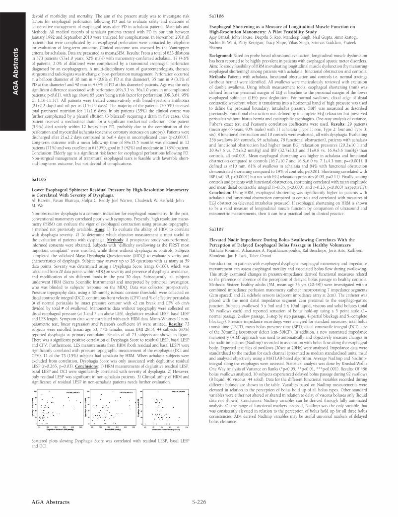
Non-obstructive dysphagia is a common indication for esophageal manometry. In the past,conventional manometry correlated poorly with symptoms. Presently, high resolution mano-metry (HRM) can evaluate the vigor of esophageal contraction using pressure topography,a method not previously available. Aims: 1) To evaluate the ability of HRM to correlatewith dysphagia severity. 2) To determine which objective measurement is most useful inthe evaluation of patients with dysphagia. Methods: A prospective study was performed;informed consents were obtained. Subjects with “difficulty swallowing as the FIRST mostimportant complaint” were enrolled, while those without dysphagia as controls. Subjectscompleted the validated Mayo Dysphagia Questionnaire (MDQ) to evaluate severity andcharacteristics of dysphagia. Subject may answer up to 28 questions with as many as 59data points. Severity was determined using a Dysphagia Score (range 0-100), which wascalculated from 20 data points within MDQ on severity and presence of dysphagia, avoidance,and modification of six different foods in the past 30 days. Subsequently, all subjectsunderwent HRM (Sierra Scientific Instruments) and interpreted by principal investigator,who was blinded to subjects' response on the MDQ. Data was collected prospectively.Pressure topography data, using a 30-mmHg isobaric contour threshold, were collected ondistal contractile integral (DCI), continuous front velocity (CFV) and % of effective peristalsis(# of normal peristalsis by intact pressure contour with <2 cm break and CFV <8 cm/sdivided by total # of swallows). Manometric data without topography were collected ondistal esophageal pressure (at 3 and 7 cm above LES), deglutitive residual LESP, basal LESPand LES length. Symptom data were correlated with each HRM data. Mann-Whitney U non-parametric test, linear regression and Pearson's coefficient (r) were utilized. Results: 73subjects were enrolled (mean age 53, 77% females, mean BMI 28.5); 44 subjects (60%)reported dysphagia as primary complaint. Results of all 73 subjects are shown in figure.There was a significant positive correlation of Dysphagia Score to residual LESP, basal LESPand CFV. Furthermore, LES measurements from HRM (both residual and basal LESP) weresignificantly correlated with pressure topographic measurement of the esophagus (DCI andCFV). 11 of the 73 (15%) subjects had achalasia by HRM. When achalasia subjects wereexcluded from correlation, Dysphagia Score was only associated with deglutitive residualLESP (r=0.265, p=0.03). Conclusions: 1) HRM measurements of deglutitive residual LESP,basal LESP and DCI were significantly correlated with severity of dysphagia. 2) However,only residual LESP was significant in non-achalasia patients. 3) Clinical utility of HRM andsignificance of residual LESP in non-achalasia patients needs further evaluation.

Scattered plots slowing Dysphagia Score was correlated with residual LESP, basal LESPand DCI.

S-226AGA Abstracts

Sa1106

Esophageal Shortening as a Measure of Longitudinal Muscle Function onHigh-Resolution Manometry: A Pilot Feasibility StudyAjay Bansal, John House, Deepthi S. Rao, Mandeep Singh, Neil Gupta, Amit Rastogi,Sachin B. Wani, Patty Kerrigan, Tracy Shipe, Vikas Singh, Srinivas Gaddam, PrateekSharma

Background: Based on probe based ultrasound evaluation, longitudinal muscle dysfunctionhas been reported to be highly prevalent in patients with esophageal spastic motor disorders.Aim: To study feasibility of HRM in evaluating longitudinal muscle dysfunction (by measuringesophageal shortening) among patients with achalasia, functional obstruction and controls.Methods: Patients with achalasia, functional obstruction and controls i.e. normal tracings(without hernia) were identified. All swallows were meticulously reviewed with exclusionof double swallows. Using inbuilt measurement tools, esophageal shortening (mm) wasdefined from the proximal margin of EGJ at baseline to the proximal margin of the loweresophageal sphincter (LES) post deglutition. For normal swallows, distal edge of distalcontractile wavefront where it transforms into a horizontal band of high pressure was usedto define the proximal boundary. Intrabolus pressure (IBP) was measured as describedpreviously. Functional obstruction was defined by incomplete EGJ relaxation but preservedperistalsis without hiatus hernia and eosinophilic esophagitis. One-way analysis of variance,Fisher's exact test and Pearson's correlation coefficients were used. Results: 29 patients(mean age 65 years, 90% males) with 11 achalasia (Type 1: one, Type 2: four and Type 3:six), 8 functional obstruction and 10 controls were evaluated, all with dysphagia. Evaluating256 swallows (84 controls, 94 achalasia, 78 functional obstruction), patients with achalasiaand functional obstruction had higher mean EGJ relaxation pressures (20.2±10.1 and20.9±7.6 vs. 7.5±3.2 mmHg) and IBP (32.7±13.2 and 31±9.8 vs. 16.9±3.6 mmHg) thancontrols, all p<0.001. Mean esophageal shortening was higher in achalasia and functionalobstruction compared to controls (16.7±10.7 and 16.8±6.0 vs. 7.1±4.3 mm; p=<0.001). Ifdefined as ≥10 mm, 61% of swallows in achalasia and 84% with functional obstructiondemonstrated shortening compared to 19% of controls, p<0.001. Shortening correlated withIBP (r=0.39, p<0.0001) but not with EGJ relaxation pressures (0.09, p=0.11). Finally, amongcontrols and patients with functional obstruction, shortening correlated with mean amplitudeand mean distal contractile integral (r=0.35, p<0.0001 and r=0.23, p<0.003) respectively).Conclusion: Using HRM, esophageal shortening was significantly higher in patients withachalasia and functional obstruction compared to controls and correlated with measures ofEGJ obstruction (elevated intrabolus pressure). If esophageal shortening on HRM is shownto be a valid measure of longitudinal muscle function by comparison of ultrasound andmanometric measurements, then it can be a practical tool in clinical practice.

Sa1107

Elevated Nadir Impedance During Bolus Swallowing Correlates With thePerception of Delayed Esophageal Bolus Passage in Healthy VolunteersNathalie Rommel, Athanasios A. Papathanasopoulos, Raf Bisschops, Joris Arts, KathleenBlondeau, Jan F. Tack, Taher Omari

Introduction: In patients with esophageal dysphagia, esophageal manometry and impedancemeasurement can assess esophageal motility and associated bolus flow during swallowing.This study examined changes in pressure-impedance derived functional measures relatedto the presence or absence of the perception of delayed bolus passage in healthy controls.Methods: Sixteen healthy adults (5M, mean age 33 yrs (20-48)) were investigated with acombined impedance perfusion manometry catheter incorporating 7 impedance segments(2cm spaced) and 22 sidehole sensors (adjacent impedance array at 2cm). The catheter wasplaced with the most distal impedance segment 2cm proximal to the esophago-gastricjunction. Subjects swallowed 5 x 5ml and 5 x 10ml liquid, viscous and solid boluses (total30 swallows each) and reported sensation of bolus hold-up using a 5 point scale (1=normal passage, 2=slow passage, 3=step by step passage, 4=partial blockage and 5=completeblockage). Pressure-impedance recordings were analysed for standard measures; total bolustransit time (TBTT), mean bolus presence time (BPT), distal contractile integral (DCI), sizeof the 30mmHg isocontour defect (cm<30ICP). In addition, a new automated impedancemanometry (AIM) approach was used to automatically and objectively measure changes inthe nadir impedance (NadImp) recorded in association with bolus flow along the esophagealbody. Exported text files of swallows (30sec at 20Hz) were analysed. Impedance data werestandardised to the median for each channel (presented as median standardised units, msu)and analysed objectively using a MATLAB-based algorithm. Average NadImp and NadImp-integral along the esophagus were assessed. Statistical analysis was done by Kruskal-WallisOne Way Analysis of Variance on Ranks (*p<0.05, **p<0.01, ***p<0.001). Results: Of 486bolus swallows analysed, 10 subjects experienced delayed bolus passage during 92 swallows(8 liquid, 40 viscous, 44 solid). Data for the different functional variables recorded duringdifferent boluses are shown in the table. Variables based on NadImp measurements wereelevated in relation to the perception of bolus hold up of all bolus types. Other standardvariables were either not altered or altered in relation to delay of viscous boluses only (liquiddata not shown). Conclusion: NadImp variables can be derived through fully automatedanalysis. Of the range of functional markers assessed, NadImp was the only variable thatwas consistently elevated in relation to the perception of bolus hold up for all three bolusconsistencies. AIM derived NadImp variables may be useful universal markers of delayedbolus clearance.