Embed Size (px)
Citation preview

ES145 - Systems Analysis & Physiology
Introduction to the Central Nervous System
Maurice Smith
10/18/2005
Goal: To explain behavior in terms of
neural activities in the brain.
How do the millions of nerve cells
collectively act to produce behavior?
How does our behavior and the
environment change the nerve cells?
Are particular mental functions localized
to a region of the brain?
If so, how does the anatomy and
physiology of one region relate to the
specific actions and behaviors that we
have?
Influences from six areas of science:
anatomy, embryology, physiology,
pharmacology, psychology,
mathematics.
Anatomy: the brain is composed of individual units of computation, called
neurons.
With the development of microscope, Golgi and then Cajal found a way to
stain neurons so that they could be seen. A silver solution, when put on a
region of the brain, would get picked up by only about 1% of the cells there, so
you could see a single neuron.
Brain is not a continuous web, but a network of discrete cells. Neurons are
the elementary signaling parts of the nervous system.
Embryology: Neurons have a common shape, a dendrite (input area) and an
axon (output area). During development, neurons move and axons grow to
make synapses with other neurons.
Physiology: Neurons produce electricity to send their messages along their
axon. They produce chemicals to send their messages to other neurons.
Pharmacology: Communication between neurons is via chemical
messengers. Chemicals can act on neurons through receptors that neurons
have on their outer membrane.
Psychology: Franz Gall proposed 3 radical ideas
• All behavior emanates from the brain.
• Particular regions of the brain control specific functions. He divided the
brain into 35 parts and assigned a specific mental function to each.
• The brain area associated with each function grows with use.
He focused on the external bumps on the skull.
Mathematics: How could seemingly identical units of computation (neurons)
give rise to such different functions such as perception, control of movement,
or motion? It is the connections of these units that matter.
From the different ways that simple elements that each compute the same
thing are connected, you can get different behaviors.
A Few Numbers:The human brain has about 100,000,000,000 (100 billion) neurons.
The adult human brain weighs about 3 pounds (1,300-1,400 g) - 2% of total body
weight.
The total surface area of the cerebral cortex is about 2500 sq. cm (~2.5 ft2)
Unconsciousness will occur after 8-10 seconds after loss of blood supply to the
brain.
Neurons multiply at a rate 4,000 neurons/sec during early pregnancy.
Neurons die at a rate or 85,000/day or 1 neuron/sec during adulthood.
There are 1,000 to 10,000 synapses for a "typical" neuron.
The cell bodies of neurons vary in diameter from 4 microns (granule cell) to 100
microns (motor neuron in spinal cord).
Total number of neurons in cerebral cortex = 10 billion
Total number of synapses in cerebral cortex = 60 trillion
Massive Convergence and Divergence of Information in the Brain.
Vision:
Number of retinal receptor cells = 5-6 million cones; 120-140 million rods
Number of fibers in optic nerve = 1,200,000
Number of cells in visual cortex (area 17) = 538,000,000
Hearing:
Number of hair cells in cochlea = 3,500 inner hair cells; 12,000 outer hair cells
Number of fibers in auditory nerve = 28,000-30,000
Number of neurons in cochlear nucleus = 8,800
Number of neurons in auditory cortex = 100,000,000
Neurons have four functional regions:
• Input component (dendrite)
• Trigger area (soma)
• Conductive component (axon)
• Output component (synapse) Cell body
Nucleus
Inhibitory
synapse
Apical
dendrites
Excitatory
synapse
basal
dendrites
Axon
hillock
axon
Node of Ranviermyelin
axon
Pre
synaptic
ce
ll
Po
sts
yn
ap
tic
ce
ll
Presynaptic terminal
Synaptic cleft
Postsynaptic dendrite
Kandel et al. (2000) Principles of Neural Science
Neurons
Neurons in different parts of the CNS are very similar in their properties. Yet
the brain has specialized function at each place.
The specialized function comes from the way that neurons are connected with
sensory receptors, with muscles, and with each other.
Kandel et al. (2000) Principles of Neural Science
Glia: support cells for neurons
• Produce myelin to insulate the nerve cell axon.
• Take up chemical transmitters released by neurons at the synapse.
• Form a lining around blood vessels: blood-brain barrier.
Kandel et al. (2000) Principles of Neural Science
Relative amounts of gray matter (cortex) and white matter
(connecting tracts of axons) across different species.
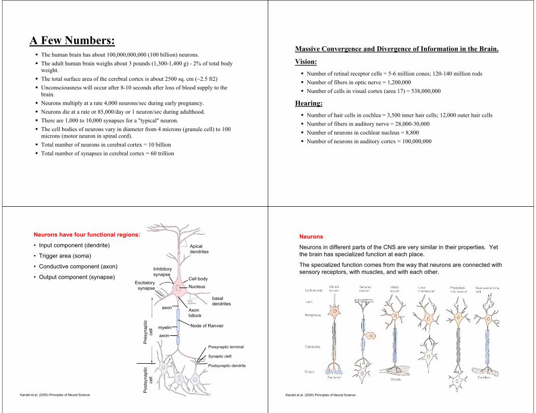
Injury in a peripheral nerve
When a peripheral nerve is cut, the
portion of the axon that was separated
from the cell body dies.
The glia cells that produce the myelin
sheath around the dying axon shrink,
but stay mostly in place.
As the cell body re-grows the axon, it
uses the path that is marked by the
glia cells.
In this way, the glia cells act as a road
map for the injured neuron to find its
previous destination.
Node of
Ranvier
injury
Myelin
sheath
Myelin
sheath
Cell body
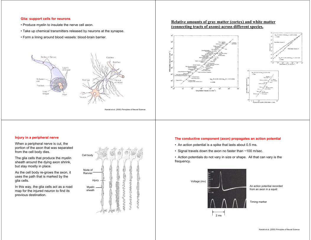
The conductive component (axon) propagates an action potential
• An action potential is a spike that lasts about 0.5 ms.
• Signal travels down the axon no faster than ~100 m/sec.
• Action potentials do not vary in size or shape. All that can vary is the
frequency.
Kandel et al. (2000) Principles of Neural Science
2 ms
An action potential recorded
from an axon in a squid
Timing marker
Voltage (mv)

Kandel et al. (2000) Principles of Neural Science
A simple neuronal circuit can produce behavior: stretch reflex
The sequence of signals that produce a reflex action.
Kandel et al. (2000) Principles of Neural Science

Modifiability of connections results in learning and adaptation
A neuron can produce only one kind of neurotransmitter at its synapse.
The post-synaptic neuron will have receptors for this neurotransmitter that
will either cause either an increase or decrease in membrane potential.
With repeated activation of pre- and post-synaptic neuron, their connection
via the synapse gets stronger.
Over the long-term, a neuron can grow and make more synapses or shrink
and prune its synapses.
The central nervous system is
divided into seven parts:
1. The spinal cord. Receives
and processes sensory
information from skin and
muscles; sends commands to
muscles; houses simple reflexes
and controls locomotion.
2. Medulla. Autonomic
function: digestion, breathing,
heart rate. Sleep function:
maintaining quiet.
3. Pons. Control of posture
and balance.
4. Cerebellum. Learning,
memory, and control of
movement.
Kandel et al. (2000) Principles of Neural Science
5. Midbrain. Eye movements.
6. Diencephalon:
• Thalamus. Nearly all
sensory information arrives
here first.
• Hypothalamus. Regulates
autonomic and endocrine
function.
7. Cerebral hemispheres.
• Hippocampus. Memory of
facts, events, places, faces,
etc.
• Basal ganglia. Control of
movement.
• Amygdala. Autonomic and
endocrine response in
emotional states.
• Cerebral cortex.
Kandel et al. (2000) Principles of Neural Science
Cerebral cortex:
• Frontal lobe. Planning of action and control of movement.
• Temporal lobe. Hearing. In its deep structures lies the
hippocampus, an important location for memory.
• Occipital lobe. Vision.
• Parietal lobe. Sense of position.

Sulci and Gyri
The surface of the brain develops crevasses and humps as it grows. We are
born with fairly smooth brains, and within 40 weeks get deeper sulci and
higher gyri.
axons pull densely
connected brain areas
together to form gyri
less dense connections
result in sulci
R. Carter (1998) Mapping the Mind R. Graham (xxxx) Physiological Psychology
R. Graham (xxxx) Physiological Psychology
dorsal
rostral caudalPrinciple of contralateral control
In 1870, it was discovered that when one electrically stimulates the cortex of
a dog, movements occur with the contralateral limb.
Hints of localization of function: stimulation in the frontal lobe most easily
and repeatedly produced limb movements.
Study of language as a window to localization of function in the brain.
1. A place in the brain that is critical for producing language.
Paul Broca thought that instead of studying bumps on the brain, maybe one
should look for specialization by finding if damage to a specific region of the
brain causes a discrete loss of function.
Broca found a patient who was unable to speak, but could understand. His
name was Tan. Tan could only say the word “Tan”. When asked what was his
name, he would say Tan. When asked if he was hungry, he would say Tan.
Tan could understand speech normally.
These patients had no problems moving their mouth or tongue. They could
whistle or sing a melody without difficulty. But they could not speak a
complete sentence, or write it down.
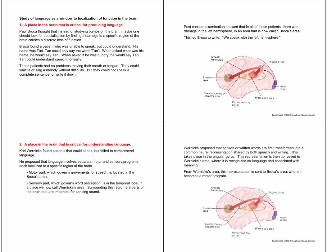
Post-mortem examination showed that in all of these patients, there was
damage in the left hemisphere, in an area that is now called Broca’s area.
This led Broca to write: “We speak with the left hemisphere.”
Kandel et al. (2000) Principles of Neural Science
2. A place in the brain that is critical for understanding language.
Karl Wernicke found patients that could speak, but failed to comprehend
language.
He proposed that language involves separate motor and sensory programs,
each localized to a specific region of the brain.
• Motor part, which governs movements for speech, is located in the
Broca’s area.
• Sensory part, which governs word perception, is in the temporal lobe, in
a place we now call Wernicke’s area. Surrounding this region are parts of
the brain that are important for sensing sound.
Wernicke proposed that spoken or written words are first transformed into a
common neural representation shared by both speech and writing. This
takes place in the angular gyrus. This representation is then conveyed to
Wernicke’s area, where it is recognized as language and associated with
meaning.
From Wernicke’s area, this representation is sent to Broca’s area, where it
becomes a motor program.
Kandel et al. (2000) Principles of Neural Science
Study of deaf people who lost their ability to use sign language.
Signing is also dependent on the left cerebral hemisphere.
Damage to the Broca’s area causes the deaf person to be unable to sign with
their hands.
Damage to the Wernicke’s area causes the deaf person to be unable to
comprehend sign language.
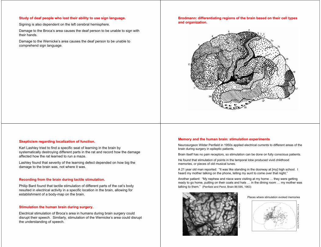
Brodmann: differentiating regions of the brain based on their cell types
and organization.
Skepticism regarding localization of function.
Karl Lashley tried to find a specific seat of learning in the brain by
systematically destroying different parts in the rat and record how the damage
affected how the rat learned to run a maze.
Lashley found that severity of the learning defect depended on how big the
damage to the brain was, not where it was.
Recording from the brain during tactile stimulation.
Philip Bard found that tactile stimulation of different parts of the cat’s body
resulted in electrical activity in a specific location in the brain, allowing for
establishment of a body-map on the brain.
Stimulation the human brain during surgery.
Electrical stimulation of Broca’s area in humans during brain surgery could
disrupt their speech. Similarly, stimulation of the Wernicke’s area could disrupt
the understanding of speech.
Memory and the human brain: stimulation experiments
Neurosurgeon Wilder Penfield in 1950s applied electrical currents to different areas of the
brain during surgery in epileptic patients.
Brain itself has no pain receptors, so stimulation can be done on fully conscious patients.
He found that stimulation of points in the temporal lobe produced vivid childhood
memories, or pieces of old musical tunes.
A 21 year old man reported: “It was like standing in the doorway at [my] high school. I
heard my mother talking on the phone, telling my aunt to come over that night.”
Another patient: “My nephew and niece were visiting at my home … they were getting
ready to go home, putting on their coats and hats … in the dining room … my mother was
talking to them.” (Penfield and Perot, Brain 86:595, 1963)
R. C
arte
r (19
98
) Ma
pp
ing
the
Min
d
R. C
arte
r (19
98
) Ma
pp
ing
the
Min
d
Places where stimulation evoked memories
Invention of functional imaging of the brain.
When neurons are active, they consume more energy. The vascular system
responds to the change in their activity by increasing the blood in the vessels
that are near these neurons.
By imaging the blood flow, one can make a rough estimate of where in the
brain things are more active than before.
PET: Positron Emission Tomography. A radioactive substance is injected into
the blood stream. Detectors estimate amount of blood flow at a given location
in the brain by the amount of radiation detected from there.
FMRI: functional magnetic resonance imaging. Strong magnetic fields are
used to detect amount of oxyhemoglobin in a particular region of the brain.
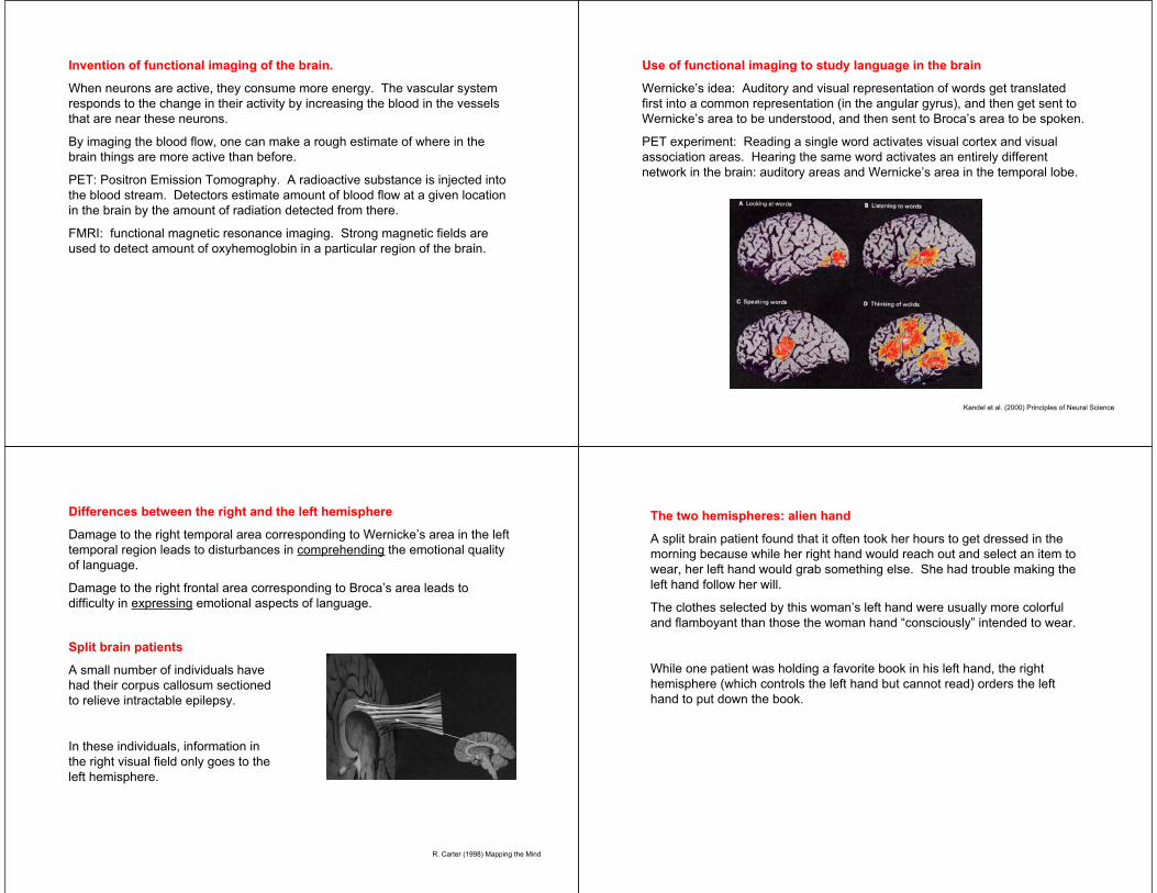
Use of functional imaging to study language in the brain
Wernicke’s idea: Auditory and visual representation of words get translated
first into a common representation (in the angular gyrus), and then get sent to
Wernicke’s area to be understood, and then sent to Broca’s area to be spoken.
PET experiment: Reading a single word activates visual cortex and visual
association areas. Hearing the same word activates an entirely different
network in the brain: auditory areas and Wernicke’s area in the temporal lobe.
Kandel et al. (2000) Principles of Neural Science
Differences between the right and the left hemisphere
Damage to the right temporal area corresponding to Wernicke’s area in the left
temporal region leads to disturbances in comprehending the emotional quality
of language.
Damage to the right frontal area corresponding to Broca’s area leads to
difficulty in expressing emotional aspects of language.
R. Carter (1998) Mapping the Mind
Split brain patients
A small number of individuals have
had their corpus callosum sectioned
to relieve intractable epilepsy.
In these individuals, information in
the right visual field only goes to the
left hemisphere.
The two hemispheres: alien hand
A split brain patient found that it often took her hours to get dressed in the
morning because while her right hand would reach out and select an item to
wear, her left hand would grab something else. She had trouble making the
left hand follow her will.
The clothes selected by this woman’s left hand were usually more colorful
and flamboyant than those the woman hand “consciously” intended to wear.
While one patient was holding a favorite book in his left hand, the right
hemisphere (which controls the left hand but cannot read) orders the left
hand to put down the book.

N.G., a woman with sectioned corpus
callosum
Experiment: she was asked to fixate her
eyes on a small dot displayed on a screen.
A pictures of a cup is briefly flashed to the
right of the dot. Because the image is to the
right, it falls on the left part of the retina of
both eyes, and goes to the left hemisphere.
The left hemisphere houses the language
centers.
She is now asked what did she see, and she
says “a cup”.
Now a picture of a spoon is shown to
the left of the dot. The picture goes to
the right hemisphere. She is asked
what she saw, and she says “nothing”.
She says this because in nearly
everyone, the language centers are in
the left hemisphere. Because the left
hemisphere has not been given the
visual information, it says that it has
seen nothing.
However, when N.G. is asked to reach
under a table with her left hand and
select, by touch only, from among a
group of concealed items the one that
was the same as the one she had just
seen, she picks a spoon. While she is
holding the spoon under the table, she
is asked what she is holding, she says
“a pencil”.
(R.W. Sperry 1968, American
Psychologist 23:723-733)
Summary
• The CNS is anatomically divided into seven regions.
• The brain has distinct functional regions.
• Language functions are usually found in the left hemisphere
• The right hemisphere influences emotional traits.
• Movements are controlled by the hemisphere contralateral to the limb.