Embed Size (px)
Citation preview
Erythrocyte Function and Marrow Regulation
in Hemoglobin Bethesda (0145 Histidine)
JOHNW. ADAMSON,AKIRA HAYASHI, GEORGESTAMATOYANNOPOULOS,andWmu3URF. BURGER
From the Hematology Service, Clinical Pathology Department, NationalInstitutes of Health, Bethesda, Maryland 20014, the Malcolm Grow U. S. AirForce Medical Center, Andrews Air Force Base, Washington, D. C. 20331,and the Divisions of Hematology and Medical Genetics, University ofWashington School of Medicine, Seattle, Washington 98195
A B S T R A C T Hemoglobin Bethesda (p145 histidine)is one of the two mutants known to affect the penulti-mate hemoglobin tyrosines. The result of this substitu-tion is extreme disorganization of the oxygenationfunction of the molecule. Red cells containing 45% HbBethesda and 55% Hb A have increased oxygen af-finity but, paradoxically, a normal Bohr effect. As isusually seen with other hemoglobins with increasedoxygen affinity, Hb Bethesda clinically is manifest inheterozygotes by erythrocytosis. Red cell production inaffected individuals is erythropoietin dependent. Thereciprocal interdependence of oxygen delivery and ef-fective erythropoiesis was documented by alterations inerythropoietin excretion, quantitative iron kinetics, andreticulocyte production in response to phlebotomy-in-duced reduction in the oxygen-carrying capacity.
INTRODUCTIONAt the present time a number of abnormal hemoglobinshave been detected because of their properties of abnor-mal oxygen affinity and the appearance of erythrocyto-sis in heterozygous carriers. The correlations betweenstructural abnormalities and the aberrant properties inmost of these hemoglobin variants have been clarified(1, 2) and the pathophysiological basis of the erythro-cytosis elucidated (3). This report describes the clinical
Dr. Adamson is the recipient of a Research Career De-velopment Award K04-AM70222-01 of the National In-stitutes of Health.
Dr. Hayashi's present address is the Third Departmentof Internal Medicine, Osaka University Hospital, Fuku-shima-Ku, Osaka, Japan.
Received for publication 25 April 1972 and in revisedform 30 June 1972.
presentation and studies of erythrocyte function anderythropoiesis in a family having one of these variants,Hb Bethesda (p145 histidine) (4).
METHODSHematologic and hemoglobin studies. All studies were
performed on b!ood less than 16 hr old at the time ofinitial processing. Routine hematologic studies were per-formed using standard techniques. Hemolysates were pre-pared by a modification of the method of Drabkin (5).Hemoglobin electrophoresis was performed on starch gelusing a Tris-EDTA-borate buffer system of pH 8.6 (3),Tris-HCl buffers at pH's 8.8 and 9.1, and phosphate buffersover a range of pH's from 6.0 to 7.2. Hemoglobin electro-phoresis on agar gel was performed using 0.05 M citratebuffer at pH 6.2 (3). Alkali-resistant hemoglobin was quan-titated by the techniques of Jonxis and Visser (6) andBetke, Marti, and Schlicht (7). Separation and quantitationof hemoglobin fractions were carried out on Amberlite CG-50 columns (Rohm & Haas Co., Philadelphia, Pa.) usingpotassium phosphate buffers at pH 7.0 and on DEAE-Sephadex using 0.05 M Tris-HCl buffer at pH 7.9.
Studies of oxygen-hemoglobin equilibria. The oxygen-hemoglobin dissociation curves and Hill's n of heparinizedwhole blood samples were determined using the mixing tech-niques of Lenfant, Ways, Aucutt, and Cruz (8). Oxygen-hemoglobin equilibria characteristics were also examinedin dilute red cell suspensions using a modification of themethod of Imai et al. (9). For these studies, 1 vol ofwashed red cells was diluted in 300 vol of isotonic phos-phate buffer solutions ranging in pH from 6.0 to 7.9. Con-tinuous oxygen dissociation curves were determined at 370Cand 600 mnu. The degree of hemolysis and proportion ofmethemoglobin were measured after each experiment andfound to be negligible. In addition, the 02 tension at 50%hemoglobin saturation was determined in red cell suspen-sions of hematocrit 27% using the gasometric method de-scribed by Van Slyke and Neill (10). Oxygen equilibriain hemoglobin solutions were determined as previously de-scribed (9, 11).
The Journal of Clinical Investigation Volume 51 November 1972 2883

1 2
1 2 3
1 ,/ 2 3 4
ffJ Hb Bethesda Q Not tested
0) Normal / Propositus0 Deceased
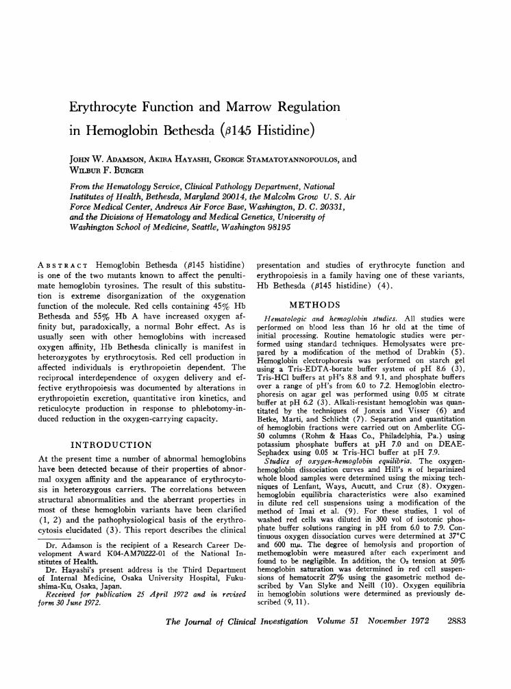
FIGURE 1 Pedigree of propositus (III-2) with Hb Bethesda.
Red cell levels of 2,3-diphosphoglycerate (DPG) 1 andATP were measured using a modification of the method ofBartlett (12).
Studies of the regulation of erythropoiesis. Total andeffective erythropoiesis, quantitated by iron kinetic studiesand serial reticulocyte counts, and the urinary excretion oferythropoietin (ESF) were monitored before and aftergraded phlebotomies designed to reduce the oxygen-carryingcapacity of the blood by approximately 15%. Hematocritsand reticulocyte counts were performed at least once daily.The reticulocyte number was first determined by screeningthe equivalent of 10' red cells using a Miller ocular (13)and then corrected for the concentration of red cells incirculation (14). The urine for ESF determination wascollected and processed by previously described means andassayed in ex-hypoxic polycythemic mice using an inter-national reference preparation (IRP) of urinary ESF asthe standard by which quantitation was achieved (15).Ferrokinetic studies were performed using '9Fe (as FeCls)bound to the transferrin in autologous plasma and injectedintravenously. The plasma iron turnover (PIT), marrowtransit time (MTT), and erythrocyte iron turnover (EIT)were determined as previously described (16).
RESULTSThe propositus. The propositus, III-2 (Fig. 1), a
23 year old Caucasian of northern European origins,was admitted to the Malcolm Grow Medical Center,Andrews Air Force Base, Washington, D. C., in March,1968, for evaluation of unexplained erythrocytosis. Pastmedical history was unremarkable. On physical exami-nation the propositus appeared healthy although pleth--oric. There were no abnormalities of the cardiovascularsystem and the spleen was not palpable. The remainderof the physical examination was entirely within normallimits. Routine hematologic examination revealed a per-sistently elevated hematocrit of 60-65%, normal leuko-cyte and platelet counts, and a normal absolute basophile
1 Abbreviations used in this paper: DPG, 2,3-diphospho-glycerate; EIT, erythrocyte iron turnover; ESF, erythro-poietin; IRP, international reference preparation; MTT,marrow transit time; PIT, plasma iron turnover.
count and leukocyte alkaline phosphatase value. Thesignificance of the elevated hematocrit. was confirmedby a standard measurement of red cell mass using '5Cr-labeled autologous red cells (49 cm5/kg). Arterial bloodgases, intravenous pyelography, chest X-ray, and elec-trocardiogram were within normal limits. An alkali de-naturation test revealed 0.6 g/100 ml resistant to alkali,within normal limits. To examine the possibility that afunctionally abnormal hemoglobin accounted for theerythrocytosis in this otherwise healthy individual, ameasurement of whole blood P5o ( Po2 in mmHg atwhich 50% of the hemoglobin exists in the oxy form)was made which gave a value of 11.5 mm Hg atstandard pH (7.4) and temperature (370C). Normalvalues for this technique (8) are 26.8+0.6 mmHg.
Electrophoretic and chromatographic studies. Hemo-globin electrophoresis on cellulose acetate at pH 8.6or on starch gels using buffers over a wide pH rangerevealed no abnormally migrating band. However, elec-trophoresis on agar gel using a citrate buffer (pH 6.2)disclosed an abnormally migrating band with cathodalmobility (Fig. 2). In addition, chromatography onAmberlite GC-50 (Rohmb & Haas Co.) disclosed thepresence of an abnormal hemoglobin comprising 46%of the total hemoglobin in solution (Fig. 3). This ab-normal fraction had the same electrophoretic and chro-matographic characteristics as Hb Rainier (3, 4) butwas clearly nonidentical because of its susceptibility toalkali denaturation. This finding prompted the detailedstructural studies resulting in the definition of HbBethesda (4).
Family studies. The family history was positive inthat the paternal grandmother was known to be poly-
A a3 F
AS
m-4 _
II - 2 .'
m-, l_
2- 1
-0 GFIGURE 2 Citrate-agar gel electrophoresis (pH 6.2) ofhemolysates with Hb Bethesda. An abnormal band withcathodal migration is seen in III-4 and II-2.
2884 J. W. Adamson, A. Hayashi, G. Stamatoyannopoulos, and W. F. Burger
Amobb..,

I0~ a0.2
414 0.1I
500 1 000 1500 2000 2500Effluent volume (ml)
FIGURE 3 Elution profile of a hemolysate containing Hb's Aand Bethesda. CG-50 Amberlite chromatography using a 2.5X 25 cm column at 50C. The flow rate was 25 ml/hr andfraction volume 8.0 ml. 430 mg of hemoglobin was appliedon the column. The first three chromatographic peaks, A3,Bethesdas, and A, were eluted with 0.1 M potassium phos-phate buffer in 10' M KCN (pH 7.0). The Bethesda peakwas eluted with 0.1 M potassium phosphate in 10'3 M KCNand 5 X 10' M NaCI (pH 7.0).
cythemic for many years. Siblings, parents, and otheravailable relatives were examined hematologically. Ery-throcytosis due to Hb Bethesda was found in threeadditional family members (Table I) spanning threegenerations (Fig. 1). There was no history of spon-taneous abortions in the affected females.
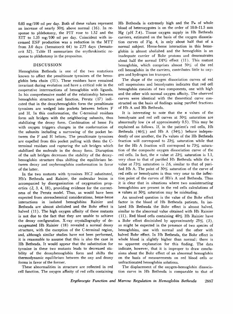
Red cell function. Oxygen-hemoglobin dissociationcurves measured in red cell suspensions with the auto-mated procedure are shown in Fig. 4. The curve ob-tained with cells from a normal sibling demonstratedthe usual sigmoid shape with P5o values of 24.2 mmHg (pH 7.4) and 35.0 mmHg (pH 7.0). In contrast,
TABLE IHematologic Values-Hb Bethesda Family
Hemo- HbSubject Age Sex Hct globin Bethesda* DPGt
yr g/100 mlI-2 77 F 56.5 19.6 +
II-1 55 M 49.0 17.0 -I I-2 50 M 52.0 17.8 +
III-1 25 F 44.0 ND§ -III-211 23 M 61.0 20.5 + 4.85111-3 21 M 45.0 16.8 - 4.69II1-4 19 F 50.0 ND +
* Documented by citrate-agar gel electrophoresis.t As micromoles per cubic centimeter packed red cells (normalrange, males: 4.840.3) (19).§ ND, Not determined.11 Propositus.
Po2(mmHg)
FIGURE 4 Continuous oxygen equilibrium curves of redcell suspensions. T1u ml volumes of washed red cells (hemato-crit 27%) were diluted to 7.0 ml in physiologic phosphatebuffer solutions, pH 7.4 and 7.0. Oxygen equilibria weredetermined at 370C and at 600 my (9). Curves 1 and 2:cells containing Hb's A and Bethesda at pH 7.4 and 7.0,respectively. Curves 3 and 4: normal cells at the same pH's.Symbols represent measurements obtained using the gaso-metric technique (10) with the patient's red cells in physio-logical phosphate buffer at pH 7.4 (U) and 7.0 (0).
the dissociation curves of the propositus were biphasic.Measured P5o's were 9.0 and 14.4 mmHg at pH 7.4and 7.0, respectively. The oxygen tensions determinedaround 50% saturation by the gasometric technique
pH
FIGURE 5 Comparison of the Bohr effect in normal (0)and Hb Bethesda (0) red cell suspensions. The Bohreffect was determined from the oxygen equilibrium curvesobtained under the conditions defined in Fig. 4 and overa pH range of 6.0-7.9 ("r" values were calculated at pH7.4).
Erythrocyte Function and Marrow Regulation in Hemoglobin Bethesda 2885

Po2 (mmHg)
FIGuRE 6 Oxygen equilibrium curves of (1) purified Hb Bethesda; (2) whole hemolysatecontaining Hb's A and Bethesda; (4) purified Hb A. The curves were obtained using theautomated method of Imai, et al. (9), with 0.2%o hemoglobin concentrations in phosphatebuffer at pH 7.0, 20'C, and at 600 mju. (3) is the curve constructed from data obtained in(1) and (4) and adjusted for a mixture containing 46%o Hb Bethesda and 54% Hb A.
were in the same range as those obtained by the auto-mated procedure (Fig. 4).
The Bohr effect factor in red cells containing HbBethesda was essentially normal (Fig. 5); "r" values(A log P5o/A pH) at pH 7.4 were 0.68 for the proposi-tus and 0.64 in normal controls.
The oxygen-hemoglobin dissociation curve at pH 7.0of the propositus's unfractionated lysate (Fig. 6) closelyresembled that obtained in red cell suspensions withthe exception that oxygen affinity was, as expected (9),greater in the lysate. A theoretical curve (Fig. 6) cal-culated on the basis of the oxygen dissociation charac-teristics of purified Hb's A and Bethesda, studied underidentical conditions, had a slope strikingly similar tothose of the hemolysate and the red cell suspension. Thefindings suggest that the dissociation curve is made upof two components having low (Hb A) and high (HbBethesda) oxygen affinity. The slope of the curve im-
t 10
8.2
t 6-
k,424-
tz
50 55Hematocrit
60
FIGURE 7 Erythropoietin excretion at various hematocritlevels for the propositus with Hb Bethesda.
plies that there is little interaction between the com-ponent hemoglobins.
Finally, measured erythrocyte DPG concentrationswere normal (Table I).
Studies of the regulation of erythropoiesis. BasalESF excretion in the prephlebotomy state averaged5.9 IRP U/day and ranged from 4.3 to 7.2 U. Afterthe initial phlebotomy, mean excretion rose to 6.7U/day (range: 4.6 to 9.0 U). After the second phle-botomy, at a hematocrit of 52.4, ESF excretion aver-aged 8.4 U/day (range: 6.4 to 12.3 U). Thus, as theoxygen-carrying capacity was reduced by 13%, averageESF excretion increased in a stepwise fashion (Fig. 7).Coincident with this was a rise in the mean reticulocytecount from a low of 0.7 to 1.7% (peak 2.2%), docu-menting an effective response to the phlebotomy. Asthe hematocrit was allowed to return toward prephle-botomy levels, the reticulocyte count fell.
Initial iron kinetic studies revealed a PIT of 0.94mg Fe/100 ml whole blood per day and an EIT of
TABLE I IErythrokinetic Response to Phlebotomy
Reticu-locyte ESF
Hct count excre-Subject N (mean) (mean) PIT* EIT* MTT tion
(days) (days) (IRPU/day)
Propositus 4 60.5 0.7 0.94 0.85 3.0 5.9t7 56.8 1.t - - - 6.79 52.4 1.7 1.52 1.35 2.75 8.4t
* Plasma iron turnover (PIT) and erythroid iron turnover (EIT) as milli-grams Fe/100 ml whole blood per day.$ Difference between groups significant with P < 0.05.
2886 J. W. Adamson, A. Hayashi, G. Stamatoyannopoulos, and W. F. Burger
1.0 10.0
0.85 mg/100 ml per day. Both of these values representan increase of nearly 50% above normal (16). In re-sponse to phlebotomy, the PIT rose to 1.52 and theEIT to 1.35 mg/100 ml per day. Coincident with in-creased ESF production was a reduction in the MTTfrom 3.0 days (hematocrit 64) to 2.75 days (hemato-crit 52). Table II summarizes the erythrokinetic re-sponse to phlebotomy in the propositus.
DISCUSSION
Hemoglobin Bethesda is one of the two mutationsknown to affect the penultimate tyrosines of the hemo-globin beta chain (11). These residues have remainedinvariant during evolution and have a critical role in thecooperative interreactions of hemoglobin with ligands.In his comprehensive model of the relationship betweenhemoglobin structure and function, Perutz (17) indi-cated that in the deoxyhemoglobin form the penultimatetyrosines are wedged into pockets between helices Fand H. In this conformation, the C-terminal residuesform salt bridges with the neighboring subunits, thusstabilizing the deoxy form. Combination of heme Fewith oxygen triggers changes in the conformation ofthe subunits including a narrowing of the pocket be-tween the F and H helices. The penultimate tyrosinesare expelled from this pocket pulling with them the C-terminal residues and rupturing the salt bridges whichstabilized the molecule in the deoxy form. Disruptionof the salt bridges decreases the stability of the deoxy-hemoglobin structure thus shifting the equilibrium be-tween deoxy and oxyhemi-oglobin conformation in favorof the latter.
In the two mutants with tyrosines HC2 substituted,Hb's Bethesda and Rainier, the molecular lesion isaccompanied by disorganization of oxygenation prop-erties (2, 3, 4, 18), providing evidence for the correct-ness of the Perutz model. Thus, as would have beenexpected from stereochemical considerations, heme-hemeinteractions in isolated hemoglobins Rainier andBethesda are almost abolished and the Bohr effect ishalved (11). The high oxygen affinity of these mutantsis not due to the fact that they are unable to achievethe deoxy configuration. X-ray crystallography of de-oxygenated Hb Rainier (18) revealed a normal deoxystructure, with the exception of the C-terminal region,and, although similar studies have not been performed,it is reasonable to assume that this is also the case inHb Bethesda. It would appear that the substitution fortyrosine in these two mutants leads to decreased sta-bility of the deoxyhemoglobin form and shifts thethermodynamic equilibrium between the oxy and deoxyforms in favor of the former.
These abnormalities in structure are reflected in redcell function. The oxygen affinity of red cells containing
Hb Bethesda is extremely high and the P55 of wholeblood of heterozygotes is on the order of 10.0-11.5 mmHg (pH 7.4). Tissue oxygen supply in Hb Bethesdacarriers, estimated on the basis of the oxygen dissocia-tion curves of Fig. 4, is only about half that of anormal subject. Heme-heme interaction in this hemo-globin is almost abolished and the hemoglobin is aninadequate carrier of Bohr protons and demonstratesabout half the normal DPG effect (11). This mutanthemoglobin, which comprises almost 50% of the redcell hemoglobin in the carriers, contributes little to oxy-gen and hydrogen ion transport.
The shape of the oxygen dissociation curves of redcell suspensions and henmolysates indicates that red cellhemoglobin consists of two components, one with highand the other with normal oxygen affinity. The observedcurves were identical with the theoretical curve con-structed on the basis of findings using purified fractionsof Hb A and Hb Bethesda.
It is interesting to note that the n values of thehemolysate and red cell curves at 50% saturation areabnormally low (n of approximately 0.5). This may beexplained as follows. If, in the patient's red cells, HbBethesda (46%) and Hb A (54%) behave indepen-dently of one another, the P50 values of the Hb Bethesdafraction will correspond to 23% saturation while thatfor the Hb A fraction will correspond to 73% satura-tion of the composite oxygen dissociation curve of thered cells. In fact, the nl value at 23% saturation is 1.1,very close to that of purified Hb Bethesda while the n
value at 73% saturation is 2.6, similar to that of puri-fied Hb A. The point of 50% saturation of the curve ofred cells or hemolysates is thus very near to the inflec-tion point of the curves of Hb's A and Bethesda. Thusit is clear that in situations where two noninteractinghemoglobins are present in the red cells calculations ofn values at 50% saturation may be misleading.
An unsolved question is the value of the Bohr effectfactor in the blood of Hb Bethesda patients. In iso-lated Hb Bethesda the Bohr effect is almost halved,similar to the abnormal value obtained with Hb Rainier(11). Red blood cells containing 40% Hb Rainier havea Bohr effect diminished by approximately 25% (3),as might be expected in the presence of two species ofhemoglobins, one with normal and the other withhalved Bohr effect. In Hb Bethesda, the Bohr effect inwhole blood is slightly higher than normal; there isno apparent explanation for this finding. The dataindicate, however, that it is improper to draw conclu-sions about the Bohr effect of an abnormal hemoglobinon the basis of measurements on red blood cells orunfractionated hemoglobin solutions.
The displacement of the oxygen-hemoglobin dissocia-tion curve in Hb Bethesda is comparable to that of
Erythrocyte Function and Marrow Regulation in Hemoglobin Bethesda 2887

Hb Rainier. However, the clinical manifestation ofthis functional abnormality, i.e. the appearance of eryth-rocytosis, is apparently dictated by more than simplythe position of the curve. Thus, there is a wide dis-crepancy between the hematocrit of the propositus andthat of his father, although both have similarly severedisplacement in the dissociation curve.
Clinically it is still uncertain whether the hypervis-cosity associated with hemoglobinopathic erythrocytosisconfers an increased risk of cardiovascular accident orother vascular complications. The affected family mem-bers with Hb Bethesda have been healthy and areasymptomatic. Reduction of the oxygen-carrying ca-pacity by 13% in the propositus was associated withno restriction in physical activity or loss of sense ofwell being. The final prospective analysis of the riskfactors associated with these abnormal hemoglobinswill only become evident over a period of years ofclinical observation.
The studies in this patient confirm the previous ob-servations of the ESF-dependence of erythropoiesis inthis setting (3). Thus, basal ESF excretion was in thehigh-normal range and increased appropriately in re-sponse to phlebotomy, demonstrating the oxygen de-pendence of ESF production. The increase in hormoneproduction was confirmed by the attendant increase inboth total (PIT) and effective (reticulocyte index)erythropoiesis and the shortening of the MTT by 0.25days. The reduction in MTT reflects the degree of pre-mature release of marrow reticulocytes and correlateswell with the degree of ESF stimulation and anemiain the normal subject (16). Thus, both a mean ESFexcretion of 8-10 U/day and MTT of 66 hr are com-patible with values obtained in normal man at a hemato-crit of 33-35 (15, 16).
ACKNOWLEDGMENTSWe are indebted to the Medical Research Council ofEngland for providing the erythropoietin IRP.
This investigation was supported in part by NationalInstitutes of Health grant No. GM-15253.
REFERENCES1. Perutz, M. F., and H. Lehmann. 1968. Molecular path-
ology of human hemoglobin. Nature (Lond.). 219: 902.2. Morimoto, H., H. Lehmann, and M. F. Perutz. 1971.
Molecular pathology of human haemoglobin: stereo-chemical interpretation of abnormal oxygen affinities.Nature (Lond.). 232: 408.
3. Adamson, J. W., J. T. Parer, and G. Stamatoyanno-poulos. 1969. Erythrocytosis associated with hemoglobinRainier: oxygen equilibria and marrow regulation. J.Clin. Invest. 48: 1376.
4. Hayashi, A., G. Stamatoyannopoulos, A. Yoshida, andJ. Adamson. 1971. Haemoglobin Rainier: p145 (HC2)Tyrosine -* Cysteine and Haemoglobin Bethesda: p145(HC2) Tyrosine -* Histidine. Nat. New Biol. 230: 264.
5. Drabkin, D. L. 1946. Spectrophotometric studies. XIV.The crystallographic and optical properties of the hemo-globin of man in comparison with those of other species.J. Biol. Chem. 164: 703.
6. Jonxis, J. H. P., and H. K. A. Visser. 1956. Determi-nation of low percentages of fetal hemoglobin in bloodof normal children. Am. J. Dis. Child. 92: 588.
7. Betke, K., H. R. Marti, and I. Schlicht. 1959. Estima-tion of small percentages of foetal haemoglobin. Nature(Lond.). 184: 1877.
8. Lenfant, C., P. Ways, C. Aucutt, and J. Cruz. 1969.Effect of chronic hypoxic hypoxia on the 02-Hb disso-ciation curve and respiratory gas transport in man.Respir. Physiol. 7: 7.
9. Imai, K., H. Morimoto, M. Kotani, H. Watari, W.Hirata, and M. Kuroda. 1970. Studies on the functionof abnormal hemoglobin. I. An improved method forthe automatic measurement of the oxygen equilibriumcurve of hemoglobins. Biochim. Biophys. Acta. 200: 189.
10. Van Slyke, D. D., and J. M. Neill. 1924. The determi-nation of gases in blood and other solutions by vacuumextraction and manometric measurement. J. Biol. Chem.61: 523.
11. Hayashi, A., and G. Stamatoyannopoulos. 1972. Role ofpenultimate tyrosine in haemoglobin 8 subunit. Nat.NewBiol. 235: 70.
12. Bartlett, G. R. 1959. Phosphorus assay in column chro-matography. J. Biol. Chem. 234: 466.
13. Brecher, G., and M. Schneiderman. 1950. A time-savingdevice for the counting of reticulocytes. Am. J. Clin.Pathol. 20: 1079.
14. Giblett, E. R., D. H. Coleman, G. Pirzio-Biroli, D. M.Donohue, A. G. Motulsky, and C. A. Finch. 1956.Erythrokinetics. Quantitative measurements of red cellproduction and destruction in normal subj ects and pa-tients with anemia. Blood. 11: 291.
15. Adamson, J. W. 1968. The erythropoietin/hematocritrelationship in normal and polycythemic man: impli-cations of marrow regulation. Blood. 32: 597.
16. Finch, C. A., K. Deubelbeiss, J. D. Cook, J. W. Esch-bach, L. A. Harker, D. D. Funk, G. Marsaglia, R. S.Hillman, S. Slichter, J. W. Adamson, A. Ganzoni, andE. R. Giblett. 1970. Ferrokinetics in man. Medicine(Baltimore). 49: 17.
17. Perutz, M. F. 1970. Stereochemistry of cooperativeeffects in haemoglobin. Nature (Lond.). 228: 726.
18. Greer, J., and M. F. Perutz. 1971. Three dimensionalstructure of Haemoglobin Rainier. Nat. New Biol. 230:261.
19. Torrance, J., P. Jacobs, A. Restrepo, J. Eschbach,C. Lenfant, and C. A. Finch. 1970. Intraerythrocyticadaptation to anemia. N. Engl. J. Med. 283: 165.
2888 J. W. Adamson, A. Hayashi, G. Stamatoyannopoulos, and W. F. Burger