Embed Size (px)
Citation preview
ERS statement on chronicthromboembolic pulmonary hypertension
Marion Delcroix 1,2,29, Adam Torbicki 3,27, Deepa Gopalan4,27, Olivier Sitbon 5,27,Frederikus A. Klok 6,27, Irene Lang7,27, David Jenkins8,27, Nick H. Kim9,27,Marc Humbert 5,27, Xavier Jais5,27, Anton Vonk Noordegraaf 10,27,Joanna Pepke-Zaba8,27, Philippe Brénot11, Peter Dorfmuller12,13,14, Elie Fadel15,Hossein-Ardeschir Ghofrani 12,13,14, Marius M. Hoeper 15, Pavel Jansa16,Michael Madani17, Hiromi Matsubara 18, Takeshi Ogo19, Ekkehard Grünig20,Andrea D’Armini21, Nazzareno Galie22, Bernhard Meyer23, Patrick Corkery24,30,Gergely Meszaros25,30, Eckhard Mayer26,28,29 and Gérald Simonneau5,28,29
@ERSpublicationsThe ERS statement on chronic thromboembolic pulmonary hypertension outlines a review of theliterature and current practice concerning diagnosis and management of CTEPH https://bit.ly/34dIjot
Cite this article as: Delcroix M, Torbicki A, Gopalan D, et al. ERS statement on chronic thromboembolicpulmonary hypertension. Eur Respir J 2021; 57: 2002828 [https://doi.org/10.1183/13993003.02828-2020].
ABSTRACT Chronic thromboembolic pulmonary hypertension (CTEPH) is a rare complication of acutepulmonary embolism, either symptomatic or not. The occlusion of proximal pulmonary arteries by fibroticintravascular material, in combination with a secondary microvasculopathy of vessels <500 µm, leads toincreased pulmonary vascular resistance and progressive right heart failure. The mechanism responsiblefor the transformation of red clots into fibrotic material remnants has not yet been elucidated. In patientswith pulmonary hypertension, the diagnosis is suspected when a ventilation/perfusion lung scan showsmismatched perfusion defects, and confirmed by right heart catheterisation and vascular imaging. Today,in addition to lifelong anticoagulation, treatment modalities include surgery, angioplasty and medicaltreatment according to the localisation and characteristics of the lesions.
This statement outlines a review of the literature and current practice concerning diagnosis andmanagement of CTEPH. It covers the definitions, diagnosis, epidemiology, follow-up after acutepulmonary embolism, pathophysiology, treatment by pulmonary endarterectomy, balloon pulmonaryangioplasty, drugs and their combination, rehabilitation and new lines of research in CTEPH.
It represents the first collaboration of the European Respiratory Society, the International CTEPHAssociation and the European Reference Network-Lung in the pulmonary hypertension domain. Thestatement summarises current knowledge, but does not make formal recommendations for clinicalpractice.
The institutions and patient representatives associated with affiliation numbers 1–5, 8, 10, 12–16 and 22–25 are part ofthe European Reference Network for rare lung diseases (ERN-Lung).
This ERS statement was endorsed by the ERS Executive Committee on 19 Nov 2020, by ERN-LUNG on 30 Nov 2020and by the International CTEPH Association (ICA) on 07 Dec 2020.
This article has supplementary material available from erj.ersjournals.com
Received: 17 July 2020 | Accepted: 5 Nov 2020
Copyright ©ERS 2021. For reproduction rights and permissions contact [email protected]
https://doi.org/10.1183/13993003.02828-2020 Eur Respir J 2021; 57: 2002828
ERS OFFICIAL DOCUMENTSERS STATEMENT
Scope of documentSince the publication of the 2015 European Society of Cardiology (ESC)/European Respiratory Society(ERS) guidelines for the diagnosis and treatment of pulmonary hypertension (PH) [1], considerableadvances have been made in the understanding, diagnosis and management of chronic thromboembolicpulmonary hypertension (CTEPH). Thus, the ERS considered a statement paper was required in order totake into account recent developments in the field. A multidisciplinary task force, with members from theERS, the International CTEPH Association (ICA) and the European Reference Network (ERN)-Lung,including specialists in pneumology, cardiology, cardiothoracic surgery, radiology and pathology, togetherwith patient representatives, defined and answered key questions related to the management of CTEPH.The task force searched for a clear definition; reviewed imaging techniques, epidemiology, follow-up ofacute pulmonary embolism (PE), microvasculopathy, indications of pulmonary endarterectomy (PEA),balloon pulmonary angioplasty (BPA) and medical treatment and the multimodality approach; anddefined research questions and priorities.
MethodsTask force chairs, supported by the ICA board members, compiled a list of 11 topics that they consideredimportant and relevant to the diagnosis and management of CTEPH. Task force members (27 experts andtwo patient representatives) were divided into subgroups targeting these topics (supplementary table S1).Each group identified three or four key focused questions relevant for their topic. These were discussed ina first face-to-face meeting in March 2019. Once the key questions were agreed, the subgroups preparedindividual subsections which were then presented and discussed in a second face-to-face meeting duringthe ERS congress in Madrid (September 2019) and subsequently revised until consent among allco-authors was reached (February 2020). All co-authors critically revised and approved the final statement(March 2020). The manuscript was finalised and submitted to the European Respiratory Journal afterapproval by the ERS Guidelines Committee (April–May 2020). The statement was presented at the ERSvirtual International Congress (September 2020).
The present ERS statement combines an evidence-based approach with the clinical expertise of the taskforce members, based on both literature search and discussions during face-to-face meetings. The taskforce members reviewed current knowledge and new scientific advances through identifying relevantindividual studies and reviews from systematic searches in MEDLINE (via PubMed) and Embase (viaOvid), performed by subgroup members, mostly with no date limits. The last search was performed inFebruary 2020; however, task force members also tracked any relevant citation which appeared beyond thisdate and until submission in April 2020. The search was limited to English literature with no datelimitations. Literature search was conducted using “chronic thromboembolic pulmonary hypertension”and the relevant key words for each respective section (supplementary table S2). Additionally, manualsearches of articles mentioned in the reference lists were performed.
Affiliations: 1Clinical Dept of Respiratory Diseases, Pulmonary Hypertension Center, UZ Leuven, Leuven,Belgium. 2BREATHE, Dept CHROMETA, KU Leuven, Leuven, Belgium. 3Dept of Pulmonary Circulation,Thrombo-embolic Diseases and Cardiology, Center of Postgraduate Medical Education, ECZ-Otwock, Otwock,Poland. 4Dept of Radiology, Imperial College Hospitals NHS Trusts, London, UK. 5Université Paris-Saclay;Inserm UMR_S 999, Service de Pneumologie, Hôpital Bicêtre (AP-HP), Le Kremlin-Bicêtre, France. 6Dept ofMedicine – Thrombosis and Hemostasis, Leiden University Medical Center, Leiden, The Netherlands. 7MedicalUniversity of Vienna, Vienna, Austria. 8Royal Papworth Hospital, Cambridge University Hospital, Cambridge,UK. 9Division of Pulmonary, Critical Care, and Sleep Medicine, University of California San Diego, La Jolla, CA,USA. 10Dept of Pulmonary Medicine, Amsterdam UMC, Vrije Universiteit Amsterdam, AmsterdamCardiovascular Sciences, Amsterdam, The Netherlands. 11Marie Lannelongue Hospital, Paris-SouthUniversity, Le Plessis Robinson, France. 12University of Giessen and Marburg Lung Center, German Center ofLung Research (DZL), Giessen, Germany. 13Dept of Medicine, Imperial College London, London, UK. 14Dept ofPneumology, Kerckhoff-Clinic Bad Nauheim, Bad Nauheim, Germany. 15Hannover Medical School, Hannover,Germany. 162nd Department of Medicine, Dept of Cardiovascular Medicine, First Faculty of Medicine, CharlesUniversity and General University Hospital in Prague, Prague, Czech Republic. 17Sulpizio CardiovascularCentre, University of California, San Diego, CA, USA. 18National Hospital Organization Okayama MedicalCenter, Okayama, Japan. 19National Cerebral and Cardiovascular Centre, Osaka, Japan. 20ThoraxklinikHeidelberg at Heidelberg University Hospital, Heidelberg, Germany. 21Unit of Cardiac Surgery, IntrathoracicTransplantation and Pulmonary Hypertension, University of Pavia School of Medicine, Foundation I.R.C.C.S.Policlinico San Matteo, Pavia, Italy. 22University of Bologna, Bologna, Italy. 23Institute for Diagnostic andInterventional Radiology, Hannover Medical School, Hannover, Germany. 24PHA Ireland, Dublin, Ireland. 25PHAEurope, Hungary. 26Dept of Thoracic Surgery, Kerckhoff Clinic Bad Nauheim, Bad Nauheim, Germany.27Section editors. 28Equal contribution. 29Co-chair. 30Patient representative.
Correspondence: Marion Delcroix, Dept of Respiratory Diseases, University Hospitals of Leuven, Herestraat49, 3000 Leuven, Belgium. E-mail [email protected]
https://doi.org/10.1183/13993003.02828-2020 2
ERS STATEMENT | M. DELCROIX ET AL.
BOX 1 Summary of statements
1 Definitions1.1 All patients in whom symptoms can be attributed to post-thrombotic deposits within pulmonary arteries could be considered to have
CTEPD with or without PH. CTEPH remains the preferred term in patients with PH.1.2 CPET and exercise-RHC could contribute to the definition of CTEPD helping to identify the main cause of exercise limitation in the
presence of comorbidities and in patients without resting PH.1.3 In many patients with CTEPH, resting mPAP is normalised by surgical or multimodality treatment and patients feel healthy, but it is
unlikely to return all pulmonary vessels back to normal.2 Diagnosis2.1 V′/Q′ scintigraphy remains the most effective screening tool to exclude CTEPD. V′/Q′ SPECT has been shown to be superior to planar
imaging and is the methodology of choice. Because the transition from planar imaging to SPECT may not be easy for clinicians unfamiliarwith the three-dimensional anatomy, two-dimensional planar images can be reprojected from SPECT without losing diagnostic accuracy.
2.2 Alternative perfusion techniques such as dual energy computed tomography and magnetic resonance perfusion have numeroustheoretical advantages over V′/Q′, but are more technically challenging and expensive, with limited availability and lacking multicentrevalidation. Hence, they are not replacing V′/Q′ in current clinical practice.
2.3 High-quality CTPA is adequate for diagnosis of proximal CTEPH, but a negative CTPA, even if high-quality, does not exclude CTEPH, asdistal disease can be missed.
2.4 Cone beam computed tomography and area detector computed tomography allow for more accurate visualisation of subsegmentalvasculature and have been shown to be useful for procedural guidance for BPA. The benefits of the technology require validation inprospective trials before recommendation for routine clinical use.
2.5 MRI is not yet fully integrated into the CTEPH diagnostic algorithm, because its use is heavily dependent on local practice and isroutinely implemented only in a few high-volume institutions where there is clustering of expertise.
3 Epidemiology3.1 CTEPH is a rare and underdiagnosed complication of PE and, upon first presentation, may be misclassified as acute PE.3.2 Persistent perfusion defects after acute PE are common, but have highly variable clinical relevance, ranging from completely
asymptomatic to established CTEPH.3.3 We have no evidence to suggest that CTEPD without PH is an early stage of CTEPH, because of a lack of data on its natural history.3.4 Current literature could not demonstrate any difference in CTEPH incidence in patients with venous thromoboembolism treated with
VKAs and NOACs, but rigorous studies have not been done.3.5 Screening for CTEPH in asymptomatic patients with specific risk factors could be effective, but is not supported by any data.
4 Follow-up after acute PE4.1 Earlier diagnosis of CTEPH may be relevant for patient outcomes. It can be achieved by targeting patients with acute PE with risk factors
for CTEPH or with radiological findings suggesting CTEPH.4.2 Patients at risk of CTEPH can be identified by accurate assessment of the CTPA images used to diagnose PE, individual risk factors for
CTEPH, and symptoms of functional limitations and/or right heart failure in the course of PE.4.3 Echocardiography is the test of choice in patients with suspected CTEPH. Other tests such as Leiden CTEPH rule-out criteria, V′/Q′ scan
or CPET could be used to exclude the presence of CTEPH and/or to establish an alternative diagnosis.4.4 The optimal timing for considering CTEPH in patients with PE is at the routine 3-month follow-up visit, but earlier work-up may be
necessary in highly symptomatic or deteriorating patients.5 Pathophysiology5.1 Current knowledge suggests that two types of vascular lesions exist in CTEPH: proximal fibrotic obstruction in large elastic pulmonary
arteries and a secondary microvasculopathy in pulmonary vessels <500 µm.5.2 Morphological delineation of pulmonary and bronchial circulation is possible with CTPA, while MRI permits both anatomical visualisation
and semi-quantitative analysis of the extent of bronchopulmonary shunting. None of these techniques can quantify the microvasculopathy.5.3 The right ventricle is less adapted in CTEPH than in idiopathic PAH, but recovers largely after successful PEA.
6 PEA6.1 Requirements for a PEA centre are based on expert opinion only, but ⩾50 cases as centre annual volume, and a properly trained surgeon
have been proposed.6.2 There is probably considerable anatomical overlap between BPA and PEA, but segmental and subsegmental disease are suitable for
surgery in expert, high-volume centres.6.3 Successful outcome after PEA is multifactorial and assumes in-hospital mortality <5%, survival of 90% at 3 years, improved functional
class and quality of life.6.4 Residual PH after PEA presents in ∼50% and represents a target for recently introduced medical therapies and BPA. There is some
evidence for their use if post-PEA mPAP remains >30 mmHg in the presence of symptoms.7 BPA7.1 Inoperable CTEPH patients can benefit from BPA. Optimal CTEPH treatment requires a multidisciplinary team approach considering
PEA, medical therapy and BPA.7.2 Long-term results after BPA are available out to 8 years after the procedure. Safety and efficacy of BPA correlate with centre experience.7.3 Vascular injury rather than reperfusion pulmonary oedema is the likely cause of any severity of lung injury after BPA.7.4 Similar to PEA, proper training in a high-volume centre is critical for BPA.
8 Medical treatment8.1 According to current guidelines, CTEPH patients should be treated with lifelong anticoagulation; VKAs are the mainstay of anticoagulant
treatment in CTEPH, but NOACs are increasingly used, with no safety issues reported yet. Antiphospholipid syndrome is acontraindication to NOACs.
Continued
https://doi.org/10.1183/13993003.02828-2020 3
ERS STATEMENT | M. DELCROIX ET AL.
All members of the task force disclosed their conflicts of interest before initiation of the project and uponsubmission of the manuscript.
1 Definitions1.1 Current definition: is there a need to change it in view of new diagnostic methods and theproposed reduced mean pulmonary arterial pressure threshold for PH? How to best definesymptomatic patients with post-thrombotic deposits but without PH at rest?Two terms are currently used to describe symptomatic patients with chronic thromboembolic occlusionsof pulmonary arteries according to the presence or the absence of PH at rest: chronic thromboembolicpulmonary hypertension (CTEPH) and chronic thromboembolic disease (CTED).
The definition provided in the 2015 ESC/ERS guidelines for the diagnosis and treatment of pulmonaryhypertension [1] stated that “The diagnosis of CTEPH is based on findings obtained after at least3 months of effective anticoagulation in order to discriminate this condition from ‘subacute’ PE. Thesefindings are mPAP ⩾25 mmHg with PAWP ⩽15 mmHg, mismatched perfusion defects on lung scan andspecific diagnostic signs for CTEPH seen by multidetector CTPA, MRI or conventional pulmonarycineangiography, such as ring-like stenoses, webs/slits and chronic total occlusions (pouch lesions ortapered lesions)”. More recently, cohorts of symptomatic patients, with exercise limitation, have beendescribed showing similar obstructive lesions as CTEPH but no resting PH [2, 3]. However, they maybenefit from the same treatment, including lifelong anticoagulation, PEA and BPA. The reporting authorsproposed the term “chronic thromboembolic disease (CTED)”, which is unclear because it does notspecify that it concerns pulmonary vessels.
The only difference between CTEPH and CTED is the presence or not of PH at rest. However, based onscientific evidence, a change in the definition of PH has been proposed with a decrease of the thresholdmean pulmonary arterial pressure (mPAP) from 25 to 21 mmHg, and in the definition of pre-capillary PH
Continued
8.2 Riociguat, an oral guanylate cyclase stimulator, and treprostinil, a subcutaneous prostacyclin analogue, are approved for patients withinoperable CTEPH or persistent/recurrent PH after PEA; other PH medications have been tested in CTEPH and are used off-label.
8.3 Oral combination therapy is a common practice in CTEPH patients with severe haemodynamic compromise.8.4 Withdrawal of PH medications is usually considered after successful BPA and/or PEA.
9 Multimodal treatment9.1 There is no consensus on eligibility criteria for multimodal therapy, which is dependent on the expertise and judgement of each individual
CTEPH centre. Patient selection for multimodal therapy is performed in expert centres through a multidisciplinary team approach.9.2 There is no convincing evidence that pre-operative pharmacological treatment is beneficial in patients with operable CTEPH.
Nevertheless, PH-targeted drugs as bridging therapy to PEA is sometimes considered in selected high-risk patients (with high pre-operative pulmonary vascular resistance) after multidisciplinary team assessment in expert centres.
9.3 The role of BPA as bridging therapy prior to PEA in patients with mixed surgically accessible and inaccessible lesions needs to beexplored. The potential benefit of hybrid procedures simultaneously combining PEA and BPA in such patients needs to be confirmed byfurther studies. There is insufficient evidence to support the use of BPA as a rescue procedure for early failure of PEA. Additional BPA,most often in combination with PH-targeted medical therapy, is common practice for patients with persistent symptomatic PH after PEA.
9.4 Most task force members use PH-targeted therapy prior to BPA to improve the safety of BPA, although there have been no clinical trialsinvestigating the benefit of this approach on the rate of BPA-related complications. Some data suggest that the combination ofPH-targeted therapy and BPA provides better results than medical therapy alone.
10 Rehabilitation10.1 Although exercise physiology is slightly different in CTEPH in comparison to PAH, it is reasonable to believe that the positive effects of
rehabilitation are similar for both conditions.10.2 Rehabilitation seems to be effective and safe in inoperable CTEPH patients.10.3 Although most of the pivotal trials on rehabilitation in CTEPH and PAH were based on in-hospital training in tertiary referral set-ups,
small nonrandomised studies have shown that home-based rehabilitation might also be an effective and safe option.10.4 A carefully monitored, low-dose rehabilitation programme of CTEPH patients after PEA or BPA might be considered standard of care.
11 Global research lines11.1 To achieve new genetic insights and basic science progress, international collaboration through biobanking should be established to
allow researchers access to the data which represents a large number of patients. Integration of clinical and -omic measurements willidentify clinically valuable diagnostic signatures and pathways for potential future interventions.
11.2 There is a need to address CTEPH in paediatric patients by a prospective global registry.
CTEPD: chronic thromboembolic pulmonary disease; PH: pulmonary hypertension; CTEPH: chronic thromboembolic pulmonary hypertension;CPET: cardiopulmonary exercise testing; RHC: right heart catheterisation; mPAP: mean pulmonary arterial pressure; V′/Q′: ventilation/perfusion; SPECT: single photon emission computed tomography; CTPA: computed tomography pulmonary angiography; BPA: balloonpulmonary angioplasty; MRI: magnetic resonance imaging; PE: pulmonary embolism; VKA: vitamin K antagonist; NOAC: non-vitamin Kantagonist oral anticoagulant; PAH: pulmonary arterial hypertension; PEA: pulmonary endarterectomy.
https://doi.org/10.1183/13993003.02828-2020 4
ERS STATEMENT | M. DELCROIX ET AL.
with a decrease of the threshold pulmonary vascular resistance (PVR) from 3 to 2 Wood units [4]. Theselowered thresholds, if adopted in upcoming guidelines, may influence the designation of former CTEDpatients as CTEPH patients [2, 5–9] (supplementary table S3).
To solve this terminology dilemma, a survey was conducted among the task force members. After collationof all propositions for names of the overarching disease and the subentities with or without PH, athree-round voting process was conducted until consensus was reached. For all symptomatic patients whopresent mismatched perfusion defects on ventilation/perfusion (V′/Q′) scan and specific signs of organisedfibrotic clots on computed tomography pulmonary angiography (CTPA), magnetic resonance imaging(MRI) or conventional pulmonary cineangiography, such as ring-like stenoses, webs/slits and chronic totalocclusions (pouch lesions or tapered lesions), after ⩾3 months of effective anticoagulation, the task forcechose the general term “chronic thromboembolic pulmonary disease (CTEPD)”. Some of these patientshave no PH at rest whatever the definition accepted. For the others, the term “chronic thromboembolicpulmonary hypertension (CTEPH)” was maintained. The presence of PH at rest in this setting is not onlythe consequence of proximal vessel obstruction by organised thrombi, but can also be related to asecondary microvasculopathy, as well as to the presence of underlying lung or left heart disease.
1.2 Should cardiopulmonary exercise testing and exercise-right heart catheterisation be includedin the definition and with what thresholds?Since symptoms in CTEPH appear during exercise, the tests performed at rest, including right heartcatheterisation (RHC), may lack sensitivity. Long-awaited recently formulated criteria for exercise PH arenot optimal for detection of “exercise CTEPH”, as they take into account total PVR (mPAP/cardiacoutput) and not PVR, ignoring pulmonary artery wedge pressure (PAWP) changes at exercise [10].However, reliable measurement of PAWP at peak exercise may be difficult due to large respiratory shifts inintrathoracic pressure. Much attention is given to cardiopulmonary exercise testing (CPET) performed notonly for prognostic purposes, but for differential diagnosis of dyspnoea in patients with suspectedpulmonary vascular disease [11–15]. The candidate CPET diagnostic parameters are reflecting inefficientventilation and include high ventilatory equivalent for carbon dioxide (minute ventilation/carbon dioxideproduction) slope and low end-tidal carbon dioxide tension, further decreasing at exercise [16]. Both arepromising for the differential diagnosis of symptomatic patients without or with only mild PH who haveimaging results that may suggest CTEPD. In addition, it is not clear whether CTEPH can be diagnosed inthe presence of suggestive intravascular changes at imaging but in patients who also have increased leftventricular (LV) filling pressures. Whether treatment removing pre-capillary obstruction would lead topulmonary congestion or, in contrast, would improve cardiorespiratory reserve by restoring flow throughventilated, but underperfused lung areas, remains unclear.
New imaging tools and increasing experience of radiologists in CTEPH referral centres make it possible toidentify patients with chronic thromboembolic obstructions clinically presenting as acute PE or withlong-standing progressive exercise limitation. The definition of CTEPH required 3 months ofanticoagulation. This is certainly still reasonable in haemodynamically stable patients and those whoclearly improve during initial anticoagulant therapy. However, in individual severely compromised caseswith significant contribution of unequivocal chronic intravascular changes, the diagnosis and specifictreatment for CTEPH could be considered by experienced CTEPH teams, regardless of the duration ofanticoagulant therapy.
1.3 Can a patient be cured of CTEPH?Despite optimal, and apparently successful, surgical, interventional and medical treatment, pulmonarycirculation does not return entirely to normal, which can be seen at V′/Q′ scans performed duringfollow-up. Thus, treatment may make patients with CTEPH feel healthy, but is unlikely to completely cureall the affected pulmonary vessels: arteries, veins and systemic collaterals [17, 18]. Furthermore, thepossibility to restore normal life expectancy is somewhat limited by potential complications related tolifelong anticoagulation.
Statements: definitions1.1 All patients in whom symptoms can be attributed to post-thrombotic deposits within pulmonary
arteries could be considered to have CTEPD with or without PH. CTEPH remains the preferred termin patients with PH.
1.2 CPET and exercise-RHC could contribute to the definition of CTEPD helping to identify the maincause of exercise limitation in the presence of comorbidities and in patients without resting PH.
1.3 In many patients with CTEPH, resting mPAP is normalised by surgical or multimodality treatmentand patients feel healthy, but it is unlikely to return all pulmonary vessels back to normal.
https://doi.org/10.1183/13993003.02828-2020 5
ERS STATEMENT | M. DELCROIX ET AL.
Proposal for future research in CTEPH definitiona) Both exercise-RHC as well as CPET still require establishment and validation of thresholds which could
be incorporated into CTEPD definitionb) Research on definition of CTPED and CTEPH in patients with concomitant significant left heart and/
or lung disease
2 DiagnosisIn patients with PH, the diagnosis of CTEPH is suspected when V′/Q′ scanning shows mismatchedperfusion defects, and is confirmed by RHC and pulmonary vascular imaging [1, 19, 20]. Consideringmajor advances in imaging technologies, the following aspects were discussed.
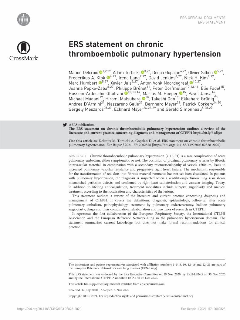
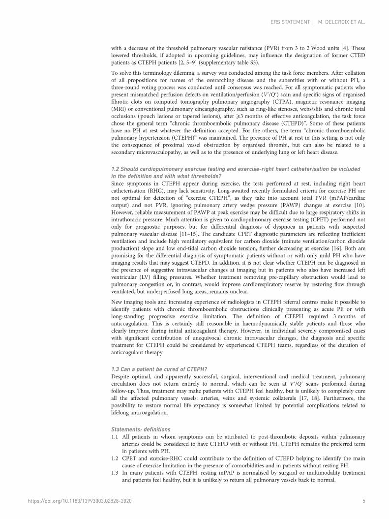
2.1 Should single photon emission computed tomography replace planar V′/Q′?V′/Q′ scintigraphy is consistently acknowledged to be the most effective screening tool in the CTEPHdiagnostic algorithm [1]. There is an emerging trend in clinical practice to replace planar V′/Q′ with singlephoton emission computed tomography (SPECT) V′/Q′. While most of the systematic reviews andmeta-analyses professing the superiority of SPECT over planar imaging are based on acute PE [21–23], thehigher sensitivity of V′/Q′ SPECT for CTEPH detection has also been demonstrated [24]. V′/Q′ SPECTimparts lower doses of isotopes per correct diagnosis [22]. The addition of low-dose computedtomography (CT) to V′/Q′ SPECT improves specificity by identifying concomitant parenchymal lungdisease [25]. SPECT quantitation can provide a measure of CTEPH disease severity [26]. Based on thereview of the available literature [23–28] as well as clinical practice (supplementary table S4), most taskforce members use V′/Q′ SPECT as the methodology of choice where possible, with adherence to thesimplified dichotomous interpretation criteria. The authors acknowledge that the transition from planarimaging to SPECT may not be easy, particularly for clinicians who may not be familiar with thethree-dimensional anatomy. This could be overcome by generating two-dimensional planar imagesreprojected from SPECT data without losing diagnostic accuracy [27] (figure 1).
2.2 Are dual-energy computed tomography or MR perfusion practical alternatives to V′/Q′ forCTEPH diagnosis?Over the past decade since its introduction, the role of dual-energy computed tomography (DECT) inCTEPH has evolved in its ability to detect parenchymal arterial perfusion and measure pulmonaryvascular reserve. DECT iodine maps visualise parenchymal iodine distribution and are surrogate markers
Anterior
a)
i)
ii)
i)
ii)
b)
Posterior
Coronal Axial Sagittal
Anteriorperfusion
99mTc-MAA lung perfusion
81mKr lung ventilation
Anteriorventilation
Posteriorperfusion
Posteriorventilation
RPOperfusion
RPOventilation
LPOperfusion
LPOventilation
FIGURE 1 Ventilation/perfusion (V′/Q′) scan. a) i) Anterior and posterior views from a planar V′/Q′ perfusion scan and ii) selected coronal, axial andsagittal perfusion images of the right lung from the corresponding single photon emission computed tomography (SPECT) show bilateralperfusion defects in a 60-year-old female with chronic thromboembolic pulmonary hypertension. Most of the left lung is nonperfused. Theperfusion defects in the right lung are much better delineated on the SPECT series compared to the planar images. b) Simulated planar imagesgenerated from the SPECT data in the same patient for i) perfusion and ii) ventilation. 99mTc: technetium-99m; MAA: macroagreggated albumin;RPO: right posterior; LPO: left posterior; 81mKr: krypton-81m.
https://doi.org/10.1183/13993003.02828-2020 6
ERS STATEMENT | M. DELCROIX ET AL.
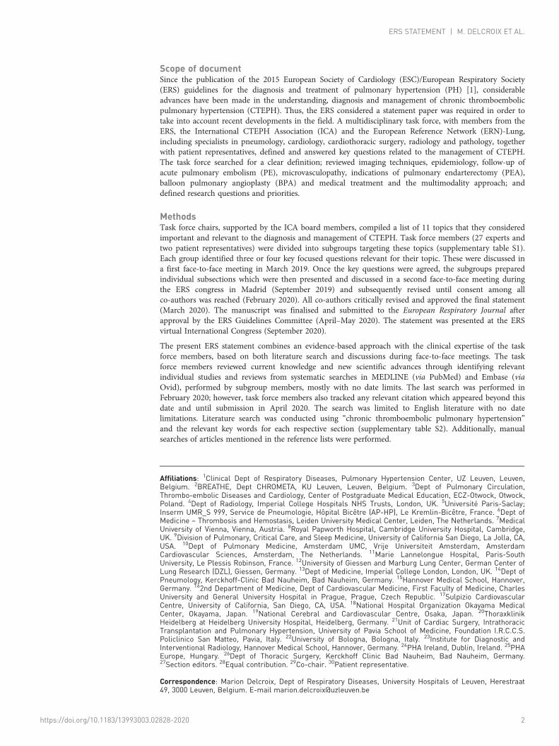
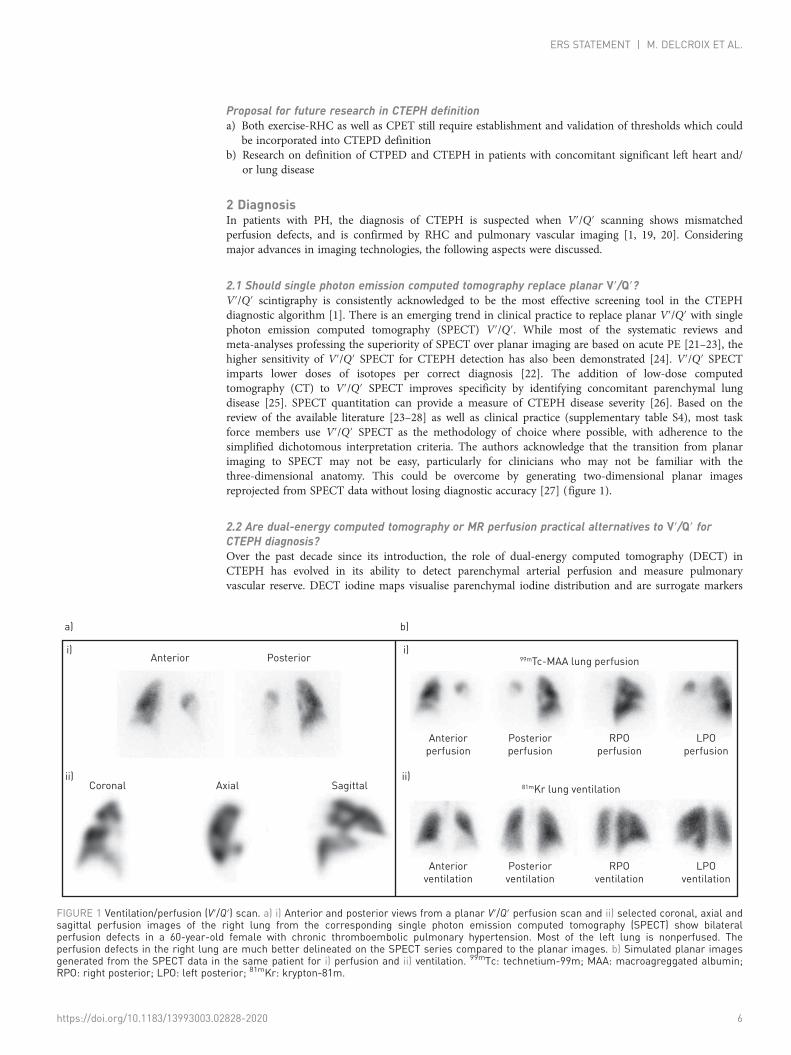
of lung perfusion [29]. DECT appearances in CTEPH is significantly different from pulmonary arterialhypertension (PAH) [30] (figure 2). DECT can be useful to differentiate acute PE and CTEPH based onthe attenuation value of emboli [31]. In addition, detection of distal CTEPH is improved as perfusiondefects are demonstrated even in the absence of visible morphological arterial abnormalities [32].Automated quantification of pulmonary perfused blood volume (PBV) maps can provide objectivemeasure of CTEPH severity [33, 34]. There are several small volume studies comparing PBV maps toplanar and V′/Q′ SPECT imaging with modest-to-good correlation [30, 35–38]. The lack of completeconcordance is not surprising, as the two techniques are not physiologically equivalent.
The task force members acknowledge the advantages of DECT and its ability to provide anatomical andfunctional information for CTEPH in a single test, but do not currently replace V′/Q′ as a screening testfor various reasons in their practice (supplementary table S4). The main limitation is the availability of CTsystems capable of performing DECT and the attendant cost implications. In addition, there is a lack ofstandardised protocols for acquisition of PBV images that are consistent and reproducible. Further studiesare required to overcome such limitations and validate the role of DECT in CTEPH.
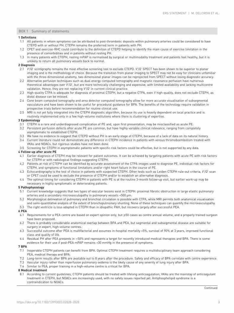
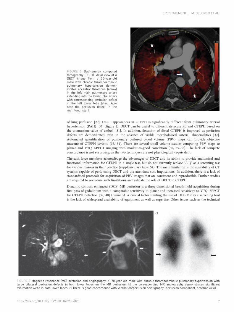
Dynamic contrast enhanced (DCE)-MR perfusion is a three-dimensional breath-hold acquisition duringfirst pass of gadolinium with a comparable sensitivity to planar and increased sensitivity to V′/Q′ SPECTfor CTEPH detection [39, 40] (figure 3). A crucial factor limiting the use of DCE-MR as a screening toolis the lack of widespread availability of equipment as well as expertise. Other issues such as the technical
FIGURE 2 Dual-energy computedtomography (DECT). Axial view of aDECT image from a 50-year-oldmale with chronic thromboembolicpulmonary hypertension demon-strates eccentric thrombus (arrow)in the left main pulmonary arteryextending into the lower lobe arterywith corresponding perfusion defectin the left lower lobe (star). Alsonote the perfusion defect in theright lung (star).
a) b) c)
FIGURE 3 Magnetic resonance (MR) perfusion and angiography. a) 70-year-old male with chronic thromboembolic pulmonary hypertension withlarge bilateral perfusion defects in both lower lobes on the MR perfusion; b) the corresponding MR angiography demonstrates significanttrifurcation webs in both lower lobes. c) There is good concordance with ventilation/perfusion scintigraphy (perfusion component, anterior view).
https://doi.org/10.1183/13993003.02828-2020 7
ERS STATEMENT | M. DELCROIX ET AL.
demands and challenges involved in the evaluation of the lung parenchyma need to be surmounted beforeroutine implementation of MR as a single imaging test in CTEPH.
2.3 Is high-quality CTPA adequate for diagnosis or is there a need for digital subtractionangiography?Ongoing technological advances continue to refine the role of CT in the CTEPH diagnostic algorithm.Modern CTPA may be noninferior to V′/Q′ in diagnosing CTEPH [41], but a fundamental reservation isthat a negative CTPA cannot exclude CTEPH, as small vessel disease can be missed. CTPA technicalquality is a significant contributor to the variation in its usage, as is the inconsistency in CT interpretationin non-CTEPH expert centres [42]. A recent meta-analysis demonstrated that high-quality CT studies hada pooled sensitivity of 99% and specificity of 97% for CTEPH detection on a vessel-based analysis [43].Some high-volume CTEPH expert centres use CT data to provide a roadmap of the location and extent ofdisease for operability assessment.
Most task force members use a high-quality CTPA (supplementary table S5) as a good noninvasivesubstitute for diagnosis of proximal CTEPH. Additionally, experienced operators use CTPA for evaluationof suitability for PEA in selected cases where the disease distribution is proximal.
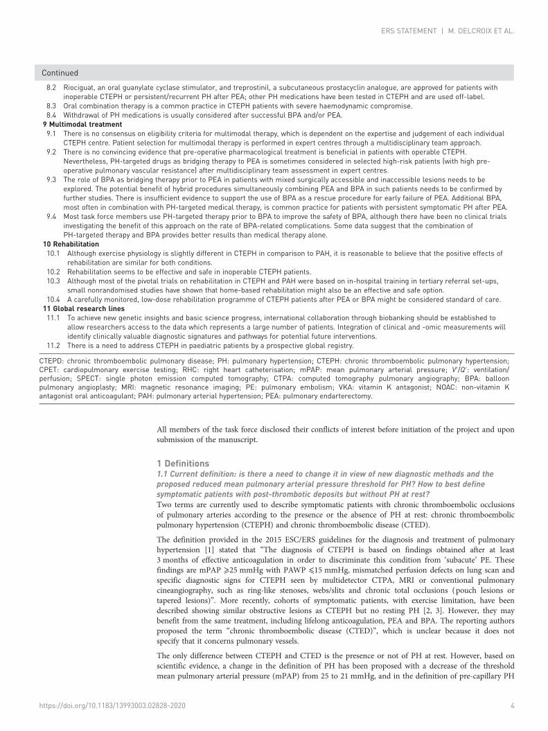
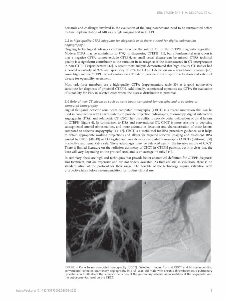
2.4 Role of new CT advances such as cone beam computed tomography and area detectorcomputed tomographyDigital flat-panel detector cone beam computed tomography (CBCT) is a recent innovation that can beused in conjunction with C-arm systems to provide projection radiography, fluoroscopy, digital subtractionangiography (DSA) and volumetric CT. CBCT has the ability to provide better delineation of distal lesionsin CTEPH (figure 4). In comparison to DSA and conventional CT, CBCT is more sensitive in depictingsubsegmental arterial abnormalities, and more accurate in detection and characterisation of these lesionscompared to selective angiography [44–47]. CBCT is a useful tool for BPA procedure guidance, as it helpsto obtain appropriate working projections and allows for targeted selective imaging and treatment. BPAguided by CBCT [48, 49] or ECG-gated and area detector computed tomography (ADCT) (320-row) [50]is effective and remarkably safe. These advantages must be balanced against the invasive nature of CBCT.There is limited literature on the radiation dosimetry of CBCT in CTEPH patients, but it is clear that thedose will vary depending on the protocol used and is on average ∼3 mSv [44].
In summary, these are high-end techniques that provide better anatomical definition for CTEPH diagnosisand treatment, but are expensive and are not widely available. As they are still in evolution, there is nostandardisation of the protocol for their usage. The benefits of the technology require validation withprospective trials before recommendation for routine clinical use.
b)a)
FIGURE 4 Cone beam computed tomography (CBCT). Selected images from a) CBCT and b) correspondingconventional catheter pulmonary angiography in a 45-year-old male with chronic thromboembolic pulmonaryhypertension to illustrate the superior depiction of the pulmonary arterial abnormalities at the segmental andthe subsegmental level on the CBCT.
https://doi.org/10.1183/13993003.02828-2020 8
ERS STATEMENT | M. DELCROIX ET AL.
2.5 What is the utility of cardiac MRI in the diagnostic algorithm?Although well suited for CTEPH diagnosis and follow-up, MRI is not yet fully integrated into the CTEPHdiagnostic algorithm. Its use is heavily dependent on local practice with routine implementation only in afew high-volume institutions where there is clustering of expertise.
MR angiography for diagnosisA comprehensive stepwise protocol of MR perfusion followed by high spatial resolution MR angiographyimproves the diagnostic performance of MR. It is essential to review the source images and usemultiplanar reformations to evaluate the complex vascular anatomy. MR angiography maximum intensityprojections (MIP) provide an overview of the pulmonary arterial tree in a format that is comparable toDSA.
Perfusion MRI has sensitivity of 97%, specificity 92%, positive predictive value 95% and negative predictivevalue 96% for detecting CTEPH, a performance comparable to V′/Q′ scintigraphy and CTPA [40]. In astudy comparing the efficacy of contrast-enhanced MR pulmonary angiography (CE-MRA), CTPA andDSA, CE-MRA demonstrated a sensitivity and specificity of 83.1% and 98.6% at the main/lobar level and87.7% and 98.1% at segmental level for diagnosing CTEPH, but was outperformed by DSA for depictionof subsegmental arteries [51]. Besides, CE-MRA was demonstrated to identify proximal and distal CTEPHwith a high sensitivity and specificity of 98% and 94%, respectively [52].
Right ventricular function in evaluating PH severity at the time of diagnosisCardiac MR is the reference standard for the assessment of right ventricle (RV) size and systolic function[53] and is well equipped to consistently provide accurate evaluation of RV function in the diagnosticwork-up and follow-up of patients with CTEPH. MRI has been used to noninvasively demonstrate thetechnical success of PEA as evidenced by the RV reverse remodelling and improvement in theinterventricular septal dyssynchrony and pulmonary clearance [54–56]. BPA-induced RV reverseremodelling has also been demonstrated using cardiac MR in patients with inoperable disease [57]. Lategadolinium enhancement (LGE) at the RV insertion points is strongly associated with elevated PAP, butits prevalence in CTEPH is not clearly defined, and its prognostic significance remains uncertain. In asubset of 39 patients with CTEPH from the ASPIRE registry [52], 97% had RV hinge point enhancement,leading the authors to conclude that its presence is highly sensitive for the detection of severe increases inRV afterload. Parametric mapping techniques have shown that native T1 values in the septum are elevatedin CTEPH even in the absence of LGE [58]. Larger trials are needed to understand the potential ofquantification of septal fibrosis as a prognostication and therapy-monitoring tool in the future.
Is noninvasive (MR-based metrics) measurement of haemodynamic parameters prime time? i.e. canwe replace RHC with MR-based data?Attempts to correlate MR-based haemodynamic parameters with RHC data have thus far met with limitedsuccess [52, 59–64]. Various multiparametric models have been introduced to estimate mPAP and PVRfrom cardiac MR data, but validation is challenged by the small number of comparable studies and paucityof prospective clinical cohorts. However, it is likely that advances such as strain imaging, parametricmapping and four-dimensional flow MR hold great potential in providing key metrics for CTEPHscreening and monitoring.
MRI has the potential to offer a noninvasive holistic approach to CTEPH diagnosis and follow-up, butthere is widespread lack of acceptance in the clinical community due to perceived as well as genuine issuesregarding access to the technique, bias about protracted acquisition times and lack of expertise ininterpretation. Multicentre prospective trials and focused training are required to make a paradigm shiftbefore MR can be routinely integrated into the CTEPH diagnostic algorithm.
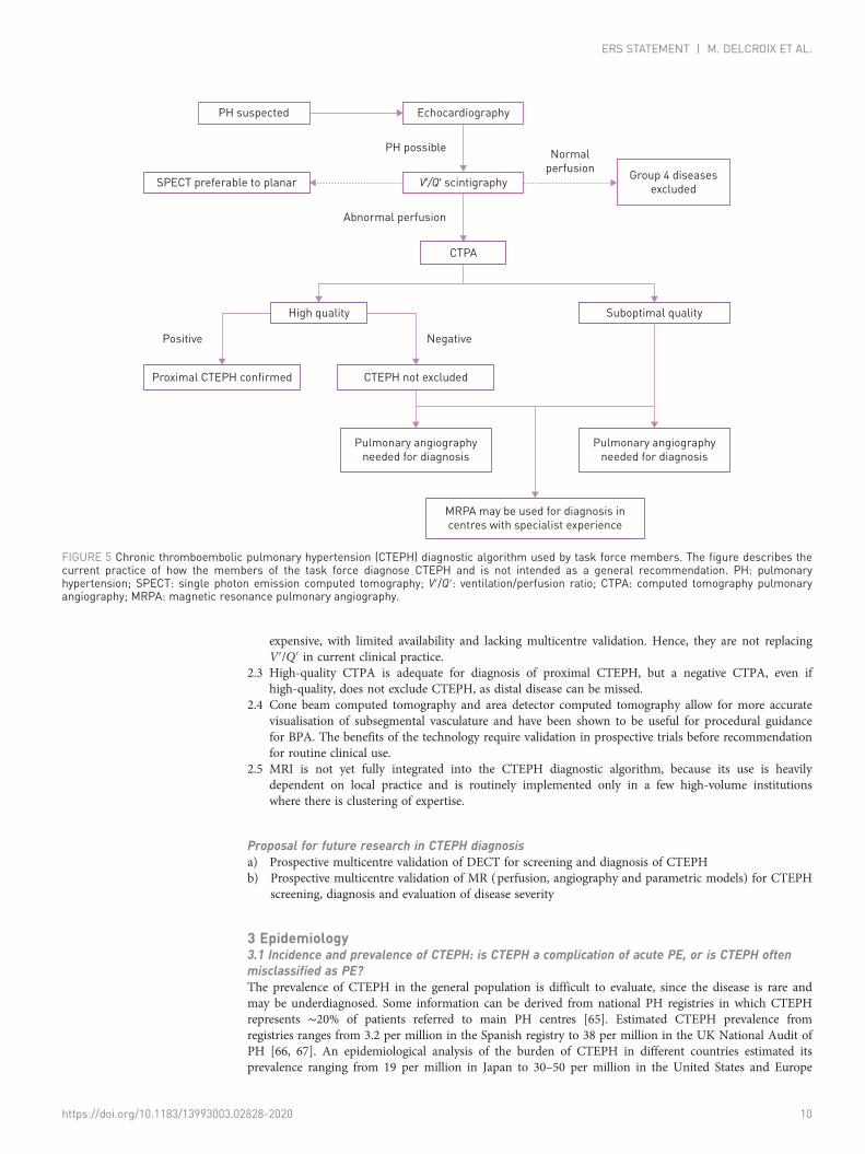
2.6 Diagnostic algorithmSummarising recent data, the diagnostic algorithm for CTEPH used by task force members in theirpractice is shown in figure 5.
Statements: diagnosis2.1 V′/Q′ scintigraphy remains the most effective screening tool to exclude CTEPD. V′/Q′ SPECT has
been shown to be superior to planar imaging and is the methodology of choice. Because the transitionfrom planar imaging to SPECT may not be easy for clinicians unfamiliar with the three-dimensionalanatomy, two-dimensional planar images can be reprojected from SPECT without losing diagnosticaccuracy.
2.2 Alternative perfusion techniques such as dual energy computed tomography and magnetic resonanceperfusion have numerous theoretical advantages over V′/Q′, but are more technically challenging and
https://doi.org/10.1183/13993003.02828-2020 9
ERS STATEMENT | M. DELCROIX ET AL.
expensive, with limited availability and lacking multicentre validation. Hence, they are not replacingV′/Q′ in current clinical practice.
2.3 High-quality CTPA is adequate for diagnosis of proximal CTEPH, but a negative CTPA, even ifhigh-quality, does not exclude CTEPH, as distal disease can be missed.
2.4 Cone beam computed tomography and area detector computed tomography allow for more accuratevisualisation of subsegmental vasculature and have been shown to be useful for procedural guidancefor BPA. The benefits of the technology require validation in prospective trials before recommendationfor routine clinical use.
2.5 MRI is not yet fully integrated into the CTEPH diagnostic algorithm, because its use is heavilydependent on local practice and is routinely implemented only in a few high-volume institutionswhere there is clustering of expertise.
Proposal for future research in CTEPH diagnosisa) Prospective multicentre validation of DECT for screening and diagnosis of CTEPHb) Prospective multicentre validation of MR (perfusion, angiography and parametric models) for CTEPH
screening, diagnosis and evaluation of disease severity
3 Epidemiology3.1 Incidence and prevalence of CTEPH: is CTEPH a complication of acute PE, or is CTEPH oftenmisclassified as PE?The prevalence of CTEPH in the general population is difficult to evaluate, since the disease is rare andmay be underdiagnosed. Some information can be derived from national PH registries in which CTEPHrepresents ∼20% of patients referred to main PH centres [65]. Estimated CTEPH prevalence fromregistries ranges from 3.2 per million in the Spanish registry to 38 per million in the UK National Audit ofPH [66, 67]. An epidemiological analysis of the burden of CTEPH in different countries estimated itsprevalence ranging from 19 per million in Japan to 30–50 per million in the United States and Europe
Abnormal perfusion
PH suspected
SPECT preferable to planar
High quality
Proximal CTEPH confirmed CTEPH not excluded
Pulmonary angiographyneeded for diagnosis
Pulmonary angiographyneeded for diagnosis
MRPA may be used for diagnosis incentres with specialist experience
Echocardiography
Normalperfusion
PH possible
NegativePositive
V’/Q’ scintigraphy
CTPA
Group 4 diseasesexcluded
Suboptimal quality
FIGURE 5 Chronic thromboembolic pulmonary hypertension (CTEPH) diagnostic algorithm used by task force members. The figure describes thecurrent practice of how the members of the task force diagnose CTEPH and is not intended as a general recommendation. PH: pulmonaryhypertension; SPECT: single photon emission computed tomography; V′/Q′: ventilation/perfusion ratio; CTPA: computed tomography pulmonaryangiography; MRPA: magnetic resonance pulmonary angiography.
https://doi.org/10.1183/13993003.02828-2020 10
ERS STATEMENT | M. DELCROIX ET AL.
[68]. A recent study estimated the prevalence of CTEPH based on a case ascertainment algorithm withinthe French exhaustive hospital discharge database (PMSI) [69]. Based on 3138 patients hospitalised forCTEPH in 2015 in France, and assuming that patients were hospitalised at least once a year, theprevalence of CTEPH was estimated 47 cases per million (range 43–50 cases per million).
CTEPH is usually considered as a complication of acute PE, with 50–75% of patients with CTEPH havinga documented history of acute PE [70–72]. In Japanese patients, only 15% have a history of acute PE [73],supporting the hypothesis of a different phenotype of CTEPH in the Japanese population, together withthe observed female preponderance, a lower proportion of coagulopathies [72] and less proximal thrombusand fresh red thrombus components in PEA specimens [74].
The cumulative incidence of CTEPH after acute PE is not exactly known and is reported as ranging from0.1% to 11.8% in the first 2 years after symptomatic PE [75–79]. This wide range could be explained byreferral bias, the paucity of early symptoms and the difficulty of differentiating acute PE symptoms frompre-existing CTEPH [77–79]. A systematic review and meta-analysis of studies including consecutive PEpatients followed for CTEPH (16 studies including 4047 PE patients who were followed-up for >2 years)showed a CTEPH incidence of 0.56% (95% CI 0.1–1.0%) in all comers. In survivors of PE and in survivorswithout major comorbidities, incidence of CTEPH was 3.2% (95% CI 2.0–4.4%), and 2.8% (95% CI 1.5–4.1%), respectively [79]. The authors suggested that the ∼3% incidences in the survivor categories may bemore relevant for daily clinical practice. They emphasised that studies that assessed CTEPH diagnosis bytests other than RHC provided an overestimated incidence of CTEPH, underscoring the need of anaccurate workup in patients who remain symptomatic after an acute PE. However, in a more recentmulticentre observational screening survey for the detection of CTEPH following PE (screening performedat 6, 12 and 24 months using a stepwise algorithm including dyspnoea phone-based survey,echocardiography, RHC and radiological confirmation of CTEPH), the CTEPH incidence was estimated as3.7 per 1000 patient-years with a 2-year cumulative incidence of 0.79% [80].
Finally, there are very strong arguments for frequent misclassification of CTEPH as PE [75, 77, 79]. In aprospective multicentre study conducted in three centres in France, 146 patients with an acute PE werefollowed-up for a median 26 months. Among the seven patients diagnosed with CTEPH (incidence 4.8%,95% CI 2.3–9.6%), echocardiography and CT performed at the time of the acute PE suggest that amajority of them had previously unknown PH, indicating that a first clinical presentation of CTEPH maymimic acute PE [77].
3.2 How many patients with persistent perfusion defects after acute PE have CTEPD?According to V′/Q′ scan studies, up to 50% of patients have persistent perfusion defects after an acute PE[81–83], with discrepant clinical relevance. Some studies reported an impact of residual obstruction onpulmonary haemodynamics, functional class, exercise capacity and outcome [83, 84], while the morerecent ELOPE study showed no correlation with exercise capacity [85]. Some of these “persistent perfusiondefects” could also be pre-existent to the acute PE, precluding any clear conclusion. Considering the“pyramid” of complications [65], CTEPD without PH probably occurs more often than CTEPH. So, if theprevalence of CTEPH after PE in survivors is 2–3% [79], the prevalence of CTEPD is probably higherthan that.
3.3 Is CTEPD without PH an early stage of CTEPH?There is no definitive answer to that question. Our limited knowledge of the natural history of CTEPH isbased on historical data prior to the current treatment era. Patients with mild PH (mPAP <30 mmHg) and“borderline PH” (mPAP 21–24 mmHg) were thought to have a good prognosis, with low probability ofprogressing to severe PH when treated solely with anticoagulation [86, 87], although these studies lackinformation on the severity of the thrombotic burden. According to clinical experience, some patientsremain stable while others experience a rapid unexplained deterioration. When focusing on CTEPDpatients without PH but extensive thromboembolic disease who underwent surgery, PEA specimens wereno different from CTEPH patients [2].
3.4 Could CTEPD incidence be different in patients treated with vitamin K antagonists andnon-vitamin K antagonist oral anticoagulants? Could it be different in patients treated withdifferent NOACs? Could the incidence be influenced by dosing strategy in NOACs?Current literature does not suggest any difference in CTEPD incidence in patients with venousthromoboembolism (VTE) treated with vitamin K antagonist (VKAs) and non-vitamin K antagonist oralanticoagulants (NOACs). Previous studies have shown that persistent perfusion defects occur despiteadequate anticoagulation (mostly with VKA; in up to 50% of patients) [81–83]. In the literature onthrombus resolution in deep vein thrombosis, some industry-sponsored studies have suggested that
https://doi.org/10.1183/13993003.02828-2020 11
ERS STATEMENT | M. DELCROIX ET AL.
rivaroxaban may lead to better venous patency than VKA [88, 89]; however, more recent independentstudies did not confirm this [90, 91]. The fact that the risk of early recurrent VTE is no different betweenNOAC- versus VKA-treated patients in randomised controlled trials (RCTs) is a strong argument to saythat the choice of anticoagulant drug should not influence the risk of CTEPH. By preventing recurrentVTE, due to more patients getting lifelong treatment, it may lower the prevalence of CTEPH (half- orfull-dose NOAC has same efficacy in reducing the risk of recurrent VTE).
However, in some specific situations, treatment with NOACs (which is recommended by current ESCguidelines for diagnosis and treatment of acute PE in preference to VKA, due to their ease of use andbetter tolerability [92, 93]) could be associated with higher risk of recurrent thromboembolic events thanVKA therapy (recurrent PE is a risk factor associated with CTEPH [65]). It was confirmed in patients withhigh-risk antiphospholipid syndrome (lupus anticoagulant, anticardiolipin antibodies, andanti-β2-glycoprotein I antibodies) [94]. In other specific clinical situations (e.g. obesity with a body massindex >40 kg·m−2 or a body weight >120 kg, concomitant medication with proton pump inhibitors or H2-antagonists which can reduce gastrointestinal absorption of dabigatran) pharmacokinetic aspects ofNOACs have to be carefully taken into account to optimise the risk–benefit profile of these drugs inprevention of recurrent thromboembolic events and perhaps CTEPD [95, 96].
3.5 Should the incidence and prevalence of CTEPD be analysed in specific subpopulations? Couldthis information be useful for the screening recommendation?Clinical conditions such as permanent intravascular devices (pacemaker, infusion chamber, ventroarticularshunts), inflammatory bowel diseases and essential thrombocythaemia have been identified as risk factorsfor CTEPH [97, 98]. Prevalence of CTEPH in this population of patients is not sufficiently described [68],precluding recommendations for screening in nonsymptomatic patients.
Statements: epidemiology3.1 CTEPH is a rare and underdiagnosed complication of PE and, upon first presentation, may be
misclassified as acute PE.3.2 Persistent perfusion defects after acute PE are common, but have highly variable clinical relevance,
ranging from completely asymptomatic to established CTEPH.3.3 We have no evidence to suggest that CTEPD without PH is an early stage of CTEPH, because of a
lack of data on its natural history.3.4 Current literature could not demonstrate any difference in CTEPH incidence in patients with venous
thromoboembolism treated with VKAs and NOACs, but rigorous studies have not been done.3.5 Screening for CTEPH in asymptomatic patients with specific risk factors could be effective, but is not
supported by any data.
4 Follow-up after acute PE4.1 Is earlier detection of CTEPH relevant for outcome?In daily practice, it may take 14–24 months from symptom onset for CTEPH to be diagnosed [71, 99–101]. Although this diagnostic delay was not associated with operability in the European CTEPH registry,longer delay was shown to correlate to higher systolic pulmonary arterial pressure (sPAP) at diagnosis andincreased risk of death [71, 99, 102]. Even in the relatively short time elapsed between diagnosis ofCTEPH and referral to a surgical centre (mean 4.4 months), it was observed that cardiac output decreasedand mPAP increased slightly [103]. In addition, progressive increases of PAP and PVR were observed 1–15 years after diagnosis in patients with initial mPAP >30 mmHg [86]. This can be attributed to thenatural course of CTEPH involving the development of a secondary microvasculopathy, further discussedin section 5, and confirms that a timely diagnosis of CTEPH is important.
4.2 How can a patient at high risk of CTEPH after acute PE be identified, and how can we betteridentify patients who already have CTEPH while presenting with what appears to be acute PE?CTEPH is usually considered at two time points: 1) when presenting with acute PE, if radiological signssuggest CTEPH on the CTPA performed to diagnose PE (figures 6 and 7 and supplementary table S6),and/or if estimated sPAP >60 mmHg [77, 104–107]; and 2) more classically, when dyspnoea or functionallimitations persist in the clinical course of PE [108–111]. A third condition could concern asymptomaticpatients with risk factors for CTEPH or a high pre-test probability [112] (supplementary table S7).
Clearly, the presence of CTEPH characteristics on a CTPA performed in the setting of acute PE promptsthe suspicion of CTEPH as primary diagnosis. CTPA signs of CTEPH include eccentric wall-adherentfilling defect, abrupt tapering and truncation, complete occlusion and pouch defects, intimal irregularity,intravascular webs and bands, stenosis, post-stenotic dilatation and vascular tortuosity [93]. It has been
https://doi.org/10.1183/13993003.02828-2020 12
ERS STATEMENT | M. DELCROIX ET AL.
shown that most patients with CTEPH after PE had several signs of CTEPH on the index CTPA,indicating acute-on-chronic events rather than acute PE [77, 106]. If evaluated by experts, the presence ofthree or more radiological characteristics of CTEPH on CTPA for suspected PE was virtually diagnostic(specificity 96%), although less-experienced radiologists may be less likely to recognise this [42, 106].
Notably, CTPA protocols are largely dependent on the available hardware and vary according to patientage, size, motion and cardiac function. Their fundamental goal is to achieve adequate pulmonary arterialenhancement such that thrombus can be distinguished from intraluminal contrast medium. The theoreticminimum intraluminal attenuation of blood required to visualise acute and chronic pulmonary
a) b)
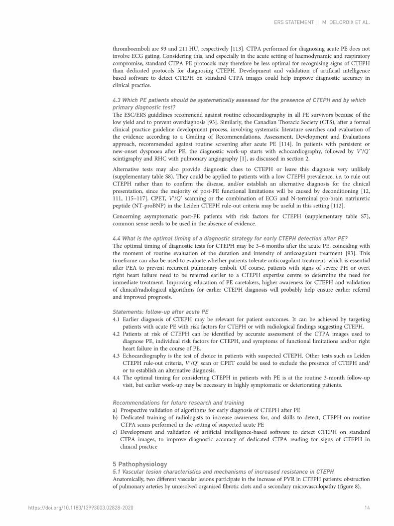
FIGURE 7 a) Coronal computed tomography pulmonary angiography (CTPA) reconstruction to show segmentalstenosis and post-stenotic dilatation in left lower lobe in a chronic thromboembolic pulmonary hypertensioncase (arrow); b) coronal CTPA lung window demonstrates mosaic attenuation.
a) b)
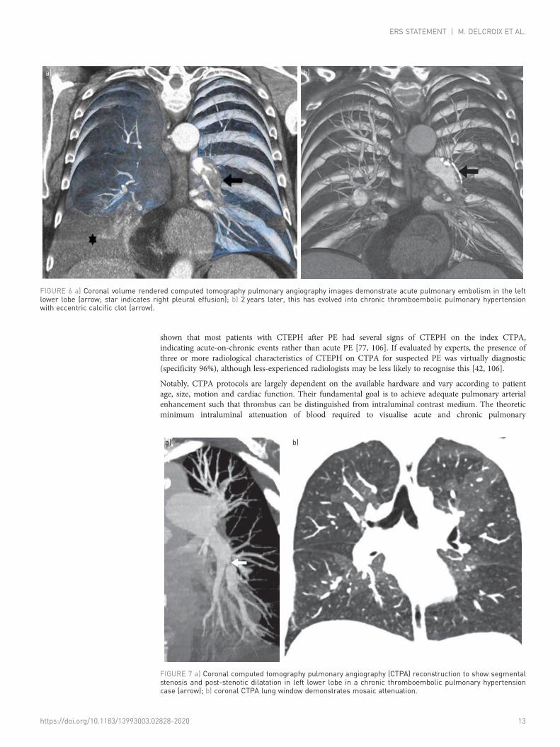
FIGURE 6 a) Coronal volume rendered computed tomography pulmonary angiography images demonstrate acute pulmonary embolism in the leftlower lobe (arrow; star indicates right pleural effusion); b) 2 years later, this has evolved into chronic thromboembolic pulmonary hypertensionwith eccentric calcific clot (arrow).
https://doi.org/10.1183/13993003.02828-2020 13
ERS STATEMENT | M. DELCROIX ET AL.
thromboemboli are 93 and 211 HU, respectively [113]. CTPA performed for diagnosing acute PE does notinvolve ECG gating. Considering this, and especially in the acute setting of haemodynamic and respiratorycompromise, standard CTPA PE protocols may therefore be less optimal for recognising signs of CTEPHthan dedicated protocols for diagnosing CTEPH. Development and validation of artificial intelligencebased software to detect CTEPH on standard CTPA images could help improve diagnostic accuracy inclinical practice.
4.3 Which PE patients should be systematically assessed for the presence of CTEPH and by whichprimary diagnostic test?The ESC/ERS guidelines recommend against routine echocardiography in all PE survivors because of thelow yield and to prevent overdiagnosis [93]. Similarly, the Canadian Thoracic Society (CTS), after a formalclinical practice guideline development process, involving systematic literature searches and evaluation ofthe evidence according to a Grading of Recommendations, Assessment, Development and Evaluationsapproach, recommended against routine screening after acute PE [114]. In patients with persistent ornew-onset dyspnoea after PE, the diagnostic work-up starts with echocardiography, followed by V′/Q′scintigraphy and RHC with pulmonary angiography [1], as discussed in section 2.
Alternative tests may also provide diagnostic clues to CTEPH or leave this diagnosis very unlikely(supplementary table S8). They could be applied to patients with a low CTEPH prevalence, i.e. to rule outCTEPH rather than to confirm the disease, and/or establish an alternative diagnosis for the clinicalpresentation, since the majority of post-PE functional limitations will be caused by deconditioning [12,111, 115–117]. CPET, V′/Q′ scanning or the combination of ECG and N-terminal pro-brain natriureticpeptide (NT-proBNP) in the Leiden CTEPH rule-out criteria may be useful in this setting [112].
Concerning asymptomatic post-PE patients with risk factors for CTEPH (supplementary table S7),common sense needs to be used in the absence of evidence.
4.4 What is the optimal timing of a diagnostic strategy for early CTEPH detection after PE?The optimal timing of diagnostic tests for CTEPH may be 3–6 months after the acute PE, coinciding withthe moment of routine evaluation of the duration and intensity of anticoagulant treatment [93]. Thistimeframe can also be used to evaluate whether patients tolerate anticoagulant treatment, which is essentialafter PEA to prevent recurrent pulmonary emboli. Of course, patients with signs of severe PH or overtright heart failure need to be referred earlier to a CTEPH expertise centre to determine the need forimmediate treatment. Improving education of PE caretakers, higher awareness for CTEPH and validationof clinical/radiological algorithms for earlier CTEPH diagnosis will probably help ensure earlier referraland improved prognosis.
Statements: follow-up after acute PE4.1 Earlier diagnosis of CTEPH may be relevant for patient outcomes. It can be achieved by targeting
patients with acute PE with risk factors for CTEPH or with radiological findings suggesting CTEPH.4.2 Patients at risk of CTEPH can be identified by accurate assessment of the CTPA images used to
diagnose PE, individual risk factors for CTEPH, and symptoms of functional limitations and/or rightheart failure in the course of PE.
4.3 Echocardiography is the test of choice in patients with suspected CTEPH. Other tests such as LeidenCTEPH rule-out criteria, V′/Q′ scan or CPET could be used to exclude the presence of CTEPH and/or to establish an alternative diagnosis.
4.4 The optimal timing for considering CTEPH in patients with PE is at the routine 3-month follow-upvisit, but earlier work-up may be necessary in highly symptomatic or deteriorating patients.
Recommendations for future research and traininga) Prospective validation of algorithms for early diagnosis of CTEPH after PEb) Dedicated training of radiologists to increase awareness for, and skills to detect, CTEPH on routine
CTPA scans performed in the setting of suspected acute PEc) Development and validation of artificial intelligence-based software to detect CTEPH on standard
CTPA images, to improve diagnostic accuracy of dedicated CTPA reading for signs of CTEPH inclinical practice
5 Pathophysiology5.1 Vascular lesion characteristics and mechanisms of increased resistance in CTEPHAnatomically, two different vascular lesions participate in the increase of PVR in CTEPH patients: obstructionof pulmonary arteries by unresolved organised fibrotic clots and a secondary microvasculopathy (figure 8).
https://doi.org/10.1183/13993003.02828-2020 14
ERS STATEMENT | M. DELCROIX ET AL.
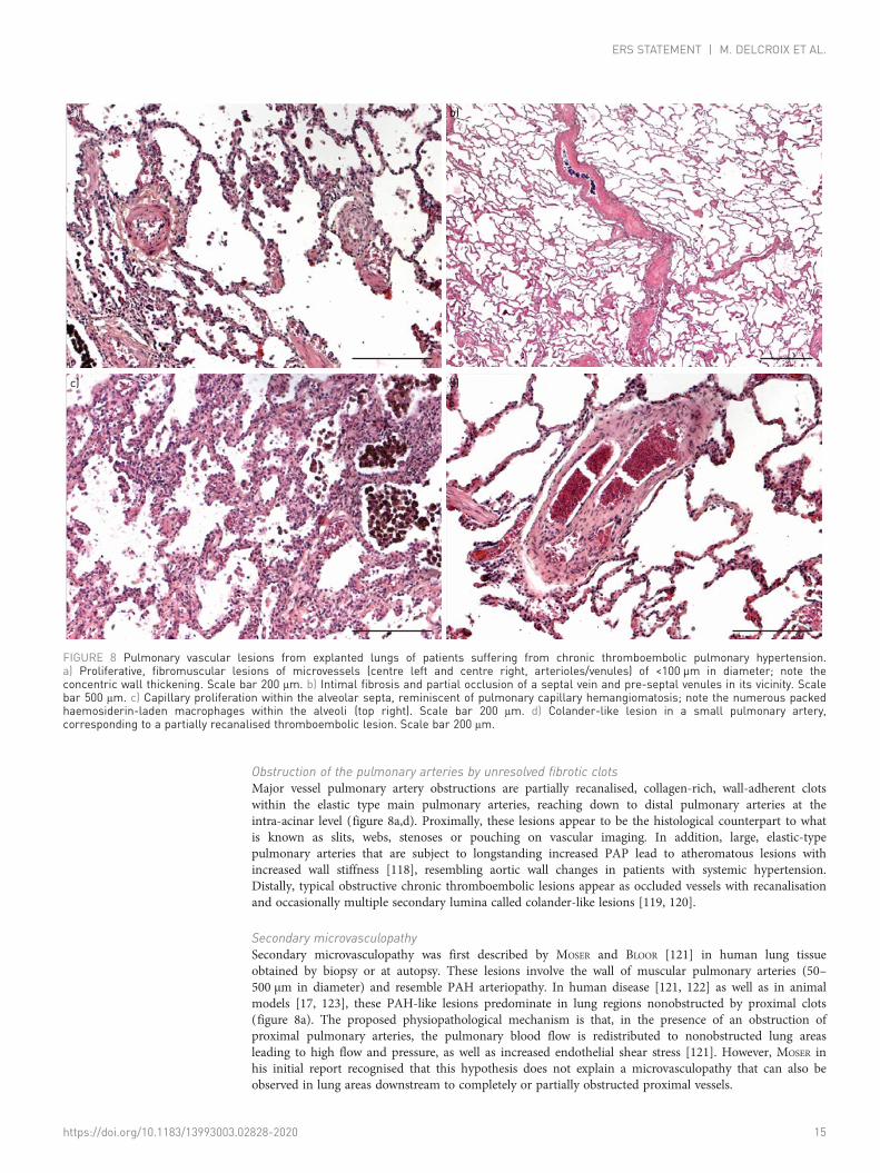
Obstruction of the pulmonary arteries by unresolved fibrotic clotsMajor vessel pulmonary artery obstructions are partially recanalised, collagen-rich, wall-adherent clotswithin the elastic type main pulmonary arteries, reaching down to distal pulmonary arteries at theintra-acinar level (figure 8a,d). Proximally, these lesions appear to be the histological counterpart to whatis known as slits, webs, stenoses or pouching on vascular imaging. In addition, large, elastic-typepulmonary arteries that are subject to longstanding increased PAP lead to atheromatous lesions withincreased wall stiffness [118], resembling aortic wall changes in patients with systemic hypertension.Distally, typical obstructive chronic thromboembolic lesions appear as occluded vessels with recanalisationand occasionally multiple secondary lumina called colander-like lesions [119, 120].
Secondary microvasculopathySecondary microvasculopathy was first described by MOSER and BLOOR [121] in human lung tissueobtained by biopsy or at autopsy. These lesions involve the wall of muscular pulmonary arteries (50–500 µm in diameter) and resemble PAH arteriopathy. In human disease [121, 122] as well as in animalmodels [17, 123], these PAH-like lesions predominate in lung regions nonobstructed by proximal clots(figure 8a). The proposed physiopathological mechanism is that, in the presence of an obstruction ofproximal pulmonary arteries, the pulmonary blood flow is redistributed to nonobstructed lung areasleading to high flow and pressure, as well as increased endothelial shear stress [121]. However, MOSER inhis initial report recognised that this hypothesis does not explain a microvasculopathy that can also beobserved in lung areas downstream to completely or partially obstructed proximal vessels.
a) b)
c) d)
FIGURE 8 Pulmonary vascular lesions from explanted lungs of patients suffering from chronic thromboembolic pulmonary hypertension.a) Proliferative, fibromuscular lesions of microvessels (centre left and centre right, arterioles/venules) of <100 µm in diameter; note theconcentric wall thickening. Scale bar 200 μm. b) Intimal fibrosis and partial occlusion of a septal vein and pre-septal venules in its vicinity. Scalebar 500 μm. c) Capillary proliferation within the alveolar septa, reminiscent of pulmonary capillary hemangiomatosis; note the numerous packedhaemosiderin-laden macrophages within the alveoli (top right). Scale bar 200 μm. d) Colander-like lesion in a small pulmonary artery,corresponding to a partially recanalised thromboembolic lesion. Scale bar 200 μm.
https://doi.org/10.1183/13993003.02828-2020 15
ERS STATEMENT | M. DELCROIX ET AL.
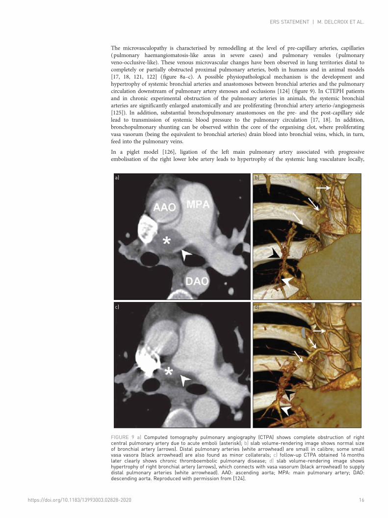
The microvasculopathy is characterised by remodelling at the level of pre-capillary arteries, capillaries(pulmonary haemangiomatosis-like areas in severe cases) and pulmonary venules (pulmonaryveno-occlusive-like). These venous microvascular changes have been observed in lung territories distal tocompletely or partially obstructed proximal pulmonary arteries, both in humans and in animal models[17, 18, 121, 122] (figure 8a–c). A possible physiopathological mechanism is the development andhypertrophy of systemic bronchial arteries and anastomoses between bronchial arteries and the pulmonarycirculation downstream of pulmonary artery stenoses and occlusions [124] (figure 9). In CTEPH patientsand in chronic experimental obstruction of the pulmonary arteries in animals, the systemic bronchialarteries are significantly enlarged anatomically and are proliferating (bronchial artery arterio-/angiogenesis[125]). In addition, substantial bronchopulmonary anastomoses on the pre- and the post-capillary sidelead to transmission of systemic blood pressure to the pulmonary circulation [17, 18]. In addition,bronchopulmonary shunting can be observed within the core of the organising clot, where proliferatingvasa vasorum (being the equivalent to bronchial arteries) drain blood into bronchial veins, which, in turn,feed into the pulmonary veins.
In a piglet model [126], ligation of the left main pulmonary artery associated with progressiveembolisation of the right lower lobe artery leads to hypertrophy of the systemic lung vasculature locally,
a) b)
c) d)
FIGURE 9 a) Computed tomography pulmonary angiography (CTPA) shows complete obstruction of rightcentral pulmonary artery due to acute emboli (asterisk); b) slab volume-rendering image shows normal sizeof bronchial artery (arrows). Distal pulmonary arteries (white arrowhead) are small in calibre; some smallvasa vasora (black arrowhead) are also found as minor collaterals; c) follow-up CTPA obtained 16 monthslater clearly shows chronic thromboembolic pulmonary disease; d) slab volume-rendering image showshypertrophy of right bronchial artery (arrows), which connects with vasa vasorum (black arrowhead) to supplydistal pulmonary arteries (white arrowhead). AAO: ascending aorta; MPA: main pulmonary artery; DAO:descending aorta. Reproduced with permission from [124].
https://doi.org/10.1183/13993003.02828-2020 16
ERS STATEMENT | M. DELCROIX ET AL.
but not in the unobstructed territory (right upper lobe). To summarise, the connection of high-pressuresystemic blood flow to the low-pressure pulmonary circulation is suspected to lead to muscular andfibrotic remodelling of pre-capillary arteries, capillaries and venules in obstructed vascular beds of humanCTEPH [18], while pulmonary arterial overflow is held responsible for changes in small arteries andarterioles in unobstructed territories. In this piglet model of CTEPH, 6 weeks after surgical reperfusion ofoccluded left PA, secondary microvasculopathy regressed both in previously occluded and nonoccludedterritories [17].
While MOSER described plexiform lesions in lungs from CTEPH patients [121, 127], others have notobserved these lesions that are typical for severe PAH [18]. Interestingly, recent reports suggest thatplexiform lesions in PAH might represent bronchopulmonary shunts through abnormally developedanastomoses [128, 129].
Impact of proximal obstructive fibrotic clots and secondary microvasculopathy on haemodynamicseverityThe presence of a severe microvasculopathy in a patient with CTEPH is usually suspected when the extentof mechanical obstruction by fibrotic organised clots does not correlate with the haemodynamic severityassessed by PVR. Some patients with CTEPH have very high PVR despite limited perfusion defects onlung scintigraphy, suggesting the presence of extensive microvasculopathy [130]. The haemodynamicimpact of secondary microvasculopathy may also be evaluated with the pulmonary occlusion techniqueused to partition PVR into upstream resistance (due to proximal fibrotic organised clots) and downstreamresistance (due to microvasculopathy). A lower pre-operative upstream resistance, suggesting extensivemicrovasculopathy, has been associated with a worse outcome after PEA and nonoperability [122, 131,132]. However, the method is technically challenging and may lack discrimination power; it has not beenincorporated into routine assessment.
Clinical consequences of microvasculopathyThe presence of a secondary microvasculopathy downstream of nonoccluded proximal pulmonary arteriesmay play a role in the progressive clinical deterioration of some patients in the absence of PE recurrence.The timing of the development of CTEPH microvasculopathy is unknown [121], but probably highlyvariable. The presence of a secondary microvasculopathy in CTEPH may explain persistence/recurrence ofPH and poor outcome after PEA [133], and provides a rationale for the use of drugs approved for PAH.
Hypothetically, the occurrence of pulmonary oedema after PEA and BPA may be related to the restorationof normal flow to a previously occluded lung region with a microvasculopathy similar to pulmonaryveno-occlusive disease and pulmonary capillary haemangiomatosis.
5.2 Imaging of the pulmonary and bronchial circulationsWhile CT can help in the visualisation of bronchial collaterals, MR can be used for both morphologicalevaluation as well as quantification of systemic to pulmonary shunt flow.
CTPAAs standard CTPA is optimised for pulmonary opacification, a longer delay from the injection of contrastmedium to image acquisition is necessary for depicting bronchial collaterals (figure 10). ECG gating willpermit sharper delineation of all vascular territories without pulsation artefacts. Limiting the acquisition toa single cardiac phase with prospective triggering can yield radiation doses that are comparable tonon-ECG gated acquisition.
DECT improves vascular enhancement allowing assessment of pulmonary arterial perfusion including thatof the collateral circulation; the latter is most conspicuous in the low kilovoltage component. PBV mapscalculate the iodine distribution in the lung parenchyma and can be a surrogate marker of the underlyingvascular reserve (figure 11).
MRIMR-based phase-contrast measurements can be regarded as a link between macro- and microcirculation.High temporal resolution phase-contrast MRI allows for the calculation of bronchopulmonary shunting bymeasuring the flow difference between the pulmonary and systemic arterial circulation. The differentialflow has been correlated with bronchial artery enlargement on CT, and the shunt size decreases inproportion to the technical success of PEA [134, 135].
Four-dimensional flow MRI is a time-resolved tridirectional velocity encoded cine sequence that canprovide comprehensive assessment of blood flow with colour-coded multiplanar reformations, streamlinesand velocity vectors. Using this technique, it has been established that in contrast to the normal central
https://doi.org/10.1183/13993003.02828-2020 17
ERS STATEMENT | M. DELCROIX ET AL.
laminar flow along the main pulmonary artery, patients with PH have a vortical blood flow >14.3% of thecardiac interval with a positive correlation between the duration of vortices and mPAP [136]. Diastolicvorticity was indicative of mild PH while systolic vorticity was seen in severe PH. Small CTEPH case serieshave shown improvement in flow vortices in the main pulmonary artery following successful BPA [137].
5.3 The RV in CTEPHIf exposed to similar loading conditions, the RV in patients with CTEPH is on average less adapted thanin patients with PAH. This becomes clear by comparing haemodynamic data from CTEPH patients withPAH patients [138, 139]. At a similar PVR, PAP is lower in CTEPH, indicative of a less adapted RV incomparison to PAH [139, 140]. In comparison to PAH, mPAP is less in CTEPH patients. Several factorsmight contribute to this difference. First, patients with CTEPH are usually older than PAH patients.Second, the time course of increase in RV load may be different in CTEPH and PAH over time. CTEPH isthought to start with an acute episode of pressure overload at the time of acute PE, which may have animpact on RV remodelling. Finally, even when PVR is similar in a CTEPH and a PAH patient, the loadfor the RV may be different due to differences in wave reflection patterns [141].
a) b)
DA
c)
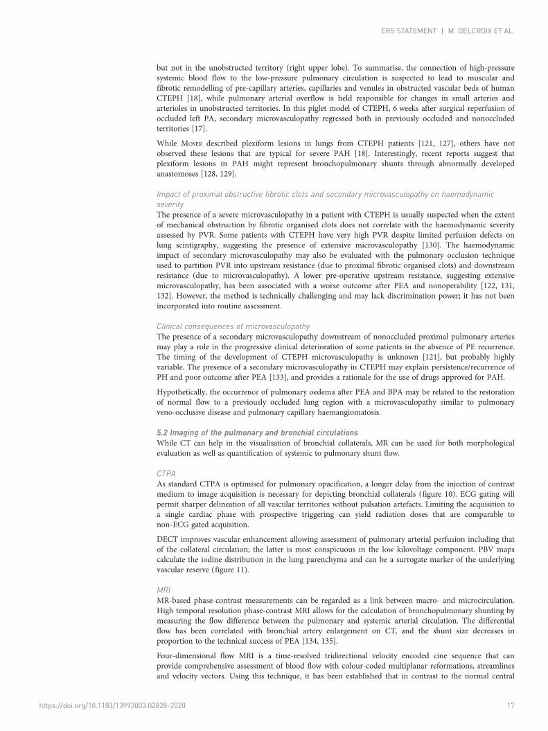
FIGURE 10 a) Axial and b) coronal computed tomography pulmonary angiography and c) magnetic resonance pulmonary angiography imagesshow the enlarged bronchial arteries (arrows) in a chronic thromboembolic pulmonary hypertension patient. DA: descending thoracic aorta.
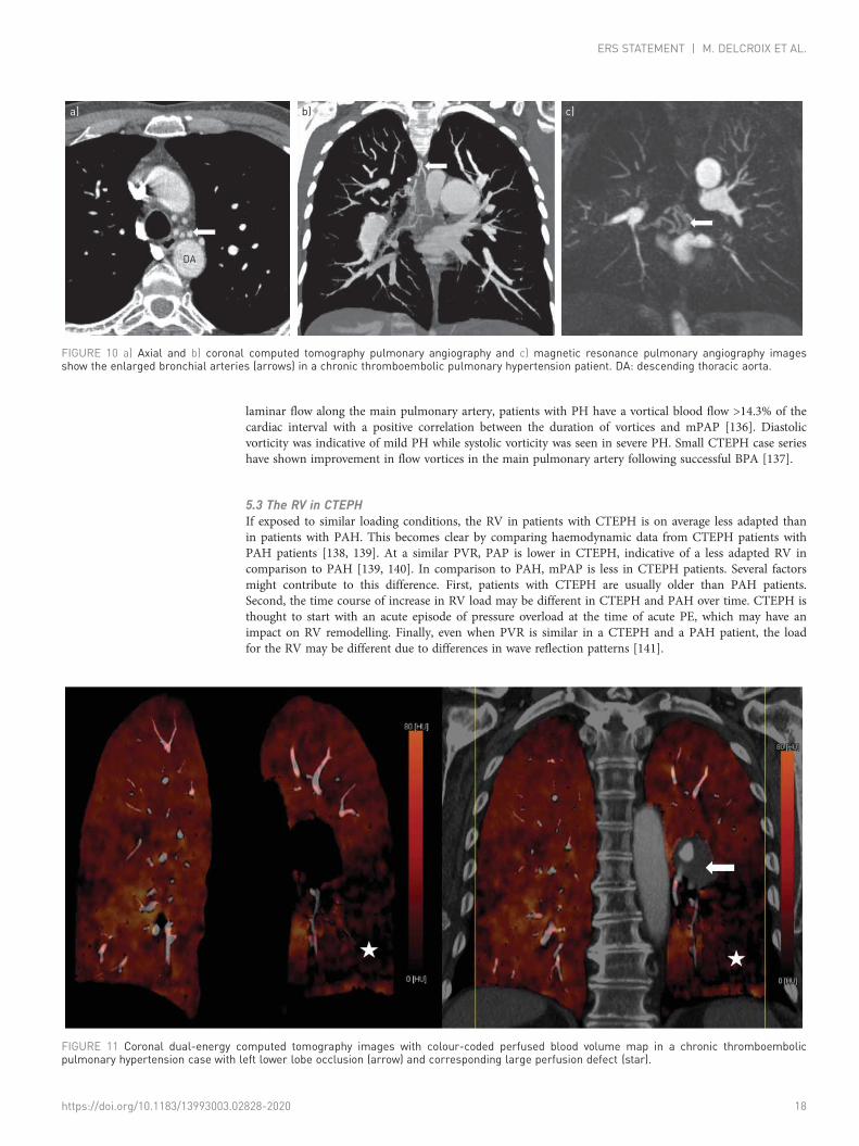
FIGURE 11 Coronal dual-energy computed tomography images with colour-coded perfused blood volume map in a chronic thromboembolicpulmonary hypertension case with left lower lobe occlusion (arrow) and corresponding large perfusion defect (star).
https://doi.org/10.1183/13993003.02828-2020 18
ERS STATEMENT | M. DELCROIX ET AL.
Restoring the load of the RV in CTEPH by successful PEA brings a profound reduction of ventricular sizeand a recovery of systolic function, for the RV and LV [142]. What remains is an abnormal response toexercise [7, 143].
Statements: pathophysiology5.1 Current knowledge suggests that two types of vascular lesions exist in CTEPH: proximal fibrotic
obstruction in large elastic pulmonary arteries and a secondary microvasculopathy in pulmonaryvessels <500 µm.
5.2 Morphological delineation of pulmonary and bronchial circulation is possible with CTPA, while MRIpermits both anatomical visualisation and semi-quantitative analysis of the extent ofbronchopulmonary shunting. None of these techniques can quantify the microvasculopathy.
5.3 The right ventricle is less adapted in CTEPH than in idiopathic PAH, but recovers largely aftersuccessful PEA.
Proposal for future research in CTEPH pathophysiologya) Search for biomarkers of fibrotic thrombus transformationb) Explore modifiers of the transforming growth factor-β pathway in thrombosis
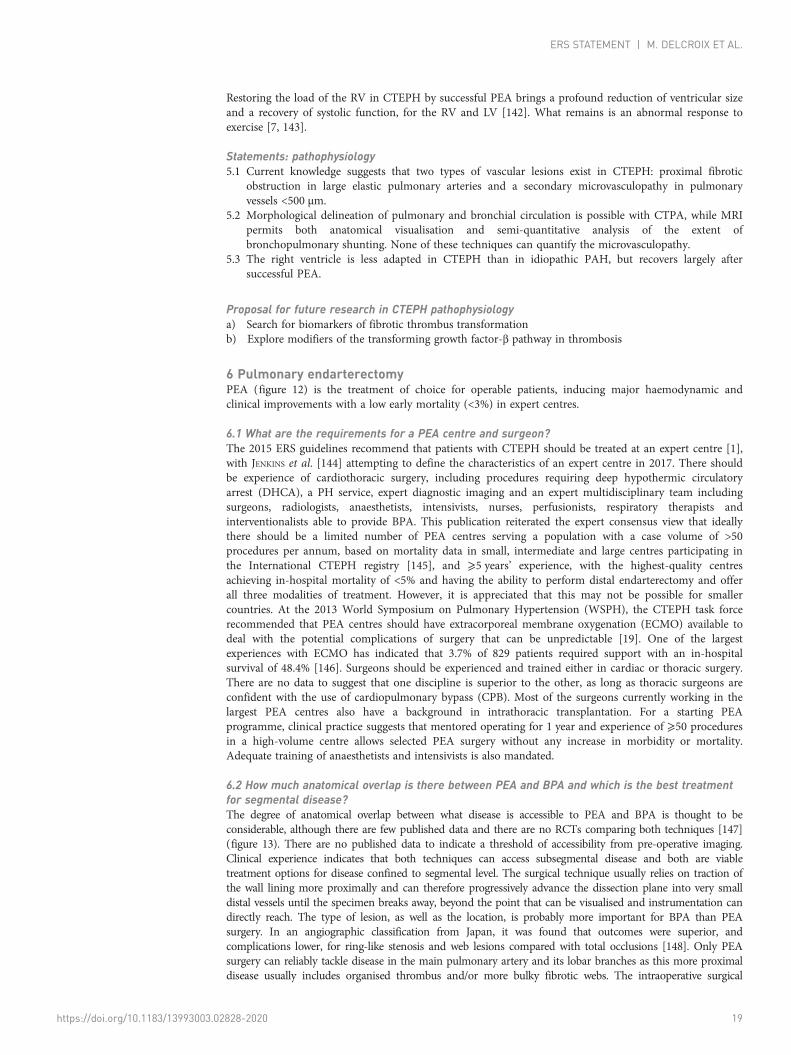
6 Pulmonary endarterectomyPEA (figure 12) is the treatment of choice for operable patients, inducing major haemodynamic andclinical improvements with a low early mortality (<3%) in expert centres.
6.1 What are the requirements for a PEA centre and surgeon?The 2015 ERS guidelines recommend that patients with CTEPH should be treated at an expert centre [1],with JENKINS et al. [144] attempting to define the characteristics of an expert centre in 2017. There shouldbe experience of cardiothoracic surgery, including procedures requiring deep hypothermic circulatoryarrest (DHCA), a PH service, expert diagnostic imaging and an expert multidisciplinary team includingsurgeons, radiologists, anaesthetists, intensivists, nurses, perfusionists, respiratory therapists andinterventionalists able to provide BPA. This publication reiterated the expert consensus view that ideallythere should be a limited number of PEA centres serving a population with a case volume of >50procedures per annum, based on mortality data in small, intermediate and large centres participating inthe International CTEPH registry [145], and ⩾5 years’ experience, with the highest-quality centresachieving in-hospital mortality of <5% and having the ability to perform distal endarterectomy and offerall three modalities of treatment. However, it is appreciated that this may not be possible for smallercountries. At the 2013 World Symposium on Pulmonary Hypertension (WSPH), the CTEPH task forcerecommended that PEA centres should have extracorporeal membrane oxygenation (ECMO) available todeal with the potential complications of surgery that can be unpredictable [19]. One of the largestexperiences with ECMO has indicated that 3.7% of 829 patients required support with an in-hospitalsurvival of 48.4% [146]. Surgeons should be experienced and trained either in cardiac or thoracic surgery.There are no data to suggest that one discipline is superior to the other, as long as thoracic surgeons areconfident with the use of cardiopulmonary bypass (CPB). Most of the surgeons currently working in thelargest PEA centres also have a background in intrathoracic transplantation. For a starting PEAprogramme, clinical practice suggests that mentored operating for 1 year and experience of ⩾50 proceduresin a high-volume centre allows selected PEA surgery without any increase in morbidity or mortality.Adequate training of anaesthetists and intensivists is also mandated.
6.2 How much anatomical overlap is there between PEA and BPA and which is the best treatmentfor segmental disease?The degree of anatomical overlap between what disease is accessible to PEA and BPA is thought to beconsiderable, although there are few published data and there are no RCTs comparing both techniques [147](figure 13). There are no published data to indicate a threshold of accessibility from pre-operative imaging.Clinical experience indicates that both techniques can access subsegmental disease and both are viabletreatment options for disease confined to segmental level. The surgical technique usually relies on traction ofthe wall lining more proximally and can therefore progressively advance the dissection plane into very smalldistal vessels until the specimen breaks away, beyond the point that can be visualised and instrumentation candirectly reach. The type of lesion, as well as the location, is probably more important for BPA than PEAsurgery. In an angiographic classification from Japan, it was found that outcomes were superior, andcomplications lower, for ring-like stenosis and web lesions compared with total occlusions [148]. Only PEAsurgery can reliably tackle disease in the main pulmonary artery and its lobar branches as this more proximaldisease usually includes organised thrombus and/or more bulky fibrotic webs. The intraoperative surgical
https://doi.org/10.1183/13993003.02828-2020 19
ERS STATEMENT | M. DELCROIX ET AL.
classification has been recently revised to recognise the level at which disease starts and separate segmentaland subsegmental disease (levels III and IV) to reflect modern surgical practice [147]. One series hasspecifically investigated the outcome of PEA surgery for more distal disease [149]. In this series of 331patients operated by a single surgeon between 2008 and 2013, PEA for Jamieson type 3 disease (segmental)was performed without an increase in operative mortality or morbidity. There were equivalent haemodynamicresults at discharge, and importantly at 1 year, to those achieved for patients with more proximal disease.
FIGURE 12 Pulmonary endarterectomy. The right pulmonary artery is opened, and the suction dissector isintroduced between the artery wall and the fibrosis. Following the inside of the artery down to segmental andsubsegmental level, the fibrotic material is subsequently freed from the wall and removed with forceps.Reproduced with permission from Maren Hötten, Master Thesis in Scientific Illustration "ChronicThromboembolic Pulmonary Hypertension: Diagnosis and Treatment”, University of Maastricht, 2016.
https://doi.org/10.1183/13993003.02828-2020 20
ERS STATEMENT | M. DELCROIX ET AL.
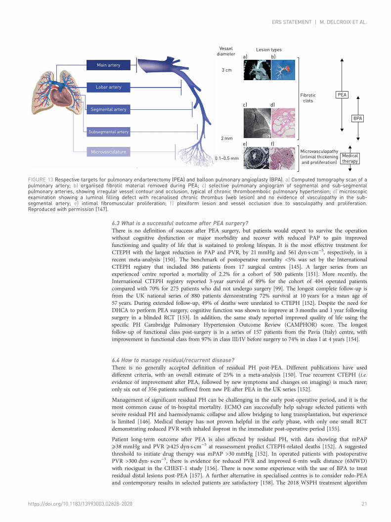
6.3 What is a successful outcome after PEA surgery?There is no definition of success after PEA surgery, but patients would expect to survive the operationwithout cognitive dysfunction or major morbidity and recover with reduced PAP to gain improvedfunctioning and quality of life that is sustained to prolong lifespan. It is the most effective treatment forCTEPH with the largest reduction in PAP and PVR, by 21 mmHg and 561 dyn·s·cm−5, respectively, in arecent meta-analysis [150]. The benchmark of postoperative mortality <5% was set by the InternationalCTEPH registry that included 386 patients from 17 surgical centres [145]. A larger series from anexperienced centre reported a mortality of 2.2% for a cohort of 500 patients [151]. More recently, theInternational CTEPH registry reported 3-year survival of 89% for the cohort of 404 operated patientscompared with 70% for 275 patients who did not undergo surgery [99]. The longest complete follow-up isfrom the UK national series of 880 patients demonstrating 72% survival at 10 years for a mean age of57 years. During extended follow-up, 49% of deaths were unrelated to CTEPH [152]. Despite the need forDHCA to perform PEA surgery, cognitive function was shown to improve at 3 months and 1 year followingsurgery in a blinded RCT [153]. In addition, the same study reported improved quality of life using thespecific PH Cambridge Pulmonary Hypertension Outcome Review (CAMPHOR) score. The longestfollow-up of functional class post-surgery is in a series of 157 patients from the Pavia (Italy) centre, withimprovement in functional class from 97% in class III/IV before surgery to 74% in class I at 4 years [154].
6.4 How to manage residual/recurrent disease?There is no generally accepted definition of residual PH post-PEA. Different publications have useddifferent criteria, with an overall estimate of 25% in a meta-analysis [150]. True recurrent CTEPH (i.e.evidence of improvement after PEA, followed by new symptoms and changes on imaging) is much rarer;only six out of 356 patients suffered from new PE after PEA in the UK series [152].
Management of significant residual PH can be challenging in the early post-operative period, and it is themost common cause of in-hospital mortality. ECMO can successfully help salvage selected patients withsevere residual PH and haemodynamic collapse and allow bridging to lung transplantation, but experienceis limited [146]. Medical therapy has not proven helpful in the early phase, with only one small RCTdemonstrating reduced PVR with inhaled iloprost in the immediate post-operative period [155].
Patient long-term outcome after PEA is also affected by residual PH, with data showing that mPAP⩾38 mmHg and PVR ⩾425 dyn·s·cm−5 at reassessment predict CTEPH-related deaths [152]. A suggestedthreshold to initiate drug therapy was mPAP >30 mmHg [152]. In operated patients with postoperativePVR >300 dyn· s·cm−5, there is evidence for reduced PVR and improved 6-min walk distance (6MWD)with riociguat in the CHEST-1 study [156]. There is now some experience with the use of BPA to treatresidual distal lesions post-PEA [157]. A further alternative in specialised centres is to consider redo-PEAand contemporary results in selected patients are satisfactory [158]. The 2018 WSPH treatment algorithm
Vesseldiameter
Main artery
Lobar artery
Segmental artery
Subsegmental artery
Microvasculature
3 cm
2 mm
0.1–0.5 mm
Lesion typesa)
c)
e)
d)
f)
b)
Fibroticclots
Microvasculopathy(intimal thickeningand proliferation)
PEA
BPA
Medicaltherapy
FIGURE 13 Respective targets for pulmonary endarterectomy (PEA) and balloon pulmonary angioplasty (BPA). a) Computed tomography scan of apulmonary artery; b) organised fibrotic material removed during PEA; c) selective pulmonary angiogram of segmental and sub-segmentalpulmonary arteries, showing irregular vessel contour and occlusion, typical of chronic thromboembolic pulmonary hypertension; d) microscopicexamination showing a luminal filling defect with recanalised chronic thrombus (web lesion) and no evidence of vasculopathy in the sub-segmental artery; e) intimal fibromuscular proliferation; f ) plexiform lesion and vessel occlusion due to vasculopathy and proliferation.Reproduced with permission [147].
https://doi.org/10.1183/13993003.02828-2020 21
ERS STATEMENT | M. DELCROIX ET AL.
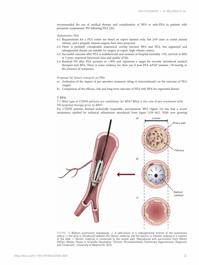
recommended the use of medical therapy and consideration of BPA or redo-PEA in patients withpersistent symptomatic PH following PEA [20].
Statements: PEA6.1 Requirements for a PEA centre are based on expert opinion only, but ⩾50 cases as centre annual
volume, and a properly trained surgeon have been proposed.6.2 There is probably considerable anatomical overlap between BPA and PEA, but segmental and
subsegmental disease are suitable for surgery in expert, high-volume centres.6.3 Successful outcome after PEA is multifactorial and assumes in-hospital mortality <5%, survival of 90%
at 3 years, improved functional class and quality of life.6.4 Residual PH after PEA presents in ∼50% and represents a target for recently introduced medical
therapies and BPA. There is some evidence for their use if post-PEA mPAP remains >30 mmHg inthe presence of symptoms.
Proposal for future research on PEAa) Evaluation of the impact of pre-operative treatment (drug or interventional) on the outcome of PEA
surgeryb) Comparison of the efficacy, risk and long-term outcome of PEA with BPA for segmental disease
7 BPA7.1 What type of CTEPH patients are candidates for BPA? What is the role of pre-treatment withPH-targeted therapy prior to BPA?For CTEPH patients deemed technically inoperable, percutaneous BPA (figure 14) has had a recentrenaissance sparked by technical refinements introduced from Japan [159–161]. With now growing
Lumena)
b)
c)
Artery wall
Ballooncatheter
Fibrosis
FIGURE 14 Balloon pulmonary angioplasty. a) A web-lesion in a subsegmental branch of the pulmonaryartery; b) the wire is introduced between the fibrotic material and the balloon is inflated, leading to a ruptureof the web; c) fibrotic material is connected to the vessel wall. Reproduced with permission from MarenHötten, Master Thesis in Scientific Illustration "Chronic Thromboembolic Pulmonary Hypertension: Diagnosisand Treatment”, University of Maastricht, 2016.
https://doi.org/10.1183/13993003.02828-2020 22
ERS STATEMENT | M. DELCROIX ET AL.
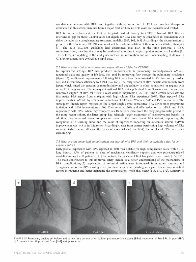
worldwide experience with BPA, and together with advances both in PEA and medical therapy asoverviewed in this series, there has been a major reset on how CTEPH cases are evaluated and treated.
BPA is not a replacement for PEA or targeted medical therapy in CTEPH. Instead, BPA fills anintervention gap for those CTEPH cases not eligible for PEA and may be considered in conjunction witheither therapies as a complementary treatment modality [147, 162, 163]. Accordingly, the determination toproceed with BPA in any CTEPH case must not be made in isolation of these other established therapies[1]. The 2015 ESC/ERS guidelines had determined that BPA at the time garnered a IIb-Crecommendation, meaning that it may be considered according to expert opinion and/or small studies [1].This will require updating in the next guidelines as the intervention and our understanding of its role inCTEPH treatment have evolved at a rapid pace.
7.2 What are the clinical outcomes and expectations of BPA for CTEPH?In experienced settings, BPA has produced improvements in pulmonary haemodynamics, 6MWD,functional class and quality of life [162, 164–166] by improving flow through the pulmonary circulation(figure 15). Additional improvements following BPA have been demonstrated in RV function by cardiacMR and in ventilatory efficiency by CPET [57, 168]. The early reports of BPA benefit were initially fromJapan, which raised the question of reproducibility and applicability to other populations or regions withactive PEA programmes. The subsequent national BPA series published from Germany and France havereinforced support of BPA for CTEPH cases deemed inoperable [169, 170]. The German series was thefirst major BPA report from a region with high-volume PEA experience [169]. They reported BPAimprovements in 6MWD by +33 m and reductions of 18% and 26% in mPAP and PVR, respectively. Thesubsequent French report represented the largest single-centre consecutive BPA series since programmeinitiation with 1006 interventions [170]. They reported 26% and 43% reduction in mPAP and PVR,respectively, with BPA. When they compared results between cases from the early programmatic period tothe more recent cohort, the latter group had relatively larger magnitude of haemodynamic benefit. Inaddition, they observed lower complication rates in the more recent BPA cohort, supporting therecognition of a learning curve and the value of experience impacting on outcomes. Overall 6MWDimprovement was +45 m in this series. Accordingly, even from centres performing high volumes of PEAsurgeries (which may influence the types of cases selected for BPA) the results of BPA have beenencouraging.
7.3 What are the important complications associated with BPA and their acceptable rates for anexpert centre?Early pivotal experience with BPA reported in 2001 was notable for high complication rates, with 61.1%lung injury, 16.7% of patients in need of mechanical ventilatory support and one procedure-relatedmortality among the 18 patients [171]. In contrast, the new era of BPA has yielded safer results [164, 165].The main contributors to this improved safety include 1) a better understanding of the mechanisms ofBPA complications; 2) application of technical refinements introduced from expert centres; and3) appreciation of the BPA learning curve and team experience (starting with patient selection) as criticalfactors in reducing and better managing the complications when they occur [148, 170, 172]. Contrary to
a)
Pre-BPA Post BPA 2 months later
b) c)
FIGURE 15 Pulmonary angiogram before and at two time periods after balloon pulmonary angioplasty (BPA) treatment. a) Pre-BPA; b) post-BPA;c) 2 months later. Reproduced from [167] with permission.
https://doi.org/10.1183/13993003.02828-2020 23
ERS STATEMENT | M. DELCROIX ET AL.
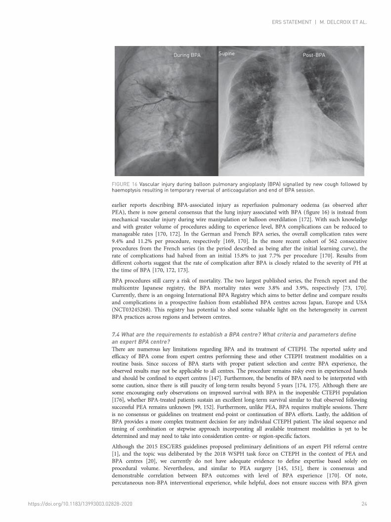
earlier reports describing BPA-associated injury as reperfusion pulmonary oedema (as observed afterPEA), there is now general consensus that the lung injury associated with BPA (figure 16) is instead frommechanical vascular injury during wire manipulation or balloon overdilation [172]. With such knowledgeand with greater volume of procedures adding to experience level, BPA complications can be reduced tomanageable rates [170, 172]. In the German and French BPA series, the overall complication rates were9.4% and 11.2% per procedure, respectively [169, 170]. In the more recent cohort of 562 consecutiveprocedures from the French series (in the period described as being after the initial learning curve), therate of complications had halved from an initial 15.8% to just 7.7% per procedure [170]. Results fromdifferent cohorts suggest that the rate of complication after BPA is closely related to the severity of PH atthe time of BPA [170, 172, 173].
BPA procedures still carry a risk of mortality. The two largest published series, the French report and themulticentre Japanese registry, the BPA mortality rates were 3.8% and 3.9%, respectively [73, 170].Currently, there is an ongoing International BPA Registry which aims to better define and compare resultsand complications in a prospective fashion from established BPA centres across Japan, Europe and USA(NCT03245268). This registry has potential to shed some valuable light on the heterogeneity in currentBPA practices across regions and between centres.
7.4 What are the requirements to establish a BPA centre? What criteria and parameters definean expert BPA centre?There are numerous key limitations regarding BPA and its treatment of CTEPH. The reported safety andefficacy of BPA come from expert centres performing these and other CTEPH treatment modalities on aroutine basis. Since success of BPA starts with proper patient selection and centre BPA experience, theobserved results may not be applicable to all centres. The procedure remains risky even in experienced handsand should be confined to expert centres [147]. Furthermore, the benefits of BPA need to be interpreted withsome caution, since there is still paucity of long-term results beyond 5 years [174, 175]. Although there aresome encouraging early observations on improved survival with BPA in the inoperable CTEPH population[176], whether BPA-treated patients sustain an excellent long-term survival similar to that observed followingsuccessful PEA remains unknown [99, 152]. Furthermore, unlike PEA, BPA requires multiple sessions. Thereis no consensus or guidelines on treatment end-point or continuation of BPA efforts. Lastly, the addition ofBPA provides a more complex treatment decision for any individual CTEPH patient. The ideal sequence andtiming of combination or stepwise approach incorporating all available treatment modalities is yet to bedetermined and may need to take into consideration centre- or region-specific factors.
Although the 2015 ESC/ERS guidelines proposed preliminary definitions of an expert PH referral centre[1], and the topic was deliberated by the 2018 WSPH task force on CTEPH in the context of PEA andBPA centres [20], we currently do not have adequate evidence to define expertise based solely onprocedural volume. Nevertheless, and similar to PEA surgery [145, 151], there is consensus anddemonstrable correlation between BPA outcomes with level of BPA experience [170]. Of note,percutaneous non-BPA interventional experience, while helpful, does not ensure success with BPA given
During BPA Supine Post-BPA
FIGURE 16 Vascular injury during balloon pulmonary angioplasty (BPA) signalled by new cough followed byhaemoptysis resulting in temporary reversal of anticoagulation and end of BPA session.
https://doi.org/10.1183/13993003.02828-2020 24
ERS STATEMENT | M. DELCROIX ET AL.
the differences between the systemic and pulmonary vasculature, nature of complications and targetedpathology [170]. Furthermore, and worth re-emphasising, aside from technical proficiency that comes withprocedural volume, success of BPA and individual case outcome also relies importantly on proper patientselection within the context of a multidisciplinary CTEPH team considering all current and availabletreatment modalities [1, 170]. For a starting BPA programme, clinical practice suggests that directmentorship from a high-volume centre followed by experience of ⩾50 procedures allows results withoutexcessive morbidity or mortality.
Within a relatively short period of time, BPA has evolved from an experimental modality to becoming anestablished treatment option for inoperable CTEPH. Looking ahead, thresholds for successful BPAoutcomes, acceptable rates of complications and optimal incorporation with the other treatment modalitiesneed to be investigated and established. This will require concerted effort and collaboration amongCTEPH centres.
Statements: BPA7.1 Inoperable CTEPH patients can benefit from BPA. Optimal CTEPH treatment requires a
multidisciplinary team approach considering PEA, medical therapy and BPA.7.2 Long-term results after BPA are available out to 8 years after the procedure. Safety and efficacy of BPA
correlate with centre experience.7.3 Vascular injury rather than reperfusion pulmonary oedema is the likely cause of any severity of lung
injury after BPA.7.4 Similar to PEA, proper training in a high-volume centre is critical for BPA.
Recommendations for research and traininga) Define the role of PH-targeted medical therapy in relation to BPAb) Identify treatment goals and key end-points for completing BPA interventions per individual patientc) Create standardised definitions of BPA complicationsd) Establish criteria or expected outcome thresholds to be recognised as an expert BPA centre
8 Medical treatment8.1 Long-term anticoagulation: what is the current standard of care in CTEPH (VKAs versusNOACs)?Besides lifelong anticoagulation, CTEPH basic therapy includes diuretics in case of right heart failure andoxygen in hypoxaemic patients [92, 93]. Traditionally, VKAs have been the mainstay of anticoagulanttreatment in PAH and CTEPH, but NOACs are increasingly used [90, 92, 93, 177]. There are no strongdata on the efficacy and safety of NOACs in CTEPH. In addition, robust information is needed on drug–drug interaction between NOACs and pulmonary vasodilators used in CTEPH. Because NOACs areregularly used in some centres, registry-based information has been released recently [90, 177].
Consecutive CTEPH patients undergoing PEA between 2007 and 2018 at the Royal Papworth Hospital(Cambridge, UK) have been included in a retrospective analysis (794 patients were treated with VKAs and206 with NOACs following PEA) [90]. Haemodynamic and functional improvements were observed inboth groups following PEA. In terms of safety, major bleeding events were equivalent in those treated withVKAs and NOACs (0.7% per person-year), but the VTE recurrence rate was proportionately higher withNOACs (4.62% per person-year) than VKAs (0.76% per person-year), although survival did not differ [90].Additionally, in a retrospective analysis of 405 consecutive PEA cases, CTEPH patients on NOACs weretwice as likely to have associated acute or subacute thrombi removed at the time of surgery compared withthose on conventional anticoagulant therapies [178].
In the setting of BPA, continued VKA treatments without interruptions appear to be practiced by themajority of expert centres.
EXPERT, an international, multicentre, prospective, noninterventional registry study, assessing riociguatsafety in clinical practice (NCT02092818) has recently released data from PAH/CTEPH patients whoreceived riociguat with either concomitant NOACs (n=227) or VKAs (n=617) at baseline [177]. In termsof adverse events, haemorrhages were reported in 26 (11%) patients in the NOACs group and 81 (13%)patients in the VKAs group, and thromboembolic events were reported in nine (4%) and 24 (4%) patients,respectively. In terms of serious adverse events, haemorrhages were reported in 13 (6%) patients in theNOACs group and 39 (6%) patients in the VKAs group, fatal haemorrhages in one (0.4%) and six (1%)patients, respectively, and thromboembolic events were reported in eight (4%) and 23 (4%) patients,respectively. Thus, serious bleeding/thrombotic events were uncommon, and their frequency was similarbetween PAH/CTEPH patients receiving concomitant NOACs or VKAs at baseline. Rates of serious
https://doi.org/10.1183/13993003.02828-2020 25
ERS STATEMENT | M. DELCROIX ET AL.
haemorrhage or thromboembolic serious adverse events were similar with baseline NOACs or VKAs forPAH and CTEPH, separately [177].
A subgroup of CTEPH patients present with antiphospholipid syndrome. In that setting, VKAs(international normalised ratio target 2.5) have been demonstrated as carrying more benefit and less riskthan NOACs in high-risk patients with thrombotic antiphospholipid syndrome (triple-positive for lupusanticoagulant, anticardiolipin, and anti-β2-glycoprotein I antibodies of the same isotype) [94].
8.2 PH medication: what is the available evidence on approved and not (yet) approved PHmedications in CTEPH?Acute pulmonary vasodilator testing is not recommended in CTEPH, even if PH reversibility could be apredictor of residual PH after PEA [179], and there is no indication of calcium channel blocker use in thiscondition [180]. In CTEPH, there is evidence of small pulmonary vessels disease indistinguishable fromthat of PAH, and similar treatment targeting the endothelin-1, nitric oxide and prostacyclin pathways havebeen studied [120].
In the CHEST-1 RCT of 261 patients with inoperable CTEPH or persistent/recurrent PH after PEA, anoral guanylase cyclase stimulator (sGC; riociguat, targeting the nitric oxide pathway) significantly increased6MWD and reduced PVR, as compared to placebo [156]. These data led to the approval of riociguat forpatients with inoperable CTEPH or persistent/recurrent PH after PEA. Currently, riociguat is being testedin RCTs, addressing its efficacy and safety as bridging therapy for patients scheduled to undergo PEA(NCT03273257). Another study compares riociguat to BPA in inoperable patients (NCT02634203).
Sildenafil, a phosphodiesterase type 5 inhibitor (PDE5i), has been tested in a small RCT in inoperableCTEPH [181]. Sildenafil (40 mg three times daily) or matching placebo was given to nine and 10inoperable CTEPH patients, respectively. At 12 weeks, no improvement was seen in the primary end-point(6MWD) despite a positive effect on PVR [181]. This pilot study was insufficiently powered to test theprimary end-point, and larger-scale trials of sildenafil in inoperable CTEPH have not been performedsince then. Sildenafil is thus not approved for CTEPH.
The dual endothelin receptor antagonist (ERA) bosentan showed no improvement in 6MWD (despite apositive effect on PVR) in an RCT of 157 patients with inoperable CTEPH or persistent/recurrent PH afterPEA (BENEFIT study) [182]. Bosentan is thus not approved for CTEPH. Safety and efficacy of the dualERA macitentan was assessed in a phase 2 RCT, showing significantly improved 6MWD and PVRcompared to placebo in 80 CTEPH patients adjudicated as inoperable (MERIT-1) [183]. Interestingly, thiswas the first RCT in CTEPH assessing the combination of an ERA with other medications approved forPAH, but excluding riociguat (61% of patients received off-label PDE5i or oral/inhaled prostanoids).
Besides off-label use, there is a single RCT studying prostacyclin analogues in inoperable CTEPH(CTREPH study) [184]. This RCT showed that long-term subcutaneous treprostinil is safe and effective in105 severe inoperable CTEPH patients, with improvements of 6MWD, PVR, functional class andNT-proBNP. In this study, 30% of patients received other pulmonary vasodilator therapies. These data ledto the marketing authorisation of treprostinil for patients with inoperable CTEPH or persistent/recurrentPH after PEA.
8.3 Combination therapy: is there a role for initial or sequential combination therapy in CTEPH?Sequential combination therapy of PH medications was allowed in CTEPH RCTs such as MERIT-1 [183]and CTREPH [184]. By analogy with PAH [177], initial dual oral combination therapy is currentlyconsidered in expert centres for patients with severe CTEPH. This is particularly true in inoperableCTEPH prior to BPA, in order to perform BPA in patients with less severe haemodynamic impairmentand presumably at lower risk of procedure-related complications. Recent single-centre registry data ofinitial dual oral combination therapy using an ERA in association with either a PDE5i or riociguat ininoperable CTEPH patients have been reported [185]. These data indicate that initial oral dualcombination therapy resulted in more pronounced reduction in PVR, when compared to monotherapy.
8.4 De-escalation: when and how to stop/reduce PH drugs after successful BPA/PEA?Most CTEPH patients improve markedly clinically and haemodynamically after BPA and/or PEA [73,170]. Lifelong anticoagulation is mandatory even if PEA and/or BPA have normalised pulmonaryhaemodynamics at rest, while the use of diuretics and oxygen are often no longer needed. Similarly,stopping or de-escalating PH medications may be considered after successful BPA and/or PEA. TheJapanese multicentre registry for BPA indicates that PH medications were used in 72% of 308 cases beforeBPA, and 45% of 196 cases at last follow-up (at a mean±SD 425±281 days after the final BPA procedures)[73]. In the French experience, 62% of 181 patients undergoing BPA received PH medications prior to
https://doi.org/10.1183/13993003.02828-2020 26
ERS STATEMENT | M. DELCROIX ET AL.
BPA (26.6% monotherapy, 28.3% double combination and 7.1% triple combination therapy) [170]. Theongoing International BPA Registry will provide long-term information on this procedure and moredetails on the use of PH medications before and after BPA (NCT03245268).
Statements: medical treatment8.1 According to current guidelines, CTEPH patients should be treated with lifelong anticoagulation;
VKAs are the mainstay of anticoagulant treatment in CTEPH, but NOACs are increasingly used, withno safety issues reported yet. Antiphospholipid syndrome is a contraindication to NOACs.
8.2 Riociguat, an oral guanylate cyclase stimulator, and treprostinil, a subcutaneous prostacyclin analogue,are approved for patients with inoperable CTEPH or persistent/recurrent PH after PEA; other PHmedications have been tested in CTEPH and are used off-label.
8.3 Oral combination therapy is a common practice in CTEPH patients with severe haemodynamiccompromise.
8.4 Withdrawal of PH medications is usually considered after successful BPA and/or PEA.
Proposal for future research on medical therapy for CTEPHa) Prospective registry data and/or studies to evaluate the safety of NOACs in CTEPH are encouragedb) Studies testing the efficacy and safety of initial dual oral combination therapy are needed in CTEPH
patients with severe haemodynamic compromisec) A dedicated trial of carefully monitored withdrawal of PH medications should be considered after
successful BPA and/or PEA
9 Multimodality treatment9.1 What are the criteria for selecting candidates?Many patients may have mixed anatomical lesions with lobar, segmental and microvascular lesions. Withthree effective treatments now available, the current treatment paradigm has evolved to include amultimodal approach to management. Such an approach involves employing combinations of PEA, BPAand pharmacological therapy to target proximal lesions, distal lesions and microvasculopathy, respectively.Currently, there is no guidance or standardisation regarding assessment of suitability for multimodaltherapy and patient selection is performed on a case-by-case basis in expert centres through amultidisciplinary team approach.
Given the various treatment options for CTEPH, lung transplantation is now rarely considered in thisgroup of patients.
9.2 Is there a potential role for PH-targeted medical therapy prior to PEA in patients withoperable CTEPH?The pre-operative PVR has been shown to be a predictor of post-operative mortality [145, 151, 186]. Inthe International CTEPH Registry, including 386 operated patients, an almost linear relationship betweenpre-operative PVR and in-hospital mortality was observed [145]. The use of PH-targeted therapy inpatients with high pre-operative PVR may improve pulmonary haemodynamics before surgery andpotentially reduce the morbidity and mortality associated with PEA. Two case series examined a total of21 patients treated with intravenous epoprostenol prior to PEA. An improvement in pre-PEAhaemodynamics, but no change in post-PEA haemodynamics was observed compared to controls [187,188]. In a randomised, controlled single-blind study including 25 patients with total pulmonary resistance(TPR) >900 dyn·s·cm−5, pre-treatment with bosentan for 16 weeks demonstrated a reduction in pre-PEATPR and mPAP, but no difference in post-PEA haemodynamics compared to controls [189]. In this study,four patients died during or after PEA (n=1 (7.6%) in the bosentan group and n=3 (25%) in the controlgroup). In a retrospective analysis of CTEPH patients referred to the University of California at San Diego,JENSEN et al. [103] assessed the effects of pre-PEA PH-targeted therapy on pre-PEA haemodynamics andpost-PEA outcomes. For this analysis, the subjects were divided into two groups: those who were onPH-targeted therapy before referral for PEA and those who were not (control group). The authors showedthat the use of PH medications delayed the time to referral for PEA with only a minimal improvement inpre-operative mPAP. In addition, their use did not result in any differences in post-PEA haemodynamicsor outcomes [103]. In the International CTEPH Registry mentioned earlier, 29% of operated CTEPHpatients had received PH-targeted therapy prior to PEA. These patients had more severe haemodynamicprofile than those who did not receive bridging therapy, while no significant difference was observed ineither post-operative PVR or post-operative complications between the two groups. More interestingly, theauthors found that bridging therapy with PH-targeted drugs was associated with an increased risk ofdeath [99]. However, a recently published retrospective, single-centre analysis suggested that the use of
https://doi.org/10.1183/13993003.02828-2020 27
ERS STATEMENT | M. DELCROIX ET AL.
targeted therapy was associated with a significantly better 1-year survival (89.1% versus 64.1%) in patientswith a more severe haemodynamic impairment [190], relaunching the discussion on whether PH-targetedtherapies before surgery in patients with operable CTEPH may result in better survival. Unfortunately, therandomised, placebo-controlled, multicentre PEA bridging study looking at safety and efficacy of riociguatprior to PEA in patients with operable CTEPH and high pre-operative PVR (>800 dyn·s·cm−5) had to beterminated because of poor recruitment (NCT03273257).
9.3 Is there a role for combined PEA and BPA in patients with CTEPH?Some CTEPH patients may have mixed anatomical lesions with surgically accessible lesions for one lungand inoperable lesions affecting the contralateral lung. Depending on the severity of PH, low-risk patientswith mixed anatomical lesions may undergo PEA, and if necessary, BPA during follow-up after surgery.High-risk patients (with high pre-operative PVR) might benefit from a combined therapy with BPA (priorto or at the same time as surgery) and PEA to decrease the surgical risk and improve the final result.However, data regarding the impact of this strategy on post-operative outcome are very limited so far andfurther studies are needed to confirm its clinical relevance [163, 191].
In large PEA cohorts, it has been shown that a significant proportion of patients may have residual or recurrentPH following PEA, which can adversely affect their quality of life and outcome [145, 151, 152, 186]. Three caseseries, including a total of 34 patients, have specifically investigated the safety and efficacy of additional BPA forthe treatment of residual or recurrent PH after PEA [157, 192, 193]. The majority of patients were treated withPH-targeted drugs before BPA and the time between PEA and first BPA session ranged from a few months toseveral years. In these case series, BPA improved haemodynamics and exercise capacity with a rate ofcomplication that was similar to that observed in inoperable CTEPH patients treated with BPA.
BPA has been reported as a rescue treatment for early failure of PEA, without producing convincingresults [194, 195].
9.4 Is there a role for combined use of PH-targeted therapy and BPA in patients with inoperableCTEPH?Most BPA series reported to date included a high proportion of patients who were treated withPH-targeted therapy prior to BPA. In initial Japanese reports, PH-targeted drugs were initiated to improvethe safety of BPA by optimising pre-BPA haemodynamics [159, 161]. This strategy was based on thefinding of FEINSTEIN et al. [171] who first showed that a pre-BPA mPAP >35 mmHg was correlated withthe occurrence of lung injury known as one of the main complications related to BPA.
This approach has been applied in more recent studies, although there have been no clinical trialsspecifically designed to investigate its effectiveness [49, 73, 169, 170, 172, 175]. In two of these studies therelationship between high pre-BPA mPAP and occurrence of lung injury was also demonstrated, thussupporting the use of pharmacological treatments prior to BPA [170, 172]. The effects of this strategy ininoperable CTEPH have been examined in two studies. In a single-centre study, consecutive patients withinoperable CTEPH were treated with medical therapy prior to BPA, which resulted in improved cardiacindex, PVR and exercise capacity, but little change in mPAP [175]. The patients then underwent BPA andhad significant further reductions in mPAP and PVR and further improvement in symptoms and exercisecapacity. In an observational cohort study, WIEDENROTH et al. [196] investigated more specifically theeffects of a sequential treatment with riociguat and BPA. The authors showed that riociguat improvedhaemodynamics in patients eligible for BPA and that BPA led to further improvement in exercise capacityand haemodynamics. These data suggest that medical therapy combined with BPA may have an advantageover medical therapy alone, although larger prospective studies are needed to confirm this finding.
Statements: multimodal treatment9.1 There is no consensus on eligibility criteria for multimodal therapy, which is dependent on the
expertise and judgement of each individual CTEPH centre. Patient selection for multimodal therapy isperformed in expert centres through a multidisciplinary team approach.
9.2 There is no convincing evidence that pre-operative pharmacological treatment is beneficial in patientswith operable CTEPH. Nevertheless, PH-targeted drugs as bridging therapy to PEA is sometimesconsidered in selected high-risk patients (with high pre-operative pulmonary vascular resistance) aftermultidisciplinary team assessment in expert centres.
9.3 The role of BPA as bridging therapy prior to PEA in patients with mixed surgically accessible andinaccessible lesions needs to be explored. The potential benefit of hybrid procedures simultaneouslycombining PEA and BPA in such patients needs to be confirmed by further studies. There isinsufficient evidence to support the use of BPA as a rescue procedure for early failure of PEA.
https://doi.org/10.1183/13993003.02828-2020 28
ERS STATEMENT | M. DELCROIX ET AL.
Additional BPA, most often in combination with PH-targeted medical therapy, is common practicefor patients with persistent symptomatic PH after PEA.
9.4 Most task force members use PH-targeted therapy prior to BPA to improve the safety of BPA,although there have been no clinical trials investigating the benefit of this approach on the rate ofBPA-related complications. Some data suggest that the combination of PH-targeted therapy and BPAprovides better results than medical therapy alone.
Proposal for future research in multimodal treatmentStudies are needed to investigate the most appropriate approach to combination of BPA and PEA
10 Rehabilitation10.1 What is the cause of exercise limitation in CTEPH?The role of rehabilitation in PAH and CTEPH has been well established [197, 198]. Despite the fact that thebeneficial effects of rehabilitation are not fully understood, increased capillarisation of the skeletal muscles aswell as improvement in RV function as a consequence of rehabilitation have been measured in both animalmodels of pulmonary hypertension and PAH patients [198, 199]. Although exercise physiology is slightlydifferent in CTEPH in comparison to PAH [200], it is reasonable to believe that the positive effects ofrehabilitation are similar for both conditions. Indeed, in the first randomised large trials showing thebeneficial effects of rehabilitation, PAH and inoperable CTEPH patients were grouped together [197, 198].
10.2 Is rehabilitation safe in CTEPH?In a further study, NAGEL et al. [201] showed that rehabilitation was effective and safe in a groupconsisting of inoperable CTEPH patients.
10.3 What are the requirements for rehabilitation in CTEPH?Most of the pivotal trials on rehabilitation in CTEPH and PAH were based on in-hospital training intertiary referral set-ups. Small nonrandomised studies showed that home-based rehabilitation might alsobe an effective and safe option [202]. However, exercise training in PH should be individually adjusted andclosely supervised by both PH experts and physiatrists who are experienced in the rehabilitation of severelycompromised patients. Patients should be on optimised targeted treatment and in a stable clinicalcondition before performing a supervised rehabilitation programme [1].
10.4 Is rehabilitation clinically relevant and safe pre- and post-PEA and BPA?Another potential role of rehabilitation is in the recovery phase after PEA and BPA. Until now, thepotential role of rehabilitation after PEA has only been studied in a small nonrandomised retrospectivestudy [203]. Although significant changes in 6MWD were observed during the 3-month rehabilitationperiod, it remains unclear whether these beneficial effects were the sole consequence of rehabilitation. In aprospective randomised study FUKUI et al. [204] studied the impact of rehabilitation directly after BPA.Although the patient number was small, results showed that the combined approach of BPA andrehabilitation programme was more successful than BPA alone. Based on these studies, a carefullymonitored, low-dose rehabilitation programme after surgical or BPA treatment of CTEPH patients mightbe considered standard of care.
10.5 Is there a potential role for rehabilitation in CTEPD patients without PH?Although of interest, the role of rehabilitation in symptomatic CTEPD patients has not been studied so far.
Statements: rehabilitation10.1 Although exercise physiology is slightly different in CTEPH in comparison to PAH, it is reasonable
to believe that the positive effects of rehabilitation are similar for both conditions.10.2 Rehabilitation seems to be effective and safe in inoperable CTEPH patients.10.3 Although most of the pivotal trials on rehabilitation in CTEPH and PAH were based on in-hospital
training in tertiary referral set-ups, small nonrandomised studies have shown that home-basedrehabilitation might also be an effective and safe option.
10.4 A carefully monitored, low-dose rehabilitation programme of CTEPH patients after PEA or BPAmight be considered standard of care.
Proposal for future research in CTEPH rehabilitationa) Larger-scale RCTs are needed to define the optimal training methodology and point in time (e.g.
time after PEA) of exercise training in CTEPHb) Studies on mechanisms of action of exercise training after PEA are needed
https://doi.org/10.1183/13993003.02828-2020 29
ERS STATEMENT | M. DELCROIX ET AL.
11 Global research lines11.1 Biobank and basic scienceStudies investigating the underlying molecular and genetic alterations responsible for contributing to thedevelopment of CTEPH are sparse, have included small numbers of patients, and mostly lack the inclusionof independent validation cohorts [205]. In rare conditions such as CTEPH, new genetic insights andbasic science progress can be achieved only by international collaboration and establishing biobanks/biorepositories allowing researchers access to data representing a large number of patients. Currently thereis no established international CTEPH biobank; however, CTEPH patients have been included into PHprojects such as PVDOMICS [206]. International collaborations/biobanks between PAH centres whichhave been successful in establishing genetic inside to PAH have been extended looking into CTEPHthrough genome-wide association studies and circulating -omics studies [207].
Treatment with PEA/BPA offers a unique opportunity to study the reversibility of disease-associatedphenotypes. Changes in metabolic and proteomic profiles in response to PEA/BPA, improvement inpulmonary haemodynamics, linking with clinical outcomes and therefore tracking -omic changes inresponse to therapeutic interventions could be a valuable outcome measure. Integration of clinical and-omic measurements and examining for clusters that identify subgroups of CTEPH may identify clinicallyvaluable diagnostic signatures [208].
11.2 Paediatric CTEPHWhile CTEPH is a rare disease in adults, it is quite exceptional in the paediatric population. There arevery few data published on chronic thromboembolism and only few case reports/series with CTEPH [209–211]. This important knowledge gap should be addressed by international prospective registries.
Statements: global research lines11.1 To achieve new genetic insights and basic science progress, international collaboration through
biobanking should be established to allow researchers access to the data which represents a largenumber of patients. Integration of clinical and -omic measurements will identify clinically valuablediagnostic signatures and pathways for potential future interventions.
11.2 There is a need to address CTEPH in paediatric patients by a prospective global registry.
Acknowledgements: We thank Thomy Tonia, ERS Senior Methodologist, for her methodological overview and ValérieVaccaro, ERS Scientific Activities Project Lead, for organising the task force meetings. With thanks to Patrick Corkery,PHA Ireland and Gergely Meszaros, PHA Europe, who provided input on patient views and preferences viateleconferences, attendance at face to face meetings, and in writing. Mr Corkery and Mr Meszaros contributed toformulating and prioritising the key questions, identifying aspects important to patients, and in reviewing the finalmanuscript. Patrick Corkery underwent PEA surgery in Royal Papworth Hospital in January 2016, recovered and swamthe English Channel in September 2017.
Conflict of interest: M. Delcroix reports grants and other (investigator, speaker and consultant fees received by theinstitution) from Actelion/J&J, other (investigator, speaker and consultant fees received by the institution) from Bayer,other (speaker and consultant fees received by the institution) from MSD, other (investigator fees received by theinstitution) from Reata, other (investigator and consultant fees received by the institution) from Bellarophon, other(consultant fees received by the institution) from Acceleron, outside the submitted work. A. Torbicki reports grants andpersonal fees for lectures and consultancy from Actelion/Janssen, Bayer and MSD, personal fees for lectures from AOP,personal fees for lectures and consultancy from Pfizer, outside the submitted work. D. Gopalan reports other (speakerfees) from Actelion/J&J, other (consultancy work and speaker fees) from Bayer, outside the submitted work. O. Sitbonreports grants, personal fees and non-financial support from Actelion Pharmaceuticals and MSD, grants fromGlaxoSmithKline, personal fees from Bayer, Acceleron Pharmaceuticals, Gossamer Bio and Ferrer, outside the submittedwork. F.A. Klok reports research grants from Bayer, Bristol-Myers Squibb, Boehringer Ingelheim, Daiichi-Sankyo, MSDand Actelion, the Dutch Heart Foundation and the Dutch Thrombosis association, outside the submitted work. I. Langreports grants and personal fees from Actelion-Janssen and AOP Orphan Pharma, personal fees from Medtronic, Ferrerand United Therapeutics, outside the submitted work. D. Jenkins reports grants from Bayer, personal fees for advisoryboard work from Actelion, outside the submitted work. N.H. Kim reports personal fees for consultancy from Actelion,Bayer and Merck, outside the submitted work. M. Humbert reports grants and personal fees from Actelion and Bayer,personal fees from Acceleron, GSK, Merck, Novartis, AstraZeneca and Sanofi, outside the submitted work. X. Jaisreports personal fees and non-financial support from Actelion and MSD, grants from Bayer, outside the submittedwork. A. Vonk Noordegraaf is supported by the Netherlands CardioVascular Research Initiative (CVON-2012-08PHAEDRA, CVON-2017-10 DOLPHIN-GENESIS) and the Netherlands Organization for Scientific Research(NWO-VICI: 918.16.610), has received speakers’ money from Johnson & Johnson and Ferrer in the past 3 years, andserved as a member of the scientific advisory board of Morphogen-XI. J. Pepke-Zaba has received speaker fees andhonoraria for consultations from Actelion, Merck and Bayer, and her institution received research and educationalgrants from Actelion and Merck. P. Brénot has nothing to disclose. P. Dorfmuller has nothing to disclose. E. Fadel hasnothing to disclose. H-A. Ghofrani reports personal fees and other (consultancy fees) from Actelion, Bayer AG,GlaxoSmithKline, Novartis and Pfizer, other (consultancy fees) from Bellerophon Pulse Technologies and MSD, grantsfrom Deutsche Forschungsgemeinschaft (DFG), during the conduct of the study. M.M. Hoeper reports personal fees for
https://doi.org/10.1183/13993003.02828-2020 30
ERS STATEMENT | M. DELCROIX ET AL.
consultancy and lectures from Bayer AG, MSD, Actelion, Jansen, Acceleron and Pfizer, during the conduct of the study.P. Jansa reports other (investigator) from Actelion, personal fees and other (investigator) from Bayer Pharma AG andReata Pharmaceuticals, personal fees from AOP and MSD, outside the submitted work. M. Madani reports personal feesfor consultancy from Actelion and Wexler Surgical, outside the submitted work. H. Matsubara reports personal feesfrom Actelion Pharmaceuticals Japan, Ltd, AOP Orphan Pharmaceuticals AG, Bayer Yakuhin, Ltd, Pfizer Japan, Inc.,Nippon Shinyaku, Co., Ltd, Kaneka Medix Corporation, GlaxoSmithKline Pharmaceuticals, Ltd and UnitedTherapeutics Corporation, outside the submitted work. T. Ogo has nothing to disclose. E. Grünig reports fees forlectures and/or consultations from Actelion, Bayer AG, GSK, MSD, United Therapeutics and Pfizer, outside thesubmitted work. A. D’Armini reports personal fees from Actelion Phamaceuticals, Bayer AG and Merck Sharp &Dohme, outside the submitted work. N. Galie reports grants and personal fees from Actelion and Janssen, personal feesfrom Pfizer and Ferrer, outside the submitted work. B. Meyer reports personal fees for lectures from Bayer AG, outsidethe submitted work. P. Corkery has nothing to disclose. G. Meszaros reports personal fees from ActelionPharmaceuticals, outside the submitted work. E. Mayer reports personal fees for lectures and consultancy from Actelion,Bayer and MSD, during the conduct of the study. G. Simonneau reports personal fees and non-financial support fromActelion, Bayer and MSD, outside the submitted work.
Support statement: Travel support was provided by the International CTEPH Association (ICA) for its board members:M. Delcroix, E. Fadel, D. Jenkins, N. Kim, I. Lang, M. Madani, E. Mayer, H. Matsubara, J. Pepke-Zaba, andG. Simonneau. M. Humbert is supported by the Investissement d’Avenir programme managed by the French NationalResearch Agency under the grant contract ANR-18-RHUS-0006 (DESTINATION 2024). The project was supported byEuropean Respiratory Society grant TF 2018-04. Funding information for this article has been deposited with theCrossref Funder Registry.
References1 Galiè N, Humbert M, Vachiéry J-L, et al. 2015 ESC/ERS Guidelines for the diagnosis and treatment of
pulmonary hypertension: The Joint Task Force for the Diagnosis and Treatment of Pulmonary Hypertension ofthe European Society of Cardiology (ESC) and the European Respiratory Society (ERS). Eur Respir J 2015; 46:903–975.
2 Taboada D, Pepke-Zaba J, Jenkins DP, et al. Outcome of pulmonary endarterectomy in symptomatic chronicthromboembolic disease. Eur Respir J 2014; 44: 1635–1645.
3 de Perrot M, Mayer E. Chronic thromboembolic pulmonary hypertension: do we need a new definition? EurRespir J 2014; 44: 1401–1403.
4 Simonneau G, Montani D, Celermajer DS, et al. Haemodynamic definitions and updated clinical classification ofpulmonary hypertension. Eur Respir J 2019; 53: 1801913.
5 van Kan C, van der Plas MN, Reesink HJ, et al. Hemodynamic and ventilatory responses during exercise inchronic thromboembolic disease. J Thorac Cardiovasc Surg 2016; 152: 763–771.
6 Held M, Kolb P, Grün M, et al. Functional characterization of patients with chronic thromboembolic disease.Respiration 2016; 91: 503–509.
7 Claeys M, Claessen G, La Gerche A, et al. Impaired cardiac reserve and abnormal vascular load limit exercisecapacity in chronic thromboembolic disease. JACC Cardiovasc Imaging 2019; 12: 1444–1456.
8 Guth S, Wiedenroth CB, Rieth A, et al. Exercise right heart catheterisation before and after pulmonaryendarterectomy in patients with chronic thromboembolic disease. Eur Respir J 2018; 52: 1800458.
9 Swietlik EM, Ruggiero A, Fletcher AJ, et al. Limitations of resting haemodynamics in chronic thromboembolicdisease without pulmonary hypertension. Eur Respir J 2019; 53: 1801787.
10 Kovacs G, Herve P, Barbera JA, et al. An official European Respiratory Society statement: pulmonaryhaemodynamics during exercise. Eur Respir J 2017; 50: 1700578.
11 Schwaiblmair M, Faul C, von Scheidt W, et al. Detection of exercise-induced pulmonary arterial hypertension bycardiopulmonary exercise testing. Clin Cardiol 2012; 35: 548–553.
12 Held M, Grün M, Holl R, et al. Cardiopulmonary exercise testing to detect chronic thromboembolic pulmonaryhypertension in patients with normal echocardiography. Respiration 2014; 87: 379–387.
13 Quezada CA, Jiménez D. Cardiopulmonary exercise testing with ventilatory gas analysis for evaluation of chronicthromboembolic pulmonary hypertension: unmasking its role after a therapeuthic intervention. Int J Cardiol2019; 296: 155–156.
14 Weatherald J, Farina S, Bruno N, et al. Cardiopulmonary exercise testing in pulmonary hypertension. Ann AmThorac Soc 2017; 14: S84–S92.
15 Zhao Q-H, Wang L, Pudasaini B, et al. Cardiopulmonary exercise testing improves diagnostic specificity inpatients with echocardiography-suspected pulmonary hypertension. Clin Cardiol 2017; 40: 95–101.
16 Pinkstaff SO, Burger CD, Daugherty J, et al. Cardiopulmonary exercise testing in patients with pulmonaryhypertension: clinical recommendations based on a review of the evidence. Expert Rev Respir Med 2016; 10:279–295.
17 Boulate D, Perros F, Dorfmüller P, et al. Pulmonary microvascular lesions regress in reperfused chronicthromboembolic pulmonary hypertension. J Heart Lung Transplant 2015; 34: 457–467.
18 Dorfmüller P, Günther S, Ghigna M-R, et al. Microvascular disease in chronic thromboembolic pulmonaryhypertension: a role for pulmonary veins and systemic vasculature. Eur Respir J 2014; 44: 1275–1288.
19 Kim NH, Delcroix M, Jenkins DP, et al. Chronic thromboembolic pulmonary hypertension. J Am Coll Cardiol2013; 62: Suppl. 25, D92–D99.
20 Kim NH, Delcroix M, Jais X, et al. Chronic thromboembolic pulmonary hypertension. Eur Respir J 2019; 53:1801915.
21 Kan Y, Yuan L, Meeks JK, et al. The accuracy of V/Q SPECT in the diagnosis of pulmonary embolism: ameta-analysis. Acta Radiol 2015; 56: 565–572.
22 Phillips JJ, Straiton J, Staff RT. Planar and SPECT ventilation/perfusion imaging and computed tomography forthe diagnosis of pulmonary embolism: a systematic review and meta-analysis of the literature, and cost and dosecomparison. Eur J Radiol 2015; 84: 1392–1400.
https://doi.org/10.1183/13993003.02828-2020 31
ERS STATEMENT | M. DELCROIX ET AL.
23 Hess S, Frary E, Gerke O, et al. State-of-the-art imaging in pulmonary embolism: ventilation/perfusionsingle-photon emission computed tomography versus computed tomography angiography – controversies,results, and recommendations from a systematic review. Semin Thromb Hemost 2016; 42: 833–845.
24 Soler X, Hoh CK, Test VJ, et al. Single photon emission computed tomography in chronic thromboembolicpulmonary hypertension. Respirology 2011; 16: 131–137.
25 Simanek M, Koranda P. The benefit of personalized hybrid SPECT/CT pulmonary imaging. Am J Nucl Med MolImaging 2016; 6: 215–222.
26 Derlin T, Kelting C, Hueper K, et al. Quantitation of perfused lung volume using hybrid SPECT/CT allowsrefining the assessment of lung perfusion and estimating disease extent in chronic thromboembolic pulmonaryhypertension. Clin Nucl Med 2018; 43: e170–e177.
27 Roach PJ, Bailey DL, Schembri GP, et al. Transition from planar to SPECT V/Q scintigraphy: rationale,practicalities, and challenges. Semin Nucl Med 2010; 40: 397–407.
28 Bajc M, Neilly JB, Miniati M, et al. EANM guidelines for ventilation/perfusion scintigraphy: part 1. Pulmonaryimaging with ventilation/perfusion single photon emission tomography. Eur J Nucl Med Mol Imaging 2009; 36:1356–1370.
29 Fuld MK, Halaweish AF, Haynes SE, et al. Pulmonary perfused blood volume with dual-energy CT as surrogatefor pulmonary perfusion assessed with dynamic multidetector CT. Radiology 2013; 267: 747–756.
30 Giordano J, Khung S, Duhamel A, et al. Lung perfusion characteristics in pulmonary arterial hypertension(PAH) and peripheral forms of chronic thromboembolic pulmonary hypertension (pCTEPH): dual-energy CTexperience in 31 patients. Eur Radiol 2016; 27: 1631–1639.
31 Kim S-S, Hur J, Kim YJ, et al. Dual-energy CT for differentiating acute and chronic pulmonarythromboembolism: an initial experience. Int J Cardiovasc Imaging 2014; 30: 113–120.
32 Le Faivre J, Duhamel A, Khung S, et al. Impact of CT perfusion imaging on the assessment of peripheral chronicpulmonary thromboembolism: clinical experience in 62 patients. Eur Radiol 2016; 26: 4011–4020.
33 Takagi H, Ota H, Sugimura K, et al. Dual-energy CT to estimate clinical severity of chronic thromboembolicpulmonary hypertension: comparison with invasive right heart catheterization. Eur J Radiol 2016; 85: 1574–1580.
34 Meinel F, Graef A, Thierfelder K, et al. Automated quantification of pulmonary perfused blood volume bydual-energy CTPA in chronic thromboembolic pulmonary hypertension. Rofo 2014; 186: 151–156.
35 Renapurkar RD, Bolen MA, Shrikanthan S, et al. Comparative assessment of qualitative and quantitativeperfusion with dual-energy CT and planar and SPECT-CT V/Q scanning in patients with chronicthromboembolic pulmonary hypertension. Cardiovasc Diagn Ther 2018; 8: 414–422.
36 Nakazawa T, Watanabe Y, Hori Y, et al. Lung perfused blood volume images with dual-energy computedtomography for chronic thromboembolic pulmonary hypertension: correlation to scintigraphy withsingle-photon emission computed tomography. J Comput Assist Tomogr 2011; 35: 590–595.
37 Masy M, Giordano J, Petyt G, et al. Dual-energy CT (DECT) lung perfusion in pulmonary hypertension:concordance rate with V/Q scintigraphy in diagnosing chronic thromboembolic pulmonary hypertension(CTEPH). Eur Radiol 2018; 28: 5100–5110.
38 Dournes G, Verdier D, Montaudon M, et al. Dual-energy CT perfusion and angiography in chronicthromboembolic pulmonary hypertension: diagnostic accuracy and concordance with radionuclide scintigraphy.Eur Radiol 2014; 24: 42–51.
39 Johns CS, Swift AJ, Rajaram S, et al. Lung perfusion: MRI vs. SPECT for screening in suspected chronicthromboembolic pulmonary hypertension. J Magn Reson Imaging 2017; 46: 1693–1697.
40 Rajaram S, Swift AJ, Telfer A, et al. 3D contrast-enhanced lung perfusion MRI is an effective screening tool forchronic thromboembolic pulmonary hypertension: results from the ASPIRE Registry. Thorax 2013; 68: 677–678.
41 He J, Fang W, Lv B, et al. Diagnosis of chronic thromboembolic pulmonary hypertension: comparison ofventilation/perfusion scanning and multidetector computed tomography pulmonary angiography withpulmonary angiography. Nucl Med Commun 2012; 33: 459–463.
42 Rogberg AN, Gopalan D, Westerlund E, et al. Do radiologists detect chronic thromboembolic disease oncomputed tomography? Acta Radiol 2019; 60: 1576–1583.
43 Dong C, Zhou M, Liu D, et al. Diagnostic accuracy of computed tomography for chronic thromboembolicpulmonary hypertension: a systematic review and meta-analysis. PLoS One 2015; 10: e0126985.
44 Hinrichs J, Werncke T, Kaireit T, et al. C-arm computed tomography adds diagnostic information in patientswith chronic thromboembolic pulmonary hypertension and a positive V/Q SPECT. Rofo 2017; 189: 49–56.
45 Hinrichs JB, von Falck C, Hoeper MM, et al. Pulmonary artery imaging in patients with chronicthromboembolic pulmonary hypertension: comparison of cone-beam CT and 64-row multidetector CT. J VascInterv Radiol 2016; 27: 361–368.
46 Fukuda T, Ogo T, Nakanishi N, et al. Evaluation of organized thrombus in distal pulmonary arteries in patientswith chronic thromboembolic pulmonary hypertension using cone-beam computed tomography. Jpn J Radiol2016; 34: 423–431.
47 Sugiyama M, Fukuda T, Sanda Y, et al. Organized thrombus in pulmonary arteries in patients with chronicthromboembolic pulmonary hypertension; imaging with cone beam computed tomography. Jpn J Radiol 2014;32: 375–382.
48 Hinrichs JB, Renne J, Hoeper MM, et al. Balloon pulmonary angioplasty: applicability of C-arm CT forprocedure guidance. Eur Radiol 2016; 26: 4064–4071.
49 Ogo T, Fukuda T, Tsuji A, et al. Efficacy and safety of balloon pulmonary angioplasty for chronicthromboembolic pulmonary hypertension guided by cone-beam computed tomography andelectrocardiogram-gated area detector computed tomography. Eur J Radiol 2017; 89: 270–276.
50 Maschke SK, Hinrichs JB, Renne J, et al. C-arm computed tomography (CACT)-guided balloon pulmonaryangioplasty (BPA): evaluation of patient safety and peri- and post-procedural complications. Eur Radiol 2018; 29:1276–1284.
51 Ley S, Ley-Zaporozhan J, Pitton MB, et al. Diagnostic performance of state-of-the-art imaging techniques formorphological assessment of vascular abnormalities in patients with chronic thromboembolic pulmonaryhypertension (CTEPH). Eur Radiol 2012; 22: 607–616.
https://doi.org/10.1183/13993003.02828-2020 32
ERS STATEMENT | M. DELCROIX ET AL.
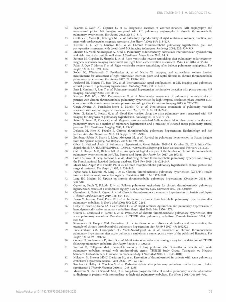
52 Rajaram S, Swift AJ, Capener D, et al. Diagnostic accuracy of contrast-enhanced MR angiography andunenhanced proton MR imaging compared with CT pulmonary angiography in chronic thromboembolicpulmonary hypertension. Eur Radiol 2012; 22: 310–317.
53 Grothues F, Moon JC, Bellenger NG, et al. Interstudy reproducibility of right ventricular volumes, function, andmass with cardiovascular magnetic resonance. Am Heart J 2004; 147: 218–223.
54 Kreitner K-FJ, Ley S, Kauczor H-U, et al. Chronic thromboembolic pulmonary hypertension: pre- andpostoperative assessment with breath-hold MR imaging techniques. Radiology 2004; 232: 535–543.
55 Mauritz GJ, Vonk-Noordegraaf A, Kind T. Pulmonary endarterectomy normalizes interventricular dyssynchronyand right ventricular systolic wall stress. J Cardiovasc Magn Reson 2012; 14: 5.
56 Berman M, Gopalan D, Sharples L, et al. Right ventricular reverse remodeling after pulmonary endarterectomy:magnetic resonance imaging and clinical and right heart catheterization assessment. Pulm Circ 2014; 4: 36–44.
57 Fukui S, Ogo T, Morita Y, et al. Right ventricular reverse remodelling after balloon pulmonary angioplasty. EurRespir J 2014; 43: 1394–1402.
58 Roller FC, Wiedenroth C, Breithecker A, et al. Native T1 mapping and extracellular volume fractionmeasurement for assessment of right ventricular insertion point and septal fibrosis in chronic thromboembolicpulmonary hypertension. Eur Radiol 2017; 27: 1980–1991.
59 Roeleveld RJ, Marcus JT, Faes TJC, et al. Interventricular septal configuration at MR imaging and pulmonaryarterial pressure in pulmonary hypertension. Radiology 2005; 234: 710–717.
60 Sanz J, Kuschnir P, Rius T, et al. Pulmonary arterial hypertension: noninvasive detection with phase-contrast MRimaging. Radiology 2007; 243: 70–79.
61 Kreitner K-F, Wirth GM, Krummenauer F, et al. Noninvasive assessment of pulmonary hemodynamics inpatients with chronic thromboembolic pulmonary hypertension by high temporal resolution phase-contrast MRI:correlation with simultaneous invasive pressure recordings. Circ Cardiovasc Imaging 2013; 6: 722–729.
62 García-Alvarez A, Fernández-Friera L, Mirelis JG, et al. Non-invasive estimation of pulmonary vascularresistance with cardiac magnetic resonance. Eur Heart J 2011; 32: 2438–2445.
63 Reiter G, Reiter U, Kovacs G, et al. Blood flow vortices along the main pulmonary artery measured with MRimaging for diagnosis of pulmonary hypertension. Radiology 2015; 275: 71–79.
64 Reiter G, Reiter U, Kovacs G, et al. Magnetic resonance-derived 3-dimensional blood flow patterns in the mainpulmonary artery as a marker of pulmonary hypertension and a measure of elevated mean pulmonary arterialpressure. Circ Cardiovasc Imaging 2008; 1: 23–30.
65 Delcroix M, Kerr K, Fedullo P. Chronic thromboembolic pulmonary hypertension. Epidemiology and riskfactors. Ann Am Thorac Soc 2016; 13: Suppl. 3, S201–S206.
66 Escribano-Subias P, Blanco I, López-Meseguer M, et al. Survival in pulmonary hypertension in Spain: insightsfrom the Spanish registry. Eur Respir J 2012; 40: 596–603.
67 Gibbs S. National Audit of Pulmonary Hypertension, Great Britain, 2018–19. October 24, 2019. https://files.digital.nhs.uk/BA/4EF20E/NAPH%2010AR%20-%20Main%20Report.pdf Date last accessed: February 24, 2020.
68 Gall H, Hoeper MM, Richter MJ, et al. An epidemiological analysis of the burden of chronic thromboembolicpulmonary hypertension in the USA, Europe and Japan. Eur Respir Rev 2017; 26: 160121.
69 Cottin V, Avot D, Lévy-Bachelot L, et al. Identifying chronic thromboembolic pulmonary hypertension throughthe French national hospital discharge database. PLoS One 2019; 14: e0214649.
70 Moser KM, Auger WR, Fedullo PF, et al. Chronic thromboembolic pulmonary hypertension: clinical picture andsurgical treatment. Eur Respir J 1992; 5: 334–342.
71 Pepke-Zaba J, Delcroix M, Lang I, et al. Chronic thromboembolic pulmonary hypertension (CTEPH): resultsfrom an international prospective registry. Circulation 2011; 124: 1973–1981.
72 Lang IM, Madani M. Update on chronic thromboembolic pulmonary hypertension. Circulation 2014; 130:508–518.
73 Ogawa A, Satoh T, Fukuda T, et al. Balloon pulmonary angioplasty for chronic thromboembolic pulmonaryhypertension: results of a multicenter registry. Circ Cardiovasc Qual Outcomes 2017; 10: e004029.
74 Chausheva S, Naito A, Ogawa A, et al. Chronic thromboembolic pulmonary hypertension in Austria and Japan.J Thorac Cardiovasc Surg 2019; 158: 604–614.
75 Pengo V, Lensing AWA, Prins MH, et al. Incidence of chronic thromboembolic pulmonary hypertension afterpulmonary embolism. N Engl J Med 2004; 350: 2257–2264.
76 Golpe R, Pérez-de-Llano LA, Castro-Añón O, et al. Right ventricle dysfunction and pulmonary hypertension inhemodynamically stable pulmonary embolism. Respir Med 2010; 104: 1370–1376.
77 Guérin L, Couturaud F, Parent F, et al. Prevalence of chronic thromboembolic pulmonary hypertension afteracute pulmonary embolism. Prevalence of CTEPH after pulmonary embolism. Thromb Haemost 2014; 112:598–605.
78 Simonneau G, Hoeper MM. Evaluation of the incidence of rare diseases: difficulties and uncertainties, theexample of chronic thromboembolic pulmonary hypertension. Eur Respir J 2017; 49: 1602522.
79 Ende-Verhaar YM, Cannegieter SC, Vonk-Noordegraaf A, et al. Incidence of chronic thromboembolicpulmonary hypertension after acute pulmonary embolism: a contemporary view of the published literature. EurRespir J 2017; 49: 1601792.
80 Coquoz N, Weilenmann D, Stolz D, et al. Multicentre observational screening survey for the detection of CTEPHfollowing pulmonary embolism. Eur Respir J 2018; 51: 1702505.
81 Wartski M, Collignon M-A. Incomplete recovery of lung perfusion after 3 months in patients with acutepulmonary embolism treated with antithrombotic agents. THESEE Study Group. Tinzaparin ou HeparinStandard: Evaluation dans l’Embolie Pulmonaire Study. J Nucl Med 2000; 41: 1043–1048.
82 Nijkeuter M, Hovens MMC, Davidson BL, et al. Resolution of thromboemboli in patients with acute pulmonaryembolism: a systematic review. Chest 2006; 129: 192–197.
83 Sanchez O, Helley D, Couchon S, et al. Perfusion defects after pulmonary embolism: risk factors and clinicalsignificance. J Thromb Haemost 2010; 8: 1248–1255.
84 Meneveau N, Ider O, Seronde M-F, et al. Long-term prognostic value of residual pulmonary vascular obstructionat discharge in patients with intermediate- to high-risk pulmonary embolism. Eur Heart J 2013; 34: 693–701.
https://doi.org/10.1183/13993003.02828-2020 33
ERS STATEMENT | M. DELCROIX ET AL.
85 Ma KA, Kahn SR, Akaberi A, et al. Serial imaging after pulmonary embolism and correlation with functionallimitation at 12 months: results of the ELOPE Study. Res Pract Thromb Haemost 2018; 2: 670–677.
86 Riedel M, Stanek V, Widimsky J, et al. Longterm follow-up of patients with pulmonary thromboembolism. Lateprognosis and evolution of hemodynamic and respiratory data. Chest 1982; 81: 151–158.
87 Lewczuk J, Piszko P, Jagas J, et al. Prognostic factors in medically treated patients with chronic pulmonaryembolism. Chest 2001; 119: 818–823.
88 Prandoni P, Ageno W, Mumoli N, et al. Recanalization rate in patients with proximal vein thrombosis treatedwith the direct oral anticoagulants. Thromb Res 2017; 153: 97–100.
89 Piazza G, Mani V, Goldhaber SZ, et al. Magnetic resonance venography to assess thrombus resolution withedoxaban monotherapy versus parenteral anticoagulation/warfarin for symptomatic deep vein thrombosis: amulticenter feasibility study. Vasc Med 2016; 21: 361–368.
90 Bunclark K, Newnham M, Chiu Y-D, et al. A multicenter study of anticoagulation in operable chronicthromboembolic pulmonary hypertension. J Thromb Haemost 2020; 18: 114–122.
91 Houghton DE, Lekah A, Macedo TA, et al. Resolution of acute lower extremity deep vein thrombosis withrivaroxaban compared to warfarin. J Thromb Thrombolysis 2020; 49: 199–205.
92 Konstantinides SV, Meyer G, Becattini C, et al. 2019 ESC Guidelines for the diagnosis and management of acutepulmonary embolism developed in collaboration with the European Respiratory Society (ERS): the task force forthe diagnosis and management of acute pulmonary embolism of the European Society of Cardiology (ESC).Eur Respir J 2019; 54: 1901647.
93 Konstantinides SV, Meyer G, Becattini C, et al. 2019 ESC Guidelines for the diagnosis and management of acutepulmonary embolism developed in collaboration with the European Respiratory Society (ERS). Eur Heart J 2020;41: 543–603.
94 Pengo V, Denas G, Zoppellaro G, et al. Rivaroxaban vs warfarin in high-risk patients with antiphospholipidsyndrome. Blood 2018; 132: 1365–1371.
95 Moll S, Crona DJ, Martin K. Direct oral anticoagulants in extremely obese patients: OK to use? Res Pract ThrombHaemost 2019; 3: 152–155.
96 Ingrasciotta Y, Crisafulli S, Pizzimenti V, et al. Pharmacokinetics of new oral anticoagulants: implications for usein routine care. Expert Opin Drug Metab Toxicol 2018; 14: 1057–1069.
97 Bonderman D, Jakowitsch J, Adlbrecht C, et al. Medical conditions increasing the risk of chronicthromboembolic pulmonary hypertension. Thromb Haemost 2005; 93: 512–516.
98 Lang I, Simonneau G, Pepke-Zaba J, et al. Factors associated with diagnosis and operability of chronicthromboembolic pulmonary hypertension. A case-control study. Thromb Haemost 2013; 110: 83–91.
99 Delcroix M, Lang I, Pepke-Zaba J, et al. Long-term outcome of patients with chronic thromboembolicpulmonary hypertension: results from an international prospective registry. Circulation 2016; 133: 859–871.
100 Ende-Verhaar YM, Huisman MV, Klok FA. To screen or not to screen for chronic thromboembolic pulmonaryhypertension after acute pulmonary embolism. Thromb Res 2017; 151: 1–7.
101 Ende-Verhaar YM, van den Hout WB, Bogaard H-J, et al. Healthcare utilization in chronic thromboembolicpulmonary hypertension after acute pulmonary embolism. J Thromb Haemost 2018; 16: 2168–2174.
102 Klok FA, Barco S, Konstantinides SV, et al. Determinants of diagnostic delay in chronic thromboembolicpulmonary hypertension: results from the European CTEPH Registry. Eur Respir J 2018; 52: 1801687.
103 Jensen KW, Kerr KM, Fedullo PF, et al. Pulmonary hypertensive medical therapy in chronic thromboembolicpulmonary hypertension before pulmonary thromboendarterectomy. Circulation 2009; 120: 1248–1254.
104 Nishiyama KH, Saboo SS, Tanabe Y, et al. Chronic pulmonary embolism: diagnosis. Cardiovasc Diagn Ther 2018;8: 253–271.
105 Rajaram S, Swift AJ, Condliffe R, et al. CT features of pulmonary arterial hypertension and its major subtypes: asystematic CT evaluation of 292 patients from the ASPIRE Registry. Thorax 2015; 70: 382–387.
106 Ende-Verhaar YM, Meijboom LJ, Kroft LJM, et al. Usefulness of standard computed tomography pulmonaryangiography performed for acute pulmonary embolism for identification of chronic thromboembolic pulmonaryhypertension: results of the InShape III study. J Heart Lung Transplant 2019; 38: 731–738.
107 McIntyre KM, Sasahara AA. The hemodynamic response to pulmonary embolism in patients without priorcardiopulmonary disease. Am J Cardiol 1971; 28: 288–294.
108 Klok FA, Delcroix M, Bogaard H-J. Chronic thromboembolic pulmonary hypertension from the perspective ofpatients with pulmonary embolism. J Thromb Haemost 2018; 16: 1040–1051.
109 Sista AK, Klok FA. Late outcomes of pulmonary embolism: the post-PE syndrome. Thromb Res 2018; 164:157–162.
110 Sista AK, Miller LE, Kahn SR, et al. Persistent right ventricular dysfunction, functional capacity limitation,exercise intolerance, and quality of life impairment following pulmonary embolism: systematic review withmeta-analysis. Vasc Med 2017; 22: 37–43.
111 Klok FA, van der Hulle T, den Exter PL, et al. The post-PE syndrome: a new concept for chronic complicationsof pulmonary embolism. Blood Rev 2014; 28: 221–226.
112 Klok FA, Dzikowska-Diduch O, Kostrubiec M, et al. Derivation of a clinical prediction score for chronicthromboembolic pulmonary hypertension after acute pulmonary embolism. J Thromb Haemost 2016; 14:121–128.
113 Wittram C. How I do it: CT pulmonary angiography. AJR Am J Roentgenol 2007; 188: 1255–1261.114 Helmersen D, Provencher S, Hirsch AM, et al. Diagnosis of chronic thromboembolic pulmonary hypertension: a
Canadian Thoracic Society clinical practice guideline update. Can J Respir Crit Care Sleep Med 2019; 3: 177–198.115 Klok FA, van Kralingen KW, van Dijk APJ, et al. Prevalence and potential determinants of exertional dyspnea
after acute pulmonary embolism. Respir Med 2010; 104: 1744–1749.116 Kahn SR, Akaberi A, Granton JT, et al. Quality of life, dyspnea, and functional exercise capacity following a first
episode of pulmonary embolism: results of the ELOPE cohort study. Am J Med 2017; 130: 990.e9–990.e21.117 Kahn SR, Hirsch AM, Akaberi A, et al. Functional and exercise limitations after a first episode of pulmonary
embolism: results of the ELOPE prospective cohort study. Chest 2017; 151: 1058–1068.118 Sznajder M, Dzikowska-Diduch O, Kurnicka K, et al. Increased systemic arterial stiffness in patients with chronic
thromboembolic pulmonary hypertension. Cardiol J 2020; 27: 742–748.
https://doi.org/10.1183/13993003.02828-2020 34
ERS STATEMENT | M. DELCROIX ET AL.
119 Pietra GG, Capron F, Stewart S, et al. Pathologic assessment of vasculopathies in pulmonary hypertension. J AmColl Cardiol 2004; 43: 25S–32S.
120 Humbert M, Guignabert C, Bonnet S, et al. Pathology and pathobiology of pulmonary hypertension: state of theart and research perspectives. Eur Respir J 2019; 53: 1801887.
121 Moser KM, Bloor CM. Pulmonary vascular lesions occurring in patients with chronic major vesselthromboembolic pulmonary hypertension. Chest 1993; 103: 685–692.
122 Gerges C, Gerges M, Friewald R, et al. Microvascular disease in chronic thromboembolic pulmonaryhypertension: hemodynamic phenotyping and histomorphometric assessment. Circulation 2020; 141: 376–386.
123 Noly P-E, Guihaire J, Coblence M, et al. Chronic thromboembolic pulmonary hypertension and assessment ofright ventricular function in the piglet. J Vis Exp 2015; 105: e53133.
124 Tsai H-Y, Chin C-S, Tsai I-C. Imaging as a biomarker in Behçet disease: prognostic and therapeutic implicationsof serial pulmonary CT angiography. AJR Am J Roentgenol 2011; 196: W105–W106.
125 Perino MG, Moldobaeva A, Jenkins J, et al. Chemokine localization in bronchial angiogenesis. PLoS One 2013; 8:e66432.
126 Mercier O, Fadel E. Chronic thromboembolic pulmonary hypertension: animal models. Eur Respir J 2013; 41:1200–1206.
127 Yi ES, Kim H, Ahn H, et al. Distribution of obstructive intimal lesions and their cellular phenotypes in chronicpulmonary hypertension. A morphometric and immunohistochemical study. Am J Respir Crit Care Med 2000;162: 1577–1586.
128 Galambos C, Sims-Lucas S, Abman SH, et al. Intrapulmonary bronchopulmonary anastomoses and plexiformlesions in idiopathic pulmonary arterial hypertension. Am J Respir Crit Care Med 2016; 193: 574–576.
129 Ghigna M-R, Guignabert C, Montani D, et al. BMPR2 mutation status influences bronchial vascular changes inpulmonary arterial hypertension. Eur Respir J 2016; 48: 1668–1681.
130 Azarian R, Wartski M, Collignon MA, et al. Lung perfusion scans and hemodynamics in acute and chronicpulmonary embolism. J Nucl Med 1997; 38: 980–983.
131 Kim NHS, Fesler P, Channick RN, et al. Preoperative partitioning of pulmonary vascular resistance correlateswith early outcome after thromboendarterectomy for chronic thromboembolic pulmonary hypertension.Circulation 2004; 109: 18–22.
132 Toshner M, Suntharalingam J, Fesler P, et al. Occlusion pressure analysis role in partitioning of pulmonaryvascular resistance in CTEPH. Eur Respir J 2012; 40: 612–617.
133 Simonneau G, Torbicki A, Dorfmüller P, et al. The pathophysiology of chronic thromboembolic pulmonaryhypertension. Eur Respir Rev 2017; 26: 160112.
134 Ley S, Kreitner K-F, Morgenstern I, et al. Bronchopulmonary shunts in patients with chronic thromboembolicpulmonary hypertension: evaluation with helical CT and MR imaging. AJR Am J Roentgenol 2002; 179:1209–1215.
135 Miller F, Coulden RA, Sonnex E, et al. The use of MR flow mapping in the assessment of pulmonary arteryblood flow following pulmonary thrombo-endarterectomy. Radiological Society of North America 2003 ScientificAssembly and Annual Meeting. December 2003: 426P.
136 Reiter U, Reiter G, Kovacs G, et al. Evaluation of elevated mean pulmonary arterial pressure based on magneticresonance 4D velocity mapping: comparison of visualization techniques. PLoS One 2013; 8: e82212.
137 Ota H, Sugimura K, Miura M, et al. Four-dimensional flow magnetic resonance imaging visualizes drastic changein vortex flow in the main pulmonary artery after percutaneous transluminal pulmonary angioplasty in a patientwith chronic thromboembolic pulmonary hypertension. Eur Heart J 2015; 36: 1630.
138 Polanowski T, Kurzyna M, Kuca P, et al. Róznice w hemodynamice pierwotnego i zatorowego nadciśnieniapłucnego. [Differences in hemodynamics of thromboembolic and primary pulmonary hypertension]. Pol ArchMed Wewn 2000; 104: 741–745.
139 Quarck R, Nawrot T, Meyns B, et al. C-reactive protein: a new predictor of adverse outcome in pulmonaryarterial hypertension. J Am Coll Cardiol 2009; 53: 1211–1218.
140 Axell RG, Messer SJ, White PA, et al. Ventriculo-arterial coupling detects occult RV dysfunction in chronicthromboembolic pulmonary vascular disease. Physiol Rep 2017; 5: e13227.
141 MacKenzie Ross RV, Toshner MR, Soon E, et al. Decreased time constant of the pulmonary circulation inchronic thromboembolic pulmonary hypertension. Am J Physiol Heart Circ Physiol 2013; 305: H259–H264.
142 Menzel T, Wagner S, Kramm T, et al. Pathophysiology of impaired right and left ventricular function in chronicembolic pulmonary hypertension: changes after pulmonary thromboendarterectomy. Chest 2000; 118: 897–903.
143 Bonderman D, Martischnig AM, Vonbank K, et al. Right ventricular load at exercise is a cause of persistentexercise limitation in patients with normal resting pulmonary vascular resistance after pulmonaryendarterectomy. Chest 2011; 139: 122–127.
144 Jenkins D, Madani M, Fadel E, et al. Pulmonary endarterectomy in the management of chronic thromboembolicpulmonary hypertension. Eur Respir Rev 2017; 26: 160111.
145 Mayer E, Jenkins D, Lindner J, et al. Surgical management and outcome of patients with chronicthromboembolic pulmonary hypertension: results from an international prospective registry. J Thorac CardiovascSurg 2011; 141: 702–710.
146 Boulate D, Mercier O, Mussot S, et al. Extracorporeal life support after pulmonary endarterectomy as a bridge torecovery or transplantation: lessons from 31 consecutive patients. Ann Thorac Surg 2016; 102: 260–268.
147 Madani M, Ogo T, Simonneau G. The changing landscape of chronic thromboembolic pulmonary hypertensionmanagement. Eur Respir Rev 2017; 26: 170105.
148 Kawakami T, Ogawa A, Miyaji K, et al. Novel angiographic classification of each vascular lesion in chronicthromboembolic pulmonary hypertension based on selective angiogram and results of balloon pulmonaryangioplasty. Circ Cardiovasc Interv 2016; 9: e003318.
149 D’Armini AM, Morsolini M, Mattiucci G, et al. Pulmonary endarterectomy for distal chronic thromboembolicpulmonary hypertension. J Thorac Cardiovasc Surg 2014; 148: 1005–1011.
150 Hsieh WC, Jansa P, Huang WC, et al. Residual pulmonary hypertension after pulmonary endarterectomy: ameta-analysis. J Thorac Cardiovasc Surg 2018; 156: 1275–1287.
https://doi.org/10.1183/13993003.02828-2020 35
ERS STATEMENT | M. DELCROIX ET AL.
151 Madani MM, Auger WR, Pretorius V, et al. Pulmonary endarterectomy: recent changes in a single institution’sexperience of more than 2,700 patients. Ann Thorac Surg 2012; 94: 97–103.
152 Cannon JE, Su L, Kiely DG, et al. Dynamic risk stratification of patient long-term outcome after pulmonaryendarterectomy: results from the United Kingdom national cohort. Circulation 2016; 133: 1761–1771.
153 Vuylsteke A, Sharples L, Charman G, et al. Circulatory arrest versus cerebral perfusion during pulmonaryendarterectomy surgery (PEACOG): a randomised controlled trial. Lancet 2011; 378: 1379–1387.
154 Corsico AG, D’Armini AM, Cerveri I, et al. Long-term outcome after pulmonary endarterectomy. Am J RespirCrit Care Med 2008; 178: 419–424.
155 Kramm T, Eberle B, Guth S, et al. Inhaled iloprost to control residual pulmonary hypertension followingpulmonary endarterectomy. Eur J Cardiothorac Surg 2005; 28: 882–888.
156 Ghofrani HA, D’Armini AM, Grimminger F, et al. Riociguat for the treatment of chronic thromboembolicpulmonary hypertension. N Engl J Med 2013; 369: 319–329.
157 Shimura N, Kataoka M, Inami T, et al. Additional percutaneous transluminal pulmonary angioplasty for residualor recurrent pulmonary hypertension after pulmonary endarterectomy. Int J Cardiol 2015; 183: 138–142.
158 Ali JM, Dunning J, Ng C, et al. The outcome of reoperative pulmonary endarterectomy surgery. InteractCardiovasc Thorac Surg 2018; 26: 932–937.
159 Mizoguchi H, Ogawa A, Munemasa M, et al. Refined balloon pulmonary angioplasty for inoperable patients withchronic thromboembolic pulmonary hypertension. Circ Cardiovasc Interv 2012; 5: 748–755.
160 Kataoka M, Inami T, Hayashida K, et al. Percutaneous transluminal pulmonary angioplasty for the treatment ofchronic thromboembolic pulmonary hypertension. Circ Cardiovasc Interv 2012; 5: 756–762.
161 Sugimura K, Fukumoto Y, Satoh K, et al. Percutaneous transluminal pulmonary angioplasty markedly improvespulmonary hemodynamics and long-term prognosis in patients with chronic thromboembolic pulmonaryhypertension. Circ J 2012; 76: 485–488.
162 Lang I, Meyer BC, Ogo T, et al. Balloon pulmonary angioplasty in chronic thromboembolic pulmonaryhypertension. Eur Respir Rev 2017; 26: 160119.
163 Wiedenroth CB, Liebetrau C, Breithecker A, et al. Combined pulmonary endarterectomy and balloon pulmonaryangioplasty in patients with chronic thromboembolic pulmonary hypertension. J Heart Lung Transplant 2016;35: 591–596.
164 Ogo T. Balloon pulmonary angioplasty for inoperable chronic thromboembolic pulmonary hypertension. CurrOpin Pulm Med 2015; 21: 425–431.
165 Zoppellaro G, Badawy MR, Squizzato A, et al. Balloon pulmonary angioplasty in patients with chronicthromboembolic pulmonary hypertension – a systematic review and meta-analysis. Circ J 2019; 83: 1660–1667.
166 Darocha S, Pietura R, Pietrasik A, et al. Improvement in quality of life and hemodynamics in chronicthromboembolic pulmonary hypertension treated with balloon pulmonary angioplasty. Circ J 2017; 81: 552–557.
167 Ogawa A, Matsubara H. After the dawn – balloon pulmonary angioplasty for patients with chronicthromboembolic pulmonary hypertension. Circ J 2018; 82: 1222–1230.
168 Fukui S, Ogo T, Goto Y, et al. Exercise intolerance and ventilatory inefficiency improve early after balloonpulmonary angioplasty in patients with inoperable chronic thromboembolic pulmonary hypertension. Int JCardiol 2015; 180: 66–68.
169 Olsson KM, Wiedenroth CB, Kamp J-C, et al. Balloon pulmonary angioplasty for inoperable patients withchronic thromboembolic pulmonary hypertension: the initial German experience. Eur Respir J 2017; 49: 1602409.
170 Brenot P, Jaïs X, Taniguchi Y, et al. French experience of balloon pulmonary angioplasty for chronicthromboembolic pulmonary hypertension. Eur Respir J 2019; 53: 1802095.
171 Feinstein JA, Goldhaber SZ, Lock JE, et al. Balloon pulmonary angioplasty for treatment of chronicthromboembolic pulmonary hypertension. Circulation 2001; 103: 10–13.
172 Ejiri K, Ogawa A, Fujii S, et al. Vascular injury is a major cause of lung injury after balloon pulmonary angioplastyin patients with chronic thromboembolic pulmonary hypertension. Circ Cardiovasc Interv 2018; 11: e005884.
173 Inami T, Kataoka M, Shimura N, et al. Incidence, avoidance, and management of pulmonary artery injuries inpercutaneous transluminal pulmonary angioplasty. Int J Cardiol 2015; 201: 35–37.
174 Inami T, Kataoka M, Yanagisawa R, et al. Long-term outcomes after percutaneous transluminal pulmonaryangioplasty for chronic thromboembolic pulmonary hypertension. Circulation 2016; 134: 2030–2032.
175 Aoki T, Sugimura K, Tatebe S, et al. Comprehensive evaluation of the effectiveness and safety of balloonpulmonary angioplasty for inoperable chronic thrombo-embolic pulmonary hypertension: long-term effects andprocedure-related complications. Eur Heart J 2017; 38: 3152–3159.
176 Taniguchi Y, Jaïs X, Jevnikar M, et al. Predictors of survival in patients with not-operated chronicthromboembolic pulmonary hypertension. J Heart Lung Transplant 2019; 38: 833–842.
177 Humbert M, Simonneau G, Pittrow D, et al. Safety of riociguat in patients with pulmonary arterial hypertensionand chronic thromboembolic pulmonary hypertension with concomitant novel oral anticoagulants or vitamin Kantagonist use: data from the EXPERT Registry. Am J Respir Crit Care Med 2020; 201: A6043.
178 Jeong I, Fernandes T, Alotaibi M, et al. Direct oral anticoagulant use and thrombus detection in patients withchronic thromboembolic pulmonary hypertension referred for pulmonary thromboendarterectomy. Eur Respir J2019; 54: OA5161.
179 Skoro-Sajer N, Hack N, Sadushi-Koliçi R, et al. Pulmonary vascular reactivity and prognosis in patients withchronic thromboembolic pulmonary hypertension: a pilot study. Circulation 2009; 119: 298–305.
180 Suntharalingam J, Hughes RJ, Goldsmith K, et al. Acute haemodynamic responses to inhaled nitric oxide andintravenous sildenafil in distal chronic thromboembolic pulmonary hypertension (CTEPH). Vascul Pharmacol2007; 46: 449–455.
181 Suntharalingam J, Treacy CM, Doughty NJ, et al. Long-term use of sildenafil in inoperable chronicthromboembolic pulmonary hypertension. Chest 2008; 134: 229–236.
182 Jaïs X, D’Armini AM, Jansa P, et al. Bosentan for treatment of inoperable chronic thromboembolic pulmonaryhypertension: BENEFiT (Bosentan Effects in iNopErable Forms of chronIc Thromboembolic pulmonaryhypertension), a randomized, placebo-controlled trial. J Am Coll Cardiol 2008; 52: 2127–2134.
https://doi.org/10.1183/13993003.02828-2020 36
ERS STATEMENT | M. DELCROIX ET AL.
183 Ghofrani HA, Simonneau G, D’Armini AM, et al. Macitentan for the treatment of inoperable chronicthromboembolic pulmonary hypertension (MERIT-1): results from the multicentre, phase 2, randomised,double-blind, placebo-controlled study. Lancet Respir Med 2017; 5: 785–794.
184 Sadushi-Koliçi R, Jansa P, Kopeć G, et al. Subcutaneous treprostinil for the treatment of severe non-operablechronic thromboembolic pulmonary hypertension (CTREPH): a double-blind, phase 3, randomised controlledtrial. Lancet Respir Med 2019; 7: 239–248.
185 Gabrielly M, Bourlier D, Taniguchi Y, et al. Initial dual oral combination therapy in inoperable chronicthromboembolic pulmonary hypertension (CTEPH). Eur Respir J 2018; 52: PA3053.
186 Tromeur C, Jaïs X, Mercier O, et al. Factors predicting outcome after pulmonary endarterectomy. PLoS One2018; 13: e0198198.
187 Nagaya N, Sasaki N, Ando M, et al. Prostacyclin therapy before pulmonary thromboendarterectomy in patientswith chronic thromboembolic pulmonary hypertension. Chest 2003; 123: 338–343.
188 Bresser P, Fedullo PF, Auger WR, et al. Continuous intravenous epoprostenol for chronic thromboembolicpulmonary hypertension. Eur Respir J 2004; 23: 595–600.
189 Reesink HJ, Surie S, Kloek JJ, et al. Bosentan as a bridge to pulmonary endarterectomy for chronicthromboembolic pulmonary hypertension. J Thorac Cardiovasc Surg 2010; 139: 85–91.
190 Castro MA, Piloto B, Dos Santos Fernandes CJC, et al. Use of medical therapies before pulmonaryendarterectomy in chronic thromboembolic pulmonary hypertension patients with severe hemodynamicimpairment. PLoS One 2020; 15: e0233063. PMID 32442171.
191 Kawashima T, Yoshitake A, Kawakami T, et al. Two-stage treatment using balloon pulmonary angioplasty andpulmonary endarterectomy in a patient with chronic thromboembolic pulmonary hypertension. Ann Vasc Surg2018; 49: 315.e5–315.e7.
192 Yanaka K, Nakayama K, Shinke T, et al. Sequential hybrid therapy with pulmonary endarterectomy andadditional balloon pulmonary angioplasty for chronic thromboembolic pulmonary hypertension. J Am HeartAssoc 2018; 7: e008838.
193 Araszkiewicz A, Darocha S, Pietrasik A, et al. Balloon pulmonary angioplasty for the treatment of residual orrecurrent pulmonary hypertension after pulmonary endarterectomy. Int J Cardiol 2019; 278: 232–237.
194 Nakamura M, Sunagawa O, Tsuchiya H, et al. Rescue balloon pulmonary angioplasty under veno-arterialextracorporeal membrane oxygenation in a patient with acute exacerbation of chronic thromboembolicpulmonary hypertension. Int Heart J 2015; 56: 116–120.
195 Collaud S, Brenot P, Mercier O, et al. Rescue balloon pulmonary angioplasty for early failure of pulmonaryendarterectomy: The earlier the better? Int J Cardiol 2016; 222: 39–40.
196 Wiedenroth CB, Ghofrani HA, Adameit MSD, et al. Sequential treatment with riociguat and balloon pulmonaryangioplasty for patients with inoperable chronic thromboembolic pulmonary hypertension. Pulm Circ 2018; 8:2045894018783996.
197 Mereles D, Ehlken N, Kreuscher S, et al. Exercise and respiratory training improve exercise capacity and qualityof life in patients with severe chronic pulmonary hypertension. Circulation 2006; 114: 1482–1489.
198 Ehlken N, Lichtblau M, Klose H, et al. Exercise training improves peak oxygen consumption andhaemodynamics in patients with severe pulmonary arterial hypertension and inoperable chronicthrombo-embolic pulmonary hypertension: a prospective, randomized, controlled trial. Eur Heart J 2016; 37:35–44.
199 Handoko ML, De Man FS, Happé CM, et al. Opposite effects of training in rats with stable and progressivepulmonary hypertension. Circulation 2009; 120: 42–49.
200 Zhai Z, Murphy K, Tighe H, et al. Differences in ventilatory inefficiency between pulmonary arterialhypertension and chronic thromboembolic pulmonary hypertension. Chest 2011; 140: 1284–1291.
201 Nagel C, Prange F, Guth S, et al. Exercise training improves exercise capacity and quality of life in patients withinoperable or residual chronic thromboembolic pulmonary hypertension. PLoS One 2012; 7: e41603.
202 Inagaki T, Terada J, Tanabe N, et al. Home-based pulmonary rehabilitation in patients with inoperable orresidual chronic thromboembolic pulmonary hypertension: a preliminary study. Respir Investig 2014; 52:357–364.
203 La Rovere MT, Pinna GD, Pin M, et al. Exercise training after pulmonary endarterectomy for patients withchronic thromboembolic pulmonary hypertension. Respiration 2019; 97: 234–241.
204 Fukui S, Ogo T, Takaki H, et al. Efficacy of cardiac rehabilitation after balloon pulmonary angioplasty for chronicthromboembolic pulmonary hypertension. Heart 2016; 102: 1403–1409.
205 Opitz I, Kirschner MB. Molecular research in chronic thromboembolic pulmonary hypertension. Int J Mol Sci2019; 20: 784.
206 Hemnes AR, Beck GJ, Newman JH, et al. PVDOMICS: a multi-center study to improve understanding ofpulmonary vascular disease through phenomics. Circ Res 2017; 121: 1136–1139.
207 Newnham M, South K, Bleda M, et al. The ADAMTS13–VWF axis is dysregulated in chronic thromboembolicpulmonary hypertension. Eur Respir J 2019; 53: 1801805.
208 Heresi GA, Mey JT, Bartholomew JR, et al. Plasma metabolomic profile in chronic thromboembolic pulmonaryhypertension. Pulm Circ 2020; 10: 2045894019890553.
209 Madani MM, Wittine LM, Auger WR, et al. Chronic thromboembolic pulmonary hypertension in pediatricpatients. J Thorac Cardiovasc Surg 2011; 141: 624–630.
210 Kumbasar U, Aypar E, Karagöz T, et al. Pulmonary thromboendarterectomy in pediatric patients: report of threecases. Turk J Pediatr 2018; 60: 604–607.
211 Verbelen T, Cools B, Fejzic Z, et al. Pulmonary endarterectomy in a 12-year-old boy with multiple comorbidities.Pulm Circ 2019; 9: 2045894019886249.
https://doi.org/10.1183/13993003.02828-2020 37
ERS STATEMENT | M. DELCROIX ET AL.