Embed Size (px)
Citation preview
Full Terms & Conditions of access and use can be found athttp://www.tandfonline.com/action/journalInformation?journalCode=taca20
International Journal of Acarology
ISSN: 0164-7954 (Print) 1945-3892 (Online) Journal homepage: http://www.tandfonline.com/loi/taca20
Eriophyid mites in the genus Aculodes Keifer(Prostigmata: Eriophyidae) from grasses – thefirst taxon from South America and world speciesinformation
Vanina Alemandri, Graciela Truol, Renata S. de Mendonça & Denise Navia
To cite this article: Vanina Alemandri, Graciela Truol, Renata S. de Mendonça & Denise Navia(2015) Eriophyid mites in the genus Aculodes Keifer (Prostigmata: Eriophyidae) from grasses – thefirst taxon from South America and world species information, International Journal of Acarology,41:5, 429-441, DOI: 10.1080/01647954.2015.1050063
To link to this article: https://doi.org/10.1080/01647954.2015.1050063
Published online: 17 Jul 2015.
Submit your article to this journal
Article views: 102
View Crossmark data

Eriophyid mites in the genus Aculodes Keifer (Prostigmata: Eriophyidae) from grasses – the firsttaxon from South America and world species information
Vanina Alemandria, Graciela Truola, Renata S. de Mendonçab and Denise Naviac
aInstituto de Patología Vegetal, Centro de Investigaciones Agropecuarias, Instituto Nacional de Tecnología Agropecuaria (IPAVE-CIAP-INTA), Córdoba, Argentina (emails: [email protected], [email protected]); bFundação de Apoio à Pesquisa(FUNAPE)/Embrapa Recursos Genéticos e Biotecnologia, Parque Estação Biológica, Brasilia, Brazil (email: [email protected]); cEmbrapa Recursos Genéticos e Biotecnologia, Parque Estação Biológica, Brasilia, Brazil (email: [email protected])
(Received 25 December 2014; accepted 6 May 2015; published online 17 July 2015)
Aculodes Keifer (Eriophyidae) mites are often associated with grasses (Poaceae) and so far all species in this genus weredescribed from the Northern hemisphere. During surveys of eriophyid mites from grasses conducted in Argentina aiming todetermine the host range of the wheat curl mite, a new Aculodes species was collected from the feather grass, Stipa sp. Thefirst Aculodes species is described from the Southern hemisphere. In addition to the traditional morphological description,DNA sequences of two genomic regions – the ITS rDNA and the 16S region mtDNA– were obtained and deposited inpublic databases; genetic distances of the new taxon with eriophyid species/genera associated with grasses are presented. Inorder to contribute to Aculodes taxonomy, a list for world grasses-associated species is provided and information on itsmorphological traits is summarized.
Keywords: Eriophyoidea; Poaceae; Argentina; DNA data; taxonomy
Introduction
The genus Aculodes Keifer, 1966b (Eriophyidae,Phyllocoptinae, Anthocoptini) presently includes 26species, most of them (21 species) associated with grasses(Poaceae) (Nalepa 1891; Keifer 1944, 1952, 1960, 1966a,1966b; Sukhareva 1972, 1981, 1985, 1986, 1994; Huang1992, 2001; Kuang 1997; Kuang and Pang 1997; Shi andBoczek 2000; Skoracka et al. 2001, 2009; Skoracka 2003,2004, 2005; Kuang et al. 2005; Skoracka and Pacyna2005; Xue et al. 2010). Until now, all Aculodes specieswere described from the Northern hemisphere, in Europe,Asia and North America. Only one species in this genus,Aculodes mckenziei (Keifer, 1944), had been reported fromthe Southern hemisphere, in Australia and New Zealand(Frost et al. 1990; Guy and Gould 1996). No Aculodesspecies had been described or reported from SouthAmerica.
Knowledge on eriophyoid mites on grasses in SouthAmerica is scarce; only eight species belonging to thegenera Aceria, Abacarus, Catarhinus, and Schizaceahave been described or reported from these plants in thecontinent (Keifer 1959, 1976, 1977, 1978; Flechtmann andAranda 1970; Flechtmann 2000; Navia et al. 2006; Pereiraet al. 2009; Castiglioni and Navia 2010). In Argentina,only the wheat curl mite, Aceria tosichella Keifer, 1969,had been reported (Navia et al. 2006).
In early 2000s, the wheat curl mite was collected forthe first time in South America, in Argentina, from wheatplants infected with Wheat streak mosaic virus (Naviaet al., 2006). Since then, surveys of eriophyid mites fromgrasses in that country have been conducted to determinethe host range of the wheat curl mite. From these surveys a
new Aculodes species was found from the feather grassStipa sp. This is the first Aculodes associated with grassesto be described from South America and even from theSouthern hemisphere.
Five eriophyoid mites have been described or reportedfrom Stipa grasses – four Eriophyidae species, three inthe genus Aceria – A. stipaespinulata Skoracka, 2004 fromS. joannis Celak and S. capillata L. from Poland; A.stipaensis Mitrofanov & Sharonov, 1988 from Stipa sp.from Ukraine (Mitrofanov et al. 1988); and A. stipaceaSukhareva, 1983 described from S. lessingiana Trin. etRupr. from Ukraine and also reported from S. capillatain Kazakhstan – and one in the genus Aculodes – A.fulleri (Keifer, 1966a) from S. californica Merr. & Burtt-Davy ex Hall. (presently referred as a synonym of S.occidentalis Thurb. ex S.Watson (The Plant List 2013))from California, USA; and one Phytoptidae species,Novophytoptus stipae Keifer, 1962 (host plantAchnatherum speciosum (Trin. & Rupr.) Barkworth, pre-sently referred as a synonym of Stipa speciosa Trin. &Rupr. (The Plant List 2013).
DNA-based resources have started to be used ineriophyoid mite systematics about 15 years ago.Important advances have been done using molecular tech-niques, which had contributed to explore some questionsthat were difficult to answer some years ago (Navajas andNavia 2010). Cryptic species have been uncovered amongeriophyid mites associated with grasses through an inte-grative approach, including DNA data (Skoracka andDabert 2010; Skoracka et al. 2012; Miller et al. 2013).Molecular information can help to distinguish species andprovide DNA data for new taxa can help to build a
International Journal of Acarology, 2015Vol. 41, No. 5, 429–441, http://dx.doi.org/10.1080/01647954.2015.1050063
© 2015 Taylor & Francis
Published online 17 Jul 2015

consistent taxonomy (Dabert et al. 2008). Among mole-cular markers successfully used for eriophyid mite sys-tematics, we can list the nuclear ribosomal InternalTranscribed Spacer (ITS) and the 16S mitochondrial(Navia et al. 2005; Navajas and Navia 2010; Skorackaet al. 2012; Miller et al. 2013).
In this paper, the description of a new Aculodes miteassociated with Stipa grass in Argentina is presented. Inaddition to the traditional morphological description,including measures and drawings, DNA sequences oftwo genomic regions – the ITS rDNA and the 16S regionmtDNA– were obtained and are presented. Genetic dis-tances of the new taxon with eriophyid species/generawere estimated.
Taxonomic literature on Aculodes species associatedwith grasses is sparse and description of some species isnot in English making difficult its access. In a way tocontribute to Aculodes taxonomy and make easier newstudies, a list for grasses-associated valid species is pre-sented as well as a table with summarized information onits main morphological traits.
Material and methods
Morphological study
Mites were collected from leaf samples by direct examina-tion under a dissecting stereomicroscope and directlymounted in modified Berlese medium (Amrine andManson 1996). Slide-mounted specimens were studiedusing a research phase and differential interference con-trast microscope (Eclipse 80i Nikon, Tokyo, Japan).Relevant structures for taxonomic purposes were measuredusing a graded eyepiece and illustrated using a cameralucida attached to the miscroscope.
Terminology follows that of Lindquist (1996) andclassification is based on Amrine et al. (2003).Measurements are given in micrometers (µm) and, unlessstated otherwise, refer to the length of the structure. In thedescription of the female, each measurement of the holo-type precedes the corresponding range for the paratypes.Some measurements of paratypes could not be takenbecause of the position in which the specimens weremounted. The specimen that was drawing in lateral viewstayed mounted in a position slightly, since then theventral seta e was not represented in its drawing. Thecount of ventral opisthosomal annuli starts from the firstfull annulus behind the genitalia. Dorsal opisthosomalannuli were counted from the first full annulus behindthe middle of the prodorsal shield rear margin. Whenthe length of cheliceral stylets is not given, it means thatit was not possible to measure this character, since theywere in a bundle that was immersed with other gnathoso-mal structures. Measurements were conducted accordingto de Lillo et al. (2010) except for the following: (1) thebody length, which was measured from the tip of thefrontal lobe to the rear end of the anal lobe, not consider-ing pedipalps; (2) the sc tubercles space measurement
(distance between the tubercles), not the sc setae distance;(3) empodium length, which includes its basal portioninserted into the tarsus.
No immature stages were found and only one malewas found and studied. Female internal genitalia was notvisible in the studied specimens.
Micrographs were obtained using a digital systemconsisting of the phase and differential interferencecontrast microscope (Nikon Eclipse 80i, Tokyo, Japan)connected to a digital camera (Nikon DS-Ri, 12.7 megapixels, Tokyo, Japan) which was in turn connected to acomputer with NIS Elements software (Nikon).
Type specimens are deposited as slide-mounted speci-mens in mite collections at Embrapa Recursos Genéticos eBiotecnologia, Brasilia, Brazil and at Departamento deEntomologia, Fitopatologia e Zoologia Agrícola, EscolaSuperior de Agricultura “Luiz de Queiroz” (ESALQ),Universidade de São Paulo, Piracicaba, São Paulo, Brazil.
Molecular characterization
Specimens were preserved in absolute ethyl alcohol andthen individualized in an eppendorf tube for DNAextraction.
DNA extraction
A Chelex method was used to extract DNA from a singlemite following Carew et al. (2004). Microcentrifuge tubescontaining mites were centrifuged at 20800 g for 5 min toensure that mites were at or near the top of the tube. Threemicrolitres of Proteinase K (Roche) was added to eachtube and mites were crushed using a plastic pestlemoulded from a pipette tip (fresh pestle used for eachmite extraction). One hundred microlitres of 5% Chelex(Bio Rad) solution was added per tube, before the tubeswere gently vortexed, and incubated, initially for 1–1.5 hat 55°C, and then for 8 min at 90°C. Mite extractions werecooled on ice and stored at −20°C (Carew et al. 2009).
Polymerase chain reaction amplification (PCR)
The ITS region (a fragment of about 900 bp) was ampli-fied using the forward and reverse primers 18S and 28SCas described by Navia et al. (2005). PCR reactions werecarried out in a final volume of 25 μl containing 1 U TaqDNA polymerase (Invitrogen), 2.5 mM MgCl2, 0.25 mMdNTP, 0.5 μM of each primer, and 6 μl of DNA. Thereactions were performed with a thermal cycler pro-grammed for one cycle of 4 min at 94°C, followed by35 cycles (30 s at 94°C, 30 s at 50°C, and 1 min at 72°C)and a final 5-min extension at 72°C. The 16S region(a fragment of about 400 bp) was amplified using theforward and reverse primers LR-J-12887 and WCM16Sas described by Carew et al. (2009). PCR reactions werecarried out in a final volume of 25 μl containing 0.75 UTaq DNA polymerase (Invitrogen), 2 mM MgCl2,0.20 mM dNTP, 0.5 μM of each primer, and 6 μl of
430 V. Alemandri et al.
DNA. The reactions were performed with a thermal cyclerprogrammed for one cycle of 7 min at 95°C, followed by40 cycles (20 s at 95°C, 45 s at 53°C, and 30 s at 72°C)and a final 5-min extension at 72°C. Amplification pro-ducts were analysed by 1.5% agarose gel electrophoresis.The amplified products were purified using QIAquickPCR Purification Kit (QIAGEN, Germany) and sequencedin both directions with the amplification primers using anABI 3130XL (Applied Biosystems) automated sequencer.
Sequence data
The genome regions were aligned using progressivemultiple-sequence alignment: ClustalX® version 1.81software (Thompson et al. 1997). All sequences gener-ated in this work were recorded in GenBank(KF648353-KF648356). The ITS and 16S sequencesgenerated here and the corresponding to predominanthaplotype A. tosichella detected in Argentina publishedby Skoracka et al. (2012) and Miller et al. (2013) wereused to calculate the distances. MEGA6 (Tamura et al.2013) was used to choose the best substitution modelfor our data, and for pairwise comparison of geneticdistances. Kimura-2-parameter (K2P) and Tamura 3-parameter (T92) were chosen as the best model for theITS and 16S data set, respectively.
Results and discussion
Taxonomy
Family Eriophyidae Nalepa, 1898Subfamily Phyllocoptinae Nalepa, 1892
Tribe Anthocoptini Amrine and Stasny, 1994Genus Aculodes Keifer, 1966b
Aculodes stipacolus Alemandri and Navia sp. nov.(Figures 1–3)
Differential diagnosis
The new species was compared to all Aculodes speciesassociated with grasses. Aculodes stipacolus sp. nov. ismost similar to A. koeleriae Sukhareva, 1985 and to A.ponticus Sukhareva, 1986 based on the prodorsal shieldornamentation pattern, with admedian lines complete andsubparalell, submedian lines I absent, and submedianlines II following lateral margins of shield. However, itdiffers from both species in the presence of a prodorsalshield short median line (absent in A. koeleriae and A.ponticus); and in the number of empodium rays (9symetrical rays in A. stipacolus, 6–7 in A. ponticus, and6–7 in A. koeleriae). The new species is also similar to A.calamaabditus Skoracka, 2003 based on the generalaspect of the prodorsal shield – pointed, acuminateslightly curved, and broad-based frontal lobe; complete,subparalell and slightly curved admedian lines; and anterolateral area finely granulated; on the scapular seta (sc)length – 22 (21–29) in A. stipacolus and 21 (12–28) in
A. calamaabditus – in the overlapped number ofdorsal annuli – 71 (64–74) in A. stipacolus and 62 (59–79) in A. calamaabditus. However, A. stipacolus differsfrom A. calamaabditus in the absence of submedian lines I(present in A. calamaabditus) and in the number of empo-dium rays (9 rays in A. stipacolus, 7–8 in A. calamaabdi-tus). The new species share the number of empodium rays(9) with Aculodes bambusae Kuang, 1997 (8–9), Aculodesdubius (Nalepa, 1891), and Aculodes multitricavusSkoracka, 2004 (9).
Description
Female (n = 10). Body wormlike, 191 (191–223), 41(37–46) wide, whitish.
Gnathosoma: 15 (14–17), projecting slightly downwards;pedipalp coxal seta (ep) 2 (2–2), dorsal pedipalp genualseta (d) simple, 8 (7–9), cheliceral stylets 18 (14–19), oralstylets 14 (12–14).
Prodorsal shield 37 (36–38), 28 (23–29) wide, subtrian-gular; frontal lobe pronounced, acuminate, slightly sinu-ous, relatively broad-based, 6 (6–8), 8 (8–9) wide; smooth.Line pattern of a short median line, faint, on rear 2/3 or 1/4(longer when seen laterally); admedian line complete,subparalell, slightly curved on rear; submedian lines Iabsent; submedian lines II following lateral margins ofshield; external antero lateral area finely granulated,some granules designing concave or diagonal lines.Scapular tubercles on rear shield margin, 16 (13–16)apart, scapular seta (sc) 22 (21–29), directed backward.
Legs: with all usual segments and setae present. Leg I29 (28–32); femur 10 (9–10), ventral basifemoral seta(bv) 8 (7–10); genu 6 (6–7), antaxial genual seta (l”) 15(15–19); tibia 7 (7–7), paraxial tibial seta (l') 8 (7–10);tarsus 8 (7–8), antaxial fastigial tarsal seta (ft”) 20(20–24), paraxial fastigial tarsal seta (ft') 12 (12–16),paraxial unguinal tarsal seta (u') 5 (5–6), tarsal empo-dium (em) 7 (7–8), simple, bilaterally symmetrical, 9rays, tarsal solenidion (ω) 9 (8–10), curved, blunt. LegII 26 (26–29); femur 10 (9–10), ventral basifemoral seta(bv) 10 (10–11); genu 5 (5–6), antaxial genual seta (l“)8 (7–10); tibia 6 (5–6); tarsus 7 (7–8), antaxial fastigialtarsal seta (ft”) 18 (18–23), paraxial fastigial tarsal seta(ft') 8 (8–9), paraxial unguinal tarsal seta 5 (5–7), tarsalempodium (em) 7 (6–7), simple, bilaterally symmetrical,8 rays, tarsal solenidion (ω) 10 (9–10), curved, blunt.
Coxigenital region with 5 (5–5) microtuberculated annuli.Coxisternal plates: sternal line (internal coxisternal apo-deme) 7 (6–7); coxisternum I and II densely ornamentedwith numerous curved short lines or dashes. Anterior setaon coxisternum I (1b) 7 (7–10), 10 (9–10) apart; proximalseta on coxisternum I (1a) 13 (12–14), 6 (5–7) apart;proximal seta on coxisternum II (2a) 28 (27–31), 20(15–20) apart; Female genitalia 13 (11–13), 18 (16–19)
International Journal of Acarology 431

wide, coverflap with one transverse row of 10 (10–12)longitudinal ridges, genital seta 3a 12 (10–19).
Opisthosoma evenly rounded, 71 (64–74) dorsal annuli,62 (59–67) ventral annuli. Dorsal annuli with minuterounded microtubercles situated on or near rear marginof each annulus; ventral annuli with bead-like microtu-bercles situated on or near rear margin of each annulus;microtubercles more elongate on the last 5–7 ventral
annuli (posteriorly ventral seta f). Seta c2 21 (20–27),on ventral annulus 2 (2–2); seta d 29 (29–43), onventral annulus 12 (12–16), 27 (19–27) apart, 20(18–22) microtubercles apart; seta e 10 (10–14), onventral annulus 31 (28–35), 12 (10–12) apart, 9 (7–10)microtubercles apart; seta f 18 (18–26), on ventral annu-lus 58 (55–63), 16 (8–16) apart, 14 (13–15) microtuber-cles apart. Caudal seta h2 56 (56–85), accessory seta h15 (4–6).
Figure 1. Aculodes stipacolus Alemandri and Navia sp. nov.: (A) dorsal habitus, female; (B) ventral habitus, female.
432 V. Alemandri et al.
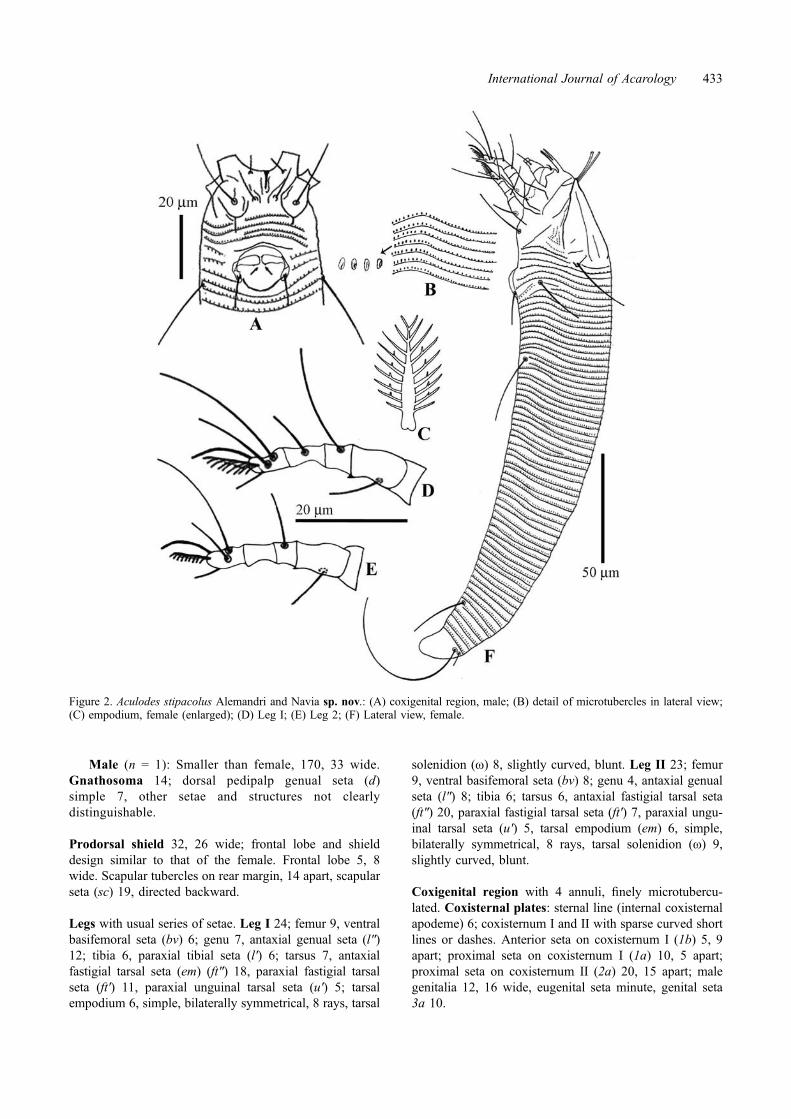
Male (n = 1): Smaller than female, 170, 33 wide.Gnathosoma 14; dorsal pedipalp genual seta (d)simple 7, other setae and structures not clearlydistinguishable.
Prodorsal shield 32, 26 wide; frontal lobe and shielddesign similar to that of the female. Frontal lobe 5, 8wide. Scapular tubercles on rear margin, 14 apart, scapularseta (sc) 19, directed backward.
Legs with usual series of setae. Leg I 24; femur 9, ventralbasifemoral seta (bv) 6; genu 7, antaxial genual seta (l″)12; tibia 6, paraxial tibial seta (l′) 6; tarsus 7, antaxialfastigial tarsal seta (em) (ft″) 18, paraxial fastigial tarsalseta (ft′) 11, paraxial unguinal tarsal seta (u′) 5; tarsalempodium 6, simple, bilaterally symmetrical, 8 rays, tarsal
solenidion (ω) 8, slightly curved, blunt. Leg II 23; femur9, ventral basifemoral seta (bv) 8; genu 4, antaxial genualseta (l″) 8; tibia 6; tarsus 6, antaxial fastigial tarsal seta(ft″) 20, paraxial fastigial tarsal seta (ft′) 7, paraxial ungu-inal tarsal seta (u′) 5, tarsal empodium (em) 6, simple,bilaterally symmetrical, 8 rays, tarsal solenidion (ω) 9,slightly curved, blunt.
Coxigenital region with 4 annuli, finely microtubercu-lated. Coxisternal plates: sternal line (internal coxisternalapodeme) 6; coxisternum I and II with sparse curved shortlines or dashes. Anterior seta on coxisternum I (1b) 5, 9apart; proximal seta on coxisternum I (1a) 10, 5 apart;proximal seta on coxisternum II (2a) 20, 15 apart; malegenitalia 12, 16 wide, eugenital seta minute, genital seta3a 10.
Figure 2. Aculodes stipacolus Alemandri and Navia sp. nov.: (A) coxigenital region, male; (B) detail of microtubercles in lateral view;(C) empodium, female (enlarged); (D) Leg I; (E) Leg 2; (F) Lateral view, female.
International Journal of Acarology 433

Opisthosoma evenly rounded, 55 dorsal annuli, 47 ventralannuli. Dorsal and ventral microtubercles similar to that ofthe female. Seta c2 20, on ventral annulus 2; seta d 33, onventral annulus 10, 21 apart, 15 microtubercles apart; setae 6, on ventral annulus 22, 9 apart, 5 microtubercles apart;seta f 14, on ventral annulus 43, 13 apart, 13 microtuber-cles apart. Caudal seta h2 broken, accessory seta h1 3.
Type material. Holotype female (slide/position 1/1 indi-cated by a red circle) and fourteen females and one maleparatypes, from Stipa sp. (Poaceae), Necochea, Provinceof Buenos Aires, Argentina (38° 45.10′ S, 58° 45.25′ W),13 January 2012, collected by Mauro Polizzi, on fivemicroscope slides. Holotype and 10 female paratypes onfour slides deposited in the mite collection at “EmbrapaRecursos Genéticos e Biotecnologia”, Brasília, DF, Brazil.One male and four female paratypes on one slide depos-ited at “Departamento de Entomologia, Fitopatologia eZoologia Agrícola, Escola Superior de Agricultura ‘Luizde Queiroz’ (ESALQ), Universidade de São Paulo”,Piracicaba, São Paulo, Brazil.
Relation to host. All specimens were collected on innerleaf blades, along midrib entire length.
Etymology. The specific designation stipacolus wasformed as a composition between stipa that refers to thegenus of the host plant, and the New Latin -colus, meaningdwelling in, inhabitting.
Molecular characterization
Three 16S and three ITS sequences of the Aculodes stipa-colus sp. nov. were obtained. One haplotype was identi-fied from 16S sequences (GenBank deposit No.KF648353) and three variants were obtained from theITS sequences (GenBank deposit No. KF648354,KF648355, KF648356). The 16S sequence KF134860 ofAbacarus hystrix (Nalepa) was incorporated in the dis-tance analysis since it is the unique 16S sequence of aneriophyid mite in grasses different to Aceria available inthe database. It was not possible to estimate the geneticdistance with other mites of the same genus since no
Figure 3. Aculodes stipacolus Alemandri and Navia sp. nov. micrographs. (A) dorsal view, female; (B) ventral view, female; (C) lateralview, female; (D) coxigenital region, male; (E) anterolateral view, female; (F) empodium; (G) coxigenital region, female, under DICmicroscope; (H) coxigenital region, female, under phase contrast microscope; (I) anterodorsal view with prodorsal shield ornamentationdetails.
434 V. Alemandri et al.
sequences were available in GenBank. The distancebetween 16S haplotype of A. stipacolus (KF648353) andthe predominant A. tosichella haplotype detected inArgentina (JQ512769) was 0.2280; and with A. hystrix(KF134860) it was 0.2539. The distance between thethree ITS variants of A. stipacolus (KF648354,KF648355, KF648356) and the predominant A. tosichellahaplotype detected in Argentina (JF960144) were respec-tively 0.2606, 0.2569, and 0.2587. These values can beconsidered as intergeneric distances in the Eriophyidaefamily.
Aculodes Keifer world species associated with grasses(Poaceae)
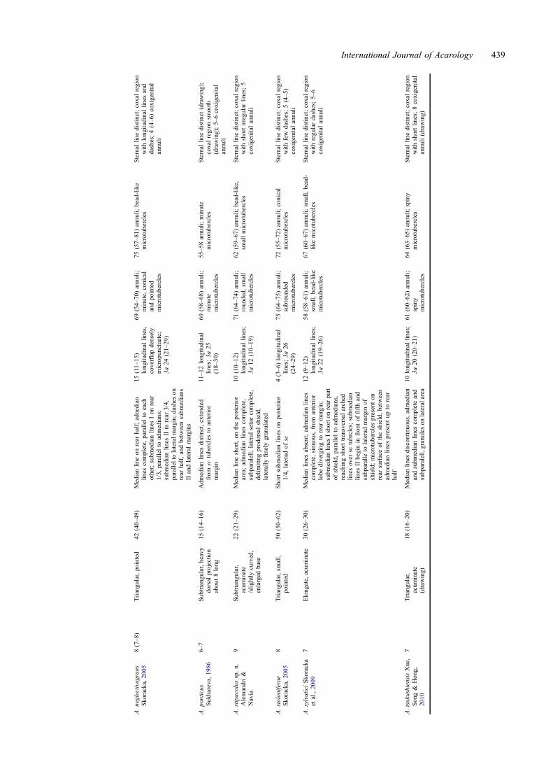
A list of the 21 Aculodes species associated with grasses inthe world is presented below. It includes information ontype host, type locality, and habitus. Main morphologicaltraits of the grass-associated Aculodes species are pre-sented in Table 1.
Aculodes agropyronis (Keifer, 1960)
Type host – Agropyron smithi Rydb., presently referred asa synonym of Elymus smithii (Rydb.) Gould (The PlantList 2013).Type locality – Texas, USA.Habitus – In young rolled leaves and on leaf plate nearsheath.
Aculodes bambusae Kuang, 1997
Type host – Bambusa sp.Type locality – Hangzhou City, Zhejiang Province, China.Habitus – Vagrant.
Aculodes calamaabditus Skoracka, 2003
Type host – Calamagrostis epigeios (L.) Roth., presentlythe accepted name is Calamagrostis epigejos (L.) Roth(The Plant List 2013).Type locality – Path in Forest, Lesna Dolina (16° 12′E, 51°44′N), Glogow, Poland.Habitus – Vagrants on upper leaf surface, often hiding infurrows.
Aculodes capillarisi Skoracka, 2003
Type host – Agrostis capillaris L.Type locality – Forest path, Biedrusko near Poznan (16°55′E, 52° 29′N), Poland.Habitus – Vagrants on upper leaf surface, mostly nearthe top.
Aculodes deschampsiae (Sukhareva, 1972)
Type host – Deschampsia cespitosa (L.) P. Beauv.Type locality – Park of the Biological Institute, LeningradUniversity in old Petergof, Leningrad Region, Russia.Habitus – Vagrant in grooves on upper leaf surface.References – Drawings and taxonomic characterizationalso in Skoracka (2004).
Aculodes dubius (Nalepa, 1891)
Type host – Helictotrichon pratense (L.) Besser ex Pilg.Type locality – Austria? (in Amrine and Stasny 1994).Habitus – In grooves between veins on surface of leaves;greening of flowers.Reference – Drawings and taxonomic characterization alsoin Skoracka (2004).
Aculodes festucae Skoracka, Labrzycka and Rector,2009
Type host – Festuca arundinaceae Schreb.Type locality – Gorski Kotar mountains, around 30 KmNE of Rijeka (14° 35.047′E, 45° 28.848′N), Croatia.Habitus – Vagrants on upper leaf surface.
Aculodes fulleri (Keifer, 1966a)
Type host – Stipa californica Merr. & Burtt-Davy ex Hall.,presently referred as a synonym of Stipa occidentalisThurb. ex S.Watson (The Plant List 2013).Type locality – Upper King’s Creek, Lassen Nat. Pk.,Shasta Co., California, USA.Habitus – The mites live in rib grooves on upper leafsurfaces.
Aculodes holcusi Skoracka, 2004
Type host – Holcus mollis L.Type locality – Mountain meadow with southern exposure,Przechyba, Beskid Sadecki Mts., Poland.Habitus – Vagrant on upper leaf surfaces.
Aculodes janboczeki Skoracka, 2005
Original description -Type host – Bromus inermis Leyss.Type locality – Meadow near Oak Forest (48° 02.730′ N,20° 28.814′ E), 580 m above sea level, Bukki Mts.,Hungary.Habitus – Vagrants on upper leaf surfaces.
Aculodes koeleriae Sukhareva, 1985
Type host – Koeleria cristata (L.)., presently referred as asynonym of Koeleria pyramidata (Lam.) P.Beauv. (ThePlant List 2013).Type locality – In steppe-like meadow, vicinity ofPutsilovska, Pogranichnyy Distr., Maritime Territory,Primorskiy Kraj, Russia.Habitus – Vagrant on upper leaf surface; on strips betweenveins on upper surfaces of leaves.
Aculodes kransnovi Sukhareva, 1994
Type host – Sasa palmata E. G. Camus.Type locality – Batumskoy Botanical Garden, Georgia.Habitus – Mites were found on the surface of the leaves, inthe gaps between the ridges.
Aculodes levis Huang, 2001
Type host – Phyllostachys makinoi Hayata.Type locality – Kaohsiung, Tengchih, Taiwan, China.
International Journal of Acarology 435

Table1.
Morph
olog
ical
traitsof
speciesin
thegenu
sAculodesKeiferassociated
with
grasses.
Species
Num
berof
empodium
rays
Frontal
lobe
Scseta
length
Prodorsal
shield
ornamentatio
n
Epigynium
longitu
dinallin
es;3a
seta
length
Num
berof
dorsal
annuli;
microtuberclesshape
Num
berof
ventralannuli;
Microtuberclesshape
Coxigenitalregion
–sternallin
e;ornamentatio
n;Num
berof
coxigenitalannuli
A.agropyronis
(Keifer,1960
)8
Shortacum
inate(text
Keifer1960);
subtriangular,
slightly
rounded
apically
(drawing)
46Designof
clearlin
es:medianlin
epresenton
rear
1/3;
admedianlin
esfrom
anterior
lobe
base,somew
hat
sinuate,
divergingto
rear
margin;
subm
edianlin
eIfrom
anterior
1/4,
runningtowards
dorsal
tubercle
andbendinglaterally
onapproachingtubercle;subm
edianII
from
anterior
lobe
base,running
back
below
tubercle;alaterallin
eandafew
granules
abovecoxae
11–1
2longitu
dinal
lines;3a
2755–6
0annuli;
microtubercles
small,pointed,
dorsally
bead-like
No.
notinform
ed;microtubercles
setaheadof
annulimargin
Strongsternallin
e(subparallel
lineon
each
side
ofsternal
line);coxalregion
with
strong
lines;8coxigenitalannuli
A.bambusaeKuang,
1997
8–9
Subcircular,apically
rounded
(drawing)
Reachingthe
fourth
annulus
(drawing)
Medianlin
ecomplete(text);adm
edian
lines
complete,
butinterruptedin
the3/4posterior;subm
edianlin
escomplete;
bifurcated
and
interruptedlin
es(discontinuous)in
therear
half(drawing)
12–1
3longitu
dinal
lines
(drawing)
45–4
8annuli;
absenceof
microtubercles
60–6
5annuli;
rounded
microtubercles
Sternal
linedistinct;coxalregion
with
shortcurved
lines;5
coxigenitalannuli
A.calamaabditu
sSkoracka,
2003
7(7–8
)Subtriangular,
pointed
21(12–28)
Medianlin
eon
rear
half;admedian
from
anterior
lobe
base
diverging
torear
margin,
slightly
concavein
themiddle;
subm
edianlin
esfrom
anterior
1/4runningtowards
outer
sctubercles,almostsubparalellto
lateralmargin
13(9–1
4)longitu
dinallin
es;
3a25
(23–25)
62(59–79)annuli;
numerousminute
microtubercles
alongannuli
margins
63(67–76)annuli;
microtubercles
notas
numerousas
dorsal,
conical,bluntedtop,
along
annulimargins
Sternal
lineslender;coxalregion
with
numerouslong
lines
and
minuteconicalmicrotubercles;
6(4–7)coxigenitalannuli
A.capilla
risi
Skoracka,
2003
8(7–8
)Subtriangular,
distinct,pointed
45(35–42)
Medianlin
eabsent;admedianlin
escomplete,
from
anterior
lobe
base,
divergingto
rear
margin;
subm
edianlin
eswith
conical
microtubercleson
rear
halfof
shield,almostparallelto
lateral
margin
12(11–13)
longitu
dinal
ridges;3a
28(17–
33)
61(54–60)annuli;
minute,
conical
butwith
blunttop
microtubercles
68(61–
71)annuli;
conical,
pointedmicrotubercles
Sternal
linedistinct;coxalregion
with
numerouslin
es(m
ostof
them
short)andnumerous
conicalmicrotubercles;
5–6
coxigenitalannuli
A.deschampsiae
(Sukhareva,
1972)
7–8(inSkoracka
2004)
Pronounced,
apically
rounded(not
acum
inate)
(in
Skoracka2004)
29–3
4(in
Skoracka
2004
)
Medianlin
eon
rear
half,dividedinto
twolin
es,at
leastoneof
them
splittin
ganteriorly;admedianlin
escomplete;
subm
edianlin
eson
rear
3/4,
parallelto
lateralmargins;
shorttransverse
lines
form
ing
obtuse
angleover
tuberclesbearing
scsetae(inSkoracka2004)
10–11longitu
dinal
lines;3a
24–30
(inSkoracka
2004)
59–6
4annuli;
conical,pointed
microtubercles(in
Skoracka2004
)
66–7
5annuli;
conical,pointed
microtubercles(inSkoracka
2004
)
Sternal
linedistinct;coxalregion
with
short,slenderlin
es;6
coxigenitalannuli(in
Skoracka
2004
)
436 V. Alemandri et al.

A.dubius
(Nalepa,
1891)
9(inSkoracka
2004)
Triangular,pointed
(inSkoracka
2004)
64–9
0(in
Skoracka
2004
)
Medianlin
eabsent;admedianlin
escomplete,
divergingfrom
base
ofanterior
lobe
torear
margin,
near
rear
marginrunningto
centre
ofshield;subm
edianlin
esIarched,
with
minuteandconical
microtubercles,on
rear
2/3of
shield,connectin
gwith
admedian
lines
inits
1/3,
with
posterior
fragmentoutsidetuberclesbearing
scseta;transverse,arched
lines
with
minuteandconical
microtuberclesover
tubercles
bearingsc
seta
(inSkoracka2004
)
9–13
longitu
dinal
lines;3a
31–45
(inSkoracka
2004)
54–6
4annuli;
conical,minute
microtubercles(in
Skoracka2004
)
74–8
3annuli;
conical,pointed
microtubercles(inSkoracka
2004
)
Sternal
lineslender;coxalregion
with
subrounded
microtuberclesandnumerous
lines
with
minute
microtubercles;
5–6
coxigenitalannuli(in
Skoracka
2004
)
A.festucae
Skoracka
etal.,2009
7Subtriangular,
acum
inate
/slig
htly
curved,
enlarged
base
21(18–24)
Medianlin
eon
therear
2/3;
admedian
lines
complete,
sinuous,diverging
torear
margin;
subm
edianlin
esbeginon
therear
2/3
10(10–13)
longitu
dinallin
es;
3a26
(20–28)
57(51–57)annuli;
smallbead-like
microtubercles
63(58–
63)annuli;
smallbead-
likemicrotubercles
Sternal
linedistinct;coxalregion
with
dashes,hipostom
iobase
denselygranulated;7
coxigenitalannuli
A.fuller(K
eifer,
1966a)
7Som
ewhatnarrow
andacute,
athin
anterior
projectio
nvisible
inside
view
32Medianlin
epresenton
rear
1/3;
admedianlin
ecomplete,
gentle
sinuous,gradually
diverging;
subm
edianlin
esIashortlin
e,subparalle
toadmedian,
inshield
centre;subm
edianlin
esIIbegining
atabout1/2andarchingback
torear
marginbelow
dorsal
tubercle,
somew
hatgranular;laterallin
efrom
side
ofanterior
lobe
topartial
ringsbelow
dorsal
tubercle
About
14longitu
dinallin
es,
sometransverse
lines
ofgranules
atbase;3a
33
44annuli;
small,
bead
like,
close
together,hardly
pointed,
orrounded
microtubercles
48–5
0annuli;
microtubercles
tendingto
beaheadof
ring
margins,morepointedthan
dorsal
Strongsternallin
e;coxalregion
with
lines
ofdashes
and
granules;7coxigenitalannuli
A.holcusiSkoracka,
2004
8Triangular,pointed
59(58–65)
Medianlin
eon
rear
half,below
medianlin
esshortlin
esform
inga
V-shapedfigure;admedianlin
escomplete,
divergingto
lateral
marginof
shield;subm
edianlin
esI
onrear
half,parallelto
admedian;
subm
edianlin
esIIarched,
connectin
gwith
admedianin
its1/4;
subrounded
andconical
microtuberclesanddashes
12(11–12)
longitu
dinallin
es;
3a40
(38–40)
50(49–51),dorsal
microtubercles
irregularly
distributed,
large,
subrounded,
setalongannuli
margins
63(60–
63);ventralmicrotuberles
minute,
conical,slightly
pointed,
aheadfrom
annuli
margins
Sternal
lineslender;coxalregion
with
wavylin
esanddashes;5
(5–6)coxigenitalannuli
A.koeleriae
Sukhareva,1985
6–7
Subtriangular,
apically
acum
inate
(drawing)
43(38–45)
Adm
edianlin
esvery
weak;
subm
edianlin
esIIfollo
winglateral
margins
8–10
longitu
dinal
lines;3a
17(15–
20)
60(57–66)annuli;
absenceof
microtubercles,
butpresence
ofunevenly
scallopedmargin
55(53–
58)annuli;
small
microtubercles
Sternal
linedistinct;coxalregion
smooth
(drawing);3–4coxi
genitalannuli(text),8
(drawing)
A.janboczeki,
Skoracka,
2005
7(7–8
)Pointed
(text),
pronounced,
triangular
(drawing)
20(18–23)
Medianlin
eabsent;admedianlin
escompleteandparallel;subm
edian
lines
Ishort,on
rear
half,parallel
toadmedian;
subm
edianlin
esIIas
shortas
subm
edianI,sinuous;
ocelar
fields
inthelateralarea
13(11–15)
longitu
dinallin
es;
3a15
(14–19)
57(55–64)annuli;
conicalandbead-
like
microtubercles
72(63–
75)annuli;
conicaland
bead-likemicrotubercles
Sternal
lineslender;coxalregion
with
numerous,minute
microtuberclesanddashes
(set
roundsetaetubercles);5(5–7
)coxigenitalannuli (Con
tinued)
International Journal of Acarology 437

Table1.
(Con
tinued).
Species
Num
berof
empodium
rays
Frontal
lobe
Scseta
length
Prodorsal
shield
ornamentatio
n
Epigynium
longitu
dinallin
es;3a
seta
length
Num
berof
dorsal
annuli;
microtuberclesshape
Num
berof
ventralannuli;
Microtuberclesshape
Coxigenitalregion
–sternallin
e;ornamentatio
n;Num
berof
coxigenitalannuli
A.kransnovi
Sukhareva,1994
5–6
Subtriangular,
slightly
acum
inate
apically
(drawing)
Reachingninth
annulus
(drawing)
Medianlin
eabsent;admedianlin
escomplete;
subm
edianlin
eson
4/5
rear;sublateral
lines
complete
(drawing)
6short,interrupted
longitu
dinallin
es46
(44–
48)annuli;
absenceof
microtubercles
(drawing)
48–5
0annuli;
absenceof
microtubercles(drawing)
Sternal
lineslender;coxalregion
smooth;8–9coxigenitalannuli
(drawing)
A.levis
Huang,2001
5Subcircular,apically
rounded
(drawing)
19Dashedlin
es;medianlin
ecomplete;
medianandadmedianlin
essubparallel;admedianand
subm
edianlin
esnotvisibleon
anterior
1/4shield
Longitudinallin
esabsent,coverflap
smooth;3a
10
56annuli;
minute,
elongated
microtubercles
(drawing)
81annuli;
rounded
microtubercles,larger
than
dorsal
(drawing)
Sternal
lineindistinct;coxal
region
with
smallgranules;2
coxigenitalannuli(drawing)
A.mckenziei
(Keifer,
1944)
7(K
eifer1944);
9(in
Skoracka
2004)
Narrow
andpointed
(textKeifer
1944
),sutriangular
(drawing);large,
pointed(in
Skoracka2004)
43(K
eifer
1944
);47
–64
(inSkoracka
2004
)
Longitudinallin
escurved
totherear,
granules
totherear
andside
(Keifer1944
);medianlin
eon
rear
half;admedianlin
escomplete,
divergingfrom
thebase
offrontal
lobe
torear
margin;
subm
edian
lines
Isubparallelto
admedian
lines,runninglaterally
infrontto
sctubercles;subm
edianlin
esIIon
rear
3/4,
subparallelto
lateral
margin;
large,
conicalpointed
microtubercleson
rear
half(in
Skoracka2004
)
12longitu
dinallin
es;
3a25
long
(in
Keifer1944).
11–15
longitu
dinallin
es;
3a38–5
0(in
Skoracka2004)
60–7
1annuli;
acum
inate
microtubercles(in
Keifer1944
);numerous,conical
andpointed
microtubercles(in
Skoracka2004
)
65annuli(inKeifer1944
);69–8
1annuli(inSkoracka2004),
conicalandpointed
microtuberclessm
allerthan
dorsal
(inSkoracka2004)
Anteriorcoxaebroadly
contiguous
(inKeifer1944),
sternallin
eslender(in
Skoracka2004);coxalregion
with
numerouslin
esand
conicalmicrotubercles;
5–8
coxigenitalannuli
A.mongolicus
Skoracka&
Shi,
2001
8(7–8
)Pronounced,
elongate
and
pointed(text),
triangular
(drawing)
45(42–45)
(text),
reaching
sixteenth
annulus
(drawing)
Medianlin
epresentin
theposterior
half;admedianlin
escomplete,
parallelto
each
otheron
the
anterior
region,divergingto
lateral
margins
intheposteriorhalf;
subm
edianlin
esIIsubparallelto
admedian,
runninglateradin
front
ofsc
tubercles;subm
edianlin
esII
beginningfrom
1/3of
the
subm
edianlin
esIandform
ing
bowsreaching
posteriormargin
(text);sparse
conicalgranules
onposteriorhalf,alongsomelin
es,
andalso
ontheantero-lateral
margin(drawing)
10(10–11)
longitu
dinallin
es;
3a38
(38–48)
64(56–
62)annuli;
triangular,
minute,
closeto
each
other
micotubercles
74(61–
70)annuli;
triangular,but
morepointedandlarger
than
dorsal
miscrotubercles
Sternal
linestrong;coxalregion
with
numerous,shortlin
es,or
triangular
microtuberclesalong
lines;8(5–7
)coxigenital
annuli
A.multitricavus
Skoracka,
2004
9Triangular,pointed
27(26–32),
reaching
tenthannulus
Medianlin
eon
rear
half;admedian
lines
complete,
from
anterior
lobe
divergingto
lateralmargin;
subm
edianlin
esIshort,on
rear
half,parallelto
admedians;
subm
edianlin
esIIbeginningon
4/5rear,parallelto
lateralmargins;
numeroustriangular
cavitieson
shield
13(12–13)
longitu
dinallin
es;
3a34
(32–35)
60(58–
60)annuli;
minute,
conical,
andpointed
microtubercles
66(62–
67)annuli;
minute,
conical,andpointed
microtubercles
Sternal
linedistinct;coxalregion
with
lines
(coxae
I)anddashes
andconicalmicrotubercles
(coxae
II);6(5–6
)coxigenital
annuli
438 V. Alemandri et al.
A.neglectivagrans
Skoracka,
2005
8(7–8
)Triangular,pointed
42(40–
49)
Medianlin
eon
rear
half;adnedian
lines
complete,
parallelto
each
other;subm
edianlin
esIon
rear
1/3,
parallelto
admedians;
subm
edianlin
esIIin
rear
3/4,
paralleltolateralm
argin;
dashes
onrear
half,andbetweensubm
edians
IIandlateralmargins
15(11–
15)
longitu
dinallin
es,
coverflap
densely
micropunctuate;
3a24
(21–29)
69(54–
70)annuli;
minute,
conical
andpointed
microtubercles
75(57–
81)annuli;
bead-like
microtubercles
Sternal
linedistinct;coxalregion
with
longitu
dinallin
esand
dashes;4(4–6)coxigenital
annuli
A.ponticus
Sukhareva,1986
6–7
Subtriangular,heavy
dorsal
projectio
nabout8long
15(14–16)
Adm
edianlin
esdistinct,extended
from
sctuberclesto
anterior
margin
11–1
2longitu
dinal
lines;3a
25(18–
30)
60(58–
68)annuli;
minute
microtubercles
55–5
8annuli;
minute
microtubercles
Sternal
linedistinct
(drawing);
coxalregion
smooth
(drawing);5–6coxigenital
annuli
A.stipacolus
sp.n.
Alemandri&
Navia
9Subtriangular,
acum
inate
/slig
htly
curved,
enlarged
base
22(21–29)
Medianlin
eshort,on
theposterior
area;admedianlin
escomplete,
subparalell;lateralsetaecomplete,
delim
iting
prodorsalshield,
laterally
finely
granulated
10(10–12)
longitu
dinallin
es;
3a12
(10–19)
71(64–
74)annuli;
rounded,
small
microtubercles
62(59–
67)annuli;
bead-like,
smallmicrotubercles
Sternal
linedistinct;coxalregion
with
shortirregularlin
es;5
coxigenitalannuli
A.stoloniferae
Skoracka,
2005
8Triangular,sm
all,
pointed
50(50–62)
Shortsubm
edianlin
eson
posterior
1/4,
lateradof
sc4(3–6)longitu
dinal
lines;3a
26(24–
29)
75(64–
75)annuli;
subrounded
microtubercles
72(55–
72)annuli;
conical
microtubercles
Sternal
linedistinct;coxalregion
with
few
dashes;5(4–5
)coxigenitalannuli
A.sylvaticiSkoracka
etal.,2009
7Elongate,
acum
inate
30(26–30)
Medianlin
esabsent;admedianlin
escomplete,
sinuous,from
anterior
lobe
divergingto
rear
margin;
subm
edianlin
esIshorto
nrear
part
ofshield,parallelto
admedians,
reaching
shorttransversalarched
lines
over
sctubrcles;subm
edian
lines
IIbeginin
frontof
fifthand
subparalle
tolateradmarginof
shield;microtuberclespresenton
rear
surfaceof
theshield,between
admedianlin
espresentup
torear
half
12(9–1
2)longitu
dinallin
es;
3a22
(19–26)
58(58–
61)annuli;
small,bead-like
micotubercles
67(60–
67)annuli;
small,bead-
likemicotubercles
Sternal
linedistinct;coxalregion
with
regulardashes;5–6
coxigenitalannuli
A.tsukushiensisXue,
Song&
Hong,
2010
7Triangular,
acum
inate
(drawing)
18(16–20)
Medianlin
esdiscontin
uous,admedian
andsubm
edianlin
escompleteand
subparalell,granules
onlateralarea
10longitu
dinallin
es;
3a20
(20–21)
61(60–
62)annuli;
spiny
microtubercles
64(63–
65)annuli;
spiny
microtubercles
Sternal
linedistinct;coxalregion
with
shortlin
es;8coxigenital
annuli(drawing)
International Journal of Acarology 439

Habitus – Vagrant on lower leaf surface. No damageobserved.
Aculodes mckenziei (Keifer, 1944)
Type host – Leymus triticoides (Buckl.) Pilg.Type locality – Sacramento, California, USA.Habitus – The mites occur in upper surface, leaf furrows.They cause some browning.Reference – Drawings and taxonomic characterization alsoin Skoracka (2004).Obs – Sukhareva (1981) considered two subspecies forthis taxon, A. mckenziei subsp. brevisetus and A. mckenzieisubsp. trivialis.
Aculodes mongolicus Skoracka & Shi, 2001
Type host – Hordeum brevisubulatum (Trin.) Link.Type locality – Arkhangy Aymag, Horgo Terhiyn TsagaanNuur National Park, Central Mongolia.Habitus – Mites are vagrants on upper leaf surface.
Aculodes multitricavus Skoracka, 2004
Type host – Bromus inermis Leyss.Type locality – Xerothermic sward, Owczary, Poland.Habitus – Vagrant on upper leaf surfaces.
Aculodes neglectivagrans Skoracka, 2005
Type host – Calamagrostis neglecta (Ehrh.) Gaertn., Meyet Schreb., presently referred as a synonym ofCalamagrostis stricta (Timm) Koeler (The Plant List2013).Type locality – Littoral dune, 14 m elev., 10 km W ofKuzrjeka Village, near Turij Cape, S. of Umba, White SeaCoast, Kola Peninsula, Russia. (66° 35.722′ N, 34°42.965′ E)Habitus – Vagrants on upper leaf surfaces.Reference – Skoracka and Pacyna (2005).
Aculodes ponticus Sukhareva, 1986
Type host – Eremopyrum distans (Koch) Nevski.Type locality – Near Kabardinka Village, KrasnodarskiKraj, Russia.Habitus – Deep grooves between veins on surface of theleaves.
Aculodes stoloniferae Skoracka, 2005
Type host – Agrostis stolonifera L. subsp. straminea(Hartm.) Tzvel.Type locality – Taiga, near a stony beach, near Turij Cape,S. of Umba, White Sea. Coast, (66° 37.822′ N, 34° 27.345′E) Kola Peninsula, Russia.Habitus – Vagrants on upper leaf surfaces.Reference – Skoracka and Pacyna (2005).
Aculodes sylvatici Skoracka, Labrzycka and Rector,2009
Type host – Brachypodium sylvaticum (Huds.) P. Beauv.
Type locality – Velebit mountains, around 24 Km SE ofSenj (15° 02.830′E, 44° 56.412′N), Croatia.Habitus – Vagrants on upper leaf surface.
Aculodes tsukushiensis Xue, Song, Hong, 2010
Type host – Elymus tsukushiensis Honda var. transiens(Hack.) Osad.Type locality – Zhouzhi County, Shaanxi Province, P.R.elevation 500 m, (34°03′54″N, 108°19′22″E), China.Habitus – Vagrant on leaf surfaces. No damage to the hostwas observed.
AcknowledgementsAuthors DN and RSM are grateful to National Council forScientific and Technological Development (CNPq), Brazil, forresearch and pos-doc fellowships, respectively. Thanks also toDr. Philipp E. Chetverikov, Saint-Petersburg State University,Russia, for traduction of Sukhareva´s papers from Russian toEnglish and to Mr. Mauro Polizzi for sample collection andhost plant identification in Argentina.
FundingThis work was supported by the Embrapa [Macroprograma 2Call 22/2011 –Embrapa/INTA].
ReferencesAmrine Jr, JW, Manson DCM. 1996. Preparation, mounting and
descriptive study of Eriophyoid mites. In: Lindquist EE,Sabelis MW, Bruin J, editors. Eriophyoid mites: their biol-ogy, natural enemies and control. Amsterdam: Elsevier,World Crop Pests; p. 383–396. n. 6.
Amrine Jr, JW, Stasny TA. 1994. Catalog of Eriophyoidea(Acari: Prostigmata) of the world. West Bloomfield (MI):Indira Publishing House.
Amrine Jr, JW, Stasny TAH, Flechtmann CHW. 2003. Revisedkeys to the world genera of the Eriophyoidea (Acari:Prostigmata). West Bloomfield (MI): Indira Publishing House.
Carew M, Schiffer M, Umina P, Weeks A, Hoffmann A. 2009.Molecular markers indicate that the wheat curl mite, Aceriatosichella Keifer, may represent a species complex inAustralia. Bulletin of Entomological Research 99:479–486.
Carew ME, Goodisman MAD, Hoffmann AA. 2004. Speciesstatus and population genetic structure of grapevine erio-phyoid mites. Entomologia Experimentalis et Applicata111:87–96.
Castiglioni E, Navia D. 2010. Presence of the Wheat Curl Mite,Aceria tosichella Keifer (Prostigmata: Eriophyidae), inUruguay. Agrociencia 14:19–26.
Dabert J, Ehrnsberger R, Dabert M. 2008. Glaucalges tytonis sp.n. (Analgoidea, Xolalgidae) from the barn owl Tyto alba(Strigiformes, Tytonidae): compiling morphology withDNA barcode data for taxon descriptions in mites (Acari).Zootaxa 1719:41–52.
de Lillo E, Craemer C, Amrine Jr, JW, Nuzzaci G. 2010.Recommended procedures and techniques for morphologicalstudies of Eriophyoidea (Acari: Prostigmata). Experimentaland Applied Acarology 51:283–307.
Flechtmann CHW. 2000. Two new species of Aceria (Acari:Eriophyidae) on grass from Brazil. International Journal ofAcarology 26:335–338.
Flechtmann CHW, Aranda BRC. 1970. New records and noteson Eriophyid mites from Brazil and Paraguay, with a list of
440 V. Alemandri et al.

Eriophyidae from South America. Proceedings of theEntomological Society of Washington 72:94–98.
Frost WE, Eagling DR, Manson DCM. 1990. Abacarus hystrix(Nalepa) (Acarina: Eriophyidae) newly recorded in Australia.Australian Journal of Entomology 29:182.
Guy PL, Gould DM. 1996. Aculodes mckenziei (Acari:Eriophyidae) newly recorded in New Zealand and its asso-ciation with other eriophyids in South Island pastures. NewZealand Entomologist 19:61–64.
Huang KW. 1992. Some new eriophyoid mites from Taiwan(Acari: Eriophyoidea). Bulletin of the National Museum ofNature and Science 3:227–228.
Huang KW. 2001. The eriophyid mites of Taiwan: description ofeighty-six species from the Tengchih Area. Bulletin of theNational Museum of Nature and Science 14:1–84.
Keifer HH. 1944. Eriophyid Studies XIV. Bulletin of theCalifornia Department of Agriculture 33:18–38.
Keifer HH. 1952. The eriophyid mites of California. Bulletin ofthe California Insect Survey 2:1–123.
Keifer HH. 1959. Eriophyid studies XXVII. Bulletin of theCalifornia Department of Agriculture.
Keifer HH. 1960. Eriophyid studies B-1. Sacramento (CA):California Department of Agriculture.
Keifer HH. 1962. Eriophyid studies B-8. Sacramento (CA):California Department of Agriculture.
Keifer HH. 1966a. Eriophyid studies B-20. Sacramento (CA):California Department of Agriculture.
Keifer HH. 1966b. Eriophyid studies B-21. Sacramento (CA):California Department of Agriculture.
Keifer HH. 1976. Eriophyid studies C-12. Sacramento (CA):California Department of Agriculture.
Keifer HH. 1977. Eriophyid studies C-13. Sacramento (CA):California Department of Agriculture.
Keifer HH. 1978. Eriophyid studies C-15. Sacramento (CA):California Department of Agriculture.
Kuang H, Pang H. 1997. Four new species of the Eriophyinae(Acari: Eriophyidae) from China. Entomotaxonomia 19:232–233.
Kuang HY. 1997. Four new species of Eriophyidae (Acari:Eriophyoidea) from China. Entomotaxonomia 19:74–78.
Kuang HY, Luo GH, Wang AW. 2005. Fauna of Eriophyid mitesfrom China (II) (Acari: Eriophyoidea). Beijing: ChinaForestry Publishing House.
Lindquist EE. 1996. External anatomy and notation of structures.In: Lindquist EE, Sabelis MW, Bruin J, editors. Eriophyoidmites: their biology, natural enemies and control.Amsterdam: Elsevier, World Crop Pests; p. 3–31. n. 6.
Miller AD, Skoracka A, Navia D, Mendonça RS, Szydło W,Schultz MB, Smith C, Truol G, Hoffmann AA. 2013.Phylogenetic analyses reveal extensive cryptic speciationand host specialization in an economically important mitetaxon. Molecular Phylogenetics and Evolution 66:928–940.
Mitrofanov VI, Sharonov AA, Badulin AV. 1988. New species ofmites of the genus Aceria (Acariformes, Eriophyoidea).Zoologicheskii Zhurnal 67:790–794.
Nalepa A. 1891. Genera und Species der Familie Phytoptida.Denkschriften, Akademie der Wissenschaften in Wien,Mathematisch-Naturwissenschaftliche Klasse 58:867–884.
Navajas M, Navia D. 2010. DNA-based methods for eriophyoidmite studies: review, critical aspects, prospects and chal-lenges. Experimental and Applied Acarology 51:257–271.
Navia D, Moraes GJ, Roderick G, Navajas M. 2005. The inva-sive coconut mite Aceria guerreronis (Acari: Eriophyidae):origin and invasion sources inferred from mitochondrial(16S) and nuclear (ITS) sequences. Bulletin ofEntomological Research 95:505–516.
Navia D, Truol G, Mendonça RS, Sagadín M. 2006. Aceriatosichella Keifer (Acari: Eriophyidae) from Wheat Streak
Mosaic Virus-infected wheat plants in Argentina.International Journal of Acarology 32:189–193.
Pereira PRVS, Navia D, Salvadori JR, Lau D. 2009. Occurrenceof Aceria tosichella in Brazil. Pesquisa AgropecuáriaBrasileira 44:539–542.
The Plant List (2013). Version 1.1. [Internet]. [cited 2015 Mar10]. Available from: http://www.theplantlist.org/
Shi A, Boczek J. 2000. Studies on eriophyoid mites (Acari:Eriophyoidea). XXXIX. Bulletin of the Polish Academy ofSciences 48:323–327.
Skoracka A. 2003. New species of Aculodes (Acari:Eriophyoidea) from grasses in Poland. Acta ZoologicaAcademiae Scientiarum Hungaricae 49:43–60.
Skoracka A. 2004. Eriophyid mites from grasses in Poland(Acari: Eriophyoidea). Genus 13:1–205.
Skoracka A. 2005. Two new species and four new records oferiophyoid mites from grasses in Hungary (Acari:Eriophyoidea). Genus 16:445–462.
Skoracka A, Dabert M. 2010. The cereal rust mite Abacarushystrix (Acari: Eriophyoidea) is a complex of species: evi-dence from mitochondrial and nuclear DNA sequences.Bulletin of Entomological Research 100:263–272.
Skoracka A, Kuczynski L, Mendonça RS, Dabert M, Szydło W,Knihinicki D, Truol G, Navia D. 2012. Cryptic species withinthe wheat curl mite Aceria tosichella (Keifer) (Acari,Eriophyoidea) revealed by mitochondrial, nuclear and mor-phometric data. Invertebrate Systematics 26:417–433.
Skoracka A, Labrzycka A, Rector BG. 2009. Three new speciesof eriophyoid mites (Acari: Prostigmata: Eriophyoidea) fromgrass hosts in Croatia. Annals of the Entomological Societyof America 102:12–19.
Skoracka A, Pacyna A. 2005. Grass associated eriophyoid mites(Acari: Eriophyoidea) from the Kola Peninsula, Russia.Annales Zoologici 55:453–466.
Skoracka A, Shi AX, Pacyna A. 2001. New eriophyoid mites(Acari: Eriophyoidea) associated with grasses fromMongolia. Zootaxa 9:1–18.
Sukhareva SI. 1972. New species of quadrupedal mites of thegenus Phytocoptes (Acarina, Eriophyoidea). ZoologicheskyZhurnal 51:296–299.
Sukhareva SI. 1981. O strukture chetireh naibolee rasprostrane-nih vidov chetirenogih klescej (Acarina: Tetrapodili) so zla-kov: Aceria tritici Shev., Aculodes mckenziei (K), Aculodesdubius (Nal.), Abacarus hystrix (Nal.). The structure of the 4most distributed eriophyid mites (Acarina: Tetrapodili) fromgrasses, etc. Vestnik Leningradskogo Universiteta 15:25–36.
Sukhareva SI. 1983. New species of Eriophyid mites of the genusAceria Keifer. (Acariformes, Tetrapodili) living on grasses.Entomologicheskoe Obozrenie 62:391–396.
Sukhareva SI. 1985. New species of mites (Acarina: Tetrapodili)on grasses from Primorskij Kraj. (Maritime Territory).Entomologicheskoe Obozrenie 64:227–234.
Sukhareva SI. 1986. New species of eriophyid mites(Acariformes: Tetrapodili) living on grasses in the SSSR.Entomologicheskoe Obozrenie 65:850–855.
Sukhareva SI. 1994. The four-legged mites (Acariformes:Tetrapodili) on the bamboos in Georgia. Vestnik Sankt-Petersburgkogo Universiteta, Seriya 3, Biologiya 1:33–38.
Tamura K, Stecher G, Peterson D, Filipski A, Kumar S. 2013.MEGA6: molecular evolutionary genetics analysis version6.0. Molecular Biology and Evolution 30:2725–2729.
Thompson JD, Gibson TJ, Plewniak F, Jeanmougin F, HigginsDG. 1997. The CLUSTALX windows interface: flexiblestrategies for multiple sequence alignment aided by qualityanalysis tools. Nucleic Acids Research 25:4876–4882.
Xue XF, Song ZW, Hong XY. 2010. Five new species ofAnthocoptini from China (Acari: Eriophyidae). Zootaxa2666:29–44.
International Journal of Acarology 441