Embed Size (px)
Citation preview
Essays in Biochemistry (2017) 61 625–635https://doi.org/10.1042/EBC20170092
Received: 19 September 2017Revised: 28 October 2017Accepted: 06 November 2017
Version of Record published:12 December 2017
Review Article
ER homeostasis and autophagyMatthew Smith and Simon WilkinsonEdinburgh Cancer Research UK Centre, MRC Institute of Genetics and Molecular Medicine, University of Edinburgh, Edinburgh EH4 2XR, U.K.
Correspondence: Simon Wilkinson ([email protected]) or Matthew Smith ([email protected])
The endoplasmic reticulum (ER) is a key site for lipid biosynthesis and folding of nascenttransmembrane and secretory proteins. These processes are maintained by careful home-ostatic control of the environment within the ER lumen. Signalling sensors within the ERdetect perturbations within the lumen (ER stress) and employ downstream signalling cas-cades that engage effector mechanisms to restore homeostasis. The most studied signallingmechanism that the ER employs is the unfolded protein response (UPR), which is known toincrease a number of effector mechanisms, including autophagy. In this chapter, we willdiscuss the emerging role of autophagy as a UPR effector pathway. We will focus on the re-cently discovered selective autophagy pathway for ER, ER-phagy, with particular emphasison the structure and function of known mammalian ER-phagy receptors, namely FAM134B,SEC62, RTN3 and CCPG1. Finally, we conclude with our view of where the future of thisfield can lead our understanding of the involvement of ER-phagy in ER homeostasis.
IntroductionThe endoplasmic reticulum (ER) is an intracellular organelle that consists of a continuous network ofmembranous sheets and tubules spanning the cytoplasm. A lipid bilayer segregates the ER lumen fromthe cytosol. The ER acts as a reservoir for calcium cations (Ca2+). These are maintained at a relatively highconcentration within the lumen and can be released during cell signalling responses [1]. The other func-tion of the ER is biosynthesis. ER membranes are divided into two conceptual types, rough and smoothER, present in different proportions and abundances in different cell lineages, although these are intercon-nected compartments and gradients of function likely exist. The smooth ER is the site for the biosynthesisof lipids and steroid hormones, and acts as a hub for detoxification enzyme activity [2]. An expansive andspecialized smooth ER, the sarcoplasmic reticulum, is present in muscle, wherein it acts as the major cal-cium store for release during contraction. The rough ER is studded with ribosomes that co-translationallyinsert nascent polypeptide chains encoding transmembrane or secretory proteins. In the lumen, theseproteins fold with the assistance of ER-luminal chaperone proteins, which bind, retain and prevent theaggregation of partially folded substrates [3]. Folding is also aided by post-translational modificationssuch as N-linked glycosylation, mediated via glycosylases, and intramolecular disulphide bond forma-tion and rearrangement, catalysed by protein disulphide isomerases (PDI) [4,5]. Chaperone binding alsoprevents secretion from the ER of incompletely folded proteins. The high luminal Ca2+ concentration fa-cilitates chaperone protein function [4]. A distinct redox potential in the ER lumen—a more oxidizingenvironment than the cytoplasm—optimizes PDI activity [5].
All cells require ER but some specialist types have a particularly heavy demand for certain ER functions.For example, hepatocytes are a major site of lipid synthesis and have expansive smooth ER. Similarly,plasma cells (effector B cells) and exocrine cells, which secrete abundant immunoglobulins and zymogensrespectively, have a high abundance of rough ER [6].
c© 2017 The Author(s). This is an open access article published by Portland Press Limited on behalf of the Biochemical Society and distributed under the Creative Commons AttributionLicense 4.0 (CC BY).
625
Essays in Biochemistry (2017) 61 625–635https://doi.org/10.1042/EBC20170092
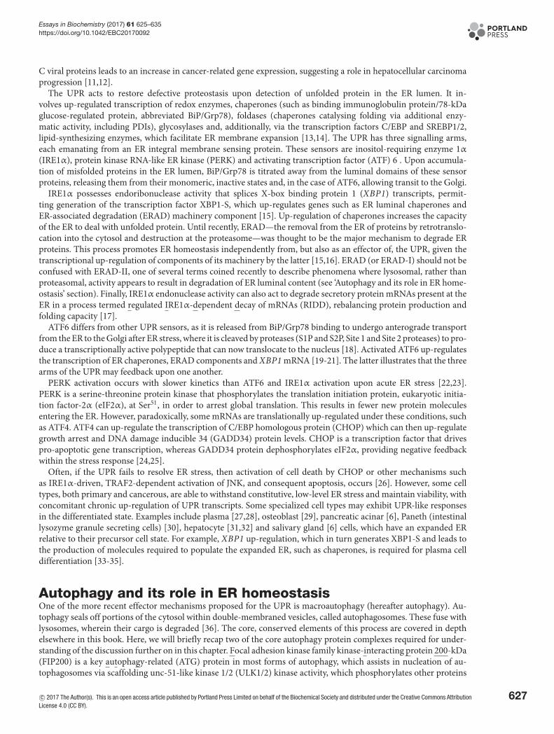
Figure 1. Outcomes of ER stress
Perturbation of ER homeostasis or ‘ER stress’ (blue), is ameliorated by the triggering of signalling cascades (yellow), which in turn
engage downstream effector mechanisms (green). Generally speaking, these mechanisms restore homeostasis. This review will
discuss these pathways and mechanisms, with particular focus on autophagy as a potential effector and, in further detail, selective
autophagic degradation of ER luminal contents or ER (ER-quality control (ERQC)-autophagy, ER-associated degradation (ERAD)-II
and ER-phagy).
ER homeostasisSignalling sensors within the ER detect perturbations within the lumen (ER stress) and employ downstream signallingcascades that engage effector mechanisms to restore homeostasis (Figure 1). These mechanisms include increasing thecapacity of the ER, increasing degradation of ER luminal proteins or up-regulating chaperones and luminal proteinmodification or folding enzymes. This review will focus on ER homeostatic pathways, with a particular emphasis onthe emerging role of autophagy as a potential effector mechanism.
Physiologically, ER stress occurs upon, for example, changes to luminal Ca2+ concentration, redox status, increasedabundance of unfolded proteins and/or hyperaccumulation of proteins. Conditions that produce this include heavybiosynthetic demand, hypoxia, redox stress, deregulated Ca2+ homeostasis and crises such as metabolite insufficiencyor low intracellular ATP levels. ER stress can also be produced experimentally with drugs that perturb calcium home-ostasis, alter redox status or inhibit glycosylation (Figure 1).
Defects in sensing and signalling pathways downstream of ER stress are associated with numerous pathologicalconditions including diabetes, non-alcoholic fatty-liver disease, Parkinson’s and Alzheimer’s diseases and cancerssuch as hepatocellular carcinoma [7,8].
Two distinct pathways for response to ER stress have been proposed, comprising the less well-understoodER-overload response (EOR) and the extensively characterized unfolded protein response (UPR). EOR is triggeredby hyperaccumulation of ER-resident proteins, not necessarily unfolded protein. Here, the ER releases luminal Ca2+,which stimulates reactive oxygen species production that in turn activates NF-κB signalling [9]. Ultimately, NF-κBup-regulates a variety of transcripts that promote proliferation and inflammation. However, little is known about themechanisms of EOR, particularly if there is any link with autophagy function. It can inhibit viral protein replica-tion, suggesting that it may act as a rapid cellular antiviral response [10,11]. Moreover, EOR activated by Hepatitis
626 c© 2017 The Author(s). This is an open access article published by Portland Press Limited on behalf of the Biochemical Society and distributed under the Creative CommonsAttribution License 4.0 (CC BY).
Essays in Biochemistry (2017) 61 625–635https://doi.org/10.1042/EBC20170092
C viral proteins leads to an increase in cancer-related gene expression, suggesting a role in hepatocellular carcinomaprogression [11,12].
The UPR acts to restore defective proteostasis upon detection of unfolded protein in the ER lumen. It in-volves up-regulated transcription of redox enzymes, chaperones (such as binding immunoglobulin protein/78-kDaglucose-regulated protein, abbreviated BiP/Grp78), foldases (chaperones catalysing folding via additional enzy-matic activity, including PDIs), glycosylases and, additionally, via the transcription factors C/EBP and SREBP1/2,lipid-synthesizing enzymes, which facilitate ER membrane expansion [13,14]. The UPR has three signalling arms,each emanating from an ER integral membrane sensing protein. These sensors are inositol-requiring enzyme 1α(IRE1α), protein kinase RNA-like ER kinase (PERK) and activating transcription factor (ATF) 6 . Upon accumula-tion of misfolded proteins in the ER lumen, BiP/Grp78 is titrated away from the luminal domains of these sensorproteins, releasing them from their monomeric, inactive states and, in the case of ATF6, allowing transit to the Golgi.
IRE1α possesses endoribonuclease activity that splices X-box binding protein 1 (XBP1) transcripts, permit-ting generation of the transcription factor XBP1-S, which up-regulates genes such as ER luminal chaperones andER-associated degradation (ERAD) machinery component [15]. Up-regulation of chaperones increases the capacityof the ER to deal with unfolded protein. Until recently, ERAD—the removal from the ER of proteins by retrotranslo-cation into the cytosol and destruction at the proteasome—was thought to be the major mechanism to degrade ERproteins. This process promotes ER homeostasis independently from, but also as an effector of, the UPR, given thetranscriptional up-regulation of components of its machinery by the latter [15,16]. ERAD (or ERAD-I) should not beconfused with ERAD-II, one of several terms coined recently to describe phenomena where lysosomal, rather thanproteasomal, activity appears to result in degradation of ER luminal content (see ‘Autophagy and its role in ER home-ostasis’ section). Finally, IRE1α endonuclease activity can also act to degrade secretory protein mRNAs present at theER in a process termed regulated IRE1α-dependent decay of mRNAs (RIDD), rebalancing protein production andfolding capacity [17].
ATF6 differs from other UPR sensors, as it is released from BiP/Grp78 binding to undergo anterograde transportfrom the ER to the Golgi after ER stress, where it is cleaved by proteases (S1P and S2P, Site 1 and Site 2 proteases) to pro-duce a transcriptionally active polypeptide that can now translocate to the nucleus [18]. Activated ATF6 up-regulatesthe transcription of ER chaperones, ERAD components and XBP1 mRNA [19-21]. The latter illustrates that the threearms of the UPR may feedback upon one another.
PERK activation occurs with slower kinetics than ATF6 and IRE1α activation upon acute ER stress [22,23].PERK is a serine-threonine protein kinase that phosphorylates the translation initiation protein, eukaryotic initia-tion factor-2α (eIF2α), at Ser51, in order to arrest global translation. This results in fewer new protein moleculesentering the ER. However, paradoxically, some mRNAs are translationally up-regulated under these conditions, suchas ATF4. ATF4 can up-regulate the transcription of C/EBP homologous protein (CHOP) which can then up-regulategrowth arrest and DNA damage inducible 34 (GADD34) protein levels. CHOP is a transcription factor that drivespro-apoptotic gene transcription, whereas GADD34 protein dephosphorylates eIF2α, providing negative feedbackwithin the stress response [24,25].
Often, if the UPR fails to resolve ER stress, then activation of cell death by CHOP or other mechanisms suchas IRE1α-driven, TRAF2-dependent activation of JNK, and consequent apoptosis, occurs [26]. However, some celltypes, both primary and cancerous, are able to withstand constitutive, low-level ER stress and maintain viability, withconcomitant chronic up-regulation of UPR transcripts. Some specialized cell types may exhibit UPR-like responsesin the differentiated state. Examples include plasma [27,28], osteoblast [29], pancreatic acinar [6], Paneth (intestinallysozyme granule secreting cells) [30], hepatocyte [31,32] and salivary gland [6] cells, which have an expanded ERrelative to their precursor cell state. For example, XBP1 up-regulation, which in turn generates XBP1-S and leads tothe production of molecules required to populate the expanded ER, such as chaperones, is required for plasma celldifferentiation [33-35].
Autophagy and its role in ER homeostasisOne of the more recent effector mechanisms proposed for the UPR is macroautophagy (hereafter autophagy). Au-tophagy seals off portions of the cytosol within double-membraned vesicles, called autophagosomes. These fuse withlysosomes, wherein their cargo is degraded [36]. The core, conserved elements of this process are covered in depthelsewhere in this book. Here, we will briefly recap two of the core autophagy protein complexes required for under-standing of the discussion further on in this chapter. Focal adhesion kinase family kinase-interacting protein 200-kDa(FIP200) is a key autophagy-related (ATG) protein in most forms of autophagy, which assists in nucleation of au-tophagosomes via scaffolding unc-51-like kinase 1/2 (ULK1/2) kinase activity, which phosphorylates other proteins
c© 2017 The Author(s). This is an open access article published by Portland Press Limited on behalf of the Biochemical Society and distributed under the Creative Commons AttributionLicense 4.0 (CC BY).
627
Essays in Biochemistry (2017) 61 625–635https://doi.org/10.1042/EBC20170092
required for autophagy [37]. It has been known for some time that the ER can platform formation of autophagosomes,potentially at sites of mitochondrial contact [38-40], generating a cradle from which the isolation membrane, the pre-cursor membrane to the autophagosome, extrudes. The isolation membrane is where ‘early’ ATG protein complexes,including those containing FIP200, are concentrated. The subsequent extension of the tubular isolation membraneand self-enclosure results in the distinctive double lipid bilayer structure of the mature autophagosome. Downstreamof ULK1 activity, a key protein grouping is ATG8-family members, of which there are six paralogues in mammals.These are microtubule-associated protein 1A/1B-light chain 3 (LC3) A, B and C and γ-aminobutyric acid receptorassociated protein (GABARAP), GABARAPL1 and GABARAPL2 [41]. These ubiquitin-like proteins are lipidatedand thus covalently attached to nascent autophagosomal membranes.
Autophagy can be a non-selective mechanism that degrades general cytosol. However, autophagy is frequentlyselective in nature, targeting damaged organelles or aberrant intracellular structures, referred to as ‘cargo’, such asperoxisomes [42], mitochondria [43,44] and lysosomes [45], ubiquitinated protein aggregates and foreign pathogens[38,39,46-48]. A key molecular component of a given selective autophagy pathway is the cargo receptor(s). The canon-ical form of this receptor class in mammals bridges cargo to ATG8 family protein(s) by binding both simultaneously.ATG8 binding is mediated via linear peptide regions called LC3-interacting region (LIR) motifs [48,49]. A primeexample of this is the well-known receptor protein, p62/SQSTM1, which links cargo to ATG8, for example duringcytosolic protein aggregate autophagy.
It is becoming evident that ER stress signals can lead to an increase in general autophagy action (Figure 2). Tuni-camycin and thapsigargin, two pharmacological inducers of the UPR, drive autophagosome formation in the humanneuroblastoma cell line SK-N-SH and increased LC3 lipidation in mouse embryonic fibroblast (MEF) cells, dependentupon the IRE1 sensor [50]. In human glioblastoma and several adenocarcinoma cell lines, it was shown that ER stressdownstream of hypoxia results in transcriptional up-regulation of MAP1LC3B (LC3B) and ATG5, via ATF4 andCHOP [51]. Here, the induced autophagy had a prosurvival role. When autophagy was induced by leucine starvationin MEFs, a range of core autophagy genes and cytosolic cargo receptors were identified as PERK-dependent ATF4transcriptional targets, including Map1lc3b, Atg5, Atg3, Atg7, Atg10, Atg12, Atg16l1, Becn1, Gabarap, Gabarapl2,p62 and Nbr1 [52]. Other pharmacologic ER stressors inducing general autophagic flux include A23187, thapsigargin,tunicamycin, brefeldin A (HCT116 human colon and DU145 human prostate carcinoma cells, and MEFs), cocaine(A172 human astrocytoma cells) and 14-deoxy-11,12-didehydroandrographolide (T47D human breast carcinomacells) [53-55]. The selectivity of autophagy was addressed in HCT116 and DU145 cells via ultrastructural character-ization of autophagosomes, which were shown to contain a variety of cargo, suggesting a general up-regulation ofrelatively non-selective autophagy [53]. Interestingly, in yeast, the specific activity of Atg1, the orthologue of ULK1, isincreased upon ER stress, without evident transcriptional induction [56]. This has not yet been observed in mammals.
None of the above studies addressed whether autophagy directly regulates ER homeostasis or showed that au-tophagy could participate in selective degradation of the ER. However, loss of all autophagy function by knockoutof conditional Atg7 flox alleles in T lymphocytes (Lck-Cre) or Atg5 flox alleles in B lymphocytes (CD19-Cre) re-sulted in expanded ER and elevated ER stress signalling, suggesting a role for autophagy in ER homeostasis [57,58].Additionally, when wild-type yeast were treated with the UPR inducer DTT, ultrastructural analysis showed, for oneof the first times, that ER in any organism could be selectively sequestered in autophagic-like vacuoles [59]. Theseearly observations generated the idea that autophagy might both regulate ER function and even do so by direct actionon the ER. In the latter instance, three pathways for putative direct regulation of ER homeostasis by selective au-tophagy are described, with the terms ‘ER-quality control autophagy’ (‘ERQC autophagy’), ERAD-II and ER-phagyproposed. The degree of overlap between these processes is currently unclear. ER-phagy is presently the mechanisti-cally best-described pathway.
ERQC autophagy was used as a term to describe a mammalian process in which disease-associated, conformermutants of proteins are removed from the lumen by autophagy without large portions of the ER itself being degraded[60,61]. An example of this is the degradation of the mutant form of the G-protein-coupled receptor E90K-GnRHR[61]. There is little mechanistic information on this phenomenon. A potentially similar phenomenon was reportedin mammals for removal of insoluble molecules of mutant protein from the ER via a lysosomal route, for which theterm ERAD-II was proposed, in analogy to proteasomal ERAD (or ERAD-I) [62].
ER-phagy involves the sequestration of portions of ER cisternae into autophagosomes and occurs in both yeast andmammals (Figure 2). Potential examples include the ER stress associated inhibition of mTOR and incorporation ofER into autophagosomes in Listeria-infected phagocytes, which may ameliorate ER stress and death [63]. Indeed, it ispresumed that general up-regulation of autophagy capacity, as described above, would translate into a commensurategreater rate of ER-phagy, but this remains unproven. However, recent findings that have given the phenomenon ofER-phagy mechanistic credence and have allowed direct testing of its role in cellular function and pathology, are the
628 c© 2017 The Author(s). This is an open access article published by Portland Press Limited on behalf of the Biochemical Society and distributed under the Creative CommonsAttribution License 4.0 (CC BY).
Essays in Biochemistry (2017) 61 625–635https://doi.org/10.1042/EBC20170092
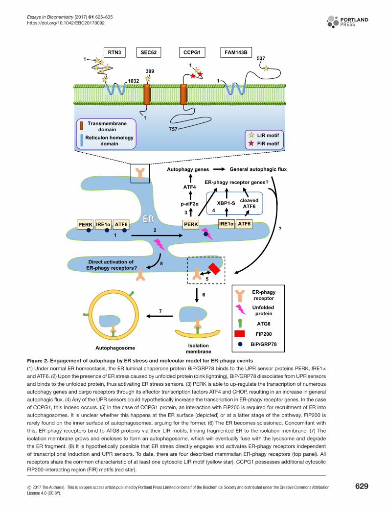
Figure 2. Engagement of autophagy by ER stress and molecular model for ER-phagy events
(1) Under normal ER homeostasis, the ER luminal chaperone protein BiP/GRP78 binds to the UPR sensor proteins PERK, IRE1α
and ATF6. (2) Upon the presence of ER stress caused by unfolded protein (pink lightning), BiP/GRP78 dissociates from UPR sensors
and binds to the unfolded protein, thus activating ER stress sensors. (3) PERK is able to up-regulate the transcription of numerous
autophagy genes and cargo receptors through its effector transcription factors ATF4 and CHOP, resulting in an increase in general
autophagic flux. (4) Any of the UPR sensors could hypothetically increase the transcription in ER-phagy receptor genes. In the case
of CCPG1, this indeed occurs. (5) In the case of CCPG1 protein, an interaction with FIP200 is required for recruitment of ER into
autophagosomes. It is unclear whether this happens at the ER surface (depicted) or at a latter stage of the pathway. FIP200 is
rarely found on the inner surface of autophagosomes, arguing for the former. (6) The ER becomes scissioned. Concomitant with
this, ER-phagy receptors bind to ATG8 proteins via their LIR motifs, linking fragmented ER to the isolation membrane. (7) The
isolation membrane grows and encloses to form an autophagosome, which will eventually fuse with the lysosome and degrade
the ER fragment. (8) It is hypothetically possible that ER stress directly engages and activates ER-phagy receptors independent
of transcriptional induction and UPR sensors. To date, there are four described mammalian ER-phagy receptors (top panel). All
receptors share the common characteristic of at least one cytosolic LIR motif (yellow star). CCPG1 possesses additional cytosolic
FIP200-interacting region (FIR) motifs (red star).
c© 2017 The Author(s). This is an open access article published by Portland Press Limited on behalf of the Biochemical Society and distributed under the Creative Commons AttributionLicense 4.0 (CC BY).
629
Essays in Biochemistry (2017) 61 625–635https://doi.org/10.1042/EBC20170092
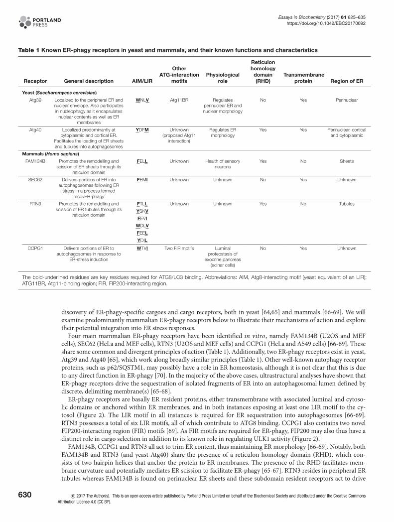
Table 1 Known ER-phagy receptors in yeast and mammals, and their known functions and characteristics
Receptor General description AIM/LIR
OtherATG-interaction
motifsPhysiological
role
Reticulonhomology
domain(RHD)
Transmembraneprotein Region of ER
Yeast (Saccharomyces cerevisiae)
Atg39 Localized to the peripheral ER andnuclear envelope. Also participatesin nucleophagy as it encapsulates
nuclear contents as well as ERmembranes
WNLV Atg11BR Regulatesperinuclear ER andnuclear morphology
No Yes Perinuclear
Atg40 Localized predominantly atcytoplasmic and cortical ER.
Facilitates the loading of ER sheetsand tubules into autophagosomes
YDFM Unknown(proposed Atg11
interaction)
Regulates ERmorphology
Yes Yes Perinuclear, corticaland cytoplasmic
Mammals (Homo sapiens)
FAM134B Promotes the remodelling andscission of ER sheets through its
reticulon domain
FELL Unknown Health of sensoryneurons
Yes No Sheets
SEC62 Delivers portions of ER intoautophagosomes following ER
stress in a process termed‘recovER-phagy’
FEMI Unknown Unknown No Yes Unknown
RTN3 Promotes the remodelling andscission of ER tubules through its
reticulon domain
FTLL Unknown Unknown Yes No Tubules
YSKV
FEVI
WDLV
FEEL
YDIL
CCPG1 Delivers portions of ER toautophagosomes in response to
ER-stress induction
WTVI Two FIR motifs Luminalproteostasis of
exocrine pancreas(acinar cells)
No Yes Unknown
The bold-underlined residues are key residues required for ATG8/LC3 binding. Abbreviations: AIM, Atg8-interacting motif (yeast equivalent of an LIR);ATG11BR, Atg11-binding region; FIR, FIP200-interacting region.
discovery of ER-phagy-specific cargoes and cargo receptors, both in yeast [64,65] and mammals [66-69]. We willexamine predominantly mammalian ER-phagy receptors below to illustrate their mechanisms of action and exploretheir potential integration into ER stress responses.
Four main mammalian ER-phagy receptors have been identified in vitro, namely FAM134B (U2OS and MEFcells), SEC62 (HeLa and MEF cells), RTN3 (U2OS and MEF cells) and CCPG1 (HeLa and A549 cells) [66-69]. Theseshare some common and divergent principles of action (Table 1). Additionally, two ER-phagy receptors exist in yeast,Atg39 and Atg40 [65], which work along broadly similar principles (Table 1). Other well-known autophagy receptorproteins, such as p62/SQSTM1, may possibly have a role in ER homeostasis, although it is not clear that this is dueto any direct function in ER-phagy [70]. In the majority of the above cases, ultrastructural analyses have shown thatER-phagy receptors drive the sequestration of isolated fragments of ER into an autophagosomal lumen defined bydiscrete, delimiting membrane(s) [65-68].
ER-phagy receptors are basally ER resident proteins, either transmembrane with associated luminal and cytoso-lic domains or anchored within ER membranes, and in both instances exposing at least one LIR motif to the cy-tosol (Figure 2). The LIR motif in all instances is required for ER sequestration into autophagosomes [66-69].RTN3 possesses a total of six LIR motifs, all of which contribute to ATG8 binding. CCPG1 also contains two novelFIP200-interacting region (FIR) motifs [69]. As FIR motifs are required for ER-phagy, FIP200 may also thus have adistinct role in cargo selection in addition to its known role in regulating ULK1 activity (Figure 2).
FAM134B, CCPG1 and RTN3 all act to trim ER content, thus maintaining ER morphology [66-69]. Notably, bothFAM134B and RTN3 (and yeast Atg40) share the presence of a reticulon homology domain (RHD), which con-sists of two hairpin helices that anchor the protein to ER membranes. The presence of the RHD facilitates mem-brane curvature and potentially mediates ER scission to facilitate ER-phagy [65-67]. RTN3 resides in peripheral ERtubules whereas FAM134B is found on perinuclear ER sheets and these subdomain resident receptors act to drive
630 c© 2017 The Author(s). This is an open access article published by Portland Press Limited on behalf of the Biochemical Society and distributed under the Creative CommonsAttribution License 4.0 (CC BY).
Essays in Biochemistry (2017) 61 625–635https://doi.org/10.1042/EBC20170092
local ER-phagy [67]. For instance, RTN3 specifically drives tubular ER-phagy. Both FAM134B and RTN3 can driveER-phagy in vitro that is stimulated by the general autophagic stimulus of amino acid starvation. In vitro, CCPG1may also participate in ER-stress driven ER-phagy, after DTT treatment. Finally, Sec62 participates specifically inER-phagy during recovery from ER stressors, clearing fragments of ER enriched in now redundant ER chaperones[68]. This process is distinguished within the larger set of ER-phagy responses by the term ‘recovER-phagy’. Thesedifferent mechanistic routes for ER-phagy, targeting distinct regions of the ER and at different stages of the ER stressresponse, suggest functional specialization of ER-phagy pathways, which may have further relevance in vivo wherethe ER exhibits large differences in form and function between different tissue types (see below).
There is emerging evidence for direct links between homeostatic ER responses and ER-phagy from in vivo mod-els. Investigations of the physiological role of autophagy have generally involved ablation of all autophagy functionby conditional knockout of core Atg genes in various tissues in mice, using tissue-specific, promoter-driven recom-binases, typically Cre or tamoxifen-inducible Cre-ERT2. Summarizing, autophagy loss per se generally disrupts ERmorphology and size, and produces stress responses. In Paneth cells, Atg7 or Atg16l were seen to be required to re-strain IRE1α activation after ER stress induced by experimental XBP1 down-regulation. Loss of this action leads tointestinal inflammation [71]. Furthermore, knockout of Atg5 or Atg7 in pancreatic acinar cells produces ER dilation,stress, inhibition of secretory protein transcription, cell death and inflammation [72,73], although not all reports agreewith these findings [74]. However, it is important to note that the contribution of ER-phagy is not precisely interro-gated when core autophagy genes are deleted; ER pathology may be an indirect consequence of damaged mitochon-dria or protein aggregate accumulation, and consequent effects on bioenergetics and signalling. The constructionof ER-phagy receptor mutant mice has begun to allow exploration of this issue. Fam134b−/− mice exhibit dilatedER and Golgi within peripheral sensory neurons and cell death [66]. Intriguingly, FAM134B is mutated in humanfamilies with heritable sensory neuropathy and these mutations ablate ER-phagy function [66]. In Ccpg1 hypomor-phic animals, the acinar cells within the exocrine pancreas exhibit distended ER and insoluble ER luminal proteinaccumulation, as well as elevated UPR [69]. Few other tissues are reported to be affected by loss of ER-phagy recep-tor function, indicating that physiologically important ER-phagy receptors remain undiscovered or untested. Thisobservation befits the functional specialization of ER within different cell lineages.
Future directionsIn the authors’ view, a major source of outstanding questions in the field of mammalian ER homeostasis centre uponthe mechanism and function of ER-phagy.
One question: is whether there is a relationship between ER-phagy and mechanistically opaque processes suchas ERQC autophagy and ERAD-II? What degree of overlap in players and mechanisms is there here? Additionally,what molecular determinants mark an ER-phagy receptor, other than LIR motifs? For example, CCPG1 additionallypossesses FIR motifs for recognition of the ER by autophagy. Linked to this, it is also crucial to consider whetherER-phagy receptors merely mark ER membrane for degradation or play an active role in scissioning ER membrane topermit sequestration or in sensing ER stress. As a potential example of the former, FAM134B and RTN3 have retic-ulon domains that might assist ER membrane breakage. In the latter category, some of these receptors have luminalpolypeptide regions and it might be conjectured that they could directly sense changes in redox, luminal [Ca2+] or un-folded protein in parallel with canonical sensors. In the case of CCPG1, the ER luminal domain contains a coiled-coilforming region, and it is possible that regulated multimerization/clustering could modify function, as suggested forRTN3 [67,69]. Cytosolic domains might also play a role within the downstream relays of ER stress signalling path-ways. For example, some cargo receptors acting in other selective autophagy paradigms bear phosphoregulable LIRmotifs [75,76].
Does ER stress specifically trigger ER-phagy, non-selective autophagy or a combination of both, and in which tissuetypes and to what ultimate purpose? The finding that the UPR can transcriptionally up-regulate CCPG1 provides amechanistic link between ER stress and selective autophagy of the ER. The Ccpg1 promoter also binds the transcrip-tion factor MIST1 [77], which is predominantly expressed in professional secretory cells such as pancreatic acinarcells. MIST1 expression is required for correct differentiation here and is itself dependent upon the IRE1α-XBP1arm of the UPR [78,79], providing a potential tissue-specific link between CCPG1 up-regulation and its role in ERhomeostasis. It is therefore of key importance to dissect whether ER stress response pathways can regulate the activityof other ER-phagy receptors, either to activate them acutely or as is likely for CCPG1 in vivo, also in part to mediatetheir tissue-specific expression.
It is difficult to say with exactitude how ER-phagy mediates ER homeostasis at a detailed mechanistic level. It ispossible that ER-phagy acts to remodel and rebalance the different regions of the ER to meet fluctuating biosynthetic
c© 2017 The Author(s). This is an open access article published by Portland Press Limited on behalf of the Biochemical Society and distributed under the Creative Commons AttributionLicense 4.0 (CC BY).
631
Essays in Biochemistry (2017) 61 625–635https://doi.org/10.1042/EBC20170092
demand. Alternatively, perhaps proteostatic defects result in localized accumulation of unfolded or aggregated pro-tein, by transport mechanisms within the ER lumen, such that specific portions of ER are ‘sacrificed’ in a piecemealmanner in order to maintain unfolded protein at a manageable level within the lumen. There is also little informationon rough ER compared with smooth ER as a cargo of autophagy. Different pathways and different receptors mayparticipate, again pointing to likely tissue-specific divergence of these mechanisms in vivo.
Finally, another potentially ER-phagy related mechanism that specialist cells might use to respond to ER stress issecretory autophagy. Paneth cells secrete ER luminal lysozyme via the ER to defend against pathogens. Here, ER stress,triggered by bacterial invasion, results in an increase in secretory autophagy of lysozyme, i.e. the use of the autophagymachinery to build secretory vesicles containing the lysozyme, rather than utilization of the default secretory pathwayvia the Golgi [80]. Investigations are required to determine if ER-phagy receptors play a role here.
ConclusionThe UPR and other ER stress response signalling pathways engage multiple effector pathways. A recently emergingeffector is autophagy and, of particular interest, ER-phagy. Taken together, the evidence points to a pivotal role forER-phagy in normal ER homeostasis and overall cell health, particularly in specialized tissue types in vivo. Cru-cially, ER-phagy helps ameliorate the effects of ER stress through the degradation of ER membranes, removal of ERluminal protein aggregates and/or removal of ER-chaperone proteins. Furthermore, ER-phagy also acts to trim ERcontent, helping maintain the dynamism that is characteristic of this key organelle. Future work will surely expandthe molecular components and role of this pathway in currently known and new tissue types.
Summary• ER is the key site for the folding of nascent transmembrane and secretory proteins.
• The lumen of ER has a specialized environment to ensure the fidelity of this process.
• Signalling sensors within the ER lumen detect stress and employ downstream cascades to engageeffector mechanisms and restore homeostasis.
• The major signalling cascade employed by the ER is the UPR, which translationally and transcrip-tionally engages a variety of effector mechanisms.
• Selective degradation of the ER by autophagy occurs via a process termed as ER-phagy.
• There are currently four known mammalian ER-phagy receptors; FAM134B, SEC62, RTN3 andCCPG1.
• ER-phagy receptors possess LIR motifs, allowing interactions with ATG8 family proteins.
• ER-phagy has important roles in the physiology of secretory cells in vivo.
• General autophagic flux and direct ER-phagy might both be transcriptionally up-regulated by theUPR.
FundingThis work was supported by the Cancer Research UK Career Development Fellowship [grant number CRUK-A12825 (to S.W.)];and the BBSRC Project [grant number BB/N000315/1].
Competing interestsThe authors declare that there are no competing interests associated with the manuscript.
632 c© 2017 The Author(s). This is an open access article published by Portland Press Limited on behalf of the Biochemical Society and distributed under the Creative CommonsAttribution License 4.0 (CC BY).
Essays in Biochemistry (2017) 61 625–635https://doi.org/10.1042/EBC20170092
AbbreviationsATF, activating transcription factor; ATG, autophagy-related; Bip/Grp78, binding immunoglobulin protein/78-kDaglucose-regulated protein; CHOP, C/EBP homologous protein; eIF2α, eukaryotic initiation factor-2α; EOR, ER-overload re-sponse; ER, endoplasmic reticulum; ERAD, ER-associated degradation; ERQC, ER quality control; FIR, FIP200-interactingregion; GADD34, growth arrest and DNA damage inducible 34; IRE1α, inositol-requiring enzyme 1α; LC3, light chain 3; LIR,LC3-interacting region; MEF, mouse embryonic fibroblast; PDI, protein disulphide isomerase; PERK, protein kinase RNA-like ERkinase; RHD, reticulon homology domain; UPR, unfolded protein response; XBP1, X-box binding protein 1.
References1 Berridge, M.J. (1998) Neuronal calcium signaling. Neuron 21, 13–262 Baumann, O. and Walz, B. (2001) Endoplasmic reticulum of animal cells and its organization into structural and functional domains. Int. Rev. Cytol.
205, 149–2143 Brodsky, J.L. and Skach, W.R. (2011) Protein folding and quality control in the endoplasmic reticulum: Recent lessons from yeast and mammalian cell
systems. Curr. Opin. Cell Biol. 23, 464–4754 Braakman, I. and Hebert, D.N. (2013) Protein folding in the endoplasmic reticulum. Cold Spring Harb. Perspect. Biol. 5, a013201,
https://doi.org/10.1101/cshperspect.a0132015 Bulleid, N.J. (2012) Disulfide bond formation in the mammalian endoplasmic reticulum. Cold Spring Harb. Perspect. Biol. 4, a013219,
https://doi.org/10.1101/cshperspect.a0132196 Lee, A.H., Chu, G.C., Iwakoshi, N.N. and Glimcher, L.H. (2005) XBP-1 is required for biogenesis of cellular secretory machinery of exocrine glands.
EMBO J. 24, 4368–43807 Wang, M. and Kaufman, R.J. (2016) Protein misfolding in the endoplasmic reticulum as a conduit to human disease. Nature 529, 326–3358 Lindholm, D., Korhonen, L., Eriksson, O. and Koks, S. (2017) Recent insights into the role of unfolded protein response in ER stress in health and
disease. Front. Cell Dev. Biol. 5, 489 Pahl, H.L. and Baeuerle, P.A. (1995) A novel signal transduction pathway from the endoplasmic reticulum to the nucleus is mediated by transcription
factor NF-kappa B. EMBO J. 14, 2580–258810 Pahl, H.L. and Baeuerle, P.A. (1995) Expression of influenza virus hemagglutinin activates transcription factor NF-kappa B. J. Virol. 69, 1480–148411 Kong, L., Li, S., Huang, M., Xiong, Y., Zhang, Q., Ye, L. et al. (2015) The roles of endoplasmic reticulum overload response induced by HCV and NS4B
protein in human hepatocyte viability and virus replication. PLoS ONE 10, e0123190, https://doi.org/10.1371/journal.pone.012319012 Kong, L., Li, S., Yu, X., Fang, X., Xu, A., Huang, M. et al. (2016) Hepatitis C virus and its protein NS4B activate the cancer-related STAT3 pathway via the
endoplasmic reticulum overload response. Arch. Virol. 161, 2149–215913 Travers, K.J., Patil, C.K., Wodicka, L., Lockhart, D.J., Weissman, J.S. and Walter, P. (2000) Functional and genomic analyses reveal an essential
coordination between the unfolded protein response and ER-associated degradation. Cell 101, 249–25814 Lee, J.S., Mendez, R., Heng, H.H., Yang, Z.Q. and Zhang, K. (2012) Pharmacological ER stress promotes hepatic lipogenesis and lipid droplet formation.
Am. J. Transl. Res. 4, 102–11315 Lee, A.H., Iwakoshi, N.N. and Glimcher, L.H. (2003) XBP-1 regulates a subset of endoplasmic reticulum resident chaperone genes in the unfolded
protein response. Mol. Cell. Biol. 23, 7448–745916 Vembar, S.S. and Brodsky, J.L. (2008) One step at a time: endoplasmic reticulum-associated degradation. Nat. Rev. Mol. Cell Biol. 9, 944–95717 Hollien, J. and Weissman, J.S. (2006) Decay of endoplasmic reticulum-localized mRNAs during the unfolded protein response. Science 313, 104–10718 Ye, J., Rawson, R.B., Komuro, R., Chen, X., Dave, U.P., Prywes, R. et al. (2000) ER stress induces cleavage of membrane-bound ATF6 by the same
proteases that process SREBPs. Mol. Cell 6, 1355–136419 Wu, J., Rutkowski, D.T., Dubois, M., Swathirajan, J., Saunders, T., Wang, J. et al. (2007) ATF6alpha optimizes long-term endoplasmic reticulum function
to protect cells from chronic stress. Dev. Cell 13, 351–36420 Yoshida, H., Matsui, T., Yamamoto, A., Okada, T. and Mori, K. (2001) XBP1 mRNA is induced by ATF6 and spliced by IRE1 in response to ER stress to
produce a highly active transcription factor. Cell 107, 881–89121 Yamamoto, K., Sato, T., Matsui, T., Sato, M., Okada, T., Yoshida, H. et al. (2007) Transcriptional induction of mammalian ER quality control proteins is
mediated by single or combined action of ATF6alpha and XBP1. Dev. Cell 13, 365–37622 Lin, J.H., Li, H., Yasumura, D., Cohen, H.R., Zhang, C., Panning, B. et al. (2007) IRE1 signaling affects cell fate during the unfolded protein response.
Science 318, 944–94923 Rutkowski, D.T., Arnold, S.M., Miller, C.N., Wu, J., Li, J., Gunnison, K.M. et al. (2006) Adaptation to ER stress is mediated by differential stabilities of
pro-survival and pro-apoptotic mRNAs and proteins. PLoS Biol. 4, e374, https://doi.org/10.1371/journal.pbio.004037424 Harding, H.P., Zhang, Y., Scheuner, D., Chen, J.J., Kaufman, R.J. and Ron, D. (2009) Ppp1r15 gene knockout reveals an essential role for translation
initiation factor 2 alpha (eIF2alpha) dephosphorylation in mammalian development. Proc. Natl. Acad. Sci. U.S.A. 106, 1832–183725 Marciniak, S.J., Yun, C.Y., Oyadomari, S., Novoa, I., Zhang, Y., Jungreis, R. et al. (2004) CHOP induces death by promoting protein synthesis and
oxidation in the stressed endoplasmic reticulum. Genes Dev. 18, 3066–307726 Nishitoh, H., Matsuzawa, A., Tobiume, K., Saegusa, K., Takeda, K., Inoue, K. et al. (2002) ASK1 is essential for endoplasmic reticulum stress-induced
neuronal cell death triggered by expanded polyglutamine repeats. Genes Dev. 16, 1345–135527 Shaffer, A.L., Shapiro-Shelef, M., Iwakoshi, N.N., Lee, A.H., Qian, S.B., Zhao, H. et al. (2004) XBP1, downstream of Blimp-1, expands the secretory
apparatus and other organelles, and increases protein synthesis in plasma cell differentiation. Immunity 21, 81–93
c© 2017 The Author(s). This is an open access article published by Portland Press Limited on behalf of the Biochemical Society and distributed under the Creative CommonsAttribution License 4.0 (CC BY).
633
Essays in Biochemistry (2017) 61 625–635https://doi.org/10.1042/EBC20170092
28 Gass, J.N., Gifford, N.M. and Brewer, J.W. (2002) Activation of an unfolded protein response during differentiation of antibody-secreting B cells. J. Biol.Chem. 277, 49047–4905
29 Saito, A., Ochiai, K., Kondo, S., Tsumagari, K., Murakami, T., Cavener, D.R. et al. (2011) Endoplasmic reticulum stress response mediated by thePERK-eIF2(alpha)-ATF4 pathway is involved in osteoblast differentiation induced by BMP2. J. Biol. Chem. 286, 4809–4818
30 Kaser, A., Lee, A.H., Franke, A., Glickman, J.N., Zeissig, S., Tilg, H. et al. (2008) XBP1 links ER stress to intestinal inflammation and confers genetic riskfor human inflammatory bowel disease. Cell 134, 743–756
31 Lee, A.H., Scapa, E.F., Cohen, D.E. and Glimcher, L.H. (2008) Regulation of hepatic lipogenesis by the transcription factor XBP1. Science 320,1492–1496
32 Rutkowski, D.T., Wu, J., Back, S.H., Callaghan, M.U., Ferris, S.P., Iqbal, J. et al. (2008) UPR pathways combine to prevent hepatic steatosis caused byER stress-mediated suppression of transcriptional master regulators. Dev. Cell 15, 829–840
33 van Anken E, R.E.P., Maggioni, C., Mezghrani, A., Sitia, R., Braakman, I. et al. (2003) Sequential waves of functionally related proteins are expressedwhen B cells prepare for antibody secretion. Immunity 18, 243–253
34 Hu, C.C., Dougan, S.K., McGehee, A.M., Love, J.C. and Ploegh, H.L. (2009) XBP-1 regulates signal transduction, transcription factors and bone marrowcolonization in B cells. EMBO J. 28, 1624–1636
35 Todd, D.J., McHeyzer-Williams, L.J., Kowal, C., Lee, A.H., Volpe, B.T., Diamond, B. et al. (2009) XBP1 governs late events in plasma cell differentiationand is not required for antigen-specific memory B cell development. J. Exp. Med. 206, 2151–2159
36 Ktistakis, N.T. and Tooze, S.A. (2016) Digesting the expanding mechanisms of autophagy. Trends Cell Biol. 26, 624–63537 Lin, M.G. and Hurley, J.H. (2016) Structure and function of the ULK1 complex in autophagy. Curr. Opin. Cell Biol. 39, 61–6838 Axe, E.L., Walker, S.A., Manifava, M., Chandra, P., Roderick, H.L., Habermann, A. et al. (2008) Autophagosome formation from membrane
compartments enriched in phosphatidylinositol 3-phosphate and dynamically connected to the endoplasmic reticulum. J. Cell Biol. 182, 685–70139 Hayashi-Nishino, M., Fujita, N., Noda, T., Yamaguchi, A., Yoshimori, T. and Yamamoto, A. (2009) A subdomain of the endoplasmic reticulum forms a
cradle for autophagosome formation. Nat. Cell Biol. 11, 1433–143740 Hamasaki, M., Furuta, N., Matsuda, A., Nezu, A., Yamamoto, A., Fujita, N. et al. (2013) Autophagosomes form at ER-mitochondria contact sites. Nature
495, 389–39341 Kabeya, Y., Mizushima, N., Yamamoto, A., Oshitani-Okamoto, S., Ohsumi, Y. and Yoshimori, T. (2004) LC3, GABARAP and GATE16 localize to
autophagosomal membrane depending on form-II formation. J. Cell Sci. 117, 2805–281242 Kim, P.K., Hailey, D.W., Mullen, R.T. and Lippincott-Schwartz, J. (2008) Ubiquitin signals autophagic degradation of cytosolic proteins and peroxisomes.
Proc. Natl. Acad. Sci. U.S.A. 105, 20567–2057443 Heo, J.M., Ordureau, A., Paulo, J.A., Rinehart, J. and Harper, J.W. (2015) The PINK1-PARKIN mitochondrial ubiquitylation pathway drives a program of
OPTN/NDP52 recruitment and TBK1 activation to promote mitophagy. Mol. Cell 60, 7–2044 Lazarou, M., Sliter, D.A., Kane, L.A., Sarraf, S.A., Wang, C., Burman, J.L. et al. (2015) The ubiquitin kinase PINK1 recruits autophagy receptors to induce
mitophagy. Nature 524, 309–31445 Maejima, I., Takahashi, A., Omori, H., Kimura, T., Takabatake, Y., Saitoh, T. et al. (2013) Autophagy sequesters damaged lysosomes to control lysosomal
biogenesis and kidney injury. EMBO J. 32, 2336–234746 Thurston, T.L., Ryzhakov, G., Bloor, S., von Muhlinen, N. and Randow, F. (2009) The TBK1 adaptor and autophagy receptor NDP52 restricts the
proliferation of ubiquitin-coated bacteria. Nat. Immunol. 10, 1215–122147 Zheng, Y.T., Shahnazari, S., Brech, A., Lamark, T., Johansen, T. and Brumell, J.H. (2009) The adaptor protein p62/SQSTM1 targets invading bacteria to
the autophagy pathway. J. Immunol. 183, 5909–591648 Pankiv, S., Clausen, T.H., Lamark, T., Brech, A., Bruun, J.A., Outzen, H. et al. (2007) p62/SQSTM1 binds directly to Atg8/LC3 to facilitate degradation of
ubiquitinated protein aggregates by autophagy. J. Biol. Chem. 282, 24131–2414549 Birgisdottir, A.B., Lamark, T. and Johansen, T. (2013) The LIR motif - crucial for selective autophagy. J. Cell Sci. 126, 3237–324750 Ogata, M., Hino, S., Saito, A., Morikawa, K., Kondo, S., Kanemoto, S. et al. (2006) Autophagy is activated for cell survival after endoplasmic reticulum
stress. Mol. Cell. Biol. 26, 9220–923151 Rouschop, K.M., van den Beucken, T., Dubois, L., Niessen, H., Bussink, J., Savelkouls, K. et al. (2010) The unfolded protein response protects human
tumor cells during hypoxia through regulation of the autophagy genes MAP1LC3B and ATG5. J. Clin. Invest. 120, 127–14152 B’Chir, W., Maurin, A.C., Carraro, V., Averous, J., Jousse, C., Muranishi, Y. et al. (2013) The eIF2alpha/ATF4 pathway is essential for stress-induced
autophagy gene expression. Nucleic Acids Res. 41, 7683–769953 Ding, W.X., Ni, H.M., Gao, W., Hou, Y.F., Melan, M.A., Chen, X. et al. (2007) Differential effects of endoplasmic reticulum stress-induced autophagy on
cell survival. J. Biol. Chem. 282, 4702–471054 Periyasamy, P., Guo, M.L. and Buch, S. (2016) Cocaine induces astrocytosis through ER stress-mediated activation of autophagy. Autophagy 12,
1310–132955 Tan, H.K., Muhammad, T.S.T. and Tan, M.L. (2016) 14-Deoxy-11,12-didehydroandrographolide induces DDIT3-dependent endoplasmic reticulum
stress-mediated autophagy in T-47D breast carcinoma cells. Toxicol. Appl. Pharmacol. 300, 55–6956 Yorimitsu, T., Nair, U., Yang, Z. and Klionsky, D.J. (2006) Endoplasmic reticulum stress triggers autophagy. J. Biol. Chem. 281, 30299–3030457 Jia, W., Pua, H.H., Li, Q.J. and He, Y.W. (2011) Autophagy regulates endoplasmic reticulum homeostasis and calcium mobilization in T lymphocytes. J.
Immunol. 186, 1564–157458 Pengo, N., Scolari, M., Oliva, L., Milan, E., Mainoldi, F., Raimondi, A. et al. (2013) Plasma cells require autophagy for sustainable immunoglobulin
production. Nat. Immunol. 14, 298–30559 Bernales, S., McDonald, K.L. and Walter, P. (2006) Autophagy counterbalances endoplasmic reticulum expansion during the unfolded protein response.
PLoS Biol. 4, e423, https://doi.org/10.1371/journal.pbio.0040423
634 c© 2017 The Author(s). This is an open access article published by Portland Press Limited on behalf of the Biochemical Society and distributed under the Creative CommonsAttribution License 4.0 (CC BY).
Essays in Biochemistry (2017) 61 625–635https://doi.org/10.1042/EBC20170092
60 Teckman, J.H. and Perlmutter, D.H. (2000) Retention of mutant alpha(1)-antitrypsin Z in endoplasmic reticulum is associated with an autophagicresponse. Am. J. Physiol. Gastrointest. Liver Physiol. 279, G961–G974
61 Houck, S.A., Ren, H.Y., Madden, V.J., Bonner, J.N., Conlin, M.P., Janovick, J.A. et al. (2014) Quality control autophagy degrades soluble ERAD-resistantconformers of the misfolded membrane protein GnRHR. Mol. Cell 54, 166–179
62 Fujita, E., Kouroku, Y., Isoai, A., Kumagai, H., Misutani, A., Matsuda, C. et al. (2007) Two endoplasmic reticulum-associated degradation (ERAD) systemsfor the novel variant of the mutant dysferlin: ubiquitin/proteasome ERAD(I) and autophagy/lysosome ERAD(II). Hum. Mol. Genet. 16, 618–629
63 Moretti, J., Roy, S., Bozec, D., Martinez, J., Chapman, J.R., Ueberheide, B. et al. (2017) STING senses microbial viability to orchestrate stress-mediatedautophagy of the endoplasmic reticulum. Cell, https://doi.org/10.1016/j.cell.2017.09.034
64 Lipatova, Z. and Segev, N. (2015) A role for macro-ER-phagy in ER quality control. PLoS Genet. 11, e1005390,https://doi.org/10.1371/journal.pgen.1005390
65 Mochida, K., Oikawa, Y., Kimura, Y., Kirisako, H., Hirano, H., Ohsumi, Y. et al. (2015) Receptor-mediated selective autophagy degrades the endoplasmicreticulum and the nucleus. Nature 522, 359–362
66 Khaminets, A., Heinrich, T., Mari, M., Grumati, P., Huebner, A.K., Akutsu, M. et al. (2015) Regulation of endoplasmic reticulum turnover by selectiveautophagy. Nature 522, 354–358
67 Grumati, P., Morozzi, G., Holper, S., Mari, M., Harwardt, M.I., Yan, R. et al. (2017) Full length RTN3 regulates turnover of tubular endoplasmic reticulumvia selective autophagy. Elife 6, https://doi.org/10.7554/eLife.25555
68 Fumagalli, F., Noack, J., Bergmann, T.J., Cebollero, E., Pisoni, G.B., Fasana, E. et al. (2016) Translocon component Sec62 acts in endoplasmic reticulumturnover during stress recovery. Nat. Cell Biol. 18, 1173–1184
69 Smith, M.D., Harley, M.E. and Kemp, A.J., Wills, J., Lee, M., Arends, M. et al. (2017) CCPG1, a non-canonical autophagy cargo receptor essential forpancreatic ER proteostasis. Dev. Cell ., in press
70 Yang, H., Ni, H.M., Guo, F., Ding, Y., Shi, Y.H., Lahiri, P. et al. (2016) Sequestosome 1/p62 protein is associated with autophagic removal of excesshepatic endoplasmic reticulum in mice. J. Biol. Chem. 291, 18663–18674
71 Adolph, T.E., Tomczak, M.F., Niederreiter, L., Ko, H.J., Bock, J., Martinez-Naves, E. et al. (2013) Paneth cells as a site of origin for intestinalinflammation. Nature 503, 272–276
72 Diakopoulos, K.N., Lesina, M., Wormann, S., Song, L., Aichler, M., Schild, L. et al. (2015) Impaired autophagy induces chronic atrophic pancreatitis inmice via sex- and nutrition-dependent processes. Gastroenterology 148, 626–638.e17
73 Antonucci, L., Fagman, J.B., Kim, J.Y., Todoric, J., Gukovsky, I., Mackey, M. et al. (2015) Basal autophagy maintains pancreatic acinar cell homeostasisand protein synthesis and prevents ER stress. Proc. Natl. Acad. Sci. U.S.A. 112, E6166–E6174
74 Hashimoto, D., Ohmuraya, M., Hirota, M., Yamamoto, A., Suyama, K., Ida, S. et al. (2008) Involvement of autophagy in trypsinogen activation within thepancreatic acinar cells. J. Cell Biol. 181, 1065–1072
75 Khaminets, A., Behl, C. and Dikic, I. (2016) Ubiquitin-dependent and independent signals in selective autophagy. Trends Cell Biol. 26, 6–1676 Wild, P., Farhan, H., McEwan, D.G., Wagner, S., Rogov, V.V., Brady, N.R. et al. (2011) Phosphorylation of the autophagy receptor optineurin restricts
Salmonella growth. Science 333, 228–23377 Tian, X., Jin, R.U., Bredemeyer, A.J., Oates, E.J., Blazewska, K.M., McKenna, C.E. et al. (2010) RAB26 and RAB3D are direct transcriptional targets of
MIST1 that regulate exocrine granule maturation. Mol. Cell. Biol. 30, 1269–128478 Hess, D.A., Humphrey, S.E., Ishibashi, J., Damsz, B., Lee, A.H., Glimcher, L.H. et al. (2011) Extensive pancreas regeneration following acinar-specific
disruption of Xbp1 in mice. Gastroenterology 141, 1463–147279 Huh, W.J., Esen, E., Geahlen, J.H., Bredemeyer, A.J., Lee, A.H., Shi, G. et al. (2010) XBP1 controls maturation of gastric zymogenic cells by induction of
MIST1 and expansion of the rough endoplasmic reticulum. Gastroenterology 139, 2038–204980 Bel, S., Pendse, M., Wang, Y., Li, Y., Ruhn, K.A., Hassell, B. et al. (2017) Paneth cells secrete lysozyme via secretory autophagy during bacterial
infection of the intestine. Science, https://doi.org/10.1126/science.aal4677
c© 2017 The Author(s). This is an open access article published by Portland Press Limited on behalf of the Biochemical Society and distributed under the Creative Commons AttributionLicense 4.0 (CC BY).
635