Embed Size (px)
Citation preview
In higher-latitude areas, particularly in the
Northern Hemisphere, there are significant
temperature changes that do not appear to be
directly related to land-cover change. Al-
though statistically significant, these changes
are relatively small as compared to the pro-
jected atmospheric forcing changes. For ex-
ample, in western Russia there is reforestation
in both scenarios, which should lead to warm-
ing. However, although the additional land-
cover changes have the expected impact on
net radiation, the B1 and A2 scenarios show
strongly opposing temperature signals in De-
cember, January, and February (DJF). These
results appear to be closely linked to changes
in regional precipitation and may be the result
of teleconnections, either linked to the Asian
Monsoon circulation or indirect effects from
temperature changes over the tropical Pacific
and North Atlantic Oceans.
Results from this study suggest that the
choices humans make about future land use
could have a significant impact on regional
and seasonal climates. Some of these effects
are the result of direct impacts of land-cover
change on local moisture and energy balances.
Other impacts appear to be related to signifi-
cant indirect climate effects through telecon-
nection processes. The A2 land-cover scenario
shows that tropical rainforest conversion will
likely lead to a weakening of the Hadley cir-
culation over much of the world and to signif-
icant changes in the Asian Monsoon circulation.
Especially in the A2 2050 scenario, the inter-
play between Asian and African land-cover
change affects the Asian Monsoon circulation.
The Indian Ocean experiences a significant
reduction in surface pressure, resulting in in-
creased cloud cover and precipitation and
warmer surface temperatures, and these effects
extend over most of the Indian subcontinent.
We conclude that the inclusion of land-
cover forcing, thereby accounting for a num-
ber of additional anthropogenic climate impacts,
will improve the quality of regional climate as-
sessments for IPCC SRES scenarios. Although
land-cover effects are regional and tend to offset
with respect to global average temperatures,
they can significantly alter regional climate out-
comes associated with global warming. Beyond
local impacts, tropical land-cover change can
potentially affect extratropical climates and
nearby ocean conditions through atmospheric
teleconnections. In this respect, our fully cou-
pled experiments differ from previous fixed
ocean temperature studies (12, 13, 15). Further
study is needed to determine the exact nature
of these responses. Overall, the results demon-
strate the importance of including land-cover
change in forcing scenarios for future climate
change studies.
References and Notes1. J. J. Houghton et al., Eds., Climate Change 2000: The
Scientific Basis (IPCC Working Group I, CambridgeUniv. Press, Cambridge, 2001).
2. P. Kabat et al., Vegetation, Water, Humans and theClimate Change: A New Perspective on an InteractiveSystem (Springer, Heidelberg, Germany, 2002).
3. W. Steffen et al., Global Change and the EarthSystem: A Planet Under Pressure (Springer-Verlag,New York, 2004).
4. R. A. Betts, Atmos. Sci. Lett. 2, 39 (2001).5. L. R. Bounoua, R. DeFries, G. J. Collatz, P. Sellers, H.
Khan, Clim. Change 52, 29 (2002).6. T. N. Chase, R. A. Peilke Sr., T. G. F. Kittel, R. R.
Nemani, S. W. Running, Clim. Dyn. 16, 93 (2000).7. J. J. Feddema et al., Clim. Dyn. 25, 581 (2005).8. J. Hansen et al., Proc. Natl. Acad. Sci. U.S.A. 95,
12753 (1998).9. H. D. Matthews, A. J. Weaver, K. J. Meissner, N. P.
Gillett, M. Eby, Clim. Dyn. 22, 461 (2004).10. M. H. Costa, J. A. Foley, J. Clim. 13, 18 (2000).11. N. Gedney, P. J. Valdes, Geophys. Res. Lett. 27, 3053
(2000).12. K. McGuffie, A. Henderson-Sellers, H. Zhang, T. B.
Durbidge, A. J. Pitman, Global Planet. Change 10, 97(1995).
13. R. S. DeFries, L. Bounoua, G. J. Collatz, Global ChangeBiol. 8, 438 (2002).
14. S. Sitch et al., Global Biogeochem. Cycles 19,GB2013 (2004).
15. R. Avissar, D. Werth, J. Hydrometeorol. 6, 134 (2005).16. R. A. Pielke Sr. et al., Philos. Trans. R. Soc. London
Ser. A 360, 1705 (2002).17. G. Krinner et al., Global Biogeochem. Cycles 19,
GB1015 (2005).18. P. K. Snyder, C. Delire, J. A. Foley, Clim. Dyn. 23, 279 (2004).19. G. B. Bonan, D. Pollard, S. L. Thompson, Nature 359,
716 (1992).20. G. A. Meehl et al., Science 307, 1769 (2005).21. W. M. Washington et al., Clim. Dyn. 16, 755 (2000).22. N. Nakicenovic et al., Special Report on Emissions
Scenarios (Cambridge Univ. Press, Cambridge, 2000).23. J. Alcamo, R. Leemans, E. Kreileman, Eds., Global Change
Scenarios of the 21st Century. Results from the IMAGE2.1 Model (Pergamon Elsevier Science, London, 1998).
24. IMAGE 2.2 CD release and documentation (RijksInstituut voor Volksgezondheid en Milieu, Bilthoven,Netherlands, 2002). The IMAGE 2.2 implementationof the SRES scenarios: A Comprehensive Analysis ofEmissions, Climate Change and Impacts in the 21stCentury (see www.rivm.nl/image/index.html for fur-ther information).
25. Materials and methods are available as supportingmaterial on Science Online.
26. T. R. Karl, R. W. Knight, Bull. Am. Meteorol. Soc. 78,1107 (1997).
27. M. P. Hoerling, J. W. Hurrell, T. Xu, G. T. Bates, A. S.Phillips, Clim. Dyn. 23, 391 (2004).
28. G. B. Bonan, Ecol. Appl. 9, 1305 (1999).29. G. B. Bonan, J. Clim. 14, 2430 (2001).30. We acknowledge the large number of scientists who
have assisted in the development of the models andtools used to create the simulations used in this study.Special thanks to A. Middleton, T. Bettge, and G.Strand for their assistance in running the model andassistance with data processing and to R. Leemansfor providing the SRES data. This research was sup-ported by the Office of Science (Biological and Envi-ronmental Research Program), U.S. Department ofEnergy, under Cooperative Agreement No. DE-FC02-97ER62402; NSF (grant numbers ATM-0107404 andATM-0413540); the National Center for AtmosphericResearch Weather and Climate Impact AssessmentScience Initiative supported by NSF; and the Centerfor Research, University of Kansas, Lawrence, KS.
Supporting Online Materialwww.sciencemag.org/cgi/content/full/310/5754/1674/DC1Materials and MethodsFigs. S1 and S2References
29 July 2005; accepted 25 October 200510.1126/science.1118160
Equivalent Effects of Snake PLA2Neurotoxins and Lysophospholipid–
Fatty Acid MixturesMichela Rigoni,1 Paola Caccin,1 Steve Gschmeissner,2
Grielof Koster,3 Anthony D. Postle,3 Ornella Rossetto,1
Giampietro Schiavo,2 Cesare Montecucco1*
Snake presynaptic phospholipase A2 neurotoxins (SPANs) paralyze the neuro-muscular junction (NMJ). Upon intoxication, the NMJ enlarges and has a reducedcontent of synaptic vesicles, and primary neuronal cultures show synapticswelling with surface exposure of the lumenal domain of the synaptic vesicleprotein synaptotagmin I. Concomitantly, these neurotoxins induce exocytosis ofneurotransmitters. We found that an equimolar mixture of lysophospholipids andfatty acids closely mimics all of the biological effects of SPANs. These resultsdraw attention to the possible role of local lipid changes in synaptic vesiclerelease and provide new tools for the study of exocytosis.
SPANs are major protein components of the
venom of many snakes (1–3). They block the
NMJ in a characteristic way (3–7). The phos-
pholipase A2 (PLA2) activity varies greatly
among different SPANs, and its involvement
in the NMJ block is still debated (3, 8, 9).
There is only a partial correlation between PLA2
activity and neurotoxicity among SPANs and no
overlap of surface residues required for neuro-
toxicity with those essential for PLA2 activ-
ity (8, 10). Here, we compared the effects of
SPANs on the mouse NMJ hemidiaphragm
preparation and on neurons in culture with those
of their hydrolysis products: lysophospholipids
(LysoPL) and fatty acids (FAs). To conclusive-
1Department of Biomedical Sciences and ConsiglioNazionale Ricerche Institute of Neuroscience, Universityof Padova, Italy. 2Cancer Research UK, London ResearchInstitute, London, UK. 3School of Medicine, Universityof Southampton, UK.
*To whom correspondence should be addressed.E-mail: [email protected]
R E P O R T S
9 DECEMBER 2005 VOL 310 SCIENCE www.sciencemag.org1678
ly determine the nature of SPAN hydrolysis
products, cerebellar neurons were treated with
SPANs, and their lipid composition was de-
termined by mass spectrometry. The major
hydrolytic substrate was phosphatidylcholine,
the main phospholipid of the outer leaflet of
the plasma membrane (fig. S1). SPAN hydrol-
ysis generated several lysophosphatidylcholines
(LysoPC), including myristoyl lysophosphati-
dylcholine (mLysoPC), and FA. SPANs were
not selective for a particular FA species (such as
arachidonic acid) and released mainly oleic acid
(OA), the most abundant FA of these cells.
The incubation of cerebellar neurons with
mLysoPCþOA (30 mM each) led to the incorpo-
ration of 6.3 nmol of mLysoPC/105 cells, com-
pared with 2.3 nmol/105 cells treated with 6 nM
taipoxin. mLysoPCþOA did not cause acylation,
because the 14.0 to 16.0 ratio in PC did not
increase. The values of LysoPC associated with
neurons in the two cases were closely compa-
rable, particularly if one considers that SPANs
induce a localized release of LysoPL and FA,
whereas the incubation with mLysoPCþOA
presumably caused generalized lipid insertion.
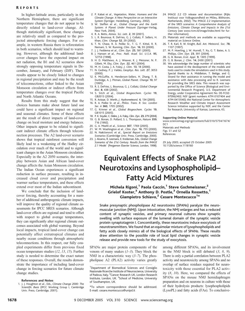
mLysoPCþOA added to a mouse hemi-
diaphragm in a physiological medium caused a
progressive NMJ paralysis with a time course
superimposable to that observed with a typical
SPAN (Fig. 1A). Four SPANs of different struc-
tural complexity and relative toxicity were used:
the single-chain notexin (14 kD, from Notechis
scutatus), the two-subunit b-bungarotoxin (21 kD,
from Bungarus multicinctus), the three-subunit
taipoxin (42 kD, from Oxyuranus scutellatus),
and the five-subunit textilotoxin (72 kD, from
Pseudonaja textilis) (1). They induced closely
similar paralysis profiles, although with slightly
different kinetics; one representative trace is
shown. When textilotoxin and mLysoPCþOA
were present at the same time (1.5 nM and 50
mM, respectively), a synergistic effect was ob-
served, with the time required to achieve 50% of
paralysis (t1/2
) shorter by a factor of 4 T 0.5
times (n 0 4) than that of textilotoxin. Pancreatic
PLA2 (at a concentration matching the activity
of textilotoxin in Fig. 1A) did paralyze the NMJ,
but with a t1/2
three times as long, presumably
because of a reduced membrane interaction.
Of the two products of SPAN phospholipid
hydrolysis, LysoPC alone was capable of
inhibiting the NMJ, although with low poten-
cy, whereas FA was poorly effective below the
threshold concentration inducing myotoxicity;
however, FA and LysoPC clearly acted syn-
ergistically (Fig. 1B).
Similar results were obtained with other
LysoPL, such as the ethanolamine, serine, and
glycerol derivatives. We also tested the effect of
LysoPC esterified with FAs of different length
and saturation obtaining similar results, but dif-
ferent kinetics, with the following order of t1/2
:
myristoyl-LysoPC (taken as 1, to normalize
the data obtained in five different experi-
ments), oleoyl-LysoPC (2.2 T 0.5 times as long),
palmitoyl-LysoPC (3.2 T 1.3), and stearoyl-
LysoPC (8.5 T 0.6). Their potency correlates
with their critical micellar concentrations (11, 12),
indicating that the more water-soluble LysoPC
equilibrates more rapidly into the membrane
and acts faster; it is also possible that the shorter
LysoPL causes a higher constraint on the mem-
brane curvature. The paralysis was not due to
an effect of mLysoPCþOA on the muscle it-
self, because direct muscle stimulation elicited
full contraction and the muscle maintained its
normal ultrastructure (Fig. 1, C and D). The
mLysoPCþOA mixture induced diagnostic al-
terations in the ultrastructure of the NMJ, in-
cluding a reduction of the number of synaptic
vesicles and an enlargement of the nerve termi-
nal (Fig. 1C), which closely mimic the changes
observed in SPAN-treated NMJs (4–7).
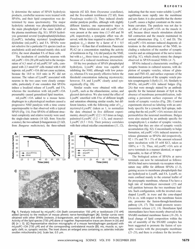
SPANs induced a characteristic swelling of
synaptic boutons in cultured neurons, with de-
pletion of synaptic vesicles, release of gluta-
mate and FM1-43, and surface exposure of the
intralumenal portion of the synaptic vesicle pro-
tein synaptotagmin I (SytI) (13, 14). Similarly to
SPANs, mLysoPCþOA induced bulges (Fig.
2A) that were strongly stained by an antibody
specific for the lumenal domain of SytI in the
absence of membrane permeabilization, indi-
cating a persistent surface exposure of the
inside of synaptic vesicles (Fig. 2B). Control
experiments showed no labeling with an anti-
body specific for a cytosolic SytI epitope, indi-
cating that the mLysoPCþOA mixture did not
permeabilize the neuronal membrane. Bulges
were also stained by an antibody specific for
synaptophysin I, a marker of synaptic vesicles,
showing that they are sites of synaptic vesicle
accumulation (fig. S2). Concomitantly to bulge
formation, mLysoPCþOA induced neurons to
release glutamate as SPANs did (respectively,
92 T 1% and 78 T 1% of the amount released
upon incubation with 55 mM KCl, taken as
100%; n 0 5). Thus, mLysoPCþOA acts at
nerve terminals in a manner identical, or super-
imposable, to that of SPANs.
The biological action of SPANs at nerve
terminals can now be rationalized as follows:
SPANs bind nerve terminals via receptors whose
nature may differ for different SPANs (1–3),
gaining access to membrane phospholipids that
are hydrolyzed to LysoPL and FA. LysoPL re-
main confined mainly to the external leaflet of
the presynaptic membrane, whereas FAs have a
high rate of transbilayer movement (15) and
will partition between the two membrane leaf-
lets. Such configuration, with the inverted cone-
shaped LysoPL in trans and the cone-shaped
FA in cis, with respect to the membrane fusion
site, promotes the fusion-through-hemifusion
pathway (16, 17). This would promote neuro-
transmitter release (13, 14). Hemifusion lipid
intermediates have been recently observed in the
SNARE-mediated membrane fusion (18–20). A
local change of lipid composition within the
site of assembly of the SNARE complex has
been suggested to promote the fusion of syn-
aptic vesicles with the presynaptic membrane
(21–23), and there is evidence for the involve-
Fig. 1. Paralysis induced by mLysoPCþOA (bath concentration 150 mM) or textilotoxin (15 nM)added (arrows) to the medium of mouse phrenic nerve-hemidiaphragm (A). Similar curves wereobtained with other SPANs (notexin, b-bungarotoxin, and taipoxin) and other lipid mixtures. (B)Activity of the lipid species (150 mM) alone or together. Representative traces are shown in (A) and(B) (n Q 5). (C) Representative electron micrographs of a mouse hemidiaphragm paralyzed withmLysoPCþOA (150 mM) and of the corresponding contralateral muscle (D). mu, muscle; sc, syn-aptic cleft; sv, synaptic vesicles. The inset shows an enlarged area containing sv; asterisks indicateswollen mitochondria (m). Scale bar, 0.5 mm.
R E P O R T S
www.sciencemag.org SCIENCE VOL 310 9 DECEMBER 2005 1679
ment of PLA2 in other exocytotic events such
as the sperm acrosomal exocytosis (24). Fur-
thermore, a SPAN microinjected into pheo-
chromocytoma cells inhibited neuroexocytosis
(25), presumably because it acted on the cyto-
solic plasma membrane side, inducing an op-
posite membrane configuration. The presence of
clathrin-coated W-shaped structures in SPAN-
poisoned NMJs (4–7) suggested that they also
inhibit synaptic vesicle fission from the plasma
membrane (3, 14). Indeed, the same SPAN-
induced lipid changes promoting membrane
fusion do inhibit membrane fission for the
same physical and topological reasons (17).
References and Notes1. R. M. Kini, Ed., Venom Phospholipase A2 Enzymes
(Wiley, Chichester, UK, 1997).2. G. Schiavo, M. Matteoli, C. Montecucco, Physiol. Rev.
80, 717 (2000).3. C. Montecucco, O. Rossetto, Trends Biochem. Sci. 25,
266 (2000).4. S. G. Cull-Candy, J. Fohlman, D. Gustavsson, R. Lullmann-
Rauch, S. Thesleff, Neuroscience 1, 175 (1976).
5. I. L. Chen, C. Y. Lee, Virchows Arch. B Cell Pathol. 6,318 (1970).
6. J. B. Harris, B. D. Grubb, C. A. Maltin, R. Dixon, Exp.Neurol. 161, 517 (2000).
7. C. Y. Lee, M. C. Tsa, Y. M. Chen, A. Ritonja, F. Gubensek,Arch. Int. Pharmacodyn. Ther. 268, 313 (1984).
8. P. Rosenberg, Venom Phospholipase A2 Enzymes, R. M.Kini, Ed. (Wiley, Chichester, UK, 1997), pp. 155-183.
9. R. M. Kini, Toxicon 42, 827 (2003).10. C. C. Yang, in Venom Phospholipase A2 Enzymes, R. M.
Kini, Ed. (Wiley, Chichester, UK,1997), pp. 185-204.11. R. E. Stafford, T. Fanni, E. A. Dennis, Biochemistry 28,
5113 (1989).12. J. Wang et al., Br. J. Pharmacol. 141, 586 (2004).13. M. Rigoni et al., J. Cell Sci. 15, 3561 (2004).14. D. Bonanomi et al., Mol. Pharmacol. 67, 1901 (2005).15. F. Kamp, D. Zakim, F. Zhang, N. Noy, J. A. Hamilton,
Biochemistry 34, 11928 (1995).16. L. V. Chernomordik, E. Leikina, V. Frolov, P. Bronk,
J. Zimmerberg, J. Cell Biol. 136, 81 (1997).17. L. V. Chernomordik, M. M. Kozlov, Annu. Rev. Biochem.
72, 175 (2003).18. Y. Xu, F. Zhang, Z. Su, J. A. McNew, Y. K. Shin, Nat.
Struct. Mol. Biol. 12, 417 (2005).19. C. G. Giraudo et al., J. Cell Biol. 170, 249 (2005).20. C. Reese, F. Heise, A. Mayer, Nature 436, 410 (2005).21. K. Farsad, P. De Camilli, Curr. Opin. Cell Biol. 15, 372
(2003).22. R. Jahn, T. Lang, T. C. Sudhof, Cell 112, 519 (2003).23. L. K. Tamm, J. Crane, V. Kiessling, Curr. Opin. Struct.
Biol. 13, 453 (2003).24. E. R. S. Roldan, Front. Biosci. 3, 1119 (1998).25. S. Wei et al., Neuroscience 121, 891 (2003).26. Supported by Telethon grant GP0272Y01, COFIN Project
2002055747, FISR-DM 16/10/00, FIRB-RBNE01RHZM,University of Padova, and Cancer Research UK.
Supporting Online Materialwww.sciencemag.org/cgi/content/full/310/5754/1678/DC1Materials and MethodsFigs. S1 and S2References
27 September 2005; accepted 4 November 200510.1126/science.1120640
Neural Systems Responding toDegrees of Uncertainty in Human
Decision-MakingMing Hsu,1 Meghana Bhatt,1 Ralph Adolphs,1,2
Daniel Tranel,2 Colin F. Camerer1*
Much is known about how people make decisions under varying levels of prob-ability (risk). Less is known about the neural basis of decision-making whenprobabilities are uncertain because of missing information (ambiguity). Indecision theory, ambiguity about probabilities should not affect choices. Usingfunctional brain imaging, we show that the level of ambiguity in choices cor-relates positively with activation in the amygdala and orbitofrontal cortex, andnegatively with a striatal system. Moreover, striatal activity correlates positivelywith expected reward. Neurological subjects with orbitofrontal lesions wereinsensitive to the level of ambiguity and risk in behavioral choices. These datasuggest a general neural circuit responding to degrees of uncertainty, contrary todecision theory.
In theories of choice under uncertainty used in
social sciences and behavioral ecology, the
only variables that should influence an un-
certain choice are the judged probabilities of
possible outcomes and the evaluation of those
outcomes. But confidence in judged probabil-
ity can vary widely. In some choices, such as
gambling on a roulette wheel, probability can
be confidently judged from relative frequen-
cies, event histories, or an accepted theory. At
the other extreme, such as the chance of a
terrorist attack, probabilities are based on
meager or conflicting evidence, where impor-
tant information is clearly missing. The two
types of uncertain events are often called risky
and ambiguous, respectively. In subjective
expected utility theory, the probabilities of out-
comes should influence choices, whereas
confidence about those probabilities should
not. But experiments show that many people
are more willing to bet on risky outcomes than
on ambiguous ones, holding judged probability
of outcomes constant (1). This empirical aver-
sion to ambiguity motivates a search for neural
distinctions between risk and ambiguity. Here,
we extend the study of the neural basis of
decision under risk to encompass ambiguity.
The difference between risky and ambigu-
ous uncertainty is illustrated by the Ellsberg
paradox (2). Imagine one deck of 20 cards
composed of 10 red and 10 blue cards (the
risky deck). Another deck has 20 red or blue
cards, but the composition of red and blue
cards is completely unknown (the ambiguous
deck). A bet on a color pays a fixed sum (e.g.,
$10) if a card with the chosen color is drawn,
and zero otherwise (Fig. 1A).
Fig. 2. Field emissionscanning electron mi-croscopy (FESEM) ofcerebellar granular neu-rons exposed to taipoxin(6 nM for 60 min) ormLysoPCþOA (30 mMfor 15 min) at lower (leftpanels) and higher (rightpanels) magnifications(A). Identical resultswere obtained withnotexin, b-bungarotoxin,and textilotoxin. Scalebar, 10 mm (left pan-els) and 2 mm (rightpanels). (B) Cerebellarneurons were exposedto 6 nM b-bungarotoxinfor 60 min or to 30 mMmLysoPCþOA for 15min and stained withan antibody specific for the lumenal domain of synaptotagmin I before fixation. Samples wereprocessed for indirect immunofluorescence without permeabilization; superimposable results wereobtained with notexin, taipoxin, and textilotoxin in cerebellar neurons and hippocampal neurons.Scale bar, 10 mm.
1Division of Humanities and Social Sciences, 228-77,California Institute of Technology, Pasadena, CA 91125,USA. 2University of Iowa Medical School, Iowa City, IA52242, USA.
*To whom correspondence should be addressed.E-mail: [email protected]
R E P O R T S
9 DECEMBER 2005 VOL 310 SCIENCE www.sciencemag.org1680