Embed Size (px)
Citation preview
EQUILIBRIUM RECEPTORS OF COCKROACH CERCI:
INTERNEURON RESPONSES TO DISPLAC2^iENT
by
WALTER WILLIAM WALTHALL, B.S.
A THESIS
IN
ZOOLOGY
Submitted to the Graduate Faculty of Texas Tech University in Partial Fulfillment of the Requirements for
the Degree of
MASTER OF SCIENCE
Approved
Accepted
Quate School foeikn olf iiie G:^
December, 1973
r
ACKNOWLEDGMENTS
I would like to thank Dr. H. Bernard Hartman for
advice and encouragement throughout this investigation.
I also would like to thank Drs. C. D. Barnes and J. K.
Wangberg for serving on my committee and for their con
structive comments on the thesis.
I am indebted to Lisa P. Bennett, Bruce W. Leander
and Randall R. Stewart for their assistance during the
course of this investigation.
11

CONTENTS
ACKNOWLEDGMENTS
III. RESULTS
IV. DISCUSSION
REFERENCES
Page
ii
LIST OF FIGURES "^
I. INTRODUCTION ^
II. MATERIALS AND METHODS "7
12
52
65
111
FIGURES
Figure Page
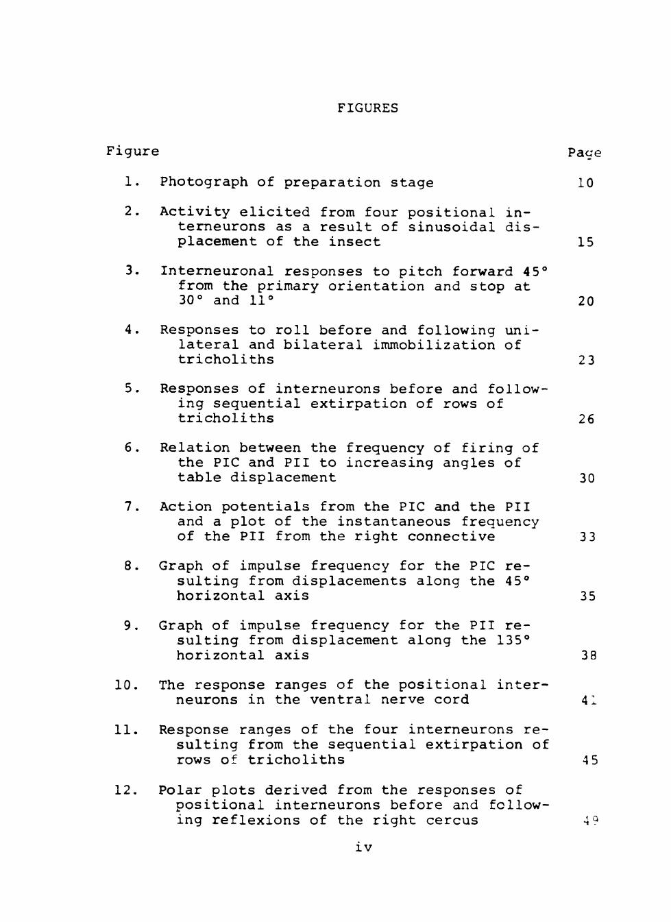
1. Photograph of preparation stage 10
2. Activity elicited from four positional in-terneurons as a result of sinusoidal displacement of the insect 15
3. Interneuronal responses to pitch forward 45° from the primary orientation and stop at 30° and 11° 20
4. Responses to roll before and following unilateral and bilateral immobilization of tricholiths 2 3
5. Responses of interneurons before and following sequential extirpation of rows of tricholiths 26
6. Relation between the frequency of firing of the PIC and PII to increasing angles of table displacement 30
7. Action potentials from the PIC and the PII and a plot of the instantaneous frequency of the PII from the right connective 3 3
8. Graph of impulse frequency for the PIC resulting from displacements along the 45° horizontal axis 35
9. Graph of impulse frequency for the PII resulting from displacement along the 135° horizontal axis 38
10. The response ranges of the positional interneurons in the ventral nerve cord 41
11. Response ranges of the four interneurons resulting from the sequential extirpation of rows of tricholiths 4 5
12. Polar plots derived from the responses of positional interneurons before and following reflexions of the right cercus 4<^
IV

Figure ^^^e
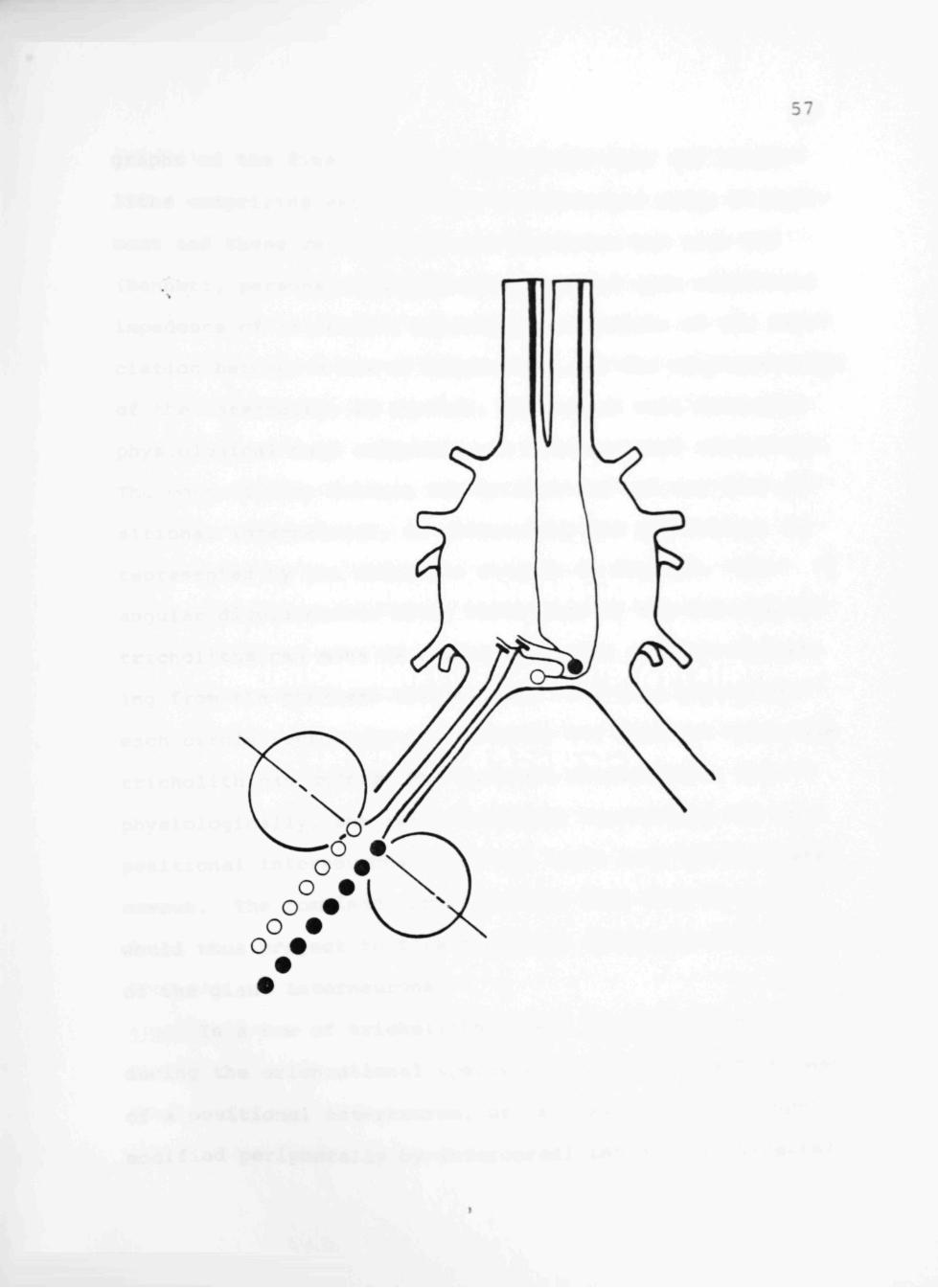
13. Schematic representation of the underlying connectivity between the rows of tricholiths on the right cercus and the associated PII and PIC 57

CHAPTER I
INTRODUCTION
The importance of afference apprising an organism
of alterations in its spatial orientation is such that
primary receptor mechanisms and statocyst organs have been
developed for virtually all of the classes in the Kingdom
Animalia. It should be recognized at the outset that the
force and direction of the gravitational field are con
stant, and as a result there is no need for the develop
ment of biological systems strictly for the reception of
gravity. However, the earth's gravitational field pro
vides a continuous source of linear acceleration. This
has allowed for the evolution of a variety of weighted
detection devices which permit the transduction of the
contact force by impeding the movement of a mass in the
gravitational field and subsequently, the extraction of
information apprising an organism of geotactic orienta
tion.
In the Class Vertebrata the labyrinth consists of
three semicircular canals, the utricle and the saccule.
Receptors in these structures deal with equilibrium and
orientation in three dimensional space. The semicircular
canals are concerned with kinetic equilibrium. They are
arranged at right angles to each other and e?ch one corres-

ponds to and represents one of the three planes of move
ment. Located at the base of each canal is a dilation,
the ampulla, containing a ridge or crista which is ori
ented transversely to the canal. Each crista is covered
by a gelatinous cupula. Angular acceleration causes dis
placement of endolymphatic fluid and movement of the cupu
la which stimulates the hair cells. The utricle and sac
cule each have a similar patch of sensory epithelium, the
macula utriculi and macula sacculi. But here the cells
are in contact with a gelatinous covering containing small
calcareous concretions or particles, the otoliths. The
utricular macula, concerned primarily with static equili
brium, responds to changes in gravitational forces, and to
linear acceleration. Macular impulses convey information
regarding the position of the head in space, the hair cells
being stimulated by the otolithic particles, whose posi
tion varies under the influence of gravity.
Crustaceans are apprised of spatial orientation by
statocysts. The statocyst is a specialized mechanorecep-
tor found at the base of the antennules or, in a few in
stances, in the abdomen (Neil, 1975). Each is a cavity
lined with sensory epithelium and contains one or more
heavy bodies, acting as liths. Some Crustacea secrete
statoliths, while others construct their liths from sand
or other particles obtained from their immediate surround-

ings. The statoliths are attached to the sensory hairs
which protrude from the walls of the cyst cavity. The
shearing force of the statolith is adequate stimulus for
the crustacean statocyst. Two parameters of this sheering
force, direction as well as magnitude, are recorded by the
statocyst receptors and contribute to the positional in
formation.
The statocysts of several species of agile crabs
have undergone extensive modification from the general
crustacean plan. They resemble morphologically and physi
ologically the semicircular canal system of vertebrates
(Sandeman and Okajima, 1972; Fraser and Sandeman, 1975).
The development of the canal system is seemingly advan
tageous in that acceleration and position, two different
types of orientational input, are separated and restricted
to independent receptors.
Investigations of equilibrium receptor systems in
the Class Insecta have revealed no structures designed
primarily for the transduction of information regarding
the insect's spatial orientation. Instead, behavioral
experiments have lead investigators to believe that terres
trial insects extract orientation information from proprio
ceptors monitoring joint position or limb loading (Horn,
1975a, b; Markl, 1971; Wendler, 1965, 1975). The major
criterion for labeling such sense organs as equilibrium

receptors depends upon experiments which measure the geo
tactic turning tendency of an insect. For example, in
his experiments, Horn (1975b) rendered the innervated
hair plates nonfunctional either by trimming, immobilizing,
or completely removing the sensilla. If the insect's geo
tactic turning tendency was significantly affected by in
capacitation of the hair plate, that hair plate was assum
ed to play a role in equilibrium reception.
Visual input provides afference which governs the
spatial orientation of some insects in certain situations.
Although this afference in no way provides a measurement
of forces related to the earth's gravitational attraction,
under natural conditions the gravitational force vector
and visual cues used by the insects maintain a constant
relationship with one another. Evidence indicates insects
through a preprocessing mechanism of pattern recognition
lock the visual receptors onto a space constant. For
locusts the space constcint has been determined to be the
bright part of the horizon (Goodman, 1965). Ants use as a
visual constant the natural pattern of polarized light in
the sky and, by recognizing the point where the highest
degree of linear polarization occurs are able to orient
themselves accordingly (Wehner, 1975).
Roth and Slifer (1973) published an anatomical de-n scription of the cerci of certain poiyphagid cockroaches.
(

They described a group of sensilla, which have terTdnally
positioned dense spheres, and are located on the ventral
side of the insect's cerci. The authors suggested that
they were modified from trichobothria; showed that each of
these sensilla inserted into an innervated socket; and
speculated that because these hairs resembled plumb bobs,
they have evolved as equilibrium receptors. Hartman et al.,
(in manuscript), using Arenivaga sp., (Dictyoptera: Poly-
phagidae) a cockroach with two rows of the specialized sen
silla on each cercus, provided physiological evidence con
firming the hypothesis of Roth and Slifer. Hartman et al_. ,
(in manuscript) found that the pendulous sensilla, termed
tricholiths, excite two ascending giant interneurons in
each of the two connectives of the ventral nerve cord.
Each interneuron in a connective can be clearly distin
guished from its counterpart based upon the amplitudes of
the spikes produced and their discrete response ranges.
It was further shown that the four interneurons occur in
two homologous pairs, a small and a large unit in each
connective.
The objectives of this work were to determine the
orientational components conveyed by the interneurons and
to investigate the association between the tricholiths and
the four giant fibers by characterizing the response ranges
of the interneurons to alterations in the insects spatial

position. While the main thrust of this research deals
with the orientation parameter of position as coded by
these giant interneurons, it is noteworthy that the data
records include clues that acceleration may also be coded
by receptors, probably trichobothria, also located on the
cerci. The experiments reported here will not include
consideration of these units.

CHAPTER II
MATERIALS AND METHODS
Adult male cockroaches of the genus Arenivaga, col
lected beneath rock outcroppings along the banks of the
Rio Grcuide in southwestern Texas, were used in this inves
tigation. Interneuronal responses evoked as a result of
rotations about horizontal axes can be obtained from both
sexes, but because the cereal sensilla of the male cock
roaches are more accessible, allowing easier observation
and manipulation, the data presented were obtained from
male insects.
To prepare the insect for recording, the wings were
removed by dissection, and the insect secured ventral
side down by stainless steel pins positioned across the
thorax and tip of the abdomen onto a bed of surgical peri
phery wax. Care was taken so that the last abdominal seg
ment and the cerci protruded unimpeded from the wax re-
strainer. A wax bridge across the restraining pins and
lateral to the abdomen provided a moat for the saline and
prevented the cerci from becoming moist. The nerve cord
was exposed by removing abdominal terga 1-4, and the heart,
hindgut and fat body. Fielden's insect saline to which
Ig/L glucose and 0.5ml/L Tris were added and having a pH
of 7.4 served to keep the nerve cord moist and alive

8
(Fielden, 1960).
En passant recordings were obtained between the first
and second abdominal ganglia by drawing the two connec
tives of the nerve cord into separate suction electrodes
(Fig. 1). Next, the platform, containing the preparation
and electrode-bearing manipulators was screwed to a tilt
table. The tilt table was designed to allow controlled
displacement over a 110° range about any designated hori
zontal axis. Table movement was achieved via a Bodine
motor controller to a Bodine D. D. motor linked to the
table via a chain. Sinusoidal or linear table displace
ments were achieved by attaching the chain from the table
to either a pivoting arm, or a wheel on the motor shaft.
In order to precisely determine the angular position of
the table (and the attached insect), a Hewlett-Packard
7DCDT linear displacement transducer was attached to the
base of the table. The output of the transducer was dis
played upon a F. Haer DDO-2 oscilloscope calibrated to
read angular position.
Signals detected by the suction electrodes were am
plified by Grass P15 A. C. preamplifiers, tape-recorded
with a Tandberg 115 FM instrumentation tape recorder, and
displayed on a Tektronix 5111 storage oscilloscope. A
pair of Grass AM7 audio monitors provided audible cues of
neuronal activity. Tape-recorded data was played back on


Fig. 1. Photograph of the preparation stage which is screwed to the tilt table. A, grounding electrode; B, three-way stopcock; C, electrode lead connection to preamplifier; D, joy stick micromanipulator; E, gross manipulator; F, wax block with roach in recording position; G, Suction electrode

10

11
a Tektronix 565 dual beam oscilloscope, the screened dis
play being filmed with a Grass C4 oscilloscope camera.
Where required, the frequencies at which the interneurons
produced spikes were analyzed using a F. Haer Window Dis
criminator and an Instantaneous Frequency Analyzer.

CHAPTER III
RESULTS
In each of the experiments that will be reported,
movement was initiated from primary orientation. This is
the position adopted by an animal when it is inactive,
and the position from which active movement generally
begins (Fraenkel and Gunn, 1961). This occurs for Areni
vaga when its longitudinal and transverse axes both lie
in the same horizontal plane. All displacements in the
following experiments will be initiated from primary ori
entation.
In the first series of experiments the orientational
component conveyed by the four interneurons which respond
to tricholith movement (Hartman et al., in manuscript)
were determined. The three orientation parameters sig
naled by equilibrium organs are velocity, acceleration,
and position (Sandeman, 1976). Velocity is a measure of
position change in relation to time. In sine wave move
ments about the primary orientation, maximum velocity is
reached as the animal passes through the primary orienta
tion. Acceleration is a measure of the rate of change of
velocity. Accordingly, acceleration reaches its peak at
the maximum rate of change of velocity. This point is
reached at the initiation of movement and 180° later
12
13
(Sandeman, 1976). In recordings made from the two con
nectives of the ventral nerve cord of Arenivaga, four
giant interneurons responded to sinusoidal movements of
the insect (Fig. 2). Upon pitch forward, from the pri
mary orientation, a small giant interneuron in each con
nective responded with a tonic output, the interpulse
interval decreasing as the table displacement angle in
creased to 55° forward. When the direction of table move
ment changed to pitch backward, the small units decreased
in firing frequency, ceased firing, and subsequently, a
large giant interneuron in each connective was activated.
The interpulse interval of these units decreased as the
table angle increased to a peak at about 55° backward
(Fig. 2A). When the pitch stimulus ceased as the insect
was stopped at primary orientation, the small units which
were activated by the movement toward pitch forward adap
ted to zero frequency in a few seconds (Fig. 2B).
Rolling the insect produced activity in these same
four giant interneurons, but in a different combination
(Fig. 2C). Upon roll to the right, first the small unit
and then the large unit of the left connective were acti
vated. As the angle of roll increased toward 55° right,
the interpulse interval of both units decreased steadily.
The change in direction of insect movement toward roll
left brought about rapid decline and cessation of firing

14
Fig. 2A-D. Activity elicited from the four positional interneurons as a result of sinusoidal displacement of the insect. The top trace in each record was obtained from the left connective and the second trace from the right connective. The third monitors table position, and the fourth is a 5/s time reference. In A, from primary orientation, the insect is first pitched forward to 55° (upward deflection of monitor) followed by pitch backward to 55° (downward deflection) . B Record showing adaptation upon cessation of movement at primary orientation the insect is first rolled right to 55° (downward deflection) and then rolled left to 55° (upward deflection). D Record showing adaptation upon cessation of movement at primary orientation following roll series. Scale: 200yV.

15

16
by the large unit followed shortly by a similar pattern
on the part of the small unit. The comparable pair of
interneurons in the right connective commenced firing as
the insect was rolled toward the left, following the pat
tern of activity previously described. When the insect
was stopped and maintained in the primary orientation af
ter the roll stimuli, the giant interneurons ceased firing
in a few seconds (Fig. 2D). Because peak neuronal output
occurred in phase with maximum insect displacement, posi
tion was confirmed as the orientation component signaled
by the four giant interneurons.
Observation of Fig. 2 revealed that the four inter
neurons appeared to be signaling position on displacements
made away from the primary orientation, but provided cim-
biguous position information upon reversal of direction
cmd return toward primary orientation. Rather, the units
exhibited hysteresis when the insect was moved in a direc
tion away from the appropriate stimulus direction. To
illustrate this ambiguity, measurements of the angle at
which responding units ceased firing and alternate units
began firing were made as the insect was sinusoidally dis
placed along the pitch axis at the rates of 5°, 10°, and
20° per second. Upon displacement from primary orientation,
activity was initiated from the two small units in each
connective as has already been described. As table move-
17
ment began its return to primary orientation, an almost
immediate increase in the interpulse interval was observed.
The activity from both small interneurons had completely
stopped by 22° (See Fig. 2A). The large unit began firing
at approximately 12° forward and continued firing with the
interpulse interval decreasing as the insect passed through
primary orientation on its rotation backward. As the table
began its return to primary orientation from displacement
55° backward, the interpulse interval of the two large
units increased rapidly until the units dropped out com
pletely at 49°. The two small units began to respond at
28° backward and continue as has been previously describ
ed. The same phenomenon occurs when the insect is oscil
lated along the roll axis (refer to Fig. 2C).
While the insect's angular position is in a transi
tional state, tricholith afference indicates the direc
tion of angular movement. It is impossible to distinguish
this fact as the insect is rotated away from primary orien
tation because during this movement, directional compon
ents and positional components both have the same neural
code. It is not until the insect is returned from an an
gular displacement toward primary orientation that these
two components separate and become distinguishable.
To illustrate this, an insect was sinusoidally oscil
lated forward to 45° and on the return to primary orienta-

18
tion was stopped at 30° in one instance, and at 11° in an
other to determine the effect that the direction of ap
proach has upon the positional component. Activity from
the small units in each connective began at the onset of
table movement and increased until the table oscillation
started to return to primary orientation. It was at this
point that the response from the small units began to wane.
When the table movement was stopped at 30°, there was a-
gain an increase in the neural activity of the small in
terneurons in response to static displacement, which con
tinued at a tonic level until table movement was resumed,
at which time they ceased responding (Fig. 3A). The two
large units started firing as the table movement approach
ed primary orientation, but when movement was halted at
11° pitch forward, the output from the large units immedi
ately stopped and spikes from the small units began at a
low but tonic level for the duration of the static dis
placement (Fig. 3B). From this experiment, it is apparent
that the tricholith afference extracts positional infor
mation during static displacements, this activity occurr
ing without regard to the direction of approach.
Hartman et al., (in manuscript) and Bennett and
Hartman (in preparation) have indicated that there are two
rows of tricholiths on each cercus. The physiological
findings by the former show that the interneurons are

19

Fig. 3A and B. Interneuronal responses to pitch forward 45° from the primary orientation and stop at 30° (A) and 11° (B) on the return toward primary orientation. Trace arrangement as detailed in Fig. 2. Time mark 5/s. Scale: 200 yV.

20
mmMmmmmmkmmif 1 1 1 1 1 1 1 1 1 1 NHM^ ^ ^ • * "
III! Ill III III I IN mi l III l l l l l l l II ti I h I i h i l
^M^ l%ii"

21
driven by the tricholiths. Are the four interneurons re
ceiving input from the tricholiths of both cerci? To as
certain this the following experiment was carried out.
The insect was subjected to rotation along the roll axis
and the results obtained were typical of those already
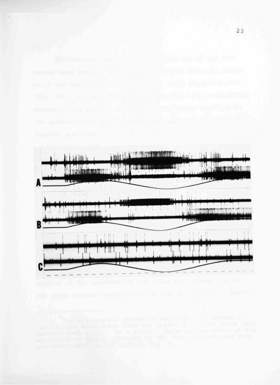
described (Fig. 4A). Following this, the entire comple
ment of tricholiths on the left cercus was immobilized
with vaseline. When rolled again, as indicated in Fig. 4B,
the large unit in the left connective and the small unit
in the right connective no longer responded to changes in
the insect's position, but the small unit in the left
connective and the large unit in the right connective con
tinued to respond as before. This indicates that each
cercus drives two interneurons. The application of vase
line to the tricholiths on the right cercus rendered the
remaining units nonfunctional (Fig. 4C).
Since there are two rows of tricholiths on each cer
cus and two interneurons driven by input from one cercus,
the possibility that each row drives an interneuron is
suggested. To test this, the rows of tricholiths were
extirpated in sequence, and the changes in the responses
of the four units examined. As a control, prior to the
operation, an insect was subjected to sinusoidal oscilla
tions in either direction along the pitch axis and a re
sponse typical of the one already described resulted. (Fie

22
i:

Fig. 4A-C. Responses to roll before and following unilateral and bilateral immobilization of tricholiths. A Neural activity detected in the ventral nerve cord to roll left and right when the tricholiths are unimpeded. B Responses to roll after the tricholiths of the left cercus have been immobilized with vaseline. C Responses to roll after the tricholiths of both cerci have been immobilized with vaseline. Time mark 2/sec. Scale: 200 yV. Trace arrangement as detailed in Fig. 2
23

24
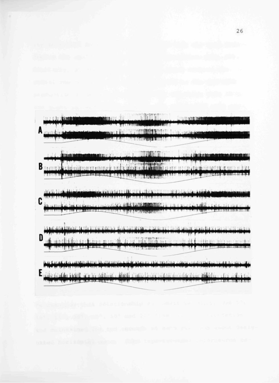
5A) .
Tricholiths composing the lateral row of the left
cercus were removed and a record obtained from the insect
as it was subjected to oscillations along the pitch axis
(Fig. 5B). It is apparent that removal of the tricholiths
composing the lateral row of the left cercus resulted in
the production of action potentials in a nonpatterned
fashion unrelated to positional displacement, by the small
interneuron in the right connective. Comparisons to the
control (Fig. 5A) indicated that the operation had no ap
parent effect on the responses of the other three units.
Continuing this experiment, the tricholiths making up the
medial row of the left cercus were removed. As indicated
by Fig. 5C, sporadic impulses unrelated to the pitch stim
ulus are produced from two interneurons - the small unit
in the right connective as before (Fig. 5B) and now the
large unit in the left connective. The small unit in the
left connective and the large unit in the right connective
continue to respond as they did in the control experiment.
Removal of the tricholiths composing the lateral row on
the right cercus resulted in the loss of meaningful activ-
The extirpation procedure requires the removal of the suction electrodes from the insect's ventral nerve cord between each trial. As a result, there is variation in the amplitude of spikes produced by the four interneurons from one experiment to the next in Fig. 5.

25

Fig. 5A-E. Responses of interneurons before and following sequential extirpation of rows of tricholiths. A Response to sinusoidal displacement along pitch axis (55° forward to 55° backward). B Responses to pitch after the extirpation of the lateral row of tricholiths on the left cercus. C Responses to pitch following the removal of both rows of tricholiths from the left cercus. D Neural activity resulting from pitch after removal of all tricholiths on the left cercus. E Responses elicited by pitching the animal after removal of all rows of tricholiths. Time mark 2/s. Scale: 200yV. Trace arrangement as detailed in Fig. 2.
26
itr'V^'fT
|)P«#H^
i i l i irii i i i i iiiiii
it)||iiiiil(tiilllilJil|iiHi!ilill!iiiill liimt injiiiimiiiiiiii I Mii|i|p<ii|ii >iii D ii ii 1 ,i 4. iij I • ' T " r * " '
1 i l f l i I •« I rf 1 * I I ' ti ( I I t I II II I • ' l l ir I I li n i l t i l l ' l l l l I I I I I I 1 li I I I r I
i>itJii(»ii>iiiiiiiiiiiii(iliijMiiii>i^|ii<iiiiiiii<iiiii>iiiiiiii»(i iiiinoiw iitii ii
27
ity in regard to angular displacement from the unit pro
ducing the small spike in the left connective (Fig. 5D).
Similarly, plucking the tricholiths which compose the
medial row on the right cercus resulted in the sporadic
production of action potentials from the large unit in
the right connective (Fig. 5E). The results of this ex
periment reveal a singular association between the four
rows of tricholiths and the four positional interneurons.
For purposes of clarity from this point forward,
the interneuron that produces the small amplitude spike in
the contralateral connective will be referred to as PIC
(Positional i nterneuron driven by excitation originating
on the contralateral cercus) . PII (positional i nterneuron
driven by excitation from the _ipsilateral cercus) will
refer to the interneurons producing the large amplitude
spike as a result of excitation from the medial rows of
tricholiths.
It is apparent from interneuron recordings presented
in Figs. 1, 2, 3, and 4 that there exists a direct rela
tionship between the degree of angular displacement and
the frequency of firing of the appropriate interneurons.
To quantify this relationship an insect was displaced 5°,
10°, 15°, 30°, 45°, 60° and 70° from primary orientation
and maintained for ten seconds at each position about desig
nated horizontal axes. From tape-recorded interneuron re-

28
sponses, the frequency of spike production for each inter
neuron was analyzed using an instantaneous frequency
counter. The relationship for the response of the PIC in
the left connective, presented in Fig. 6, resulted from
displacing the insect along the 45° horizontal axis. The
relationship for the response of the PII in the left con
nective resulted from displacement of the insect along
the 45° horizontal axis. The interneuronal results for
displacement along these two horizontal axes was presented
because, as will be shown later, it is to displacement a-
long these axes that the two interneurons are most sensi
tive. Each datum point represents the frequency of firing
for the first second after reaching the designated dis
placement. A linear relationship exists between the angle
of displacement and the frequency of firing for each of
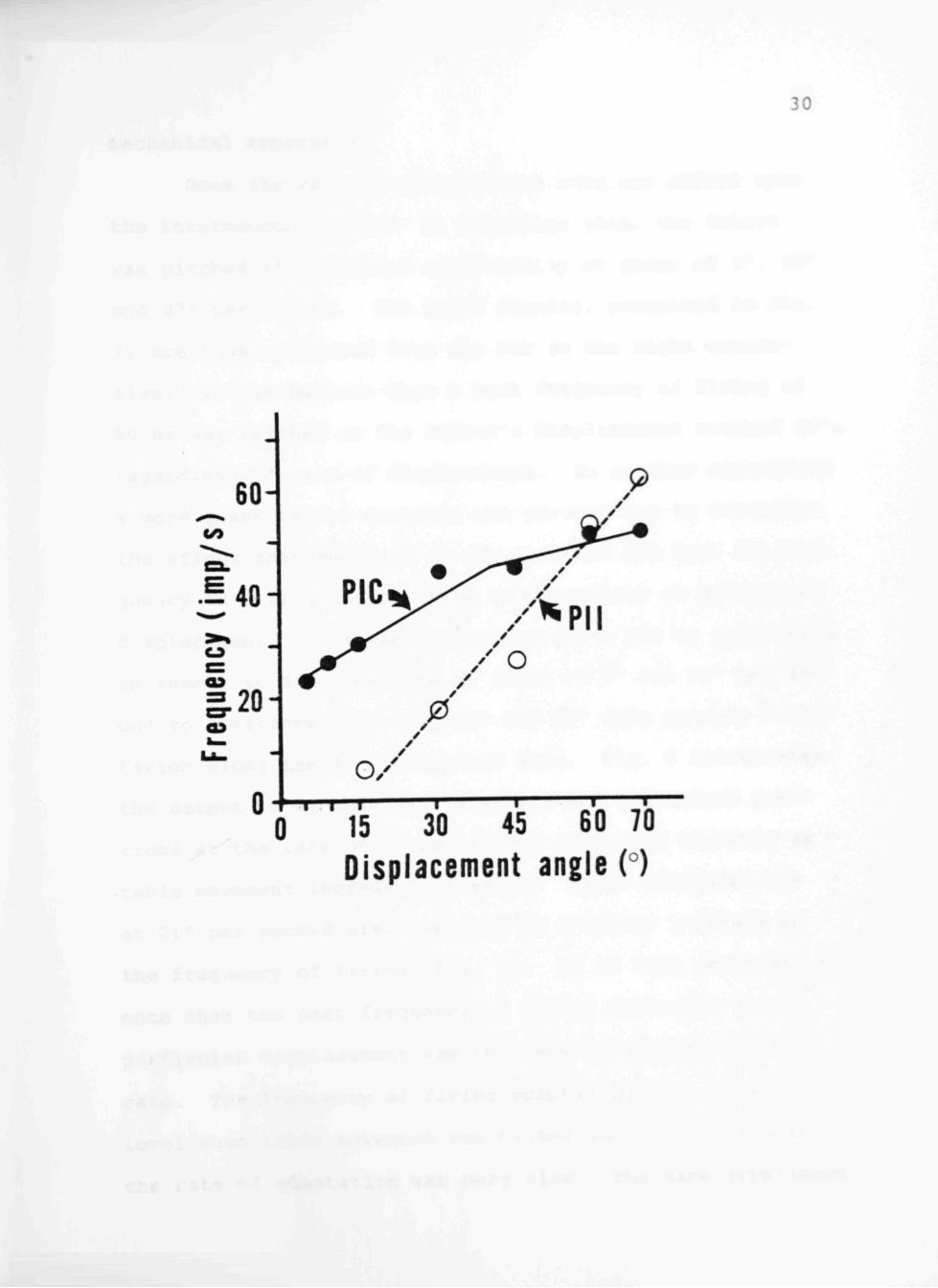
the positional interneurons. The PIC exhibited a linear
response to displacement from primary orientation up to
about 50°. The frequency of firing showed no appreciadDle
increase for displacements greater than 50°. The PII be
gan its response to table displacement from primary orien
tation at some point between 15° and 30° and exhibited a
linear output to 70°. The PIC showed greatest sensitivity
to displacements less than 50°, the PII, although insensi
tive to displacements less than 15°, had an upper ramge
exceeding displacement of 70°, the physical limit of our

29

Fig. 6. Relation between the frequency of firing of the position interneuron contralateral (PIC) and position interneuron ipsilateral (PII) to increasing angles of table displacements. The rate of table displacement was 7°/s. The graph of the PIC is the neutral responses elicited as a result of displacement along the 45° horizontal axis, and the graph of the PII is the response resulting from displacement along the 135° horizontal axis, the axes of maximum output for the interneurons. The impulse frequency was measured for the first second after static position was reached.
30
0 15 30 45 60 70
Displacement angle (°)

31
mechanical apparatus.
Does the rate of displacement have any effect upon
the interneuron output? To determine this, the insect
was pitched 45° backward sinusoidally at rates of 9°, 18°
and 27° per second. The taped results, presented in Fig.
7, are those produced from the PII in the right connec
tive. It can be seen that a peak frequency of firing of
80 Hz was reached as the insect's displacement reached 45°,
regardless of rate of displacement. In another experiment
a more quantitative analysis was carried out to determine
the effect that the rate of displacement had upon the fre
quency of firing and the rate of adaptation at maintained
displacement. This was determined for a PIC by subjecting
an insect to displacements at rates of 7° and 21° per sec
ond to positions 15°, 30°, 45° and 60° from primary orien
tation along the 45° horizontal axis. Fig. 8 illustrates
the output to dynamic displacements and maintained posi
tions at the rate of 7° per second increased linearly as
table movement increased linearly. Table displacements
at 21° per second also resulted in a linear increase in
the frequency of firing (Fig. 8). It is also important to
note that the peak frequency of firing occurring for a
particular displacement was the scime regardless of the
rate. The frequency of firing adapted quickly to a tonic
level when table movement was halted and from that point
the rate of adaptation was very slow. The same experiment

32
Fig. 7. Action potentials from the PIC and PII and a plot of the instantaneous frequency of the PII from the right connective in response to pitch backward to 45° from the primary orientation at three different oscillation frequencies. Top trace: table movement monitor with downward deflections indicating pitch backward. Middle trace: neuronal response obtained from the right connective. Bottom trace: Instantaneous frequency 0, 50 and 100 are calibrations for the interpulse interval in impulses/s. Time mark 5/s. Scale: 200yV.

33
27°/$ :'

34

Fig. 8. Graph of impulse frequency for the PIC resulting from displacements along the 45° horizontal axis. The table was displaced to 15°, 30°, 45° and 60° at rates of 7° and 21°/s. The frequency of action potentials is seen to peak at a displacement of 45°.

35
</5
CU
60
40
20
0
•
/ // '^^'--y^'--^ 30- //' ^ - ^ r ' - ^ ' — 3 0
/ ^.- 4J ^ -" " m-
/." ^
5 10 Time (s)
15
36
testing the properties of a PII was carried out by dis
placing the insect along the 135° axis (Fig. 9). The
displacement positions were changed to 30°, 45°, 60° and
70° to compensate for the different range of sensitivity
exhibited by the PIIs (Fig. 6). Events similar to those
described for the PIC were shown to be characteristic of
the PII. As the insect was displaced from primary orien
tation, the frequency of firing was determined for select
ed angles as the insect passed through them. It was ap
parent that the frequency of firing for a particular dis
placement was the same regardless of rate. Upon stopping
at a designated angle, rapid adaptation to a tonic level
was observed. This was followed by a much slower rate of
adaptation (Fig. 9).
While the pitch and roll axes were convenient refer
ences for the experiments, undoubtedly, these insects are
subjected to displacements along other horizontal axes as
they burrow, run or fly. To test the ability of the trich
oliths to apprise the insect of displacement about hori
zontal axes other than pitch and roll, the insect's posi
tion around the yaw axis was altered in 15° steps, the in
sect subjected to displacements throughout the 360° of the
horizontal plane, and the interneuronal output noted as
measure of the responsive interneuron's sensitivity for
that positional displacement. Since movement components

37

Fig. 9. Graph of impulse frequency for the PII resulting from displacements along the 135° horizontal axis. The table was displaced to 30°, 45°, 60° and 70 at rates of 7° and 21°/s. The frequency of action potentials continues to increase to 70°, the limit of linear mechanical movement of the tilt table.
o

38
</>
80
60
40
^ 2 0
/ ^ - \ / / ^ •
70 •60^
/ \
- • • /
60
45
45^
. - • - - - - - - i i n ^ .30 30
70^
70^ 45° 30^
10 15 Time (s)
20
(velocity and acceleration) have been eliminated as ade
quate stimuli for the positional component, ten second
static displacements of 15°, 30° and 45° were used to
characterize the response ranges of the positional inter
neurons.
The following polar coordinates, representing the
360° of the horizontal plane, were adopted for describing
the horizontal axes about which the insect was displaced.
The longitudinal axis bisecting the insect corresponds
anteriorly to 0° and posteriorly to 180°. The transverse
axis bisecting the insect corresponds on the right to 90°
and on the left to 270°. Each datum point on the polar
plot is the mean value for the number of spikes produced
by an interneuron during the first ten seconds of the
static displacement.
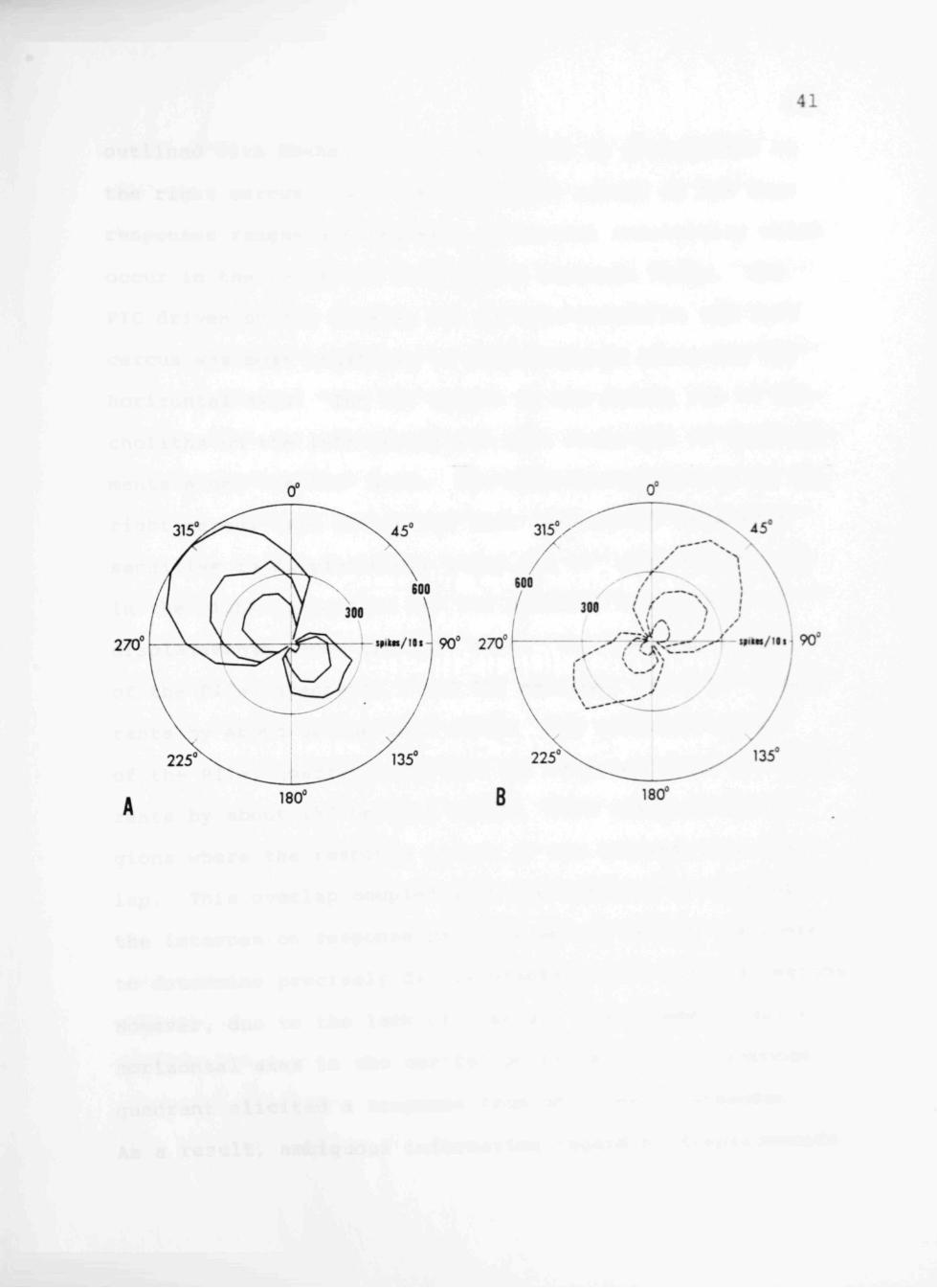
As may be seen in Fig. lOA and B, each interneuron
has a response range equivalent to a quadrant of movement.
The PIC in the right connective responded to displacements
along horizontal axes ranging from 240° - 15° as shown by
the polar plot Fig. lOA. The PII in the right connective
had a response range spanning 165° - 285° (Fig. lOB); the
PIC in the left connective responded throughout a range
from 330° - 120° (Fig. lOB); the PII in the left connec
tive had a response range spanning a quadrant from 75° -
195° (Fig. lOA). The response ranges outlined with solid
lines were driven by tricholiths on the left cercus, those

40

Fig. lOA and B. The response ranges of the positional interneurons in the ventral nerve cord as indicated by the number of spikes produced in the first ten seconds following displacements to 15°, 30° and 45° at 14°/s about designated horizontal axes. The outer circle represents 600 spikes produced in ten seconds, the inner 300. The longitudinal axis which would bisect the insect corresponds anteriorly to 0° and posteriorly to 180°. The transverse axis bisecting the insect corresponds on the right to 90° and on the left to 270°. A Interneurons driven by the tricholiths on the left cercus. The PIC responds to displacements in quadrant II; the PII to those in quadrant IV. B Interneurons driven by the tricholiths on the right cercus. The PIC unit responds to displacements in quadrant I; the PII to displacements in quadrant III.

41
41
270° i»ito./iii^ 90° 270"

42
outlined with dashed lines were driven by tricholiths on
the right cercus. Note the symmetric nature of the four
responses ranges and the axes of maximal sensitivity which
occur in the center of each unit's response range. The
PIC driven by the lateral row of tricholiths on the left
cercus was most sensitive to displacements along the 315°
horizontal axis. The PII driven by the medial row of tri
choliths on the left cercus was most sensitive to displace
ments along the 135° axis. The interneurons driven by the
right cercus (the PIC in the left connective) were most
sensitive to displacements along the 45° axis and the PII
in the right connective had its greatest sensitivity to
displacements along the 225° axis. The response ranges
of the PICs, quadrants I and II, exceeded their 90° quad
rants by about 30° on both sides. The response ranges
of the PIIs, quadrants III and IV, exceeded their 90° quad
rants by about 15° on both sides. This resulted in re
gions where the response ranges of two interneurons over
lap. This overlap coupled with the symmetry of each of
the interneuron response patterns should permit the insect
to determine precisely displacements within limited regions
However, due to the lack of overlap, displacements about
horizontal axes in the central portions of each response
quadrant elicited a response from only one interneuron.
As a result, ambiguous information regarding displacements

43
about these horizontal axes was produced, for example,
angular displacements about horizontal axes from 40° -
60° in quadrant I (Fig. IIA).
Examination of the response range of each interneur
on illustrated a decline for the interneuronal output to
displacement along horizontal axes adjacent to the unit's
axis of maximal sensitivity. This decline in the sensi
tivity from the interneuron continued as the deviation of
displacement from the axis of maximal response was in
creased. Concomitant with the decreasing responsiveness
observed in one unit was an increasing responsiveness of
the interneurons which had adjacent response ranges. One
explanation for this occurrence would be intercercal in
hibitory circuits serving to enhance the directional prop
erties of the positional interneurons. Inhibitory cir
cuits proposed by this explanation would be most effective
in areas of moderate to low sensitivity (e.g. along the
pitch and roll axes).
Removal of individual rows demonstrated the singular
association that existed between the excitability of each
interneuron and a row of tricholiths. To determine whether
inhibitory neural circuits between rows of tricholiths
modify the afferent information mediated by the four inter
neurons, comparisons of polar plots (Fig. 11) depicting
the response ranges of the other interneurons not excited

44

Fig. IIA-D. Response ranges of the four interneurons resulting from the sequential extirpation of rows of tricholiths. The table was displaced 30° about each of the designated horizontal axes at 14°/s. Each datum point is a count of the number of impulses produced in the first ten seconds following displacement. A Control responses. B Extirpation of tricholiths composing the medial row right cercus. C Additional removal of tricholiths composing the lateral row on the right cercus. D additional removal of tricholiths composing the lateral row left cercus. Polar coordinates are as described in Fig. 10.

45
270 90' 270 1 «»'»«/'•«
B
i»iiw/iii-^ 90 270 r**'^/'"

46
by the extirpated row were made before and after the oper
ation. Following extirpation of the seven tricholiths
composing the medial row of the right cercus, the response
range of the PIC in the right connective covered from
240° - 15°; the response range of the PII in the left con
nective spanned from 195° - 75°; and the response range
of the PIC in the left connective was 345° - 120° (Fig.
IIB). Next, the tricholiths in the lateral row on the
right cercus was removed. The response rcinge of the PIC
in the right connective covered a quadrant from 24 0° -
15°; and the response of the PII in the left spcinned from
210° - 60° (Fig. IIC). Finally, the seven tricholiths on
the lateral row of the left cercus were removed and the
only remaining responding unit was the PII in the left
connective. Its response range spanned from 195° - 75°
(Fig. IID). The changes that occurred are attributable
to deterioration in the viability of the preparation, and
did not result from the absence of tricholith rows. These
results suggest that if intercercal inhibitory circuits
are present, they play a very subtle role in determining
the response ranges of adjacent interneurons.
A second approach was undertaken to expose possible
intercercal inhibition. If the system is simply and direct
ly wired, reflexion of a cercus anteriorly or posteriorly
should change the range of movement of the involved tricho-

47
liths. Predictably, this change would be mirrored by a
shift in the response ranges of the two interneurons
driven by the reflected cercus and cause an increase or a
decrease in the amount of overlap that occurs between ad
jacent response ranges. If there are intercercal inhibi
tory circuits, their presence should be revealed by an
expansion of the response range in areas of low to moderate
sensitivity for interneurons driven by the unreflected cer
cus as a result of a decrease in the amount of overlap of
the response ranges. Likewise, a reduction in the fre
quency of firing for a particular horizontal axis from the
interneuron driven by the unreflected cercus would be ex
pected as a result of increasing the overlap of adjacent
response ranges. To determine this, the insect was sub
jected to three 30° displacements along each of the desig
nated axes. The first displacement occurred with the cerci
unrestrained (Fig. 12A); the second took place with the
right cercus reflected backward (Fig. 123); and the third
with the right cercus reflected forward. This experimental
procedure was followed to insure that any changes in the
responses due to deterioration of the preparation would be
negated.
The polar plots resulting from cereal reflexion were
compared with the control. As seen in Fig. 12, the re
sponse ranges of the interneurons driven by the tricholiths

48

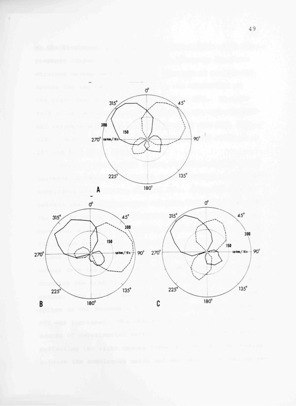
Fig. 12A-C. Polar plots derived from the responses of positional interneurons before and following reflexion forward (nearly perpendicular to the body axis) or backward (nearly parallel to the body axis) of the right cercus. The table was displaced 30° about each of the designated horizontal axes at 14°/s. Each datum point is a count of the number of impulses produced in the first ten seconds following displacement. A Control experiment. B Responses of interneurons when the right cercus is reflected backward. C Responses of the interneurons when the right cercus is reflected forward. Note that while the response ranges for the interneurons driven by the reflected cercus are altered, there is no change in output by the interneurons driven by the left cercus. Polar coordinates are as described in Fig. 10.
49
270° »•
270° mtoi / t« i ; i90° 270
B 180°
— - i«ik«/iii i 90°
50
on the displaced cercus each underwent a shift while the
response ranges of the interneurons driven by the unre
strained cercus were unchanged. The axis of niaximal re
sponse for the two units driven by the tricholiths on
the right cercus was 45° and 225° (Fig. 12A). As a re
sult of reflecting the cercus backward, the axis of maxi
mal response underwent a 45° shift to 90° and 2 70° (Fig.
12B). Moving the cercus forward caused a 30° shift to
15° and 195° (Fig. 12C).
Reflecting the right cercus backward resulted in an
increase in the overlap in the response ranges of the non
homologous interneurons (PIC and PII), while the overlap
between the homologous units had been decreased (Fig. 12B).
The PII driven by the left cercus decreased its output by
seventeen spikes in ten seconds to pitch backward and the
PIC driven by the left cercus had increased by twenty
spikes in ten seconds to pitch forward. No change in the
output from the PIC driven by left cercus accrued from in
creasing the area of overlap with the PII from the right
cercus. The PII in the left cercus produced twelve fewer
spikes in ten seconds when the overlap with the adjacent
PIC was increased. The changes observed are within the
bounds of experimental variability commonly encountered.
Reflecting the right cercus forward increased the overlap
between the homologous units and decreased the overlap be-

51
tween the nonhomologous units. As before, no overt changes
in frequences were detected. On the basis of these ex
periments it was concluded that there were no intercercal
inhibitory circuits.
The frequencies of firing of the PIC and the PII
driven by the displaced cercus show definite alterations.
These resulted from inadvertently moving the cercus out of
the insect's horizontal plane. When the right cercus was
displaced backward (Fig. 12B), the frequency of firing of
the PIC increased while that of the PII was reduced. The
reflexion of the right cercus forward (Fig. 12C) produced
a marked increase in the interneuronal output of the PII
and a decrease in the output of the PIC for displacements
along each of the appropriate horizontal axes. When the
cercus is reflected backward, it was displaced upward mak
ing it more sensitive to pitch forward and less sensitive
to pitch backward. Reflexion of the cercus forward re
sulted in the downward displacement of the cercus increas
ing the sensitivity to pitch backward and decreasing the
sensitivity to pitch forward.
CHAPTER IV
DISCUSSION
Orthopteran and dictyopteran insects possess sensory
organs, trichobothria, which are long filiform hairs lo
cated on the cerci. The trichobothria act as mechanical
transducers evoking neural responses in primary sensory
neurons. These in turn produce activity that is relayed
to the insect's higher centers via a system of giant in
terneurons. Investigations into the neurophysiological
aspects of these pathways begun by Pumphrey and Rawdon-
Smith (1937), coupled with intracellular mapping of par
ticipating units (Milburn and Bentley, 1971; Harris and
Smyth, 1971; Pitman, e_t aj . , 1973) have provided a lucid
picture of the cellular neurophysiological properties of
this afferent system. The cockroach Periplaneta americana
has fourteen longitudinal rows of trichobothria on the
ventral portion of each cercus (Nicklaus, 1965). Each
sensillum is associated with one sensory neuron and an
accompanying complement of supportive cells. The dendrite
of the sensory neuron inserts into the base of the hair
shaft, and the axon exits via the cereal nerve to make
synaptic contact with giant interneurons in the sixth ab
dominal ganglion. Each sensillum has a preferred plane
of deflection which confers upon its sensory neuron a re-
52
53
stricted range of responsiveness (Gnatzy, 1976). Intra
cellular dye injections of a few of the giant interneurons
which respond to cereal stimulation have shown the cell
bodies to be located in the sixth abdominal ganglion
(Farley and Milburn, 1969). Investigations studying the
degeneration of giant fibers show that they run continu
ously from the sixth abdominal ganglion to the subesopha-
geal ganglion (Spira et aJ., 1969).
The characterization of the neural aspects of the
cercus-to-giant interneuron system has resulted in little
data indicating the functional significance of this affer
ence. Roeder (1948) postulated the direct involvement of
excitation of these giant fibers with evasive running be
havior. Subsequent experiments showed that cereal acti
vity directly responsible for driving a motor response in
the cockroach's legs is mediated by a smaller group of
neurons (Dagan and Parnas, 1970). They suggested that
giant interneuron system function was associated with the
complicated organization of an escape response. Harris
(1977) presented evidence that the giant interneurons are
not directly responsible for initiating escape behavior in
p. americana, thus supporting the hypothesis of Dagan and
Parnas (1970). However, Harris refuted the proposal that
the function of the giant units was to squelch other on
going neural activity. Schwab and Josephson (1977) showed

54
that the giant interneurons of P. americana could convey
information arising from cereal receptors responsive to
low frequency auditory stimuli. Whether the insect makes
use of auditory stimuli via this route has not been shown.
The fourteen histologically identifiable giant in
terneurons in p. americana were examined to determine
whether anatomical restrictions on trichobothrial move
ments, described by Nicklaus (1965) and Gnatzy (1976), im
posed directional specificity on the interneuronal response
ranges. Variations in the direction of delivery of wind
puffs revealed that ten of the fourteen giant fibers have
restricted response ranges (Westin e;t ad., 1977). Data
collected from these experiments led the authors to suggest
the cercus-to-giant interneuron system evolved as a means
of rapid localization of a source of air currents.
Fraser (1977) designed experiments to determine
whether the cerci play a role in the locomotory behavior
of P. americana. He conducted experiments in which be
havioral evidence implicated cereal receptors as special
ized equilibrium organs used by the insect during flight.
It was shown that the removal of one cercus, or interfer
ence with the movement of the cereal sensory hairs on one
cercus, changed the abdominal posture, the leg position and
the beat pattern of the contralateral wing. These effects,
he stated, were similar to the compensatory wing movements

55
produced when the cockroach was tilted during flight.
The physiological evidence presented in this report
and that of Hartman et al., (in manuscript) coupled with
the behavioral experiments by Fraser on Periplaneta ameri
cana strongly suggest that the major function of the cer
cus-to-giant interneuron system is to apprise the insect
with orientational input. In my experiments, I have
characterized the responses of the four interneurons ac
tivated when the cockroach is subjected to angular dis
placements about horizontal axes. Taking advantage of the
accessibility of the tricholiths as well as the maneuver
ability of the cerci, experiments were performed which
indicate that the afference from the four interneurons
was the sole product of tricholith input, unmodified by
other elements peripheral to the recording site.
Immobilization of tricholiths on one cercus showed
that the activity conveyed by the PICs is the result of
input from tricholiths located on the cercus contralateral
to the interneuron. The PII input was from tricholiths
located on the cercus ipsilateral to the interneuron. Ex
tirpation of a single row of tricholiths revealed an even
more discrete association. The lateral row of tricholiths
on each cercus drives the PIC in the contralateral connec
tive while the medial rows of tricholiths provide excita
tory input to the PII (Fig. 13). Scanning electron micro-

56

Fig. 13. Schematic representation of the underlying connectivity between the rows of the tricholiths on the right cercus and the associated PII and PIC. The circles originating from the proximal tricholith on each row indicate the range of angular displacements to which that row responds. The line bisecting each of the circles represents the axis of maximal response for that row of tricholiths.
57
O
O o o

58
graphs of the fine structure have shown that the tricho
liths comprising each row have a restricted range or move
ment and these restrictions are different for each row
(Bennett, personal communication). Based upon anatomical
impedence of tricholith movement, predictions of the asso
ciation between a row of tricholiths and the response range
of the interneuron it excites, correspond well with the
physiological data collected via interneuronal recordings.
The connectivity between the tricholiths and the four po
sitional interneurons, as affirmed by the physiology, is
represented by the schematic diagram in Fig. 13. The
angular displacements about which each of the two rows of
tricholiths can move is indicated by the circle originat
ing from the proximal tricholiths. The line bisecting
each circle represents anatomically the angle at which the
tricholith can undergo the greatest displacement, and
physiologically, the axis of maximal sensitivity for the
positional interneurons receiving input from the indicated
cercus. The combined input from the four rows of cerci
would thus project to form the polar quadrants (Fig. 10)
of the giant interneurons.
Is a row of tricholiths solely responsible for pro
ducing the orientational specificity seen in the responses
of a positional interneuron, or is this tricholith input
modified peripherally by intercercal inhibitory circuits?

59
Inhibitory circuits between cereal receptors and some of
the giant fibers in the sixth abdominal ganglion of Peri
planeta americana have been confirmed by several investi
gators (Kerkut et al.. , 1969; Pitman and Kerkut, 1970; «
Callec, 1974). Matsumoto and Murphey (1977) showed, using
sound stimuli, that inhibition originating from transverse
ly vibrating sensilla on the cerci of the cricket Acheta
domesticus enhance the directional sensitivity of the
response ranges of the contralateral medial giant inter
neurons . Altering the response ranges of the four inter
neurons (cereal reflection and extirpation experiments) of
Arenivaga failed to demonstrate the presence of contralat
eral inhibitory circuits (Figs. 11 and 12). Matsumoto
and Murphey (1977) showed that long term unilateral depri
vation of input from cereal receptors resulted in the en
hancement of the efficacy of the inhibitory input of the
cereal receptors on the contralateral cercus. Long term
deprivation experiments, similar to those described by
Matsumoto and Murphey (1977) coupled with experiments
designed to locate and subsequently record intracellularly
from the integrating regions of the four interneurons in
Arenivaga should provide unequivocal evidence regarding
the presence of inhibitory pathways associated with the
four positional interneurons.
The niches inhabited by Periplaneta americana and
Arenivaga are vastly different, and thus their require
ments for sensory information are also different. P.
americana, an agile terrestrial insect, extracts position
al information through indirect means previously described.
It has need for a receptor system capable of precise de
tection of angular and linear accelerations as the insect
is running or flying, and the trichobothria seem especial
ly suited for this requirement.
Arenivaga, a burrowing insect, inhabits fine, quasi-
filled soil which makes the construction of permanent
burrows impossible. The uniform damped support of the
sand grains about the insect would impair the ability of
proprioceptive hair plates and that of cuticular stress
receptors to monitor geotactic orientation. The adaptive
significance underlying the evolution of the tricholiths
underscores the insect's need for a direct means of ob
taining information regarding its position in relation
to the gravitational force vector. Thus, it is not sur
prising that cereal receptors have undergone special adap
tation making possible subsistence in subterrestrial con
ditions. The employment of giant interneurons to mediate
this modality suggests that this information is of con
siderable importance in modulating locomotory behavior.
Resolution of slight angular displacements within
the response quadrants of the PIIs is not possible due to
61
the insensitivity of the Pll, the restricted area of over
lap of the two adjacent receptive fields, and the symmetry
of the response ranges of the four interneurons. This
probably does not constitute any difficulty for the insect
while burrowing. Experiments in this lab (Leander, per
sonal communication) indicate when burrowing downward,
Arenivaga maintains angular displacements within the lin
ear range of inputs provided by the PICs. When the insect
emerges or flies, these ambiguities probably force the
insect to utilize other proprioceptive systems in conjunc
tion with the tricholiths for orientation information.
It is significant to note that structurally Areni
vaga has its lowest degree of stability about its roll
axis. The overlap of the response range of a PII, which
has its greatest sensitivity to angular displacements
ranging from 30° to 70° with that of a small unit which is
maximally sensitive to angular displacements ranging from
primary orientation to 50°, occurs as the insect is dis
placed in either direction along the roll axis. From this
it can also be seen that the thresholds of the interneurons
producing the smaller spikes are lower than those of the
interneurons producing the larger spikes. This observation
concurs with those reported by Carpenter and Henneman
(1966), in which they concluded that axonal size, as in
dicated by the size of the spike produced, is inversely
62
related to the threshold of afferent neurons. However,
the difference in sensitivity between the PII and the PIC
is not wholly attributable to the differences in threshold.
It is likely that the anatomical relation between the lat
eral and medial rows on a cercus plays a role in the two
different ranges of sensitivity. Distal views looking
down the longitudinal axis of a cercus have revealed that
due to a slight rotation of the cercus the medial row was
in a slightly higher plane than the lateral row. Because
both shafts originate from above the midline between the
two rows, the medial row is closer to the inner part of
its socket, and the lateral row closer to the outer edge
of its socket. As a result of this arrangement, the
medial row requires a greater displacement than the lateral
row before reaching a point where impulse production would
begin.
A holistic look at orientation systems which have
evolved among different evolutionary lineages of the Animal
Kingdom shows that closely associated with the primary
snesory organ are neural circuits correlating complex
compensatory adjustments such as eyestalk adjustments in
crabs (Sandeman and Okajima, 1972) and compensatory counter
rolling of the eyes in cephalopods and vertebrates (Budel-
mann, 1975; Schaefer et al . , 1975). Fraser (1977) re
ported observing compensatory adjustments involving the

63
wings, legs and abdomen during flight as a result of de
priving p. americcma adults of afference from one cercus.
Neural activity originating from cereal receptors is known
to travel via the system of giant interneurons, and it is
possible that the postural changes he described are medi
ated by the giant fiber system. Intracellular stimula
tion of abdominal giant fibers has been shown to evoke
antennal movements and the associated electrical activity
from muscle fibers at the base of the antennae in £. ameri-
ccuia (Dagan and Parnas, 1970). This may be analagous to
compensatory responses which occur in the antennae of lob
sters as a result of tilting the animal (Schone et al .,
1976). Also, the high degree of directional specificity
manifested in the activity of particular giant units in
p. americana (Westin et al . , 1977), as alluded to earlier,
could underlie the coordination of compensatory responses
seen by Fraser (1977).
Each lith, in Arenivaga, is supported by a single
hair from above. This differs from the arrangement de
scribed for positional receptors in the crustacean stato
cyst as well as the vertebrate utricular muculae where,
in both instances, the lith is supported from below. De
spite this difference, the neural elements appear to ex
tract changes in the direction of the gravitational force,
manifested by the liths, in the same manner as described
64
for other positional receptors. It is also significant
to realize that the receptor system of Arenivaga is sen
sitive to stimuli other than absolute position. This fact
becomes apparent each time the table movement returns the
insect to primary orientation. Signals conveyed by the
interneurons during movement apprise the insect of the
direction of angular movement and not dynamic position.
The same observation was made for neurons driven by the
static position detectors in the lobster statocyst (Cohen,
1955) .
Finally, this study characterizes the responses of
the four interneurons mediating tricholith afference, and
along with anatomical and preliminary behavioral evidence
supports the theory that tricholiths of the cerci provide
equilibrium position information. My results provide
basic information which may be usefully applied to further
studies of different aspects of this sensory system. The
simple organization (cereal receptors-to-interneurons)
which I have found should allow firmer interpretation of
results correlating insect development with anatomic,
behavioral and physiologic changes of the cerci.
REFERENCES
Budelmann, B.: Gravity receptor function in Cephalopods with particular reference to Sepia officinalis. Fortschr. Zool. 23, 84-96 (197T)
Carpenter, D., Henneman, E.: A relation between the threshold of stretch receptors in skeletal muscle and the diameter of their axons. J. Neurophysiol. 29, 353-368 (1966)
Callec, J.: Synaptic transmission in the central nervous system of insects. In: Insect Neurobiology (ed. J. E. Treherne) pp. 119-185. North-Holland Publishing Company, Amsterdam-Oxford 197 4
Cohen, M. J.: The function of receptors in the statocyst of the lobster Homarus americanus. j. Physiol. 130, 9-34 (1955)
Dagan, D. , Parnas, I.: Giant fibre and small fibre pathways involved in the evasive response of the cockroach, Periplaneta americana. J. exp. Biol. 52, 313-324 (1$70)
Farley, R. , Milburn, N.: Structure and function of the giant fiber system in the cockroach, Periplaneta americana. J. Insect Physiol. 15, 457-476 (1969)
Fielden, A.: Transmission through the last cibdominal ganglion of the dragonfly nymph, Anax imperator. J. exp. Biol. 37, 832-844 (1960)
Fraenkel, S. G. , Gunn, D. L. : The orientation of animals (Kitneses, Taxes and Compass Reactions) Dover Publications, Inc. New York 1961
Fraser, P.: Cereal ablation modifies tethered flight behavior of cockroach. Nature 268, 523-524 (1977)
Fraser, P., Sandeman, D.: Effects of angular and linear accelerations on semicircular canal interneurons of the crab Scylla serrata. J. comp. Physiol. 96, 20 5-221 (1975)
Gnatzy, W.: The ultrastructure of the thread-hairs on the cerci of cockroach Periplaneta americana L. : The m-termolt phase. J. Ultrastruct. Res. 54, 124-134 (1976)
65
66
Hartman, H. B., Walthall, w. w., Bennett, L. P., Stewart, R. R.: Giant interneurons mediating equilibrium reception in an insect. Science (in manuscript)
Harris, C.: Giant interneurons of the cockroach neither trigger escape nor "clear all stations." Comp. Bio-chem. Physiol. 56A, 333-335 (1977)
Harris, C., Smyth, T.: Structural details of cockroach giant axons revealed by injected dye. Comp. Biochem. Physiol. 40A, 295-303 (1971)
Horn, E.: Mechanisms of gravity processing by leg and abdominal gravity receptors in bees. J. Insect Physiol 24, 673-679 (1975a)
Horn, E.: The contributions of different gravity receptors on the gravity orientation in insects. Fortschr Zool. 23, 1-16 (1975b)
Kerkut, G. A., Pitman, R. M., Walker, R. J.: Sensitivity of neurons of the insect central nervous system to ion-tophoretically applied acetylcholine or GABA. Nature 222, 1075-1076 (1969)
Markl, H.: Prpprioceptive gravity perception in Hymenop-tera. In: Gravity and the Organism (eds. S. A. Gordon, M. J. Cohen), pp. 185-194. The Univ. of Chicago Press 1971
Matsumoto, S. G., Murphey, R. K. : Sensory deprivation during development decreases the responsiveness of cricket giant interneurones. J. Physiol. (London) 268, 533-548 (1977)
Milburn, N. S., Bentley, D. R.: On the dendritic topology and activation of the cockroach giant interneurones. J. Insect Physiol. 17, 607-623 (1971)
Neil, D. M.: The mechanism of statocyst operation in the mysid shrimp Pranus flexuosus. J. exp. Biol. 62, 685-700 (1975)
Nicklaus, R.: Die Erregung einzelner fadenharre von Periplaneta americana in Abhangigkeit von der GroBe und Richtung der Auslenkung. Z. vergl. Physiol. 50, 331-362 (1965)

67
Pitman, R M., Kerkut, G. A.: Comparison of the actions of iontophoretically applied acetylcholine and GABA with the EPSP and IPSP in cockroach central neurons. Comp. Gen. Pharmacol. 1, 221-230
Pitman, R. M., Tweedle, C. D., Cohen, M. J.: The form of nerve cells: determination by cobalt impregnation. In: Intracellular Staining in Neurobiology (eds. S. B. Kater, C. Nicholson), pp. 83-97. Berlin: Spring-er-Verlag 1973
Pumphrey, R. J., Rawdon-Smith, A. J.: Synaptic transmission of nervous impulses through the last abdominal ganglion of the cockroach. Proc. R. Soc. B 122, 106-118 (1937)
Roeder, K. D. : Organization of the ascending giant fiber system in the cockroach (Periplaneta americana). J. exp. Zool. 108, 243-262 (1548)
Roth, L. M., Slifer, E. H. : Spheroid sense organs on the cerci of Poiyphagid cockroaches (Blattaria: Poly-phgidae). Int. J. Morphol. & Embryol. 2, 13-24 (1973)
Sandeman, D. C , Okajima, A.: Statocyst-induced eye movements in the crab Scylla serrata. I. The sensory input from the statocyst. J. exp. Biol. 57, 187-204 (1972)
Sandeman, D. C.: Spatial equilibrium in the arthropods. In: Structure and Function of Proprioceptors in the Invertebrates (ed. P. J. Mill) pp. 485-527. London: Chapman and Hall 1976
Schaefer, K. P., Schott, D., Meyer, D. L.: On the organization of neuronal circuits involved in the generation of the orientation response (Visual Graspreflex) (1). Fortschr. Zool. 23, 199-211 (1975)
Schwab, W. E., Josephson, R. K.: Coding of acoustic information in cockroach giant fibers. J. Insect Physiol. 23, 665-670 (1977)
Schone, H., Stein, A., Carlstead, M.: Reactions of the spiny lobster, Palinurus vulgaris, to substrate tilt (I). J. comp. Physiol. 107, 113-128 (1976)
68
Spira, M. E., Parnas, I., Bergmamn, F.: Histological and electrophysiological studies on the giant axons of cockroach Periplaneta americana. J. exp. Biol. 50, 629-634 (196Tr
Wehner, R. : Space constancy of the visual world in insects. Fortschr. Zool. 23, 148-160 (1975)
Wendler, G. : The coordination of walking movements in arthropods. Symp. Soc. exper. Biol. 20, 229-249 (1965)
Wendler, G. : Physiology and systems analysis of gravity orientation in two species (Carausius morosus, Calandra granaria). Fortschr. Zool. 23, 33-46 (1975)—
Westin, J. J., Langberg, J. J., Camhi, J. M.: Responses of giant interneurons of the cockroach Periplaneta americana to wind puffs of different directions amd velocities. J. comp. Physiol. A 121, 314-324 (1977)