Embed Size (px)
Citation preview

Electron paramagnetic resonance (EPR) spectroscopy is the only method for the direct detection of species that have unpaired electrons.
A surprisingly large number of materials have unpaired electrons. These include free radicals, many transition metal ions, and defects in materials. Free radicals are often short-lived, but play crucial roles in many significant processes such as photosynthesis, oxidation, catalysis, and polymerization reactions. As a result, the application of EPR spans one of the widest ranges of any major analytical technique, from molecular research to quality control in fields as diverse as: chemistry, quantum physics, structural biology, materials science, medical research and many more. Importantly, EPR data can be collected in seconds, and the analysis of the data provides not only the identity, but also quantitative information about the species being measured. Despite being more than 70 years old, EPR continues to deliver new breakthroughs and new insights. So, how did it all begin?
The early years – from academic to applied
In 1945, the Soviet physicist Zavoisky published a report on the first successful observation of EPR absorption. Building on this work, a group at Oxford University in England made a series of announcements on theoretical and experimental studies. Over a 10-year period this group, and others, effectively established the academic discipline of EPR.
Subsequently, chemists became interested in applying EPR methodology, and in the 1960s and 70s there was an explosion of research, mostly focused on the relationships between molecular orbital methods and EPR. Throughout the 1980s, attention focused on the importance of electron spin and the deep connection to physical properties, especially driven by the development of materials like amorphous silicon and magnetic bubble memory elements. This led to a gradual increase in the demand for EPR in fields related to materials science.
Applications widened as spin traps were developed to ‘capture’ the very short-lived free radicals, effectively fixing them into a more stable form that could be measured by EPR. A common method for spin-trapping involves the addition of radical to a nitrone spin trap resulting in the formation of a spin adduct, a
nitroxide-based persistent radical, that can be detected using EPR. The spin adduct usually yields a distinctive EPR spectrum that is characteristic of the free radical that is trapped. The result was a move for EPR to become widely used in in vitro and in vivo studies in the medical and pharmaceutical fields.
Spin traps are very efficient in cell-free systems, but to further expand EPR into the cell-based biological environment a new approach was needed. In response, cyclic hydroxylamines were developed as spin probes. They have the advantage of a much more rapid reaction with free radicals (around 100 times faster than spin traps) as well as the formation of a stable nitroxide with a much longer half-life than can be achieved with nitrone spin traps. The relationship between various diseases and the generation/annihilation mechanisms of the unstable radicals in the reactions within living organisms, as well as the interactions with pharmaceutical agents have all become important topics using this technology.
To study the structure and local dynamics of proteins, pioneering work published around 1990 gave rise to site directed spin label (SDSL) methods. The theory of SDSL is based on the specific reaction of spin labels, which always contain an unpaired electron, with amino acids. A spin label’s built-in protein structure can, therefore, be detected by EPR spectroscopy. SDSL has also proved to be a useful tool in examinations of the protein folding process. Spin labels are a unique molecular reporter and a variety of nitroxide spin labels have enjoyed widespread use for the study of macromolecular structure and dynamics because of their stability and simple EPR signal.
The expansion of the application of EPR has also been driven by developments in instrumentation. The fundamentals of sensitivity, resolution and stability are all related to the microwave or magnet technology in the spectrometer. Users and instrument manufacturers like Bruker have worked in close partnership to push the boundaries of application and create more powerful, more stable and more flexible EPR spectrometers.
Everyday accessibility
Today, with a very solid hardware platform for EPR now available, developments in workflow and software have continued to open new fields of application.
EPR – The ‘workhorse technique’ that is still enabling scientists to break new ground after 70+ years
Kalina Ranguelova, Ph.D., EPR Application Scientist at Bruker BioSpin Corp.
Accessibility is key – with packages tailored to specific applications emerging, ensuring ease of use in a routine measurement or QC monitoring situation.
Such systems include:
�� Bruker’s microESR, is a small, portable research grade instrument that can easily fit in a fume hood or glove box, or be transported to the field. It requires no special installation or regular maintenance. �� Bruker e-scan instruments are dedicated for 24/7 QC operation,
and packages for the food industry, alanine dosimetry, and for testing the freshness of beer and other beverages are now installed in leading companies around the world.
Of course, fundamental research continues and high-end instruments allow scientists the flexibility and unlimited power they need to push into new fundamental areas. For Bruker, a modular instrument series – the ELEXSYS-II EPR – meets this need. ELEXSYS-II spectrometers offer outstanding performance and flexibility and can be expanded with, for example, an imaging accessory, an FT system, ENDOR equipment, and microwave bridges from 1 GHz to 263 GHz for CW and/or FT operation that enables real multi-frequency & multi-resonance EPR.
More recently, Bruker brought research power to the bench top with the EMXnano. Launched in 2015 at the 56th annual Experimental Nuclear Magnetic Resonance Conference (ENC), EMXnano is a completely new development featuring the latest digital and microwave technologies. Combined with a new generation of magnet system with full range up to 6.5 kG and a highly efficient microwave resonator, this state-of-the-art bench-top instrument is superior in sensitivity and stability, making it ideal for a comprehensive range of analysis applications.
Identification of paramagnetic species is taken care of by Bruker’s spectrum simulation and fitting module (SpinFit) and, for the first time in this instrument category, quantitative EPR is as straightforward as it can be, thanks to a fully calibrated instrument and the inclusion of Bruker’s patented spin counting software module (SpinCount) for reference-free concentration determination.
Continued excitement
EPR spectroscopy remains a lively field of research. Advances in spectrometer and imager hardware, in experimental protocols, and in important applications in chemistry and biology continue to be made. A review of topics presented at the 2017 conference of the EPR specialist group of the Royal Society for Chemistry (RSC), and recent publications highlights a host of exciting new developments and applications, for example:
�� Moving from cryogenic to ambient temperature distance measurements in structural biology�� Elucidating the mechanism of electron transfer in respiratory complex I and ATP synthesis�� Very high field EPR studies of transition metal ions in molecular magnets�� Understanding the pharmacokinetics of drug candidates in animal models
Detailed discussion of each of these is outside the scope of this paper, but three broad areas of applied interest: monitoring the degradation and stability of pharmaceutical products, photochemistry and photoaging, and the detection of defects in materials used for solar/fuel cells and batteries, are presented here as examples of the work going on in the vibrant EPR community.
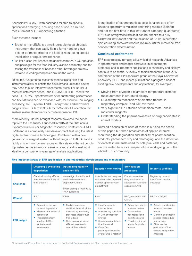
Detecting & evaluating
degradation
Optimizing stability
and shelf-lifeReaction monitoring
Sterilization
processesImpurity profiling
Challenge
Chemical stability affects the safety and efficacy of drug products
Knowledge of stability and shelf-life is essential to proper formulation.
Stress testing is required by IHC* guidelines
Chemistries involving free radicals or other unpaired electron species impact product yield
Process can cause drug inactivation or alteration of functional excipients / APIs
Regulatory drive to identify and monitor impurities
Where?R & D R & D R & D R&D, production and
QA/QCR&D and QA/QC
EPR insight
�� Determines the root cause of degradation
�� Measures the extent of degradation
�� Predicts long-term stability of APIs, excipients and formulations
�� Predicts long-term stability (chemical, photo, thermos) by monitoring processes that produce free radicals
�� Determines antioxidant efficiency required to quench free radicals
�� Identifies reaction intermediates
�� Answers key questions of yield and reaction kinetics
�� Generates data to build kinetics model
�� Quantifies paramagnetic species over course of reaction
�� Determines stability post-sterilization
�� Characterizes free radicals and identifies source
�� Provides go/no go results for product release
�� Detects and identifies traces of transition metals
�� Monitors degradation process that produce free radicals
�� Observes the production of free radicals catalyzed by impurities
*International Council for Harmonization of Technical Requirements for Pharmaceuticals for Human Use
Five important areas of EPR application in pharmaceutical development and manufacture
Photochemistry and photoaging
Several factors including pollution, stress, nutrition and light exposure challenge skin health. Skin is very susceptible to UV radiation, therefore adequate sun protection is essential to control UV-related disorders, including sunburn, photo-aging and photo-carcinogenesis. Modern multifunctional skin care products are increasingly complex and confront the industry with new problems in product development. Active ingredients and basic raw materials may increase the burden of free radicals in the formulations and/or inside the skin, even though they were designed to do just the opposite. Some of the free radical sources include:
�� Unsaturated fatty acid components, in either natural oils or emulsifiers, increase the risk of lipid peroxidation under UV light.
�� Photo-unstable UV blockers may enhance the peroxide radical concentration when applied on the skin.
�� Moisturizers may influence the radical content of skin during UV radiation by increasing the penetration of UV rays into deeper layers of the skin. With higher skin hydration, the risk for free radical formation is increased.
�� Perfumes and dyes are among the ingredients with the highest risk of peroxide radical contamination. Radical chain reactions may occur under UV radiation.
Recent work using EPR spectroscopy has shown a clear correlation between the amount of UV-inducible free radicals in a sunscreen formulation and the clinical symptoms of Acne aestivalis.
In practice, the quantification of UV-induced free radicals by EPR can now be considered as a predictive tool in the creation of new formulations.
Solar cell efficiency and stability
As the market for photovoltaic (PV) modules and batteries has developed in recent years, prices have reduced, on average, 20% for every doubling of the accumulated sales. In addition, lithium-ion batteries have become important in portable electronics due to their high working voltage and capacity in comparison with other types of batteries. The availability of sufficiently pure silicon and polymers for solar cells, and the search for novel electrodes and electrolyte materials for high power batteries, has been an important limiting factor for rapid growth. Moreover, manufacturers have looked for lower-cost technologies to synthesize silicon and polymers, however, such low-cost production may compromise purity, increase the number of free radical defects, and affect reproducibility from batch to batch.
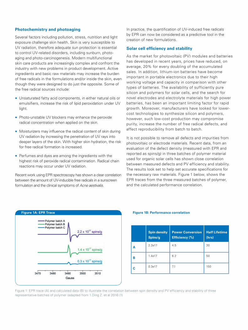
It is not possible to remove all defects and impurities from photovoltaic or electrode materials. Recent data, from an evaluation of the defect density (measured with EPR and reported as spins/g) in three batches of polymer material used for organic solar cells has shown close correlation between measured defects and PV efficiency and stability. The results look set to help set accurate specifications for the necessary raw materials. Figure 1 below, shows the EPR traces from the three measured batches of polymer, and the calculated performance correlation.
Figure 1A: EPR Trace Figure 1B: Performance correlation
Spin density
Spins/g
Power Conversion
Efficiency (%)
Half Lifetime
(hrs)
A2.2e17 4.5 30
B1.4e17 6.2 50
C0.3e17 7.1 150
Figure 1: EPR trace (A) and calculated data (B) to illustrate the correlation between spin density and PV efficiency and stability of three representative batches of polymer (adapted from 1.Ding Z. et al 2016 (1)
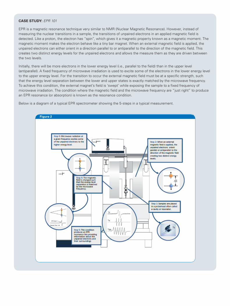
CASE STUDY: EPR 101
EPR is a magnetic resonance technique very similar to NMR (Nuclear Magnetic Resonance). However, instead of measuring the nuclear transitions in a sample, the transitions of unpaired electrons in an applied magnetic field is detected. Like a proton, the electron has “spin”, which gives it a magnetic property known as a magnetic moment. The magnetic moment makes the electron behave like a tiny bar magnet. When an external magnetic field is applied, the unpaired electrons can either orient in a direction parallel to or antiparallel to the direction of the magnetic field. This creates two distinct energy levels for the unpaired electrons and allows the measure them as they are driven between the two levels.
Initially, there will be more electrons in the lower energy level (i.e., parallel to the field) than in the upper level (antiparallel). A fixed frequency of microwave irradiation is used to excite some of the electrons in the lower energy level to the upper energy level. For the transition to occur the external magnetic field must be at a specific strength, such that the energy level separation between the lower and upper states is exactly matched by the microwave frequency. To achieve this condition, the external magnet’s field is ‘swept’ while exposing the sample to a fixed frequency of microwave irradiation. The condition where the magnetic field and the microwave frequency are “just right” to produce an EPR resonance (or absorption) is known as the resonance condition.
Below is a diagram of a typical EPR spectrometer showing the 5-steps in a typical measurement.
Figure 2

© B
ruke
r B
ioS
pin
05/1
7 T1
6506
6
References
[1] Ding Z. et al., Efficient solar cells are more stable: the impact of polymer molecular weight on performance of organic photovoltaics, J. Mater. Chem. A (2016) 4 7274
Learning and prizes
As a leading supplier of EPR instrumentation, Bruker recognizes the role it has in stimulating and disseminating information about the fundamentals of the technique. To this end, a comprehensive teaching package is available. Consisting of an easy to use continuous wave EPR spectrometer that is fully optimized for the teaching environment, an ‘Introduction to EPR’ theory and practice pack, real life samples for analysis in the lab and a suite of experiments for teaching the EPR measurement, data acquisition and processing steps required.
For more details of this unique Bruker initiative, visit: https://www.bruker.com/products/mr/epr/epr-in-education.html
In addition, Bruker supports annual research and student prizes, administered by the RSC and presented at their annual conference events. For more details, visit: http://www.esr-group.org
For information about Bruker EPR instrumentation, and to access live and on-demand webinars and other training materials please visit: https://www.bruker.com/products/mr/epr.html
For more information about the Bench-Top EPR, please visit: https://www.bruker.com/products/mr/epr/emxnano/overview.html. To download our EPR application flyers, please visit: https://www.bruker.com/products/mr/epr/epr-in-pharma.html
About Bruker Corporation (NASDAQ: BRKR)
For more than 55 years, Bruker has enabled scientists to make breakthrough discoveries and develop new applications that improve the quality of human life. Bruker’s high-performance scientific instruments and high-value analytical and diagnostic solutions enable scientists to explore life and materials at molecular, cellular and microscopic levels.
In close cooperation with our customers, Bruker is enabling innovation, productivity and customer success in life science molecular research, in applied and pharma applications, in microscopy, nanoanalysis and industrial applications, as well as in cell biology, preclinical imaging, clinical phenomics and proteomics research, clinical microbiology and molecular pathology research.
For more information, please visit:
www.bruker.com/epr
[email protected] - www.bruker.com
Bruker BioSpin
15 Fortune Drive, Billerica, MA 01821 • USA