Embed Size (px)
Citation preview
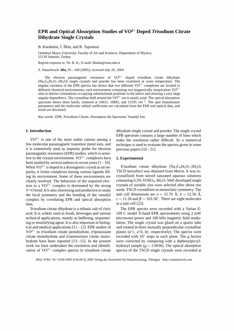
EPR and Optical Absorption Studies of VO2+ Doped Trisodium CitrateDihydrate Single Crystals
B. Karabulut, I. Ilkin, and R. Tapramaz
Ondokuz Mayıs University, Faculty of Art and Sciences, Department of Physics,55139 Samsun, Turkey
Reprint requests to Dr. B. K.; E-mail: [email protected]
Z. Naturforsch. 60a, 95 – 100 (2005); received July 20, 2004
The electron paramagnetic resonance of VO2+ doped trisodium citrate dihydrate(Na3C6H5O7·2H2O) single crystals and powder has been examined at room temperature. Theangular variation of the EPR spectra has shown that two different VO2+ complexes are located indifferent chemical environments, each environment containing two magnetically inequivalent VO2+
sites in distinct orientations occupying substitutional positions in the lattice and showing a very largeangular dependence. The crystalline field around the VO2+ ion is nearly axial. The optical absorptionspectrum shows three bands, centered at 24631, 16892, and 12195 cm−1. The spin Hamiltonianparameters and the molecular orbital coefficients are calculated from the EPR and optical data, andresult are discussed.
Key words: EPR; Trisodium Citrate; Absorption the Spectrum; Vanadyl Ion.
1. Introduction
VO2+ is one of the most stable cations among afew molecular paramagnetic transition metal ions, andit is extensively used as impurity probe for electronparamagnetic resonance (EPR) studies, which is sensi-tive to the crystal environment. VO2+ complexes havebeen studied by several authors in recent years [1 – 10].When VO2+ is doped in a diamagnetic crystal as an im-purity, it forms complexes having various ligands fill-ing its environment. Some of these environments areclearly resolved. The behaviour of the unpaired elec-tron in a VO2+ complex is dominated by the strongV=O bond. It is also interesting and productive to studythe local symmetry and the bonding of the vanadylcomplex by correlating EPR and optical absorptiondata.
Trisodium citrate dihydrate is a tribasic salt of citricacid. It is widely used in foods, beverages and varioustechnical applications, mainly as buffering, sequester-ing or emulsifying agent. It is also important in biolog-ical and medical applications [11 – 12]. EPR studies ofVO2+ in trisodium citrate pentahydrate, tripotassiumcitrate monohydrate and triammonium citrate mono-hydrate have been reported [13 – 15]. In the presentwork we have undertaken the resolution and identifi-cation of VO2+ complex spectra in trisodium citrate
0932–0784 / 05 / 0100–0095 $ 06.00 c© 2005 Verlag der Zeitschrift fur Naturforschung, Tubingen · http://znaturforsch.com
dihydrate single crystal and powder. The single crystalEPR spectrum contains a large number of lines whichmake the resolution rather difficult. So a numericaltechnique is used to evaluate the spectra given in someprevious papers [16 – 21].
2. Experimental
Trisodium citrate dihydrate [Na3C6H5O7·2H2O;TSCD hereafter] was obtained from Merck. It was re-crystallized from stirred saturated aqueous solutionscontaining 0.3% VOSO4·3H2O. Well developed singlecrystals of suitable size were selected after about oneweek. TSCD crystallizes in monoclinic symmetry. Theunit cell dimensions are a = 15.70 A, b = 12.50 A,c = 11.28 and β = 103.58◦. There are eight moleculesin a unit cell [22].
The EPR spectra were recorded with a Varian E-109 C model X-band EPR spectrometer using 2 mWmicrowave power and 100 kHz magnetic field modu-lation. The single crystal was glued on a quartz tubeand rotated in three mutually perpendicular crystallineplanes (a∗c, a∗b, bc, respectively). The spectra wererecorded with 10◦ steps in each plane. The g factorswere corrected by comparing with a diphenylpicryl-hydrazyl sample (g = 2.0036). The optical absorptionspectra of the TSCD single crystals were recorded at
96 B. Karabulut et al. · VO2+ Doped Trisodium Citrate Dihydrate Single Crystals
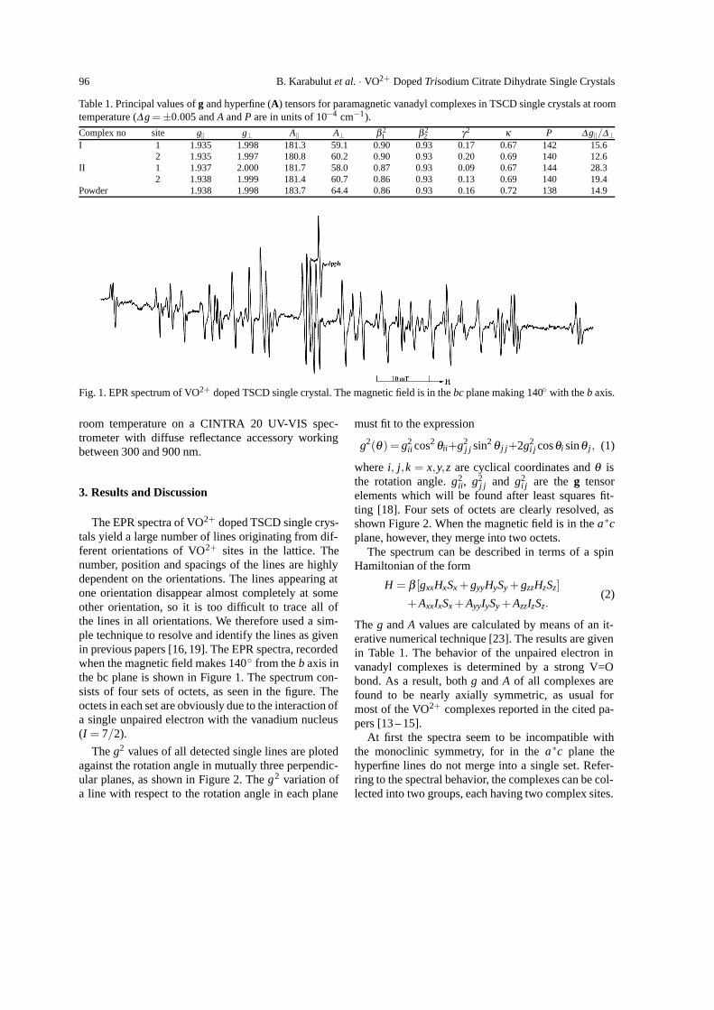
Table 1. Principal values of g and hyperfine (A) tensors for paramagnetic vanadyl complexes in TSCD single crystals at roomtemperature (∆g = ±0.005 and A and P are in units of 10−4 cm−1).
Complex no site g‖ g⊥ A‖ A⊥ β 21 β 2
2 γ2 κ P ∆ g‖/∆⊥I 1 1.935 1.998 181.3 59.1 0.90 0.93 0.17 0.67 142 15.6
2 1.935 1.997 180.8 60.2 0.90 0.93 0.20 0.69 140 12.6II 1 1.937 2.000 181.7 58.0 0.87 0.93 0.09 0.67 144 28.3
2 1.938 1.999 181.4 60.7 0.86 0.93 0.13 0.69 140 19.4Powder 1.938 1.998 183.7 64.4 0.86 0.93 0.16 0.72 138 14.9
Fig. 1. EPR spectrum of VO2+ doped TSCD single crystal. The magnetic field is in the bc plane making 140◦ with the b axis.
room temperature on a CINTRA 20 UV-VIS spec-trometer with diffuse reflectance accessory workingbetween 300 and 900 nm.
3. Results and Discussion
The EPR spectra of VO2+ doped TSCD single crys-tals yield a large number of lines originating from dif-ferent orientations of VO2+ sites in the lattice. Thenumber, position and spacings of the lines are highlydependent on the orientations. The lines appearing atone orientation disappear almost completely at someother orientation, so it is too difficult to trace all ofthe lines in all orientations. We therefore used a sim-ple technique to resolve and identify the lines as givenin previous papers [16, 19]. The EPR spectra, recordedwhen the magnetic field makes 140◦ from the b axis inthe bc plane is shown in Figure 1. The spectrum con-sists of four sets of octets, as seen in the figure. Theoctets in each set are obviously due to the interaction ofa single unpaired electron with the vanadium nucleus(I = 7/2).
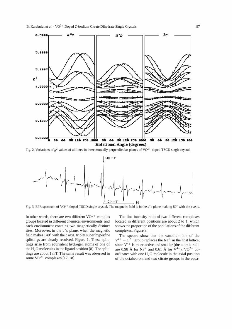
The g2 values of all detected single lines are plotedagainst the rotation angle in mutually three perpendic-ular planes, as shown in Figure 2. The g2 variation ofa line with respect to the rotation angle in each plane
must fit to the expression
g2(θ ) = g2ii cos2 θii+g2
j j sin2 θ j j+2g2i j cosθi sinθ j, (1)
where i, j,k = x,y,z are cyclical coordinates and θ isthe rotation angle. g2
ii, g2j j and g2
i j are the g tensorelements which will be found after least squares fit-ting [18]. Four sets of octets are clearly resolved, asshown Figure 2. When the magnetic field is in the a∗cplane, however, they merge into two octets.
The spectrum can be described in terms of a spinHamiltonian of the form
H = β [gxxHxSx + gyyHySy + gzzHzSz]
+ AxxIxSx + AyyIySy + AzzIzSz.(2)
The g and A values are calculated by means of an it-erative numerical technique [23]. The results are givenin Table 1. The behavior of the unpaired electron invanadyl complexes is determined by a strong V=Obond. As a result, both g and A of all complexes arefound to be nearly axially symmetric, as usual formost of the VO2+ complexes reported in the cited pa-pers [13 – 15].
At first the spectra seem to be incompatible withthe monoclinic symmetry, for in the a∗c plane thehyperfine lines do not merge into a single set. Refer-ring to the spectral behavior, the complexes can be col-lected into two groups, each having two complex sites.
B. Karabulut et al. · VO2+ Doped Trisodium Citrate Dihydrate Single Crystals 97
Fig. 2. Variations of g2 values of all lines in three mutually perpendicular planes of VO2+ doped TSCD single crystal.
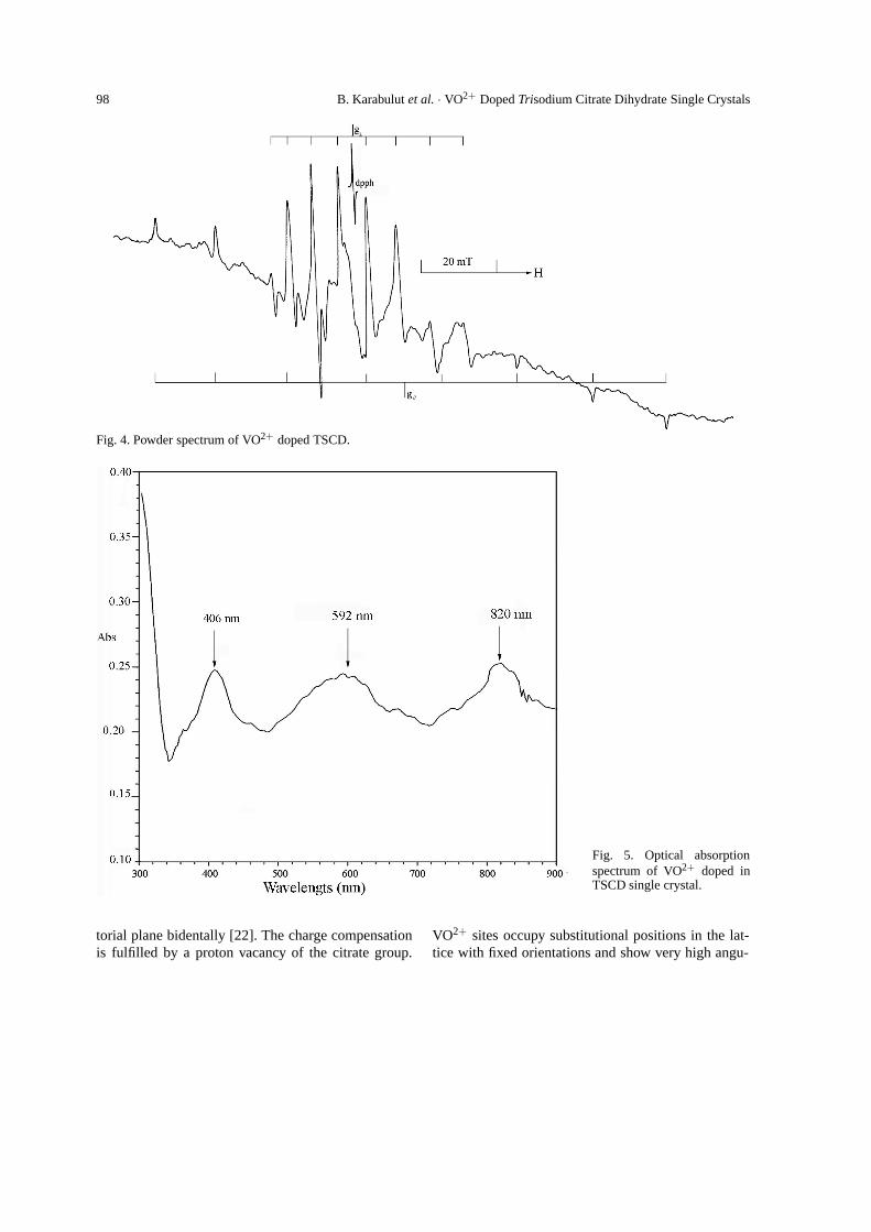
Fig. 3. EPR spectrum of VO2+ doped TSCD single crystal. The magnetic field is in the a∗c plane making 80◦ with the c axis.
In other words, there are two different VO2+ complexgroups located in different chemical environments, andeach environment contains two magnetically distinctsites. Moreover, in the a∗c plane, when the magneticfield makes 140◦ with the c axis, triplet super hyperfinesplittings are clearly resolved, Figure 1. These split-tings arise from equivalent hydrogen atoms of one ofthe H2O molecules in the ligand position [8]. The split-tings are about 1 mT. The same result was observed insome VO2+ complexes [17, 18].
The line intensity ratio of two different complexeslocated in different positions are about 2 to 1, whichshows the proportion of the populations of the differentcomplexes, Figure 3.
The spectra show that the vanadium ion of theV4+ = O2− group replaces the Na+ in the host lattice;since V4+ is more active and smaller (the atomic radiiare 0.98 A for Na+ and 0.61 A for V4+). VO2+ co-ordinates with one H2O molecule in the axial positionof the octahedron, and two citrate groups in the equa-
98 B. Karabulut et al. · VO2+ Doped Trisodium Citrate Dihydrate Single Crystals
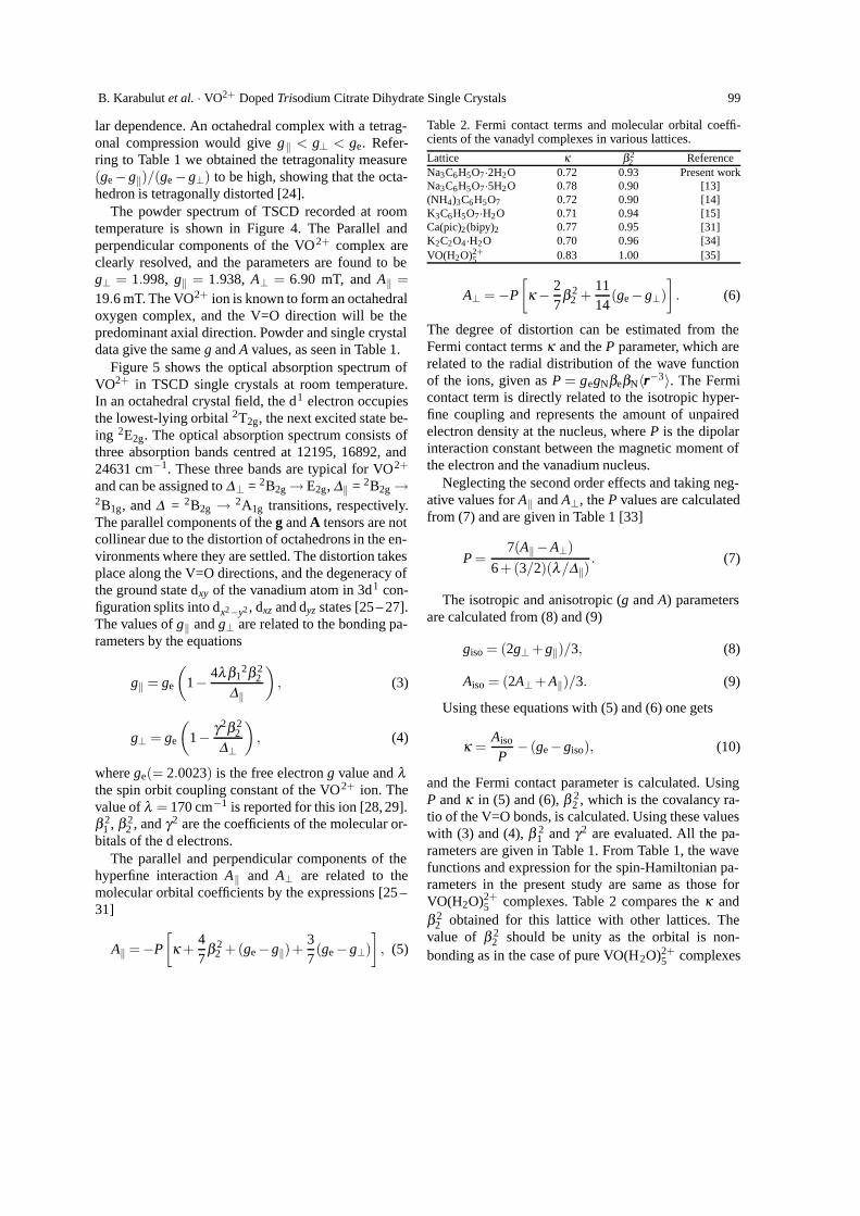
Fig. 4. Powder spectrum of VO2+ doped TSCD.
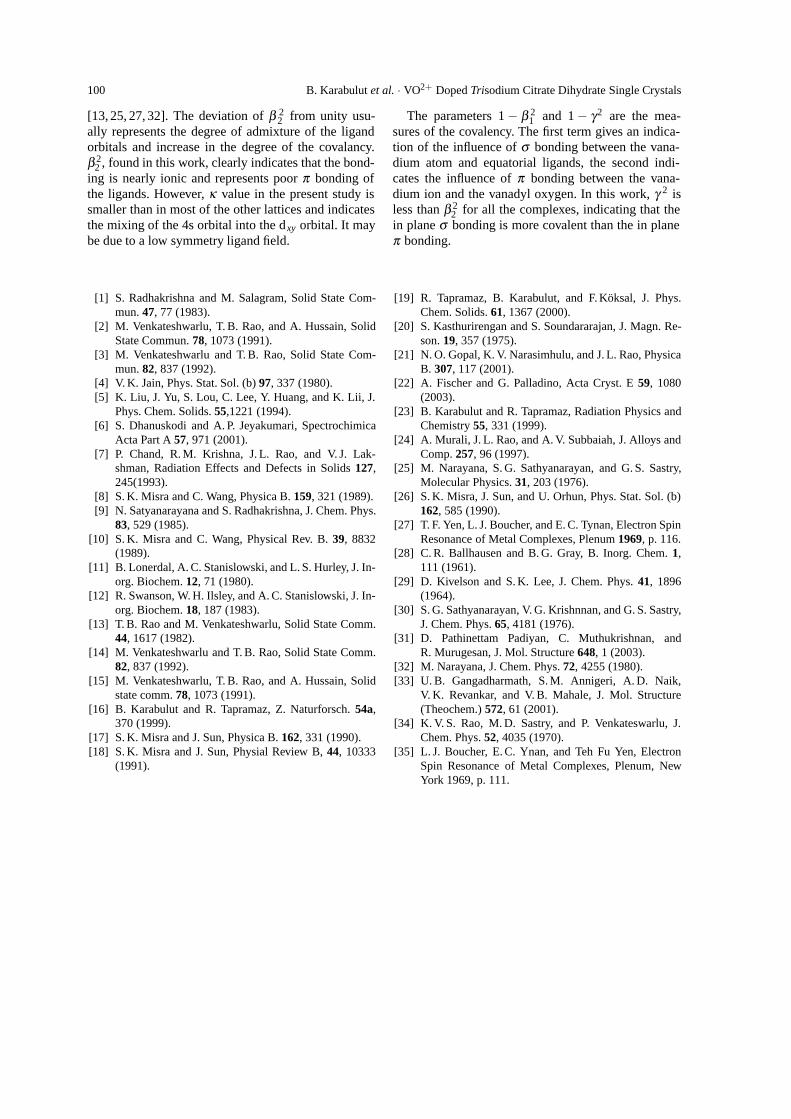
Fig. 5. Optical absorptionspectrum of VO2+ doped inTSCD single crystal.
torial plane bidentally [22]. The charge compensationis fulfilled by a proton vacancy of the citrate group.
VO2+ sites occupy substitutional positions in the lat-tice with fixed orientations and show very high angu-
B. Karabulut et al. · VO2+ Doped Trisodium Citrate Dihydrate Single Crystals 99
lar dependence. An octahedral complex with a tetrag-onal compression would give g‖ < g⊥ < ge. Refer-ring to Table 1 we obtained the tetragonality measure(ge −g‖)/(ge −g⊥) to be high, showing that the octa-hedron is tetragonally distorted [24].
The powder spectrum of TSCD recorded at roomtemperature is shown in Figure 4. The Parallel andperpendicular components of the VO2+ complex areclearly resolved, and the parameters are found to beg⊥ = 1.998, g‖ = 1.938, A⊥ = 6.90 mT, and A‖ =19.6 mT. The VO2+ ion is known to form an octahedraloxygen complex, and the V=O direction will be thepredominant axial direction. Powder and single crystaldata give the same g and A values, as seen in Table 1.
Figure 5 shows the optical absorption spectrum ofVO2+ in TSCD single crystals at room temperature.In an octahedral crystal field, the d1 electron occupiesthe lowest-lying orbital 2T2g, the next excited state be-ing 2E2g. The optical absorption spectrum consists ofthree absorption bands centred at 12195, 16892, and24631 cm−1. These three bands are typical for VO2+
and can be assigned to ∆⊥ = 2B2g → E2g, ∆‖ = 2B2g →2B1g, and ∆ = 2B2g → 2A1g transitions, respectively.The parallel components of the g and A tensors are notcollinear due to the distortion of octahedrons in the en-vironments where they are settled. The distortion takesplace along the V=O directions, and the degeneracy ofthe ground state dxy of the vanadium atom in 3d1 con-figuration splits into dx2−y2 , dxz and dyz states [25 – 27].The values of g‖ and g⊥ are related to the bonding pa-rameters by the equations
g‖ = ge
(1− 4λ β1
2β 22
∆‖
), (3)
g⊥ = ge
(1− γ2β 2
2
∆⊥
), (4)
where ge(= 2.0023) is the free electron g value and λthe spin orbit coupling constant of the VO2+ ion. Thevalue of λ = 170 cm−1 is reported for this ion [28, 29].β 2
1 , β 22 , and γ2 are the coefficients of the molecular or-
bitals of the d electrons.The parallel and perpendicular components of the
hyperfine interaction A‖ and A⊥ are related to themolecular orbital coefficients by the expressions [25 –31]
A‖ =−P
[κ +
47
β 22 +(ge −g‖)+
37(ge −g⊥)
], (5)
Table 2. Fermi contact terms and molecular orbital coeffi-cients of the vanadyl complexes in various lattices.
Lattice κ β22 Reference
Na3C6H5O7·2H2O 0.72 0.93 Present workNa3C6H5O7·5H2O 0.78 0.90 [13](NH4)3C6H5O7 0.72 0.90 [14]K3C6H5O7·H2O 0.71 0.94 [15]Ca(pic)2(bipy)2 0.77 0.95 [31]K2C2O4·H2O 0.70 0.96 [34]VO(H2O)2+
5 0.83 1.00 [35]
A⊥ = −P
[κ − 2
7β 2
2 +1114
(ge −g⊥)]. (6)
The degree of distortion can be estimated from theFermi contact terms κ and the P parameter, which arerelated to the radial distribution of the wave functionof the ions, given as P = gegNβeβN〈rrr−3〉. The Fermicontact term is directly related to the isotropic hyper-fine coupling and represents the amount of unpairedelectron density at the nucleus, where P is the dipolarinteraction constant between the magnetic moment ofthe electron and the vanadium nucleus.
Neglecting the second order effects and taking neg-ative values for A‖ and A⊥, the P values are calculatedfrom (7) and are given in Table 1 [33]
P =7(A‖−A⊥)
6+(3/2)(λ/∆‖). (7)
The isotropic and anisotropic (g and A) parametersare calculated from (8) and (9)
giso = (2g⊥ + g‖)/3, (8)
Aiso = (2A⊥+ A‖)/3. (9)
Using these equations with (5) and (6) one gets
κ =Aiso
P− (ge−giso), (10)
and the Fermi contact parameter is calculated. UsingP and κ in (5) and (6), β 2
2 , which is the covalancy ra-tio of the V=O bonds, is calculated. Using these valueswith (3) and (4), β 2
1 and γ2 are evaluated. All the pa-rameters are given in Table 1. From Table 1, the wavefunctions and expression for the spin-Hamiltonian pa-rameters in the present study are same as those forVO(H2O)2+
5 complexes. Table 2 compares the κ andβ 2
2 obtained for this lattice with other lattices. Thevalue of β 2
2 should be unity as the orbital is non-bonding as in the case of pure VO(H2O)2+
5 complexes
100 B. Karabulut et al. · VO2+ Doped Trisodium Citrate Dihydrate Single Crystals
[13, 25, 27, 32]. The deviation of β 22 from unity usu-
ally represents the degree of admixture of the ligandorbitals and increase in the degree of the covalancy.β 2
2 , found in this work, clearly indicates that the bond-ing is nearly ionic and represents poor π bonding ofthe ligands. However, κ value in the present study issmaller than in most of the other lattices and indicatesthe mixing of the 4s orbital into the dxy orbital. It maybe due to a low symmetry ligand field.
The parameters 1 − β 21 and 1 − γ2 are the mea-
sures of the covalency. The first term gives an indica-tion of the influence of σ bonding between the vana-dium atom and equatorial ligands, the second indi-cates the influence of π bonding between the vana-dium ion and the vanadyl oxygen. In this work, γ 2 isless than β 2
2 for all the complexes, indicating that thein plane σ bonding is more covalent than the in planeπ bonding.
[1] S. Radhakrishna and M. Salagram, Solid State Com-mun. 47, 77 (1983).
[2] M. Venkateshwarlu, T. B. Rao, and A. Hussain, SolidState Commun. 78, 1073 (1991).
[3] M. Venkateshwarlu and T. B. Rao, Solid State Com-mun. 82, 837 (1992).
[4] V. K. Jain, Phys. Stat. Sol. (b) 97, 337 (1980).[5] K. Liu, J. Yu, S. Lou, C. Lee, Y. Huang, and K. Lii, J.
Phys. Chem. Solids. 55,1221 (1994).[6] S. Dhanuskodi and A. P. Jeyakumari, Spectrochimica
Acta Part A 57, 971 (2001).[7] P. Chand, R. M. Krishna, J. L. Rao, and V. J. Lak-
shman, Radiation Effects and Defects in Solids 127,245(1993).
[8] S. K. Misra and C. Wang, Physica B. 159, 321 (1989).[9] N. Satyanarayana and S. Radhakrishna, J. Chem. Phys.
83, 529 (1985).[10] S. K. Misra and C. Wang, Physical Rev. B. 39, 8832
(1989).[11] B. Lonerdal, A. C. Stanislowski, and L. S. Hurley, J. In-
org. Biochem. 12, 71 (1980).[12] R. Swanson, W. H. Ilsley, and A. C. Stanislowski, J. In-
org. Biochem. 18, 187 (1983).[13] T. B. Rao and M. Venkateshwarlu, Solid State Comm.
44, 1617 (1982).[14] M. Venkateshwarlu and T. B. Rao, Solid State Comm.
82, 837 (1992).[15] M. Venkateshwarlu, T. B. Rao, and A. Hussain, Solid
state comm. 78, 1073 (1991).[16] B. Karabulut and R. Tapramaz, Z. Naturforsch. 54a,
370 (1999).[17] S. K. Misra and J. Sun, Physica B. 162, 331 (1990).[18] S. K. Misra and J. Sun, Physial Review B, 44, 10333
(1991).
[19] R. Tapramaz, B. Karabulut, and F. Koksal, J. Phys.Chem. Solids. 61, 1367 (2000).
[20] S. Kasthurirengan and S. Soundararajan, J. Magn. Re-son. 19, 357 (1975).
[21] N. O. Gopal, K. V. Narasimhulu, and J. L. Rao, PhysicaB. 307, 117 (2001).
[22] A. Fischer and G. Palladino, Acta Cryst. E 59, 1080(2003).
[23] B. Karabulut and R. Tapramaz, Radiation Physics andChemistry 55, 331 (1999).
[24] A. Murali, J. L. Rao, and A. V. Subbaiah, J. Alloys andComp. 257, 96 (1997).
[25] M. Narayana, S. G. Sathyanarayan, and G. S. Sastry,Molecular Physics. 31, 203 (1976).
[26] S. K. Misra, J. Sun, and U. Orhun, Phys. Stat. Sol. (b)162, 585 (1990).
[27] T. F. Yen, L. J. Boucher, and E. C. Tynan, Electron SpinResonance of Metal Complexes, Plenum 1969, p. 116.
[28] C. R. Ballhausen and B. G. Gray, B. Inorg. Chem. 1,111 (1961).
[29] D. Kivelson and S. K. Lee, J. Chem. Phys. 41, 1896(1964).
[30] S. G. Sathyanarayan, V. G. Krishnnan, and G. S. Sastry,J. Chem. Phys. 65, 4181 (1976).
[31] D. Pathinettam Padiyan, C. Muthukrishnan, andR. Murugesan, J. Mol. Structure 648, 1 (2003).
[32] M. Narayana, J. Chem. Phys. 72, 4255 (1980).[33] U. B. Gangadharmath, S. M. Annigeri, A. D. Naik,
V. K. Revankar, and V. B. Mahale, J. Mol. Structure(Theochem.) 572, 61 (2001).
[34] K. V. S. Rao, M. D. Sastry, and P. Venkateswarlu, J.Chem. Phys. 52, 4035 (1970).
[35] L. J. Boucher, E. C. Ynan, and Teh Fu Yen, ElectronSpin Resonance of Metal Complexes, Plenum, NewYork 1969, p. 111.