Embed Size (px)
Citation preview

Epoxide electrophiles as activity-dependent cysteine proteasepro¢ling and discovery toolsDoron Greenbaum 1, Katalin F Medzihradszky 1;2, Alma Burlingame 1;2 andMatthew Bogyo 3
Background: Analysis of global changes in gene transcription and translation bysystems-based genomics and proteomics approaches provides only indirectinformation about protein function. In many cases, enzymatic activity fails tocorrelate with transcription or translation levels. Therefore, a direct method forbroadly determining activities of an entire class of enzymes on a genome-widescale would be of great utility.
Results: We have engineered chemical probes that can be used to broadly trackactivity of cysteine proteases. The structure of the general cysteine proteaseinhibitor E-64 was used as a scaffold. Analogs were synthesized by varying thecore peptide recognition portion while adding af¢nity tags (biotin and radio-iodine)at distal sites. The resulting probes containing a P2 leucine residue (DCG-03 andDCG-04) targeted the same broad set of cysteine proteases as E-64 and wereused to pro¢le these proteases during the progression of a normal skin cell to acarcinoma. A library of DCG-04 derivatives was constructed in which the leucineresidue was replaced with all natural amino acids. This library was used to obtaininhibitor activity pro¢les for multiple protease targets in crude cellular extracts.Finally, the af¢nity tag of DCG-04 allowed puri¢cation of modi¢ed proteases andidenti¢cation by mass spectrometry.
Conclusions: We have created a simple and £exible method for functionallyidentifying cysteine proteases while simultaneously tracking their relative activitylevels in crude protein mixtures. These probes were used to determine relativeactivities of multiple proteases throughout a de¢ned model system for cancerprogression. Furthermore, information obtained from libraries of af¢nity probesprovides a rapid method for obtaining detailed functional information without theneed for prior puri¢cation/identi¢cation of targets.
1Department of Pharmaceutical Chemistry, Universityof California, San Francisco, CA 94143, USA2Mass Spectrometry Facility, University of California,San Francisco, CA 94143, USA3Department of Biochemistry and Biophysics,University of California, San Francisco, CA 94143,USA
Correspondence: Matthew BogyoE-mail: [email protected]
Keywords: Af¢nity labeling; Cysteine protease; E-64;Electrophile; Epoxide; Proteomics
Received: 14 April 2000Accepted: 9 May 2000
Published: 1 August 2000
Chemistry & Biology 2000, 7:569^581
1074-5521 / 00 / $ ^ see front matterß 2000 Elsevier Science Ltd. All rights reserved.PII: S 1 0 7 4 - 5 5 2 1 ( 0 0 ) 0 0 0 1 4 - 4
IntroductionNew approaches for studying global cellular processesmust permit the analysis of differential changes withinlarge sets of known and unknown genes or proteins.DNA microarray techniques allow analysis of genome-wide changes in mRNA transcription for a given cellularstimulus [1,2]. Advances in two-dimensional (2D) gel elec-trophoresis coupled to highly sensitive mass spectrometrytechniques now allow the rapid identi¢cation of proteinsfrom whole cells or tissue extracts [3,4]. While these tech-niques have revolutionized the global analysis of biologicalprocesses, often information about function of enzymaticproteins can only be inferred by analysis of transcriptional/translational co-regulation of sets of genes under differentstimuli. In fact, levels of transcription and translation of anenzyme, in many cases, do not correlate with its activity[5].
To assign a function to enzymatic proteins on a genome-wide scale, a method to obtain direct information about
enzymatic activity is necessary. Since the simultaneous tar-geting of all enzyme classes with a single probe is likely tobe impossible, we chose to focus our effort on proteolyticenzymes. The papain family of cysteine proteases serves asa good model system for several reasons. Firstly, most cys-teine proteases are synthesized with an inhibitory propep-tide that must be proteolytically removed to activate theenzyme [6,7], resulting in expression pro¢les that do notdirectly correlate with activity. Secondly, the largest set ofpapain-like cysteine proteases, the cathepsins, act in con-cert to digest a protein substrate. Thus, information regard-ing regulation of activity of each member relative to oneanother is critical for understanding their collective func-tion. Furthermore, the cathepsins are involved in manycritical biological processes, and biochemical studies offunction have been limited to family members that havebeen cloned and expressed or puri¢ed from crude tissue.Finally, a large body of information is available regardingcovalent, mechanism-based inhibitors that speci¢cally tar-get this family of cysteine proteases.
CHBIOL 13 11-8-00
Research Paper 569

The papain family is classi¢ed into several major groups,most notable of which are the bleomycin hydrolases, cal-pains, caspases and cathepsins. To date, 16 human cathep-sins have been cloned and sequenced. Several of theseproteases are key players in normal physiological processessuch as antigen presentation [8], bone remodeling [9] andprohormone processing [10]. In addition, several of theseproteases are involved in pathological processes such asrheumatoid arthritis [11], cancer invasion and metastasis[12] and Alzheimer's disease [13,14].
The enzymatic mechanism used by the papain family ofproteases has been well studied and is highly conserved.Thus, electrophilic substrate analogs that are only reactivein the context of this conserved active site can be used asgeneral probes of function. A wide range of electrophileshas been developed as mechanism-based, cysteine pro-tease inhibitors including diazomethyl ketones [15], £uo-romethyl ketones [16], acyloxymethyl ketones [17], O-acyl-hydroxylamines [18], vinyl sulfones [19] and epoxysuccinicderivatives [20]. These inhibitors all consist of a peptidespeci¢city determinant attached to an electrophile that be-comes irreversibly alkylated when bound in close proxim-ity to an attacking nucleophile.
Several groups have recognized the value of using irrevers-ible mechanism-based inhibitors as af¢nity labels [21^25].Addition of a reporter function, such as a radioactive io-dine, to inhibitors permits the visualization of covalentlymodi¢ed proteases in a standard SDS^PAGE gel format.Labeling intensity provides a read-out of relative enzy-matic activity. Furthermore, both known and novel pro-teases are targets for analysis by this methodology. Similaraf¢nity labeling approaches have been used extensively tostudy or identify proteases such as the proteasome[22,26,27], caspases [28,29], cathepsins [23,25] and methio-nine amino peptidase [30,31]. Recently, Cravatt and co-workers have taken advantage of the broad class-speci¢creactivity of £uorophosphonates towards serine proteases[32]. By incorporation of a simple, extended alkyl chaincapped with a biotin moiety, they have created a broadserine protease-speci¢c probe (FP-Biotin) for functionalproteomic analysis of serine proteases in crude cellular ex-tracts.
We have developed functional proteomics tools that can beused to determine global patterns of activity for the papainfamily of cysteine proteases based on the broad reactivityof the natural product E-64. These tools provide functionalinformation that can be used in concert with existing ge-
Figure 1. Structures of epoxide inhibitors and probes E-64, JPM-565, DCG-03 and DCG-04. Radiolabel attachment and af¢nity sites areindicated for each compound.
CHBIOL 13 11-8-00
570 Chemistry & Biology 2000, Vol 7 No 8

nomic and proteomic methods to correlate gene and pro-tein expression pro¢les with enzymatic activity. Further-more, diversi¢cation of core compounds using solid-phasecombinatorial chemistry provides libraries of compoundsthat can be used to obtain information about inhibitor spe-ci¢cities of targeted protease. This information is likely tobe of use in the generation of selective inhibitors withoutthe need for prior characterization and puri¢cation of pro-tease targets. Finally, the probes themselves can be used torapidly identify targets after covalent modi¢cation.
Results and discussionDesign and synthesis of DCG-04The natural product E-64 is a promiscuous irreversiblecysteine protease inhibitor that is broadly reactive towardthe papain family of cysteine proteases [20] (Figure 1). Itsleucine side chain mimics the P2 amino acid of a substrate,occupying the target's S2 binding pocket while the agma-tine moiety binds in the S3 position [33]. Rich et al. syn-thesized JPM-565 (Figure 1), a derivative in which a tyr-amine moiety replaces the agmatine side chain of E-64[34,35]. This closely related compound was found tohave similar class-speci¢c reactivity for cysteine proteasesas E-64. Since the P2 position of a substrate is consideredto be the main speci¢city determinant for many cysteineproteases, we reasoned that further extension of the non-prime binding portion of JPM-565 would not signi¢cantlyperturb binding af¢nity for a target protease. In addition,modi¢cation to the non-prime site binding element of theE-64 derivative CA-074 had little effect on binding to ca-thepsin B [23,36]. Elaboration of the peptide portion of E-64 allowed both incorporation of an af¢nity tag as well asattachment of the compound to a solid support. The re-
sulting bi-functional compounds, DCG-03 and DCG-04,contain both the iodinatable phenol ring of JPM-565 andthe additional af¢nity site created by incorporation of aside chain biotinylated lysine residue (Figure 1). Addition(DCG-04) or removal (DCG-03) of an amino hexanoic acidspacer between the af¢nity site and the electrophile wasused to determine the space requirement for binding andrecognition of the af¢nity label by support-bound avidin.
Peptide epoxides were synthesized using a combination ofsolution and solid-phase chemistries. The solution-phasesynthesis of the epoxide acid building block (Figure 2A)starting from commercially available diethyl tartrate hasbeen reported elsewhere [23]. Standard solid-phase pep-tide chemistry was used to build the peptide portion ofDCG-04 and related compounds (Figure 2B). This meth-odology provides a £exible system with which to incorpo-rate virtually any peptide sequence prior to attachment ofthe electrophilic epoxide. Surprisingly, the epoxy acidbuilding block was stable to standard solid-phase peptidesynthesis cleavage conditions (95% TFA). The use of sol-id-phase chemistry also allowed the synthesis of a diverselibrary in which the P2 leucine of DCG-04 was replacedwith each of the natural amino acids (except cysteine dueto reactivity with the epoxide and methionine due to ox-idation). The non-natural amino acid norleucine was usedas an isosteric methionine analog. The results obtainedusing this 19 member library of compounds are describedbelow.
DCG-04 is an activity-dependent af¢nity labelDendritic cells express relatively high levels of lysosomalcathepsins, making them a logical source of material for
Figure 2. Synthesis of DCG-04. (A) Epoxyacid building block (I) and (B) solid-phasesynthesis scheme for DCG-04. Details ofthe synthesis and characterization ofpeptide epoxides can be found in Materialsand methods.
CHBIOL 13 11-8-00
Research Paper Functional proteomics tools for cysteine proteases Greenbaum et al. 571

establishing parameters for the use of DCG-04. Figure 3shows the labeling pro¢le of polypeptides modi¢ed by in-cubation with either DCG-03, DCG-04, 125I-DCG-03 or125I-DCG-04 followed by SDS^PAGE analysis. Radio-iodi-nated (autoradiogram) and non-radio-iodinated (blot)DCG-03 and DCG-04 labeled multiple polypeptides inthe range of 20^40 kDa.
Although the intrinsic reactivity of the epoxide electrophileportion of DCG-04 towards free thiols is quite poor, wewanted to determine if DCG-04 and its derivatives werecapable of non-speci¢c alkylation of proteins in crude cel-lular extracts. A pre-heating control was used to reveal non-speci¢c labeling, with the assumption that denatured, in-active proteins modi¢ed by DCG-03 and DCG-04 repre-sent non-speci¢c modi¢cations. Enzymatically active pro-teins were deduced by subtraction (Figure 3). Labeling ofall of the major species in the 20^40 kDa size range waslost upon heat denaturation of samples prior to addition ofcompounds, suggesting that labeling is dependent on en-zymatic activity and that these bands correspond to themajor proteases in the extract. Several higher molecularweight species were observed by af¢nity blotting of bothdenaturing controls and samples in which no inhibitor wasadded. These species are likely to represent non-speci¢calkylations and endogenously biotinylated proteins.
Comparison of labeling, at neutral (pH 7.4) and at theacidic pH of the lysosome (pH 5.5), indicated that severalof the modi¢ed polypeptides in the 30 kDa size rangerequired reduced pH for activity. This result is consistentwith reported ¢ndings that several lysosomal cysteine pro-teases either reversibly or irreversibly lose activity uponde-acidi¢cation of lysosomal compartments [37].
Analysis of the labeling of DC2.4 lysates by both af¢nityblot and autoradiography techniques resulted in similarpro¢les of modi¢ed polypeptides, highlighting the utilityof both techniques. However, the autoradiogram showedlabeling of only enzymatically active polypeptides by radio-labeled forms of DCG-03 and DCG-04. Addition of therather bulky iodine atom to DCG-03 and DCG-04 hadonly a modest effect on target modi¢cation yet resultedin compounds with dramatically reduced background label-ing and increased sensitivity. Ultimately, the ability to useboth autoradiography as well as blot techniques enhancesthe £exibility of these protease detection reagents and fur-ther highlights the utility of bi-functional inhibitors.
DCG-04 targets cysteine proteases inhibited by E-64 andJPM-565Both direct labeling and indirect competition experimentswere performed to con¢rm that DCG-04 reacts with a sim-
Figure 3. DCG-03 and DCG-04 label active proteases in dendritic cell extracts. (A) Total cell extracts from DC2.4 cells were diluted intoeither pH 5.5 or pH 7.4 buffer, pre-heated to 100³C for 1 min (+pre-heating) or not (3pre-heating) and labeled with 50 WM DCG-03 andDCG-04. Samples were separated by SDS^PAGE (12.5% gel) and labeled bands visualized by af¢nity blotting as described in Materialsand methods. (B) Same as for (A) except 125I-labeled versions of DCG-03 and DCG-04 were used and gels analyzed by autoradiography.The locations of cathepsins B, L and S are indicated for reference based on their known molecular weights.
CHBIOL 13 11-8-00
572 Chemistry & Biology 2000, Vol 7 No 8
ilar subset of proteases to the parent compounds E-64 andJPM-565. An indirect competition experiment was re-quired to determine the polypeptides modi¢ed by E-64since it lacks an af¢nity labeling site. Extracts from thedendritic cell line DC2.4 were preincubated with an in-creasing concentration of E-64 followed by labeling withDCG-04. Final labeling intensity was used to indirectlymonitor the extent of polypeptide modi¢cation by E-64.The competition revealed that all polypeptides labeled byDCG-04 are effectively competed by E-64, indicating thatthe two compounds target the same subset of proteases(Figure 4A). A similar competition experiment was per-formed using the cathepsin B-speci¢c inhibitor MB-074[23]. These results positively identi¢ed the diffuse 30kDa polypeptide (labeled cat B in Figure 4A) as cathepsinB (data not shown).
Comparison of the speci¢city of DCG-03, DCG-04 andJPM-565 was accomplished using direct labeling ofDC2.4 cell lysates. Labeling pro¢les obtained for 125I-DCG-03, 125I-DCG-04 and 125I-JPM-565 were identicalfor all three probes and indicated that each targeted poly-peptides in the 20^40 kDa size range (Figure 4B). Analysisof non-radiolabeled DCG-03-, DCG-04- and JPM-565-treated extracts again showed the similarity of the blottingand autoradiography detection systems. As expected, JPM-
565, which lacks a biotin label, showed no labeling asdetected by af¢nity blotting. Together these results estab-lish that modi¢cations to the extended binding portion ofthe E-64 family of compounds have little effect on selec-tivity or potency. However, this region of the inhibitor maystill play an important role in establishing speci¢city ofbinding when equipped with the proper recognition se-quence. Future work is aimed at exploring the use of ex-tended peptide recognition motifs to ¢ne tune selectivityof the DCG family of inhibitors for speci¢c protease tar-gets.
Pro¢ling applicationsThe aforementioned methods established the initial pa-rameters for use of the general cysteine protease labelsDCG-03 and DCG-04. We next wanted to apply thesetechniques to pro¢le the activity and speci¢city of cysteineproteases in several different model systems. The broadlyreactive probe DCG-04 was used to generate activity pro-¢les of multiple protease targets both in a model for dis-ease progression and throughout multiple tissue types.Similarly, activity pro¢les were generated using the cathep-sin B-speci¢c probe MB-074 to provide complementaryinformation for a single, well-de¢ned cysteine protease tar-get. This information was also used to positively establishthe identity of cathepsin B in the DCG-04 labeling pro-
Figure 4. DCG-03 and DCG-04 target the same polypeptides as parent compounds E-64 and JPM-565. (A) Total cellular extracts fromDC2.4 cells were incubated with increasing concentrations of E-64 as indicated for 30 min at 25³C followed by addition of 50 WM DCG-04and further incubation for 1 h. Samples were resolved by SDS^PAGE (12.5%) and labeled bands visualized by af¢nity blotting. (B) Totalcellular extracts were labeled with either 125I-labeled forms (auto-rad) or with non-labeled forms (blot) of DCG-03, DCG-04 and JPM-565followed by separation by SDS^PAGE (12.5%) and analysis as indicated. The locations of cathepsin B and S are indicated for referencebased on their known molecular weights.
CHBIOL 13 11-8-00
Research Paper Functional proteomics tools for cysteine proteases Greenbaum et al. 573
¢les. To obtain more detailed functional information forDCG-04-modi¢ed proteases, inhibitor speci¢city pro¢leswere generated using a library of DCG-04 analogs in totalcellular extracts. The same libraries were also used in con-junction with the cathepsin B-speci¢c probe, 125I-MB-074,as well as with puri¢ed cathepsin H to determine speci¢c-ity pro¢les for individual target proteases. These results aredescribed below.
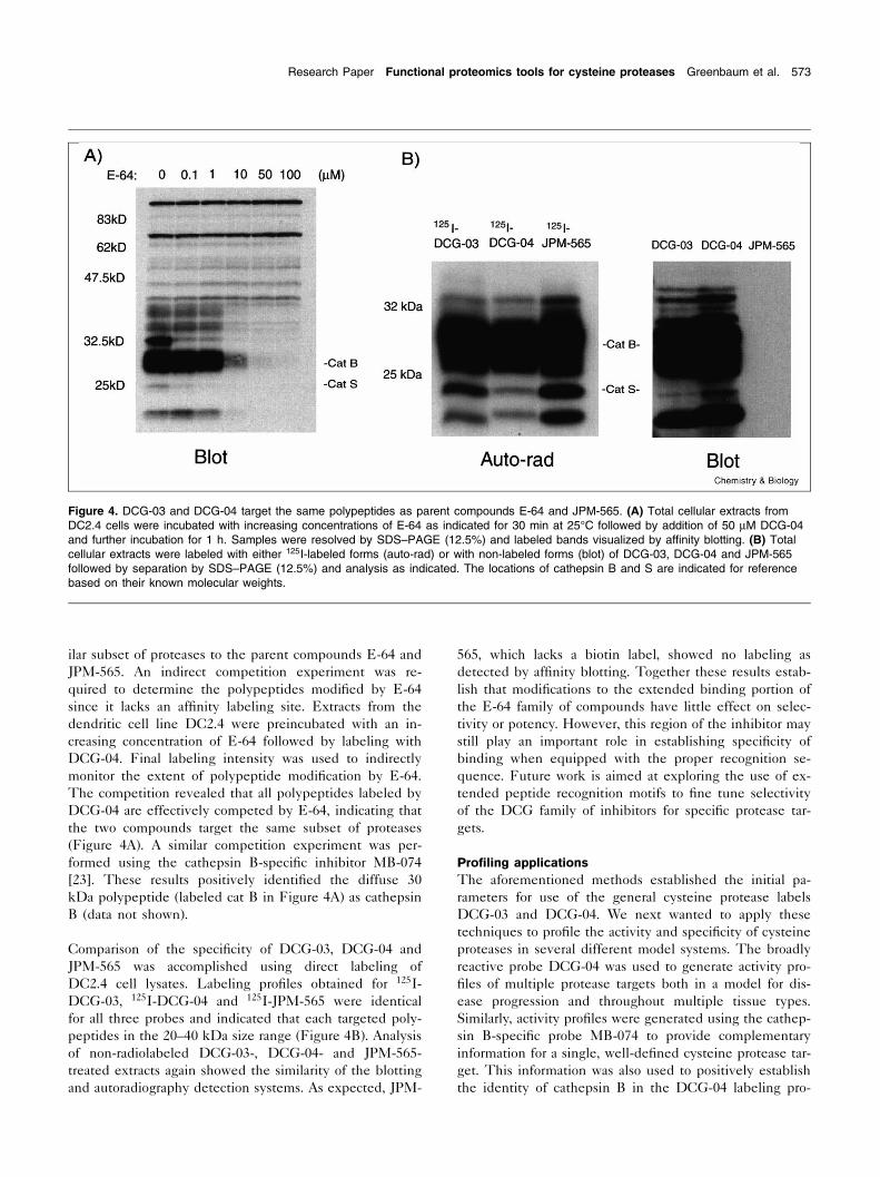
Pro¢ling across disease progression using DCG-04 and MB-074The mouse skin model of multi-stage carcinogenesis hasbeen well studied genotypically and phenotypically, hasdiscrete steps in the progression, but lacks informationon cysteine protease involvement [38,39]. The role of ca-thepsins in tumor biology has mostly focused on cathepsinsB and L. Upregulated levels of both cathepsins B and Lhave been shown to correlate with an invasive phenotype[12,40]. Furthermore, cathepsins B and L are secreted bymany types of tumorigenic cells and treatment of invasivecells with the cysteine protease inhibitor E-64 results in ablock in cellular invasion into a synthetic matrix [41,42].These data indicate that cathepsins are likely to play animportant role in the metastatic process.
Ten cell lines representing various steps in the progressionfrom benign skin cell (C5N) to highly invasive spindle cell
carcinomas (Car B and Car C) were used to analyze globalchanges in activity of cathepsins throughout this multi-stage carcinogenesis model. The carcinoma progressionalso includes benign papilloma cell lines P6, PDV andPDV-C57, and more invasive squamous cell carcinomacell lines B9, A5 and D3. Equal amounts of protein fromeach cell lysate were labeled with both the broadly reactiveprobe, 125I-DCG-04, as well as the cathepsin B-speci¢cprobe, 125I-MB-074 at pH 5.5 (Figure 5). The resultsshow that several protease activities, including cathepsinB, dramatically £uctuate across the panel of cell lines.
The broadly reactive probe 125I-DCG-04 highlights theactivity of several proteases in the lysosomal cysteine pro-tease size range in each of the cell types (Figure 5A). Thebenign cell lines C5N and P6 both contain multiple la-beled polypeptides between 28 and 45 kDa; however,the labeling intensity observed for the P6 line is dramati-cally increased for all polypeptides in this range. Interest-ingly, the major difference between these cell lines is anactivating mutation in the ras gene [43]. It has previouslybeen shown that various classes of proteases, including thecathepsins, are upregulated downstream of Ras; however,these studies were limited to analysis of expression levelsof cathepsin B and H [44].
Figure 5. Activity pro¢ling across a disease progression. Tissue culture cells were isolated from carcinomas generated by application of achemical mutagen to the skin of mice (see Materials and methods). Progression begins at the left with the non-invasive benign cells (C5Nand P6) and progresses to the right through papilloma cell lines (PDV and PDV-C57), squamous cell carcinomas (B9, A5 and D3) and¢nally highly invasive spindle cell carcinomas (Car B and Car C). Total cellular lysates were normalized with respect to proteinconcentration and labeled with (A) 125I-DCG-04 and (B) the cathepsin B-speci¢c probe 125I-MB-074. A pre-heat control from the C5N lysatewas included in (A) to show background labeling.
CHBIOL 13 11-8-00
574 Chemistry & Biology 2000, Vol 7 No 8

The papilloma cell lines PDV and PDV-C57 show nearlyidentical patterns of labeling (Figure 5A). However, thesepro¢les are dramatically different than the pro¢le observedfor C5N and P6 lysates. A predominant 30 kDa polypep-tide (cathepsin B; see below) is labeled along with a lessintensely labeled 21 kDa polypeptide. The squamous cellcarcinoma cell lines B9, A5 and D3 result in similar pro¢lesof modi¢ed polypeptides. While all three lines are nearlyidentical cancer cell types, only B9 shows appreciable la-beling of the major 30 kDa and 21 kDa polypeptides.Similarly, the two highly invasive spindle cell carcinomasCar B and Car C show similar, but not identical, labelingpro¢les. The 21 kDa species, in particular, shows differ-ential labeling in the two cell types. These ¢ndings illus-trate that cells isolated from different tumor sources havedifferent protease activities. This signature of protease ac-tivity may in fact be unique to each cell and/or tumor,much the same way genomics studies by Browne and col-leagues have shown that individual tumor cells haveunique global gene expression pro¢les [45].
The cathepsin B-speci¢c label 125I-MB-074 was used todirectly examine the pro¢le of cathepsin B activity in thesame collection of cells described (Figure 5B). This probehas been found to label cathepsin B in a highly speci¢cmanner [23]. Labeling of cathepsin B dramatically changedacross the pro¢le of cell types with the greatest activityobserved for the PDV and PDV-C57 lines. Furthermore,the apparent molecular weight, as well as the sharpness ofthe cathepsin B, band differed for the benign and spindlecell carcinomas, suggesting that this enzyme is modi¢eddifferently in these cell types. This change in migrationfor cathepsin B may be due to changes in glycosylation orother post-translational modi¢cations. Cathepsin B hasbeen found to exist as different isoforms with differingpIs in various tumor cells as a result of changes in glyco-sylation and traf¢cking [46]. Changes in the post-transla-tional modi¢cation of cathepsin B are likely to affect thelocalization of active forms of the enzyme and thereforemay play an important role in the control of cathepsin Bactivity in tumors [46]. Overall, the results obtained fromlabeling with 125I-MB-074 further highlight the variability
Figure 6. Pro¢ling protease inhibitor speci¢city. Lysates from the dendritic cell line DC2.4 (A,B) or puri¢ed cathepsin H (C) werepreincubated with 50 WM of each of the 19 derivatives of DCG-04 and then labeled with 125I-DCG-04 (A,C) or 125I-MB-074 (B) asindicated. The general structures of the inhibitors are shown with the variable amino acid side chain indicated as an X (competitor; top).The predominant labeled polypeptides in (A) are labeled with numbers, and positions of cathepsins B and S are indicated for reference.
CHBIOL 13 11-8-00
Research Paper Functional proteomics tools for cysteine proteases Greenbaum et al. 575
of cathepsin B activity found in different types of tumorcells as well as in nearly identical cell lines derived fromdifferent sources.
Pro¢ling protease speci¢city using a library of inhibitorsTo take advantage of the £exibility and ease of synthesisof the DCG-04 family of compounds, we created a smalllibrary of compounds in which the peptide recognitionportion of the molecule is modi¢ed. It has been proposedthat the main speci¢city regions within the active bindingsite of the cathepsins are S2, S1, S1P and S2P, with S2containing the main binding pocket [47]. Since the leucineresidue of E-64 binds in the critical S2 pocket of manyproteases [33], changes to this residue are likely to havethe greatest effect on speci¢city of our inhibitor for a giventarget. A complete scanning library consisting of 18 naturalamino acids and the isosteric methionine analog norleucinewas constructed. This library of inhibitors was used tocreate pro¢les of inhibitor speci¢city for proteases targetedby DCG-04 and MB-074 (Figure 6).
Competition analysis was used to determine the potency ofeach member of the P2 scanning library towards multipleprotease targets. Lysates from DC2.4 cells were preincu-bated with 50 WM of each of the 19 DCG library membersand residual activity measured for multiple proteases using125I-DCG-04 (Figure 6A). In general, residues containingnon-charged aliphatic side chains [isoleucine (I), leucine(L; DCG-04) and norleucine (n)] show the highest activityand the lowest amount of speci¢city across the pro¢le ofpolypeptides. More interesting was the apparent selectivityof several DCG family compounds for a subset of labeledpolypeptides. For example, the valine containing com-pound competed for polypeptides 1, 2 and cathepsin Bbut had little effect on the remaining species. In contrast,both the phenylalanine and tyrosine containing compoundsshowed speci¢city for polypeptides 2, 3, 4 and 5. Further-more, while the aspartic acid and glycine containing com-pounds showed relatively poor activity overall, theyshowed some degree of speci¢city against polypeptide 2.Using these data to simultaneously score inhibitors for po-tency and selectivity will be valuable for the developmentof speci¢c inhibitors.
Similar competition experiments were performed with thelibrary of DCG analogs to obtain pro¢les of single pro-teases. DC2.4 lysates were preincubated with P2 libraryand then labeled with the cathepsin B-speci¢c compound125I-MB-074 (Figure 6B). This method allowed analysis ofcathepsin B speci¢city in crude extracts. As found in the125I-DCG-04 labeling (Figure 6B), isoleucine, leucine, va-line and norleucine analogs showed the highest activityfollowed by the aromatic amino acids (W, Y, F) containingcompounds. In order to explore speci¢city pro¢les for addi-tional cysteine proteases that could not be speci¢cally la-beled in crude extracts, we performed the same competi-
tion labeling experiment described above using a puri¢edenzyme. Preincubation of puri¢ed cathepsin H with thelibrary of compounds followed by 125I-DCG-04 labelingresulted in a speci¢city pro¢le that was remarkably similarto the pro¢le observed for cathepsin B in crude extracts(Figure 6C). While these two proteases are quite differentin their biological functions, it is clear from these data thatthe two have similar inhibitor speci¢city in the S2 pocket.
Since it is unlikely that two distinct proteases will exhibitidentical reactivity across a diverse set of inhibitors, it maybe possible to use this information from inhibitor librariesto generate `speci¢city ¢ngerprints' for a series of well-characterized proteases. Establishment of a database ofprotease inhibitor pro¢les could potentially be used to es-tablish target identi¢cation by labeling of crude proteinmixtures in the presence of compound libraries. Further-more, extension of this methodology to longer, more di-verse peptide substrate analogs may further accentuate thespeci¢city differences of closely related protease species.
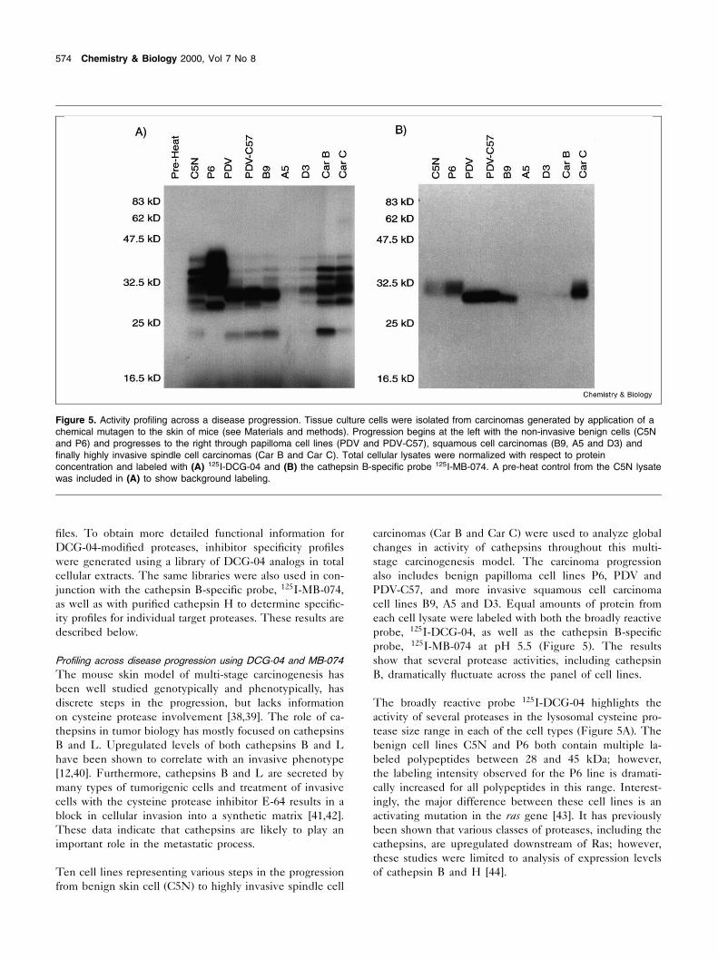
Pro¢ling across tissue typesHaving determined that both DCG-03 and DCG-04 werecapable of covalently modifying multiple papain family
Figure 7. Activity pro¢ling of cysteine proteases across tissuetypes. Labeling of total cellular extracts (100 Wg protein/lane) fromrat brain, kidney, liver, prostate and testis with 125I-DCG-04 at pH5.5. Samples were analyzed by SDS^PAGE followed byautoradiography. A pre-heating control was included for eachtissue type to indicated background labeling.
CHBIOL 13 11-8-00
576 Chemistry & Biology 2000, Vol 7 No 8

proteases in extracts generated from several cell lines, wewanted to test the utility of these reagents for pro¢lingprotease activity patterns in various tissues. In this way, acrude map of protease activities can be created for eachtissue and ultimately the identity of these major speciescan be determined by virtue of their reactivity towards theDCG-04 af¢nity probe.
Samples of rat brain, kidney, liver, prostate and testis tissuewere used to make crude homogenates at the reduced pHof the lysosome (pH 5.5). Samples were labeled with 125I-DCG-04 and analyzed by SDS^PAGE/autoradiography(Figure 7). The most intense labeling in the 20^30 kDasize range was observed for kidney and liver tissue, con-sistent with the known protein processing functions ofthese organs. Comparison of the labeling pro¢les acrosstissue samples indicated that while some of the modi¢edpolypeptides were observed in multiple tissues at nearlyidentical intensities, several polypeptides showed in-creased or speci¢c activity in a given tissue type. Thesedata are consistent with the ¢ndings that cathepsin expres-
sion patterns and activities are differentially regulatedacross tissue types [48]. In addition, the major species la-beled by 125I-DCG-04 were in the 20^30 kDa size rangeand are likely to be lysosomal cathepsins such cathepsinsB, H and L. To con¢rm this hypothesis, we chose ratkidney as a starting material for the af¢nity puri¢cationof targeted cysteine protease using DCG-04 as an af¢nitytag. The results of this puri¢cation are described below.
Identi¢cation of DCG-04-modi¢ed proteins in rat kidney byaf¢nity chromatographyPerhaps the greatest attribute of a functional proteomicstool is its ability to aid in the identi¢cation of targetedproteins. As shown above, rat kidney contains several poly-peptides that were ef¢ciently targeted by DCG-04 (Figure7). Three prominently labeled species of 23 kDa, 28 kDaand 30 kDa were identi¢ed in total kidney extract (Figure8A). When subjected to anion exchange chromatography,these polypeptides partitioned over a wide range of theelution gradient as determined by DCG-04 labeling of col-umn fractions (Figure 8B). Two pools of fractions were
Figure 8. Af¢nity puri¢cation of DCG-04-targeted proteases from rat kidney. (A) Labeling of total cellular extracts (100 Wg protein/lane) fromrat kidney with 50 WM DCG-04 at pH 5.5. Samples were analyzed by SDS^PAGE followed by af¢nity blot. (B) Anion exchangechromatography of rat kidney lysate using a gradient from 0.05 to 1 M NaCl, pH 9.0. Fractions were analyzed by addition of DCG-04 (50WM) followed by SDS^PAGE and af¢nity blotting. Fractions containing DCG-04-labeled proteins were pooled (fractions 5^7 and fractions11^13). (C) Pooled fractions were labeled with DCG-04 (50 WM), and DCG-04-modi¢ed proteins bound to a monomeric avidin column,washed with 1 M NaCl, and eluted using 2 mM biotin. A sample of material from pools prior to application to the af¢nity column (PC) alongwith column £ow through (FT) and biotin elution fractions (E1^E5) were analyzed by SDS^PAGE followed by silver staining. (D) Elutionscontaining labeled proteins were pooled, volumes reduced and analyzed by 2D IEF electrophoresis followed by silver staining. Spotslabeled with numbers were excised and used for sequencing.
CHBIOL 13 11-8-00
Research Paper Functional proteomics tools for cysteine proteases Greenbaum et al. 577

chosen based on differences in labeled protein composi-tion. Fractions 7^9 contained predominantly the 23 and 28kDa species, and fractions 11^13 contained the 23, 28 and30 kDa species. Modi¢ed proteins were af¢nity-puri¢edusing a monomeric avidin column that has a reduced bind-ing af¢nity for biotin and thus the bound proteins could becompetitively eluted with high concentrations of biotin (2mM). The af¢nity column puri¢ed all DCG-04-modi¢edpolypeptides in both pools as visualized by SDS^PAGEand silver staining of eluted fractions (Figure 8C). To fur-ther resolve DCG-04-modi¢ed polypeptides, peak fractionswere concentrated, separated by 2D SDS^PAGE and visu-alized by silver staining (Figure 8D).
The 30 kDa polypeptide (cat B) yielded a single spot nearthe acidic end of the gel, while the 28 kDa polypeptide(spot #1) resolved into a streak near the basic end of thegel. The 23 kDa band yielded three distinct spots rangingin pI from acidic to basic (spots #2^5). All spots wereexcised from the gel and subjected to in-gel trypsin diges-tion, followed by peptide extraction and analysis by massspectrometry. The protein amount in the 30 kDa spot wasnot suf¢cient for unambiguous identi¢cation based on MSdata alone. Thus its identity was con¢rmed as cathepsin Bby labeling of anion exchange column fractions with thecathepsin B-speci¢c label 125I-MB-074 [23] (data notshown).
The tryptic mass ¢ngerprint obtained for the 28 kDa bandas well as two of the three 23 kDa spots (#2, #3) indicatedthe presence of cathepsin H. Furthermore, all three digestscontained a MH� 1429.7 peptide that was sequenced bylow energy dissociation analysis (CID; Figure 9). The re-
sulting sequence, MGEDSYPYL/IGK, unequivocallymatched cathepsin H. The amino-terminus of cathepsinH is heterogeneous, explaining the presence of multiplecathepsin H isoforms at similar molecular weights [49]. Inaddition, cathepsin H exists as both single chain and two-chain isoforms differing by about 5 kDa [49]. Thus, spot #1is likely to be the single chain form of cat H while spots #2and #3 may represent heavy chain versions of the two-chain isoform.
The remaining 23 kDa spots (#4, #5) did not yield se-quence data; however, spot #5 was identi¢ed as cathepsinL based on the tryptic peptides observed in its digest, itssize and pI. Thus, DCG-04 successfully identi¢ed the pre-dominant active cysteine proteases in rat kidney as cathep-sins B, H and L in agreement with previous studies [48].
Signi¢canceFunctional proteomics methods are becoming more impor-tant as genomics efforts complete the sequences of variousorganisms. Cravatt and co-workers have established theutility of a functional proteomics tool speci¢c for the serinehydrolase family of proteases [32]. We show here that ageneral af¢nity label, DCG-04, and its radiolabeled coun-terpart, 125I-DCG-04, can be used to pro¢le cysteine pro-tease activities in crude extracts from cells and tissues, aswell as throughout multiple stages of a physiological pro-cess. Diversi¢cation of the peptide portion of the inhibitorusing solid-phase synthesis established the utility of smalllibraries of compounds for determining pro¢les of inhibitorspeci¢city for both characterized and potentially novel en-zymes. The information obtained from these libraries pro-vides a starting point for the development of protease-spe-
Figure 9. Low energy CID spectrum of tryptic peptides with MH� = 1429.7. The doubly charged ion at m/z 715.35 was selected as aprecursor ion. Only the C-terminal fragment ions used for sequence determination are labeled.
CHBIOL 13 11-8-00
578 Chemistry & Biology 2000, Vol 7 No 8

ci¢c inhibitors and also provides functional informationabout a protease target that may serve as a method forrapid identi¢cation of targets in crude protein mixtures.Furthermore, DCG-04 can be used as an af¢nity puri¢ca-tion reagent to aid in the identi¢cation of proteases se-lected by virtue of their reactivity towards our electrophilicprobes. Target identi¢cation of proteases from crude ex-tracts based on activity pro¢les will assist in the assignmentof protein function as well as potentially identify new play-ers in processes such as carcinogenesis. Finally, furtherdiversi¢cation of these reagents is likely to extend theirutility for the study of additional physiological processesthat are regulated by proteolysis.
Materials and methodsSynthesis of DCG-04, DCG-03 and P2 diverse librarySolution-phase synthesis of ethyl (2S,3S)-oxirane-2,3-dicar-boxylate. The synthesis of this compound has been reported else-where [23].
Solid-phase synthesis of DCG-04 and DCG-03. The details of thesolid-phase synthesis are shown in Figure 2. All resins and reagentswere purchased from Advanced Chemtech (Louisville, KY, USA). DryFmoc-Rink amide resin (0.7 mmol/g) was weighed into 1U10 cm col-umns (Waters). The columns were ¢tted with Te£on stopcocks andconnected to a 20 port vacuum manifold (Waters) that was used to drainsolvents and reagents from the columns. The resin was swelled usingDMF. The Fmoc protecting group was removed (deprotected) by treat-ment with a 20% piperidine solution in DMF for 15 min. The resin waswashed with 3U3 ml of DMF and 3U3 ml of CH2Cl2.
Fmoc-Lys(biotin)-OH (100 mg, 70 Wmol, 1 eq), DIC (11.4 Wl, 112 Wmol,1.5 eq), HOBT (15.1 mg, 112 Wmol, 1.5 eq) were dissolved in 2 ml ofDMF, added to the resin and the reaction was agitated for 1 h. The resinwas washed, and the N-terminal Fmoc group was deprotected. Fmoc-6-aminohexanoic acid (74.2 mg, 210 Wmol, 3 eq), DIC (21.4 Wl, 210 Wmol,3 eq) and HOBT (28.4 mg, 210 Wmol, 3 eq) were dissolved in 2 ml DMF,and agitated with the resin for 1 h, followed by washing and deprotectionof the N-terminal Fmoc group (synthesis of DCG-03 leaves this stepout). Fmoc-Tyr(But)-OH (160.8 mg, 350 Wmol, 5 eq), DIC (35.6 Wl, 350Wmol, 5 eq) and HOBT (47.2 mg, 350 Wmol, 5 eq) were dissolved in 2 mlDMF, and the reaction agitated for 1 h followed by washing andN-terminal Fmoc group deprotection. Fmoc-leucine (61.8 mg, 350Wmol, 5 eq), DIC (35.6 Wl, 350 Wmol, 5 eq) and HOBT (47.2 mg, 350Wmol, 5 eq) were dissolved in 2 ml DMF, and the reaction agitated for1 h. The resin was washed followed by deprotection of the N-terminalFmoc group. Ethyl (2S,3S)-oxirane-2,3-dicarboxylate (22.4 mg, 140Wmol, 2 eq), DIC (14.2 Wl, 140 Wmol, 2 eq) and HOBT (18.9 mg,140 Wmol, 2 eq) were dissolved in 2 ml DMF, and the reaction agitatedfor 1 h. The resin was washed with 3U3 ml of DMF and 3U3 ml ofCH2Cl2.
The inhibitors were cleaved from the resin using 1 ml of cleavage cock-tail (95% TFA, 2.5% water, 2.5% triisopropylsilane). The mix was col-lected, and the resin washed with 0.5 ml of fresh cleavage cocktail. Icecold ether (15 ml) was used to precipitate the product. The solid wascollected and dissolved in a minimal amount of DMSO. The product waspuri¢ed on a C18 reverse phase high performance liquid chromatogra-phy (HPLC) column (Waters, Delta-Pak) using a linear gradient of 0^100% water^acetonitrile. Fractions containing the product were pooled,frozen and lyophilized to dryness. The identity of the product wascon¢rmed by mass spectrometry. Electrospray mass spectrum: [M+H]calculated for DCG-03 C37H55N7O10S 791.0, found 791.0; calculated forDCG-04 C43H66N8O11S 903.1, found 903.7.
A similar protocol was used to synthesize the P2 diverse library exceptthat synthesis was performed using a 96 well manifold (Robbins Scien-ti¢c). Synthesis was carried out on 20 mg of Rink resin per well, and allcoupling conditions were identical to those described above. Each of 18natural amino acids (except cysteine and methionine) and includingnorleucine were coupled after addition of the amino hexanoic acidspacer group. All subsequent steps were performed as described aboveexcept peptides were used without HPLC puri¢cation due to the fact thatproducts were found to be pure by HPLC analysis. Identity of productswas con¢rmed by mass spectrometry. Electrospray mass spectrum:X = Ala calculated [M+H] for C40H60N8O11S 862.0, found 861.9; ArgC42H66N12O11S 946.5, found 946.7; Asn C41H61N9O12S 905.0, found904.9; Asp C41H60N8O13S 906.0, found 905.9; Glu C42H62N8O13S920.0, found 919.8; Gln C42H63N9O12S 919.0, found 918.9; GlyC39H58N8O11S 848.0, found 847.7; His C43H62N10O11S 928.0, found927.7; Ile C43H66N8O11S 904.1, found 904.0; Leu C43H66N8O11S904.1, found 904.0; Lys C43H67N9O11S 919.0, found 919.0;C46H64N8O11S 938.0, found 937.8; Pro C42H62N8O11S 888.0, found877.8; Ser C40H60N8O12S 878.0, found 877.8; Thr C41H62N8O12S892.0, found 892.0; Trp C48H65N9O11S 977.1, found 976.7; TyrC46H64N8O12S 954.1, found 953.8; Val C42H64N8O11S 890.0, found890.0; Nle C43H66N8O11S 904.1, found 903.9.
Radiolabeling of inhibitorsAll compounds were iodinated and isolated using the previously re-ported protocol [23].
Preparation of cell and tissue lysatesTissues were Dounce-homogenized in buffer A (50 mM Tris pH 5.5,1 mM dithiothreitol (DTT), 5 mM MgCl2, 250 mM sucrose) and extractscentrifuged at 1100Ug for 10 min at 4³C. The resulting supernatant wascentrifuged at 22 000Ug for 30 min at 4³C and ¢nal supernatants usedfor all labeling experiments. Cells were lysed using glass beads (6 104Wm) in buffer A and supernatants centrifuged at 15 000Ug for 15 min at4³C. The total protein concentration of the ¢nal supernatants (soluble)was determined by BCA protein quanti¢cation (Pierce).
Labeling of lysates with 125I-DCG-04, 125I-DCG-03 and125I-MB-074Equivalent amounts of radioactive inhibitor stock solutions (approxi-mately 106 cpm per sample) were used for all labeling experiments.Samples of lysates (100 Wg total protein in 100 Wl buffer; 50 mM TrispH 5.5, 5 mM MgCl2, 2 mM DTT) were labeled for 1 h at 25³C unlessnoted otherwise. Samples were quenched by dilution of 4USDS samplebuffer to 1U (for one-dimensional (1D) SDS^PAGE) or by dissolvingurea to a ¢nal concentration of 9.5 M (for 2D SDS^PAGE).
Gel electrophoresis1D SDS^PAGE, 2D IEF gels were performed as described [22].
SDS^PAGE^Western blotting detection of and autoradiographyof DCG-04-modi¢ed proteinsQuenched DCG-04-labeled samples were separated by SDS^PAGE(100 Wg/lane) and transferred to nitrocellulose using a semi-dry appara-tus. Membranes were blocked using phosphate-buffered saline (PBS)and 5% (w/v) dry milk for 30 min at 25³C. Blots were washed brie£y withPBS/0.2% Tween (PBS^Tween) and treated with avidin-horseradishperoxidase conjugate (VectaStain) in PBS^Tween for 30 min at 25³C.Blots were washed three times with PBS^Tween, treated with ECLreagents (Amersham) and exposed to ¢lm.
Competition labeling experimentsLysates from the dendritic cell line DC2.4 were prepared at pH 5.5 asdescribed above. Puri¢ed cathepsin H was purchased from Calbiochem(San Diego, CA, USA). Samples of lysates (100 Wg total protein in 100
CHBIOL 13 11-8-00
Research Paper Functional proteomics tools for cysteine proteases Greenbaum et al. 579

Wl buffer B; 50 mM Tris pH 5.5, 5 mM MgCl2, 2 mM DTT) or puri¢edcathepsin H (1 Wg protein in 100 Wl buffer A) were preincubated with 50WM of each library member (diluted from 5 mM DMSO stocks) for 2 h atroom temperature. Samples were then labeled by addition of either 125I-DCG-04 or 125I-MB-074 to each sample followed by further incubation atroom temperature for 1 h. Samples were quenched by the addition of4Usample buffer to 1U followed by boiling for 5 min. Samples wereanalyzed by SDS^PAGE followed by autoradiography.
Preparation of mouse carcinoma cell linesMouse melanoma cell lines were prepared by a single topical applica-tion of 25 Wg of the chemical mutagen dimethylbenzanthracene to theskin of mice followed by biweekly application of 100 WM of the tumorpromoter, TPA, over an extended period of time essentially as described[50^52].
Protein identi¢cation of DCG-04-modi¢ed proteinsA soluble fraction of rat kidney lysate (80 mg total protein) was dilutedinto anion exchange starting buffer (50 mM Tris, 50 mM NaCl, pH 9.0).The lysate was applied to a HitrapQ anion exchange column (Amer-sham Pharmacia Biotech) and eluted using a linear gradient of 0.05^1M NaCl, pH 9. An aliquot from each fraction (50 Wl) was incubated with50 WM DCG-04 at 25³C for 1 h and analyzed on a 12.5% SDS^PAGEgel followed by af¢nity blotting as described above.
The fractions containing peak labeling of the 25^30 kDa bands werepooled, and DCG-04 was added to a ¢nal concentration of 50 WM. Poolswere incubated at 25³C for 2 h and then 12 h at 4³C. Unbound inhibitorwas removed and buffer was exchanged with PBS using a PD-10 col-umn (Pharmacia). Samples were applied to a monomeric avidin column(1 ml bed volume; Pierce), and the column was washed with 6U1 mlfractions of 1 M NaCl. Bound proteins were eluted with 0.5 ml fractionsof 2 mM biotin/100 mM NH4HCO3 buffer. All wash and eluent fractionswere analyzed by SDS^PAGE and silver staining. The fractions contain-ing the labeled 25^30 kDa bands were pooled, the volume reduced bylyophilization and solid urea added to 9.5 M along with BME to 5%, NP-40 to 2%, pH 5^7 ampholytes to 1.6% and pH 3.5^10 ampholytes to0.4%. Samples were applied to IEF tube gels and electrophoresed at1000 V for 13 h followed by separation in the second dimension on12.5% SDS^PAGE gels.
The resulting gels were ¢xed in 12% acetic acid/50% methanol stainedwith silver according to reported protocols [22]. Spots were excised,digested with trypsin and the peptide molecular weight measurementswere carried out by matrix-assisted laser desorption ionization-massspectrometry (PE Voyager DESTR). Sequence determination was per-formed on a quadrupole time-of-£ight hybrid tandem mass spectrometer(PE QSTAR) equipped with a Protome nanospray source. This instru-ment affords high resolution and accuracy for mass measurement andthe CID data obtained allowed unambiguous sequence determination.Database searches were performed using the Protein Prospector soft-ware package (http://prospector.ucsf.edu/).
AcknowledgementsWe thank A. Balmain and B. Hahn for supplying cell lines for the tumorcell pro¢ling experiments. We thank B. Cravatt for supplying rat tissuesand for critical evaluation of the manuscript. We thank K. Williams forassistance with in-gel digestion protocols and S. Fisher for advice onsample preparation. We thank Wendell Lim for critical evaluation of themanuscript. This work was supported by funding from the Sandler Foun-dation (D.G. and M.B.). QSTAR shared instrumentation Grant NIHNCRR BRTP RR01614.
References1. Schena, M., Heller, R.A., Theriault, T.P., Konrad, K., Lachenmeier,
E. & Davis, R.W. (1998). Microarrays: biotechnology's discoveryplatform for functional genomics. Trends Biotechnol. 16, 301^306.
2. DeRisi, J.L. & Iyer, V.R. (1999). Genomics and array technology.Curr. Opin. Oncol. 11, 76^79.
3. Jungblut, P.R., et al., & Stoë f£er, G. (1999). Proteomics in humandisease: cancer, heart and infectious diseases. Electrophoresis 20,2100^2110.
4. Celis, J.E., Ostergaard, M., Jensen, N.A., Gromova, I., Rasmussen,H.H. & Gromov, P. (1998). Human and mouse proteomic data-bases: novel resources in the protein universe. FEBS Lett. 430,64^72.
5. Gygi, S.P., Rochon, Y., Franza, B.R. & Aebersold, R. (1999). Cor-relation between protein and mRNA abundance in yeast. Mol. Cell.Biol. 19, 1720^1730.
6. Cygler, M., Sivaraman, J., Grochulski, P., Coulombe, R., Storer,A.C. & Mort, J.S. (1996). Structure of rat procathepsin B: modelfor inhibition of cysteine protease activity by the proregion. Structure4, 405^416.
7. Coulombe, R., Grochulski, P., Sivaraman, J., Meènard, R., Mort, J.S.& Cygler, M. (1996). Structure of human procathepsin L reveals themolecular basis of inhibition by the prosegment. EMBO J. 15, 5492^5503.
8. Villadangos, J.A., et al., & Ploegh, H.L. (1999). Proteases involvedin MHC class II antigen presentation. Immunol. Rev. 172, 109^120.
9. Gelb, B.D., Shi, G.P., Chapman, H.A. & Desnick, R.J. (1996). Pyc-nodysostosis, a lysosomal disease caused by cathepsin K de¢c-iency. Science 273, 1236^1238.
10. Beinfeld, M.C. (1998). Prohormone and proneuropeptide process-ing. Recent progress and future challenges. Endocrine 8, 1^5.
11. Iwata, Y., Mort, J.S., Tateishi, H. & Lee, E.R. (1997). Macrophagecathepsin L, a factor in the erosion of subchondral bone in rheuma-toid arthritis. Arthritis Rheum. 40, 499^509.
12. Yan, S., Sameni, M. & Sloane, B.F. (1998). Cathepsin B and humantumor progression. Biol. Chem. 379, 113^123.
13. Golde, T.E., Estus, S., Younkin, L.H., Selkoe, D.J. & Younkin, S.G.(1992). Processing of the amyloid protein precursor to potentiallyamyloidogenic derivatives (see comments). Science 255, 728^730.
14. Munger, J.S., et al., & Chapman, H.A. (1995). Lysosomal process-ing of amyloid precursor protein to AL peptides: a distinct role forcathepsin S. Biochem. J. 311, 299^305.
15. Shaw, E. (1994). Peptidyl diazomethanes as inhibitors of cysteineand serine proteinases. Methods Enzymol. 244, 649^656.
16. Shaw, E., Angliker, H., Rauber, P., Walker, B. & Wikstrom, P.(1986). Peptidyl £uoromethyl ketones as thiol protease inhibitors.Biomed. Biochim. Acta 45, 1397^1403.
17. Pliura, D.H., Bonaventura, B.J., Smith, R.A., Coles, P.J. & Krantz,A. (1992). Comparative behaviour of calpain and cathepsin B to-ward peptidyl acyloxymethyl ketones, sulphonium methyl ketonesand other potential inhibitors of cysteine proteinases. Biochem. J.288, 759^762.
18. Broëmme, D., et al., & Demuth, H.U. (1989). Potent and selectiveinactivation of cysteine proteinases with N-peptidyl-O-acyl hydroxyl-amines. Biochem. J. 263, 861^866.
19. Palmer, J.T., Rasnick, D., Klaus, J.L. & Broëmme, D. (1995). Vinylsulfones as mechanism-based cysteine protease inhibitors. J. Med.Chem. 38, 3193^3196.
20. Barrett, A.J., et al., & Hanada, K. (1982). L-trans-Epoxysuccinyl-leu-cylamido(4-guanidino)butane (E-64) and its analogues as inhibitorsof cysteine proteinases including cathepsins B, H and L. Biochem.J. 201, 189^198.
21. Rauber, P., Wikstrom, P. & Shaw, E. (1988). Iodination of peptidylchloromethyl ketones for protease af¢nity labels. Anal. Biochem.168, 259^264.
22. Bogyo, M., Shin, S., McMaster, J.S. & Ploegh, H.L. (1998). Sub-strate binding and sequence preference of the proteasome revealedby active-site-directed af¢nity probes. Chem. Biol. 5, 307^320.
23. Bogyo, M., Verhelst, S., Bellingard-Dubouchaud, V., Toba, S. &Greenbaum, D. (2000). Selective targeting of lysosomal cysteineproteases with radiolabeled electrophilic substrate analogs. Chem.Biol. 7, 27^38.
24. Mason, R.W., Wilcox, D., Wilkstrom, P. & Shaw, E.N. (1989). Theidenti¢cation of active forms of cysteine proteinases in Kirsten-virus-transformed mouse ¢broblasts by use of a speci¢c radiolabeledinhibitor. Biochem. J. 257, 125^129.
25. Mason, R.W., Bartholomew, L.T. & Hardwick, B.S. (1989). The useof benzyloxycarbonyl[125I]iodotyrosylalanyldiazomethane as a probe
CHBIOL 13 11-8-00
580 Chemistry & Biology 2000, Vol 7 No 8

for active cysteine proteinases in human tissues. Biochem. J. 263,945^949.
26. Bogyo, M., McMaster, J.S., Gaczynska, M., Tortorella, D., Goldberg,A.L. & Ploegh, H. (1997). Covalent modi¢cation of the active sitethreonine of proteasomal L subunits and the Escherichia coli homo-log HslV by a new class of inhibitors. Proc. Natl. Acad. Sci. USA 94,6629^6634.
27. Meng, L., Mohan, R., Kwok, B.H., Elofsson, M., Sin, N. & Crews,C.M. (1999). Epoxomicin, a potent and selective proteasome inhib-itor, exhibits in vivo anti-in£ammatory activity. Proc. Natl. Acad. Sci.USA 96, 10403^10408.
28. Faleiro, L., Kobayashi, R., Fearnhead, H. & Lazebnik, Y. (1997).Multiple species of CPP32 and Mch2 are the major active caspasespresent in apoptotic cells. EMBO J. 16, 2271^2281.
29. Nicholson, D.W., et al., & Lazebnik, Y.A. et al. (1995). Identi¢cationand inhibition of the ICE/CED-3 protease necessary for mammalianapoptosis. Nature 376, 37^43.
30. Grif¢th, E.C., et al., & Liu, J.O. (1997). Methionine aminopeptidase(type 2) is the common target for angiogenesis inhibitors AGM-1470and ovalicin. Chem. Biol. 4, 461^471.
31. Sin, N., Meng, L., Wang, M.Q., Wen, J.J., Bornmann, W.G. &Crews, C.M. (1997). The anti-angiogenic agent fumagillin covalentlybinds and inhibits the methionine aminopeptidase, MetAP-2. Proc.Natl. Acad. Sci. USA 94, 6099^6103.
32. Liu, Y., Patricelli, M. & Cravatt, B. (1999). Activity-based proteinpro¢ling: The serine hydrolases. Proc. Natl. Acad. Sci. USA 96,14694^14699.
33. Matsumoto, K., Mizoue, K., Kitamura, K., Tse, W.C., Huber, C.P. &Ishida, T. (1999). Structural basis of inhibition of cysteine proteasesby E-64 and its derivatives. Biopolymers 51, 99^107.
34. Meara, J.P. & Rich, D.H. (1996). Mechanistic studies on the inacti-vation of papain by epoxysuccinyl inhibitors. J. Med. Chem. 39,3357^3366.
35. Shi, G.-P., Munger, J.S., Meara, J.P., Rich, D.H. & Chapman, H.A.(1992). Molecular cloning and expression of human aveolar macro-phage cathepsin S, an elastinolytic cysteine protease. J. Biol.Chem. 267, 7258^7262.
36. Towatari, T., et al., & Katunuma, N. (1991). Novel epoxysuccinylpeptides. A selective inhibitor of cathepsin B, in vivo. FEBS Lett.280, 311^315.
37. Barrett, A., Rawlings, N. & Woessner, J. (1998). Handbook of Pro-teolytic Enzymes, Academic Press, San Diego, CA.
38. Kemp, C.J., Burns, P.A., Brown, K. & Balmain, A. (1994). Trans-genic approaches to the analysis of ras and p53 function in multi-stage carcinogenesis. Cold Spring Harbor Symp. Quant. Biol. 59,427^434.
39. Yuspa, S.H., et al., & Weinberg, W.C. (1994). Role of oncogenes
and tumor suppressor genes in multistage carcinogenesis. J. Invest.Dermatol. 103, 90S^95S.
40. Baricos, W.H., Zhou, Y., Mason, R.W. & Barrett, A.J. (1988). Hu-man kidney cathepsins B and L. Characterization and potential rolein degradation of glomerular basement membrane. Biochem. J. 252,301^304.
41. Linebaugh, B.E., Sameni, M., Day, N.A., Sloane, B.F. & Keppler, D.(1999). Exocytosis of active cathepsin B enzyme activity at pH 7.0,inhibition and molecular mass. Eur. J. Biochem. 264, 100^109.
42. Mason, R.W., Gal, S. & Gottesman, M.M. (1987). The identi¢cationof the major excreted protein (MEP) from a transformed mouse¢broblast cell line as a catalytically active precursor form of cathep-sin L. Biochem. J. 248, 449^454.
43. Quintanilla, M., Haddow, S., Jonas, D., Jaffe, D., Bowden, G.T. &Balmain, A. (1991). Comparison of ras activation during epidermalcarcinogenesis in vitro and in vivo. Carcinogenesis 12, 1875^1881.
44. Kim, K., Cai, J., Shuja, S., Kuo, T. & Murnane, M.J. (1998). Pres-ence of activated ras correlates with increased cysteine proteinaseactivities in human colorectal carcinomas. Int. J. Cancer 79, 324^333.
45. Alizadeh, A.A., et al., & Staudt, L.M. (2000). Distinct types of diffuselarge B-cell lymphoma identi¢ed by gene expression pro¢ling (seecomments). Nature 403, 503^511.
46. Moin, K., Cao, L., Day, N.A., Koblinski, J.E. & Sloane, B.F. (1998).Tumor cell membrane cathepsin B. Biol. Chem. 379, 1093^1099.
47. Turk, D., Guncar, G., Podobnik, M. & Turk, B. (1998). Revisedde¢nition of substrate binding sites of papain-like cysteine pro-teases. Biol. Chem. 379, 137^147.
48. Kominami, E., Tsukahara, T., Bando, Y. & Katunuma, N. (1985).Distribution of cathepsins B and H in rat tissues and peripheralblood cells. J. Biochem. 98, 87^93.
49. Ishidoh, K., et al., & Kominami, E. (1998). Multiple processing ofprocathepsin L to cathepsin L in vivo. Biochem. Biophys. Res. Com-mun. 252, 202^207.
50. Bremner, R. & Balmain, A. (1990). Genetic changes in skin tumorprogression: correlation between presence of a mutant ras geneand loss of heterozygosity on mouse chromosome 7. Cell 61,407^417.
51. Burns, P.A., Kemp, C.J., Gannon, J.V., Lane, D.P., Bremner, R. &Balmain, A. (1991). Loss of heterozygosity and mutational altera-tions of the p53 gene in skin tumours of interspeci¢c hybrid mice.Oncogene 6, 2363^2369.
52. Haddow, S., Fowlis, D.J., Parkinson, K., Akhurst, R.J. & Balmain, A.(1991) Loss of growth control by TGF-L occurs at a late stage ofmouse skin carcinogenesis and is independent of ras gene ac-tivation. Oncogene 6, 1465^1470 [Erratum. Oncogene 6, 2377^2378].
CHBIOL 13 11-8-00
Research Paper Functional proteomics tools for cysteine proteases Greenbaum et al. 581