Embed Size (px)
Citation preview
Epitheliopathy in the Region of Epitheliopathy in the Region of the Eyelid Associated with the the Eyelid Associated with the
Caruncle in Normal Subjects and Caruncle in Normal Subjects and Dry-Eye PatientsDry-Eye Patients
Jack V. Greiner, M.S., D.O., Ph.D.Jack V. Greiner, M.S., D.O., Ph.D.Paula J. Oliver, A.S., Rina Wu,Paula J. Oliver, A.S., Rina Wu,
Mikhail Salganik, Ph.D.Mikhail Salganik, Ph.D.
The Schepens Eye Research InstituteMassachusetts Eye and Ear Infirmary
Harvard Medical SchoolThe Boston Ocular Surface Center
None of the authors have any financial interest to disclose
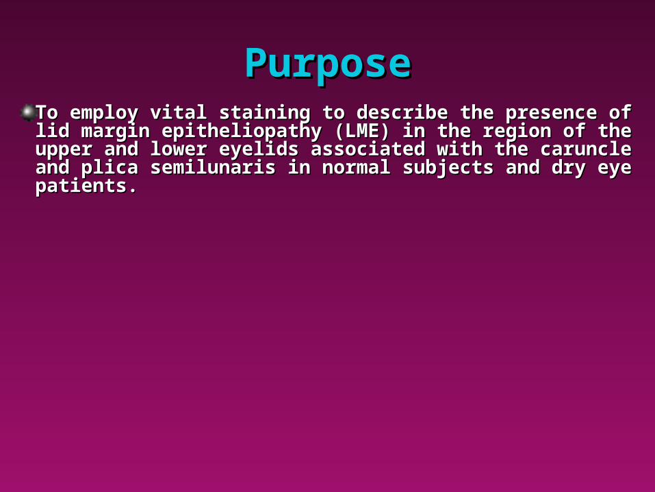
PurposePurposeTo employ vital staining to describe the presence of To employ vital staining to describe the presence of lid margin epitheliopathy (LME) in the region of the lid margin epitheliopathy (LME) in the region of the upper and lower eyelids associated with the caruncle upper and lower eyelids associated with the caruncle and plica semilunaris in normal subjects and dry eye and plica semilunaris in normal subjects and dry eye patients.patients.
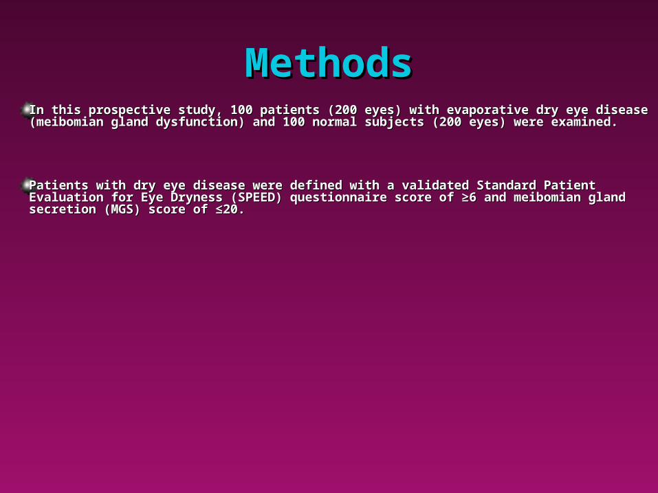
MethodsMethodsIn this prospective study, 100 patients (200 eyes) with evaporative dry eye disease In this prospective study, 100 patients (200 eyes) with evaporative dry eye disease (meibomian gland dysfunction) and 100 normal subjects (200 eyes) were examined.(meibomian gland dysfunction) and 100 normal subjects (200 eyes) were examined.
Patients with dry eye disease were defined with a validated Standard Patient Patients with dry eye disease were defined with a validated Standard Patient Evaluation for Eye Dryness (SPEED) questionnaire score of ≥6 and meibomian gland Evaluation for Eye Dryness (SPEED) questionnaire score of ≥6 and meibomian gland secretion (MGS) score of ≤20.secretion (MGS) score of ≤20.
MethodsMethodsFor the purposes of this study, the region of the eyelid For the purposes of this study, the region of the eyelid margin associated with the caruncle was defined as margin associated with the caruncle was defined as that area of the upper and lower eyelid margin medial that area of the upper and lower eyelid margin medial to the puncta and extending to the medial canthus. to the puncta and extending to the medial canthus.
BiomicroscopyBiomicroscopyBiomicroscopy of upper and lower eyelid margins from Biomicroscopy of upper and lower eyelid margins from the puncta to the medial canthus was performed at the puncta to the medial canthus was performed at 25x and 40x magnification 90 sec after instillation of 25x and 40x magnification 90 sec after instillation of Lissamine green vital stain into the conjunctival sac.Lissamine green vital stain into the conjunctival sac.
Lissamine Green Lissamine Green StainingStaining
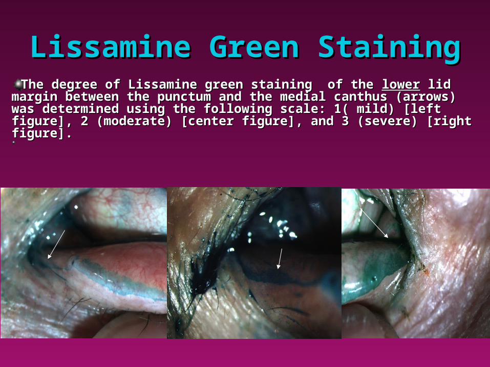
The degree of Lissamine green staining of the The degree of Lissamine green staining of the lowerlower lid lid margin between the punctum and the medial canthus (arrows) margin between the punctum and the medial canthus (arrows) was determined using the following scale: 1( mild) [left was determined using the following scale: 1( mild) [left figure], 2 (moderate) [center figure], and 3 (severe) [right figure], 2 (moderate) [center figure], and 3 (severe) [right figure].figure].
Lissamine Green Lissamine Green StainingStaining
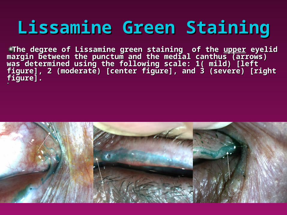
The degree of Lissamine green staining of the The degree of Lissamine green staining of the upperupper eyelid eyelid margin between the punctum and the medial canthus (arrows) margin between the punctum and the medial canthus (arrows) was determined using the following scale: 1( mild) [left was determined using the following scale: 1( mild) [left figure], 2 (moderate) [center figure], and 3 (severe) [right figure], 2 (moderate) [center figure], and 3 (severe) [right figure].figure].
ResultsResultsEssentially Essentially allall normal subjects and dry eye patients exhibited normal subjects and dry eye patients exhibited staining in this caruncular zone between the puncta and the staining in this caruncular zone between the puncta and the medial canthus, although the degree of staining in the area of medial canthus, although the degree of staining in the area of the lower lid margin was significantly greater in patients with the lower lid margin was significantly greater in patients with dry eye disease (p<0.05).dry eye disease (p<0.05).
ResultsResults
Interestingly there was a significant difference between right and left eyes Interestingly there was a significant difference between right and left eyes (p<0.5) in both normal subjects and dry eye patients with the left eye (p<0.5) in both normal subjects and dry eye patients with the left eye exhibiting a greater degree of staining than the right eye.exhibiting a greater degree of staining than the right eye.
The most significant difference between dry eye patients and controls is that The most significant difference between dry eye patients and controls is that the degree of staining in the lower eyelid margin associated with the the degree of staining in the lower eyelid margin associated with the caruncular zone is more intense in dry eye patients than controls (p<0.05).caruncular zone is more intense in dry eye patients than controls (p<0.05).
ConclusionsConclusionsIn contrast to prior results documenting a lack of correlation between vital staining in In contrast to prior results documenting a lack of correlation between vital staining in temporal regions and dry eye symptoms,temporal regions and dry eye symptoms,11 symptomatic dry eye patients have more symptomatic dry eye patients have more eyelid margin staining in the caruncular region than normal subjects.eyelid margin staining in the caruncular region than normal subjects.
Vital staining of this clinically-overlooked region of the eyelid margin (although Vital staining of this clinically-overlooked region of the eyelid margin (although anatomically comprising >10% of the eyelid margin) may have potential benefits as a anatomically comprising >10% of the eyelid margin) may have potential benefits as a marker and an additional diagnostic tool that may be particularly informative in those marker and an additional diagnostic tool that may be particularly informative in those patients with a questionable presentation of dry eye.patients with a questionable presentation of dry eye.
1Greiner JV: Association of epitheliopathy of upper and lower eyelid marginal conjunctiva with meibomian gland dysfunction and symptoms of dry-eye disease. ASCRS, April 27, 2014.
Epitheliopathy in the Region of Epitheliopathy in the Region of the Eyelid Associated with the the Eyelid Associated with the
Caruncle in Normal Subjects and Caruncle in Normal Subjects and Dry-Eye PatientsDry-Eye Patients
Jack V. Greiner, M.S., D.O., Ph.D.Jack V. Greiner, M.S., D.O., Ph.D.Paula J. Oliver, A.S., Rina WuPaula J. Oliver, A.S., Rina Wu
Mikhail Salganik, Ph.D.Mikhail Salganik, Ph.D.
The Schepens Eye Research InstituteMassachusetts Eye and Ear Infirmary
Harvard Medical SchoolThe Boston Ocular Surface Center