Embed Size (px)
Citation preview

1
EPITHELIAL TISSUE
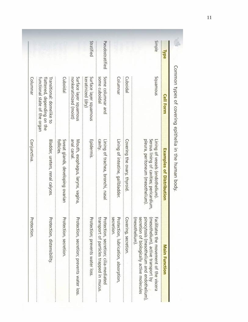
Epithelial (Covering) Tissue (See Figure EP 1- 10): Epithelial tissue covers the external and internal surfaces, including cavities and tubes of the body. Three different shapes of cells are encountered in the epithelium. These are:
1. Squamous (Flat) cells 2. Cuboidal cells and 3. Columnar cells.
These three cell types could be arranged in: • A single layer to form Simple epithelium e.g. the lining of the stomach or in • Several layers to form a Compound (Stratified) epithelium e.g. the epidermis of the
skin.
Other examples of simple epithelium include: 1. Mucosa of the intestine 2. Ependymal lining of the ventricles of the brain 3. The lining of the lumen of the kidney tubules 4. The lining of blood vessels (Endothelium)
Other examples of stratified epithelium are: 1. Mucosa of the mouth and oesophagus 2. Epidermis of the skin 3. Mucosa of the urinary bladder (Transitional) 4. Mucosa of the respiratory tract (Pseudostratified)

2
FIGURE EP 1.

3
Glands ((Figure EP2): Glands are epithelial structures/derivatives involved in secretion of substances. They are often derived by invagination of the surface epithelium and subsequent projection into the underlying connective tissue. A glandular structures which maintains it connection to the surface from which it was formed and secretes its product unto the surface is called an Exocrine Gland. (examples are: the liver, the Pancreas, Salivary glands, Sweat glands, Gastric glands). Glands which detach from their surface of origin and secrete their product directly into the blood circulatory system are referred to as Endocrine Glands. (examples are: the Pituitary, Pineal, Thyroid and Suprarenal glands). Figure EP 2
Epithelial cells interactions: Adjacent cells of the epithelium interact in various ways. Consequently several modifications occur on the contacting surfaces to facilitate cellular interaction. Certain transmembrane binding proteins (Cadherins) also participate in the establishment of these contact points

4
amongst epithelial cells. The functions of the modifications/adaptations on the cellular surfaces include:
• Communications • Adhesion • Protection • Movement of materials on the epithelial surfaces • Cell motility
TYPES OF INTERRACTIONS
Cellular Interdigitations These are interlocking membrane folds of adjacent cells, often located on the lateral surface close to the basal aspect of the cells. They facilitate intercellular adhesion.
Tight Junctions (Zonulae occludens): These are membranous junctions closest to the apex of the cells. A tight junction forms a continuous band around the cell. It is characterized by complete fusion of the adjacent membranes with closure of the intercellular space between adjacent cells. Tight junctions seal up the spaces between cells so as to prevent the passage of materials between the contacting cells.

5
Zonulae Adherens: These are also membranous junctions often located beneath tight junctions. They also form a continuous circular band around the cell. They are characterized by a thickening on the cytoplasmic surface of the membranes into which is inserted actin filaments. This junction provides for the adhesion of one cell to neighboring cells and for structural reinforcement of zonulae occludens. Desmosomes (Spot-Adhering Junction or Macula Adherens): These are membranous junctions which appear as small circular patches around the circumference of the epithelial cell immediately deep to the zonula adherens junction. The gap between adhering membranes is usually about 30nm. Desmosomes hold cells together firmly. They are commonly associated with columnar epithelial cells. Hemidesmosomes are desmosomal junctions between the basal surface of a cell and its basal lamina Gap Junctions (Communicating Junctions): These are also membranous junctions which facilitate communication between contacting cells by allowing the passage of metabolically active substances. The intercellular space between contacting cells is reduced to about 2nm. The junction is further characterized by the presence of six rods of protein subunits (Connexins) surrounding a channel which traverses the thickness of the membrane. These structures which are called Connexon form hydrophilic communication between adjacent cells. These junctions permit exchange between cells of molecules with molecular mass less than 1500 Da (dalton). E.g. hormones, cyclic AMP etc. As in the heart where they are very prominent, gap junctions facilitate highly coordinated cellular actions.

6
Figure EP 3

7
Figure EP 4
Figure EP 5

8
Figure EP 6
Fig.EP7
Figure EP 8

9
Figure EP 9
Figure EP 10

10
Specialization of the Apical and basal cellular surfaces: The apical surfaces of epithelial cells are modified in certain areas in order to facilitate movement of substances on the epithelial surface or to increase the surface area of the epithelial lining. These structures are formed by projections of the cytoplasm and include: Microvilli: These are small fingerlike cytoplasmic projections of the apical surface of cells lining absorptive surfaces. They measure about 1 micron in length and about 0.08 micron in diameter. The core of microvilial projections is filled with clusters of actin filaments. Microvilli and surface glycocalyx combine to form what is referred to as Brush or striated border of absorptive epithelial lining such as in the small intestine and the proximal convoluted tubules in the kidney. Microvilli serve to increase the surface area of the epithelial lining, thus facilitating absorption. Cilia and Flagella: Cilia are cylindrical, motile cytoplasmic apical projections of cells lining surface along which materials are transported. Cilia measure between 5-10 micron in length and about 0.2 micron in diameter. The core of cilium is filled with microtubules while the base is attached to small cylindrical structures called basal bodies. Basal bodies have structures similar to Centrioles. Cilia facilitate movement of substances along the epithelial surface usually in one direction. Flagella are similar in structure to cilia except that they are much longer than cilia. Furthermore while there could be as many as 250 cilia per cell, there is usually only one flagellum per cell as in the case of the spermatozoa. Flagella facilitate cell motility. Stereocilia: Stereocilia are similar in structure with microvilli except that they are much longer and restricted to the lining of the epididymis and vas deferens and the receptor hair cells of the auditory and vestibular system in the inner ear. They are nonmotile structure which increase epithelial surface area and facilitate movement into and out of the cells.
11