Embed Size (px)
Citation preview
CHAPTER FOUR
Epithelial Stem Cells in Adult SkinAna Mafalda Baptista Tadeu, Valerie Horsley1Department of Molecular, Cell and Developmental Biology, Yale University, New Haven, Connecticut, USA1Corresponding author: e-mail address: [email protected]
Contents
1.
CurISShttp
Introduction
rent Topics in Developmental Biology, Volume 107 # 2014 Elsevier Inc.N 0070-2153 All rights reserved.://dx.doi.org/10.1016/B978-0-12-416022-4.00004-4
109
2. Stem Cells in the Interfollicular Epidermis 111 3. Stem Cells in the Pilosebaceous Unit 114 4. Stem Cells in the Sweat Gland 116 5. Components of Adult Stem Cell Niches in the Skin 1175.1
Intrinsic regulation of stem cell function 117 5.2 Cell extrinsic regulation of SC function 1206.
Stem Cells in Epithelial Skin Cancers 121 7. Concluding Remarks 124 Acknowledgments 124 References 125Abstract
The skin is the first line of defense against dehydration and external environmentalaggressions. It constantly renews itself throughout adult life mainly due to the activityof tissue-specific stem cells. In this review, we discuss fundamental characteristics ofdifferent stem cell populations within the skin and how they are able to contribute tonormal skin homeostasis. We also examine the most recent results regarding thecell-intrinsic and -extrinsic components of the stem cell niche within the adult skin epi-thelium. Finally, we address the recent efforts to understand how abnormal regulation ofstem cell activity contributes to the initiation and progression of skin-associated cancers.
1. INTRODUCTION
The skin serves as a highly dynamic and adaptable outer coating for the
bodies of many animal species, protecting against the external environment
and providing tactile function for touch sensation. These roles are engen-
dered by multiple types of differentiated cells within the interfollicular epi-
dermis (IFE) and by the formation of epidermal appendages such as hair
109
110 Ana Mafalda Baptista Tadeu and Valerie Horsley
follicles (HFs), sebaceous glands (SGs), and sweat glands. In order for organ-
isms to navigate a continuously changing external environment, specialized
cell types in the skin continually regenerate through the action of several
distinct epithelial stem cell (SC) populations that self-renew and generate
cells with unipotent and multipotent differentiation potential (Lee &
Tumbar, 2012; Sennett & Rendl, 2012).
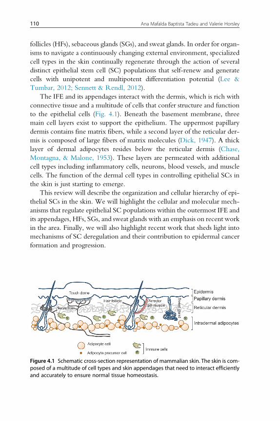
The IFE and its appendages interact with the dermis, which is rich with
connective tissue and a multitude of cells that confer structure and function
to the epithelial cells (Fig. 4.1). Beneath the basement membrane, three
main cell layers exist to support the epithelium. The uppermost papillary
dermis contains fine matrix fibers, while a second layer of the reticular der-
mis is composed of large fibers of matrix molecules (Dick, 1947). A thick
layer of dermal adipocytes resides below the reticular dermis (Chase,
Montagna, & Malone, 1953). These layers are permeated with additional
cell types including inflammatory cells, neurons, blood vessels, and muscle
cells. The function of the dermal cell types in controlling epithelial SCs in
the skin is just starting to emerge.
This review will describe the organization and cellular hierarchy of epi-
thelial SCs in the skin. We will highlight the cellular and molecular mech-
anisms that regulate epithelial SC populations within the outermost IFE and
its appendages, HFs, SGs, and sweat glands with an emphasis on recent work
in the area. Finally, we will also highlight recent work that sheds light into
mechanisms of SC deregulation and their contribution to epidermal cancer
formation and progression.
Figure 4.1 Schematic cross-section representation of mammalian skin. The skin is com-posed of a multitude of cell types and skin appendages that need to interact efficientlyand accurately to ensure normal tissue homeostasis.
111Epithelial Stem Cells in Adult Skin
2. STEM CELLS IN THE INTERFOLLICULAR EPIDERMIS
The outermost layer of mammalian skin is comprised of a multilayered
or stratified epidermis of the IFE that is anchored to the underlying papillary
dermis via integrin-mediated adhesion to a basement membrane (reviewed
in Blanpain & Fuchs, 2006).The epidermal cells that adhere to the basement
membrane are proliferative keratinocytes of the basal layer. Epidermal
keratinocytes are formed during embryonic development from the surface
ectoderm and generate differentiated suprabasal cells through asymmetric
cell divisions (Lechler & Fuchs, 2005). Cells in the outermost epidermal
layer (stratum corneum) tightly adhere to one other and form a protein–lipid
matrix that ultimately creates the skin’s essential barrier (reviewed in
Sandilands, Sutherland, Irvine, & McLean, 2009). The cells of the stratum
corneum are constantly shed and thus, proliferative basal cells fuel the con-
tinual reformation of these dedicated cells of the IFE.
Classic experiments analyzing IFE homeostasis via morphology and pro-
liferation proposed the existence of an epidermal proliferative unit (EPU) in
which a central slow-cycling basal cell generates a defined number of rapidly
dividing progenitor cells that differentiate into a restricted number of “units”
(Loeffler, Potten, & Wichmann, 1987; Mackenzie, 1969, 1970; Potten,
1981; Potten,Wichmann, Loeffler, Dobek, &Major, 1982). More recently,
extensive and quantitative analyses of basal cell progeny using genetic lineage
tracing was performed in several mouse models (Clayton et al., 2007;
Doupe, Klein, Simons, & Jones, 2010; Mascre et al., 2012).The ground-
breaking initial studies used mouse models expressing tamoxifen-regulated
cre recombinase driven by an inducible CYP1A1 promoter (AhcreERT),
crossed to a YFP reporter strain (Clayton et al., 2007; Doupe et al.,
2010). Low-dose tamoxifen administration allowed single-cell labeling
within the tail and ear IFE and the ability to follow clone generation long
term. Interestingly, the average size of persisting clones increased linearly
with time, which is contrary to the previously proposed restricted size of
the EPU. Furthermore, mathematic analysis of the clone generation in these
studies suggested that basal cells could generate proliferative or differentiated
progeny stochastically. However, whether these experiments labeled the
most primitive SC within the IFE was unclear.
More recently, comparing lineage tracing in the IFE of mouse models
expressing either an inducible CreER driven by the keratin 14 (K14) pro-
moter or a fragment of the Involucrin (Inv) promoter reveal a hieracherical
112 Ana Mafalda Baptista Tadeu and Valerie Horsley
and heterogeneous nature of progenitor cells in the IFE (Mascre et al., 2012).
In the InvCreER mouse model, persistent labeled clones followed the same
cell-fate dynamics and linear growth patterns as the clones generated in the
AhCre model (Clayton et al., 2007; Doupe et al., 2010). By contrast, the
persistent clones generated in the K14CreER mouse model displayed sto-
chastic fate decisions but were restricted in their growth potential, consistent
with an EPU-type model and supporting the existence of a slow-cycling SC
within the IFE. Molecular characterization of basal cells labeled in the
K14CreER and InvCreER mouse models further supported the labeling
of two distinct cell types within the IFE. Together, these studies reveal a
hierarchy of cells within the IFE: a slow-cycling SC (marked by K14CreER)
that gives rise to more rapidly cycling committed progenitors (marked by
AhCreER or InvCreER) that subsequently undergo terminal differentiation
(Fig. 4.2). These studies provide significant knowledge regarding the molec-
ular characteristics of the slow-cycling IFE progenitors that will allow the
future identification of novel markers as well as the further analysis of the
molecular regulation of these cells during skin homeostasis.
Heterogeneity also exists within the slow-cycling SCs of the IFE.Within
the majority of the murine epidermis, orthokeratotic differentiation gener-
ates cells that are spinous, granular, and have lost nuclei in the stratum cor-
neum (Didierjean, Wrench, & Saurat, 1983; Schweizer & Marks, 1977). In
the tail, however, IFE regions between the organized arrays of HFs postna-
tally develop a parakeratotic program postnatally, resulting in IFE regions
lacking the granular layer and retention of nuclei within the stratum cor-
neum (Didierjean et al., 1983; Schweizer & Marks, 1977). Careful genetic
lineage analysis of the clonogenic behavior of K14CreER marked cells in
these different regions revealed that IFE SCs are restricted to a particular
compartment and display distinct proliferative behavior, suggesting that het-
erogeneous, unipotent SCs exist within the IFE (Gomez, Chua, Miremadi,
Quist, & Headon, 2013).
Within the adult IFE, a separate epithelial SC niche exists for Merkel
cells, specialized sensory cells that allow mammals to respond to mechanical
stimuli during touch sensations (Fig. 4.2). Mature Merkel cells reside in
touch domes, which are clusters of cells that are innervated by afferent
somatosensory nerve fibers at the dermal–epidermal border in specialized
skin regions (Merkel, 1875). These unique cells express both cytokeratins
and neuroendocrine proteins and thus their developmental origin was
unclear until several recent studies demonstrated that these cells derive from
K14 expressing cells of the developing epidermis (Morrison, Miesegaes,
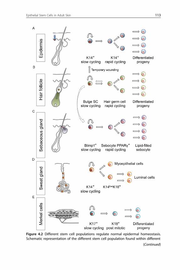
Figure 4.2 Different stem cell populations regulate normal epidermal homeostasis.Schematic representation of the different stem cell population found within different
(Continued)
113Epithelial Stem Cells in Adult Skin
114 Ana Mafalda Baptista Tadeu and Valerie Horsley
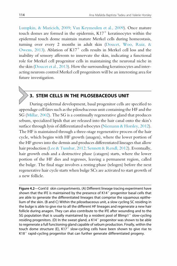
Lumpkin, & Maricich, 2009; Van Keymeulen et al., 2009). Once mature
touch domes are formed in the epidermis, K17þ keratinocytes within the
epidermal touch dome maintain mature Merkel cells during homeostasis,
turning over every 2 months in adult skin (Doucet, Woo, Ruiz, &
Owens, 2013). Ablation of K17þ cells results in Merkel cell loss and the
inability of sensory afferents to innervate the skin, indicating a functional
role for Merkel cell progenitor cells in maintaining the neuronal niche in
the skin (Doucet et al., 2013). How the surrounding keratinocytes and inter-
acting neurons control Merkel cell progenitors will be an interesting area for
future investigation.
3. STEM CELLS IN THE PILOSEBACEOUS UNIT
During epidermal development, basal progenitor cells are specified to
appendage cell fates such as the pilosebaceous unit containing theHF and the
SG (Millar, 2002). The SG is a continually regenerative gland that produces
sebum, specialized lipids that are released into the hair canal onto the skin’s
surface through lysis of differentiated sebocytes (Niemann &Horsley, 2012).
The HF is maintained through a three-stage regenerative process of the hair
cycle, which begins with HF growth (anagen), where the lower portion of
the HF grows into the dermis and produces differentiated lineages that allow
hair production (Lee & Tumbar, 2012; Sennett &Rendl, 2012). Eventually,
hair growth ends and a destructive phase (catagen) starts, where the lower
portion of the HF dies and regresses, leaving a permanent region, called
the bulge. The final stage involves a resting phase (telogen) before the next
regenerative hair cycle starts when bulge SCs are activated to start growth of
a new follicle.
Figure 4.2—Cont'd skin compartments. (A) Different lineage tracing experiment haveshown that the IFE is maintained by the presence of K14þ progenitor basal cells thatare able to generate the differentiated lineages that comprise the squamous epithe-lium of the skin. (B and C) Within the pilosebaceous unit, a slow cycling SC residing inthe bulge is able to give rise to all the different HF lineages and regenerate a new hairfollicle during anagen. They can also contribute to the IFE after wounding and to theSG population that is usually maintained by a resident pool of Blimp1þ slow-cyclingresiding progenitors. (D) In the sweat gland, a K14þ progenitor was shown to be ableto regenerate a full functioning gland capable of sebum production. Finally, within thetouch dome structure (E), K17þ slow-cycling cells have been shown to give rise toK18þ rapid-cycling progenitor that can further generate differentiated progeny.
115Epithelial Stem Cells in Adult Skin
The initial identification of the SC properties of bulge cells took advan-
tage of their slow-cycling nature, which allowed retention of nucleotide
analogs such as BrdU and tritiated thymidine during pulse-chase experi-
ments (Bickenbach, 1981; Cotsarelis, Sun, & Lavker, 1990). Innovative
genetic mouse models were also developed that allowed fluorescent labeling
of histone H2B in epithelial cells and after a lengthy chase period, bulge cells
were the primary cell-type labeled within the skin (Tumbar et al., 2004).
These experiments demonstrated that the cell cycle of bulge cells was slower
than the other epithelial cells of the skin.
Genetic lineage tracing experiments using multiple mouse models with
cell-type-specific expression of cre recombinase have demonstrated that sev-
eral populations of cells within the pilosebaceous unit have the capacity to
generate lineages of the HF and SG (Fig. 4.2) (Jaks et al., 2008; Jensen et al.,
2009;Morris et al., 2004; Petersson et al., 2011; Snippert et al., 2010), as well
as the epidermis after wounding (Ito et al., 2005; Levy, Lindon, Zheng,
Harfe, & Morgan, 2007; Snippert et al., 2010). The isolation and trans-
plantation of putative follicular SCs based on microdissection or based
on fluorescence-activated cell sorting for bulge markers such CD34 and
a6-integrin also demonstrated the multipotency of SCs within the HF
bulge (Blanpain, Lowry, Geoghegan, Polak, & Fuchs, 2004; Claudinot,
Nicolas, Oshima, Rochat, & Barrandon, 2005). In sum, these studies have
demonstrated that bulge cells are multipotent, contributing temporarily to
the epidermis during wounding and primarily to the homeostasis of HF
and SG.
The activation of HF SCs during hair cycling involves several cellular
processes including proliferation and migration of bulge cells and hair germ
cells (Greco, Chen, Rendl, Schober, & Pasolli, 2009; Rompolas et al., 2012;
Zhang, Cheong, Ciapurin, McDermitt, & Tumbar, 2009). The activated
SCs generate proliferative progeny and differentiate to form the inner root
sheath and the HF shaft (Ito et al., 2005; Oshima, Rochat, Kedzia,
Kobayashi, & Barrandon, 2001; Tumbar et al., 2004).
Similar to the heterogeneity in IFE SCs (Gomez et al., 2013), the per-
manent portion of the HF contains heterogeneous group of cells with dif-
fering capacities for contributing to homeostasis of epithelial lineages of the
skin (Goldstein & Horsley, 2012). At the junction between the IFE and HF,
cells expressing SC antigen-1 and/or leucine-rich repeats and
immunoglobulin-like domain 1 (Lrig1) exist and are able to generate IFE
and SG cells but are limited in their contribution to the HF lineage
( Jensen et al., 2009, 2008). Below the junctional zone and at the base of
116 Ana Mafalda Baptista Tadeu and Valerie Horsley
the SG, several slow-cycling cells exist such as MTS24 expressing cells
(Nijhof et al., 2006) and also leucine-rich repeat containing G protein-
coupled receptor 6 or B-lymphocyte-induced maturation protein 1
(Blimp1) positive cells that can generate SG lineages (Horsley et al., 2006;
Snippert et al., 2010). The cells that primarily generate HF lineages are
located at the base of the isthmus and throughout the CD34þ, K15þ region
of the bulge (Blanpain et al., 2004; Brownell, Guevara, Bai, Loomis, &
Joyner, 2011; Morris et al., 2004; Snippert et al., 2010; Tumbar et al.,
2004). At the base of this compartment and within the hair germ,
leucine-rich repeat containing G protein-coupled receptor 5 (Lgr5) positive
cells can give rise to different HF lineages (Jaks et al., 2008). These
populations are not restricted but dynamically interact: K15þ bulge cells
can migrate to the SG where they locally self-renew and continue to con-
tribute to the normal gland homeostasis (Petersson et al., 2011). Addition-
ally, Lgr5þdescendants of bulge cells can repopulate the bulge at the end of
hair HF regression (Hsu, Pasolli, & Fuchs, 2011).
4. STEM CELLS IN THE SWEAT GLAND
Eccrine sweat glands are an additional epidermal appendage that gen-
erate sweat for mammalian thermoregulation. Sweat fluids are generated by
specialized epithelial cells in a coiled structure and are secreted into luminal
structures into a duct that opens directly onto the skin surface (Lobitz &
Dobson, 1961). Similar to the mammary gland, the secretory coil of the
sweat gland contains an outer basal layer of myoepithelial cells that express
K5, K14, and smooth muscle actin. An inner suprabasal layer of luminal cells
is positive for K8, K18, and K19 (Langbein et al., 2005; Moll & Moll, 1992;
Schon, Benwood, O’Connell-Willstaedt, & Rheinwald, 1999). In contrast
to the dramatic cellular changes during puberty and pregnancy in the mam-
mary gland (Richert, Schwertfeger, Ryder, & Anderson, 2000), the sweat
gland presents little sign of continual renewal (Lu et al., 2012).
Initial experiments to address the existence of sweat gland progenitors
identified cell divisions in basal cells of the gland’s duct region (Lobitz,
Holyoke, & Montagna, 1954). Other studies analyzed the regenerative
potential of sweat glands in vitro by showing proliferation of dissociated gland
cells in culture and in vivo by demonstrating the contribution of sweat gland
for epidermal reconstitution after superficial skin injuries (Biedermann et al.,
2010; Miller, Burke, Rader, Coulombe, & Lavker, 1998). Recent work has
identified K14þ progenitors that can differentiate into myoepithelial cells
117Epithelial Stem Cells in Adult Skin
and can also stratify to form the suprabasal layer of K14low/K18þ cell that
give rise to luminal cells (Lu et al., 2012) (Fig. 4.2). Furthermore, lineage
tracing and EdU pulse-chase experiments were able to show that both sweat
gland progenitor cell types participate in repair of the epidermis after
wounding but the sweat gland itself remains relatively quiescent throughout
the wound repair process (Lu et al., 2012). The plasticity of sweat gland pro-
genitor cells was illustrated by transplantation studies in which progenitor
cells were injected into cleared mammary fat pads and were able to recon-
stitute an entire sweat gland. By contrast, luminal cells were unable to con-
tribute to de novo formation of sweat glands (Lu et al., 2012). Further studies
will be necessary to define the molecular mechanisms that regulate sweat
glands homeostasis and their contribution to epidermal healing.
5. COMPONENTS OF ADULT STEM CELL NICHESIN THE SKIN
The identification, isolation, and characterization of different
populations of SCs within the adult skin epithelium identified layers of
molecular mechanisms that act intrinsically to regulate SC behavior. Recent
reviews have discussed several studies that identify molecular mechanisms
that regulate SC behavior in the skin (Arwert, Hoste, & Watt, 2012;
Blanpain & Fuchs, 2006; Hsu et al., 2011; Lee & Dai, 2013; Lee &
Tumbar, 2012). Several signaling pathways are involved in regulating SC
behavior in the skin including Wnt, BMP, FGF, and PDGF (Festa et al.,
2011; Greco et al., 2009; Kandyba et al., 2013; Kobielak, Stokes, de la
Cruz, Polak, & Fuchset, 2007; Myung, Takeo, Ito, & Atit, 2013;
Oshimori & Fuchs, 2012; Plikus et al., 2008). In addition, the involvement
of dermal cell types has recently come to light (Goldstein & Horsley, 2012).
Here, we will highlight recent developments regarding the intrinsic and
extrinsic regulation of epithelial SCs.
5.1. Intrinsic regulation of stem cell functionCellular metabolism and environmental assaults can result in DNA damage,
especially in the skin, which directly receives UV irradiation from the sun
and environmental mutagens that can induce genomic instability. Interest-
ingly, HFSCs are more resistant to radiation-induced damage compared to
other epithelial skin cells (Sotiropoulou et al., 2010). To attain this resistance,
HFSCs express high levels of the antiapoptotic protein B-cell lymphoma
2 (Bcl2) and transiently express p53 to promote survival. In addition,
118 Ana Mafalda Baptista Tadeu and Valerie Horsley
breast cancer 1 (Brca1) is essential for DNA damage repair
(Gudmundsdottir & Ashworth, 2006; Moynahan & Jasin, 2010) and epider-
mal deletion of Brca1 leads to defects in HF formation as well as induction of
caspase-dependent apoptosis that leads to hyperproliferation and subsequent
exhaustion of adult SCs (Sotiropoulou et al., 2013). The differential regu-
lation of DNA damage in SCs also exists within other tissues (Mandal,
Blanpain, & Rossi, 2011) and may be similar to the mechanisms that act
in SCs in the IFE and other epidermal appendages.
Several transcriptional regulators of SC function in the HF have been
identified and are shared among SCs of other tissues including transcription
factor 3 and 4 (TCF3/4), nuclear factor of activated T-cells 1 (NFATc1) and
sex determining region Y-box 9 (Sox9) (Blanpain & Fuchs, 2006; Nguyen
et al., 2009; Nguyen, Rendl, & Fuchs, 2006; Nowak, Polak, Pasolli, &
Fuchs, 2008). In addition, Lgr5 (Barker et al., 2007) and the atypical
HOP homeobox protein Hopx are expressed by an intestinal SC epithelial
pool at the base of the crypt (Takeda, Jain, LeBoeuf, Wang, & Lu, 2011). In
the HF, Hopx is expressed within bulge cells and can contribute to all HF
lineages upon HF growth as well as to IFE cells upon wounding (Takeda,
Jain, LeBoeuf, & Padmanabhan, 2013). Lower bulge cells expressing the
SC marker Lgr5 also express Hopx, are able to escape apoptosis during
the HF death phase and contribute long-term to bulge cell maintenance
(Takeda et al., 2013).
The transcription factor LIM homeobox protein 2 (Lhx2) is another
homeobox protein that has been implicated in regulating morphogenesis
and patterning of ectodermal derivatives and in SC maintenance and quies-
cence within the HF SC niche (Mardaryev et al., 2011; Rhee, Polak, &
Fuchs, 2006; Tornqvist, Sandberg, Hagglund, & Carlsson, 2010). Lhx2 is
expressed in the bulge and secondary hair germ where it co-localizes with
the SCmarkers Sox9, Tcf4, and Lgr5. In response to skin injury, Lhx2þ cells
within the bulge and secondary hair germ proliferate and contribute to skin
re-epithelialization via positive regulation of Sox9 and Tcf4 while inhibiting
HF cycling through negatively regulating Lgr5 (Mardaryev et al., 2011).
These and many other studies have provided novel insights of how Wnt
and BMP signaling pathways and transcriptional regulation networks mod-
ulate activity of epithelial SCs during normal homeostasis and in response to
injury (Blanpain & Fuchs, 2006; Lee & Tumbar, 2012; Sennett &
Rendl, 2012).
Epithelial SCs are also regulated post-transcriptionally and translationally
in part by microRNAs (miRNAs), which are small noncoding RNAs that
119Epithelial Stem Cells in Adult Skin
alter RNA translation or stability to control gene expression. Complete
ablation of miRNA production by deletion of the upstream processing
enzyme Dicer in mice results in perinatal lethality and severe HF defects
(Andl et al., 2006; Yi et al., 2006). Among these defects are undeveloped
and misaligned HFs, increased apoptosis and lack of K15þ and CD34þ cells
within the bulge compartment suggesting that miRNAs, in general, are
important for HF SC maintenance (Andl et al., 2006).
Several miRNAs are spatiotemporally regulated within the IFE and the
HFSCs. MiR203 was shown to be preferentially enriched in the IFE versus
the HF (Andl et al., 2006; Yi, Poy, Stoffel, & Fuchs, 2008) and is sufficient to
promote IFE differentiation and suppress self-renewal in the IFE by control-
ling the expression of p63 (Andl et al., 2006; Yi et al., 2008).Additionally,
miR203 is transcriptionally activated during asymmetric cell division in the
developing epidermis, localizing to the differentiated daughter cell, where it
promotes cell cycle exit and abolishes self-renewal in a process involving
co-suppression of p63, S-phase kinase-associated protein 2 (Skp2), and
musashi RNA-binding protein 2 (Msi2) (Jackson et al., 2013).
An additional miRNA, miR125b is sufficient to alter IFE homeostasis
and abrogate hair specification (Zhang, Stokes, Polak, & Fuchs, 2011).
MiR31 can also alter HFSC activity by targeting fibroblast growth factor
10 (Fgf10), distal-less homeobox 3 (Dlx3), several keratin genes and also
components of the Wnt and BMP signaling pathways (Mardaryev et al.,
2010). The differential regulation of several miRNAs in the epithelium
of the skin suggests that roles for additional miRNAs will be defined as this
burgeoning field continues to expand.
Another level of regulation of skin SCs occurs through modification of
histones and DNA to epigenetically regulate transcription (Calo &
Wysocka, 2013). Several epigenetic factors play a role in epidermal differ-
entiation (Mulder et al., 2012). Histone acetylation andmethylation through
histone deacetylase and methyltransferase activity, respectively, regulate IFE
development (Driskell et al., 2012; LeBoeuf et al., 2010) and homeostasis
(Driskell et al., 2012). Maintenance of repressive histone modifications
via the polycomb repressor complex, enhancer of zeste homolog 1 (Ezh1)
and Ezh2 are essential for IFE differentiation and for HF morphogenesis
and maintenance (Bardot et al., 2013; Ezhkova et al., 2011). Merkel cells
also require Ezh2 proteins for their maintenance through the regulation
of the transcription factor Sox2 (Bardot et al., 2013). Histone methylation
controlled by the demethylase Jumonji domain containing 3 (JmjD3) is
essential for IFE differentiation (Sen, Webster, Barragan, Chang, & Khavari,
120 Ana Mafalda Baptista Tadeu and Valerie Horsley
2008), while the demethylase Jumonji/jmjc domain-containing protein 2
(Jarid2) is required to maintain IFE basal progenitors (Mejetta et al.,
2011). In addition, the DNA methyltransferase 1 (DNMT1) and the
ubiquitin like, containing PHD and RING finger domain-1 (UHRF1) are
expressed in basal cells and are downregulated once cells enter the differenti-
ation program suggesting that they are also involved in regulating
stemness. Deletion ofDNMT1 in human skin regeneration assays induced pre-
mature differentiation of progenitors and progressive tissue loss further demon-
strating its importance for self-renewal (Sen, Reuter, Webster, Zhu, &
Khavari, 2010).
Additional control of SC function occurs through the regulation of gene
expression by altering nucleosome positioning through the action of chro-
matin remodeling complexes such as the SWI/SNF complex (Kidder,
Palmer, & Knott, 2009). By rearranging nucleosome positions within the
chromatin, these complexes regulate RNA polymerase II occupancy and
thus transcriptional initiation in an ATP-dependent manner (Liu,
Balliano, & Hayes, 2011). At the crux of these complexes, brahma-related
gene 1 (Brg1) acts as a catalytic subunit and regulates SC proliferation and
differentiation. In the HF, it was recently shown that Brg1 is dynamically
activated after SC activation in the skin. Deletion of Brg1 with the
bulge-specific NFATc1-Cre induced precocious HF regression, loss of
HFSCs, and progressive hair loss (Xiong et al., 2013). Molecularly, Brg1
and Shh act in a molecular loop, where Brg1 regulates Shh expression
and Shh activates Brg1 expression within the follicle (Xiong et al., 2013).
Whether Brg1 regulates additional genes to control HFSC function will
be an interesting area of future investigation.
5.2. Cell extrinsic regulation of SC functionThe intrinsic regulation of epithelial SCs in the skin is influenced bymultiple
cell types, including follicle-associated melanocytes and other cells within
the dermis (Goldstein &Horsley, 2012). Of primary importance, the dermal
papillae (DP) is a mesenchymal cell population that abuts the HF, induces its
morphogenesis and remains associated with the follicle throughout its life
cycle (Driskell, Clavel, Rendl, & Watt, 2011). The association of the DP
with the follicle is essential for the activation of HF growth (Chi, Wu, &
Morgan, 2013; Rompolas et al., 2012) and the size of the DP can define
the size and shape of the hair follicle (Chi et al., 2013). Several signaling
ligands are expressed by the DP (Rendl, Lewis, & Fuchs, 2005) and the
121Epithelial Stem Cells in Adult Skin
identification of DP specific Cre lines (Enshell-Seijffers, Lindon,
Kashiwagi, & Morgan, 2010; Grisanti et al., 2013) will allow the identifica-
tion of the molecular mechanisms by which these cells control HFSC
activity.
Below the DP, a depot of dermal adipocytes displays dynamic changes in
size during the hair cycle. Both adipocyte hypertrophy and adipogenesis
contribute to the growth of the adipocyte depot in the skin. The production
of immature adipocyte precursor cells during adipogenesis is both necessary
and sufficient to drive hair cycling (Festa et al., 2011). Adipogenesis also
occurs after acute wounding and inhibition of adipogenesis can alter fibro-
blast function in the skin, leading to wound closure failure (Schmidt &
Horsley, 2013). Themolecular mechanisms by which adipocyte lineage cells
function in the skin will reveal novel components of the skin SC niche.
Permeating through the dermal cell layers, somatosensory nerve fibers
innervate the touch dome in the IFE and surround the HF in a piloneural
collar (Lumpkin, Marshall, & Nelson, 2010). The HF cells of each follicle
type may provide unknown cues for the distinct neural innervation. Inter-
estingly, each hair follicle type is innervated by distinct mechanoreceptors
which convergewithin the dorsal horn of the spinal column to process touch
sensations (Li et al., 2011). Afferents from the dorsal root ganglion produce
glutamate that is essential for the proper development of the piloneural
mechanoreceptors. The neural innervation also provides signals for the
HF, such as Shh and can promote the contribution of HF cells to wound
healing and SG homeostasis (Brownell et al., 2011).
Genome-wide association studies have implicated inflammatory cells in
the regulation of hair loss (Petukhova et al., 2010), suggesting that these cells
may be instrumental in SC function in the skin. Supporting a role for
immune cells in hair cycling, mice lacking gdT cells through deletion of
TCRd have defects in hair cycling (Kloepper, Kawai, Bertolini,
Kanekura, & Paus, 2013). Inflammatory cells also play many roles in wound
healing (Eming, Krieg, & Davidson, 2007), and gdT cells can induce HF
neogenesis from healed epithelial cells after wounding via the production
of Fgf9 (Gay et al., 2013). Functional roles of other immune cells may reveal
their functions in the control of additional epithelial SCs in the skin.
6. STEM CELLS IN EPITHELIAL SKIN CANCERS
Several types of epithelial cancers form in the skin. Papillomas, basal
cell carcinomas (BCCs), and squamous cell carcinomas (SCCs) are found in
122 Ana Mafalda Baptista Tadeu and Valerie Horsley
the IFE, while pilomatriomas, trichofolliculomas, and SG carcinomas are
associated with the pilosebaceous unit. Resident epidermal SCs have been
proposed to initiate epithelial tumorigenesis. Furthermore, maintenance of
tumors following initiation is thought to be driven by tumor cells with SC
characteristics such as self-renewal, and slow-cycling, properties (Al-Hajj &
Clarke, 2004).
BCCs comprise 80% of epithelial skin cancers and are often associated
with activating mutations in the Shh pathway via inactivation of the repres-
sive receptor Patched (Ptch), expression/activation of the transcriptional
mediators GLI family zinc finger (Gli) proteins, or activation of the Shh sig-
nal transducer, smoothened (Smo) (Athar, Tang, Lee, Kopelovich, & Kim,
2006; Rogers et al., 2010). The morphology of BCCs is variable, forming
various subtypes including superficial and nodular tumors, suggesting that
different cellular origins may exist.
Based on the similarities of keratin expression between BCCs and HF
progenitor cells (Asada, 1993) and the inability of BCCs to develop in
irradiated mice with only one allele of Ptch1 during HF SC quiescence
(Mancuso et al., 2006), it was initially hypothesized that HF progenitor
cells were the cells of origin for BCCs. However, genetic lineage-tracing
experiments in mice expressing an active Gli2 demonstrated that BCCs
can arise from several epithelial cell types including the bulge, SGs, and
IFE (Grachtchouk et al., 2011). Similarly, Lgr5þ bulge progeny required
wounding to generate BCC-like lesions upon genetic Gli1 activation
(Kasper et al., 2011). Expression of a constitutively active form of Smo in
a cell-type specific manner was shown to generate BCCs preferentially from
cells in the IFE and infundibulum rather than from bulge cells (Youssef et al.,
2010). Taken together, these data suggest that BCC may have multiple ori-
gins and that wounding promotes BCC expansion.
Abnormal Wnt signaling has been associated with the development of
epidermal tumors. In fact expression of a constitutively active form of
b-catenin promotes the development of pilomatricomas and
trichofolliculomas and mutations of lymphoid enhancer-binding factor 1
(lef1) that normally inactivates b-catenin is associated with human SG
tumors (Chan, Gat, McNiff, & Fuchs, 1999; Gat, DasGupta,
Degenstein, & Fuchs, 1998; Takeda et al., 2006). Inducible activation of
b-catenin under the K14 promoter lead to the formation of lesions similar
to pilomatricomas that regress when b-catenin is no longer activated
(Lo Celso, Prowse, & Watt, 2004). Furthermore, expression of DNLef1
in epidermis induced SG tumors (Niemann, Owens, Hulsken,
123Epithelial Stem Cells in Adult Skin
Birchmeier, &Watt, 2002; Niemann, Owens, Schettina, &Watt, 2007) and
ablation of b-catenin also under the K14 promoter caused a regression of
chemically induced papillomas (Malanchi et al., 2008). Different SC
populations react differently to Wnt signaling-associated tumor formation
stimuli. For example, sustained b-catenin activity under the K15 promoter
leads to an increase in proliferation, expression of Wnt target genes within
the bulge compartment, and inability to form pilomatricomas even when a
wounding stimulus is enforced, while sustained b-catenin activity under a
truncated K5 promoter (expressed in SG and HF bulb) causes conversion
of SG into HF structures that develop and resemble benign tumors
(Baker, Verstuyf, Jensen, & Watt, 2010).
SCCs encompass 20% of epithelia-derived tumors in the skin and
have metastatic potential following papilloma formation (Alam & Ratner,
2001). Induction of SCCs in mice can occur via UV irradiation,
chemical carcinogenesis protocols using 7,12-dimethylbenz(a)anthracene
(DMBA) to induce Ras mutations followed by promotion with 12-O-
tetradecanoylphorbol-13-acetate or genetic expression of oncogenes within
the epithelium. Clone analysis of human skin with p53 mutations suggested
that SCCs derived from the dermal–epidermal junction and from HFs
( Jonason et al., 1996). Similar results were obtained when a mutated acti-
vated form of Ras was expressed in the proliferative cells of the IFE and
HF (Brown, Strathdee, Bryson, Lambie, & Balmain, 1998).
To further elucidate the cellular origin of Ras induced SCCs, cell-type-
specific expression of mutant Kras was induced in mouse models controlled
by bulge-specific promoters, K15 or K19 (Lapouge et al., 2011;White et al.,
2011). Activation of Kras in the bulge led to papilloma formation, whereas
tumorigenesis was not induced when Kras mutations were targeted to the
transiently amplifying cells of the HF. Interestingly, activation of Kras in
InvCre-expressing cells also generated papillomas (Lapouge et al., 2011).
Since this promoter can drive expression in IFE progenitor cells (Mascre
et al., 2012), IFE cells may also contribute to SCC formation. The contri-
bution of bulge cells to papilloma formation was further confirmed by the
presence of progeny from K15þ bulge cells in murine papillomas after
induction of Ras mutations with DMBA (Kangsamaksin, Park,
Trempus, & Morris, 2007).
To determine if cells with SC potential exist within SCCs, transplanta-
tion assays of purified cells into immunocompromised mice have revealed
that CD34 or a6b1 integrin expression can enrich for tumor-initiating cells
in this assay (Malanchi et al., 2008; Schober & Fuchs, 2011). Importantly,
124 Ana Mafalda Baptista Tadeu and Valerie Horsley
ablation of TGFb signaling enhanced the proliferation of integrinhi,
CD34þ cells, and their tumorigenic potential (Schober & Fuchs, 2011).
By applying quantitative clonal analysis to SCC tumor growth in vivo,
Blanpain and colleagues found a minority of cells within papillomas with
dramatic growth potential (Driessens, Beck, Caauwe, Simons, & Blanpain,
2012). By contrast, metastatic SCCs displayed clones of cells with increased
replicative and abrogated differentiation potential. Interestingly, a hierarchical
organization of tumor cells also exists in benign intestinal adenomas and
metastatic brain tumors such as glioblastomas (Chen et al., 2012; Schepers
et al., 2012). In glioblastomas, when the highly proliferative progeny
of the cells are ablated with chemotherapeutic drugs, the other cells with
SC properties can repopulate the tumors. When these cells with more prim-
itive potential were selectively ablated with genetic tools in the presence of
chemotherapeutic drugs, tumor growth was significantly hampered.
Together, these studies provide strong evidence for the ability of cancer cells
to acquire SC properties to fuel tumorigenesis and suggest that targeting
both primitive and more developed cells within tumors will be important
for effective cancer therapies.
7. CONCLUDING REMARKS
Several advances have been made toward understanding the cellular
and molecular mechanisms that control epidermal SC quiescence and differ-
entiation in multiple lineages in the skin. Important progress achieved in
recent years to develop mouse models to target and disrupt distinct SC pools
within the epidermis have provided precious information that begins to
unravel not only the complexity of the different epidermal SC niches but
also the interactions between these niches. Future in-depth studies looking
at the different SC niche signals and how they are affected during disease will
definitely contribute to the better understanding of epidermal SC biology
and the consequent application toward treatment of SC-related pathologies
such as cancer.
ACKNOWLEDGMENTSWe thank the Horsley lab members for critical reading of the chapter and valuable discussions.
A. M. B. T. was a Fundacao para a Ciencia e Tecnologia postdoctoral fellow. V. H. is a Pew
Scholar in Biomedical Research and is funded by the NIH (AR060295) and the state of CT
(12-SCB-YALE-01 and 12-SCA-YALE-09).
125Epithelial Stem Cells in Adult Skin
REFERENCESAlam, M., & Ratner, D. (2001). Cutaneous squamous-cell carcinoma. The New England Jour-
nal of Medicine, 344, 975–983.Al-Hajj, M., & Clarke, M. F. (2004). Self-renewal and solid tumor stem cells. Oncogene, 23,
7274–7282.Andl, T., Murchison, E. P., Liu, F., Zhang, Y., Yunta-Gonzalez, M., Tobias, J. W., et al.
(2006). The miRNA-processing enzyme dicer is essential for the morphogenesis andmaintenance of hair follicles. Current Biology, 16, 1041–1049.
Arwert, E. N., Hoste, E., & Watt, F. M. (2012). Epithelial stem cells, wound healing andcancer. Nature Reviews Cancer, 12, 170–180.
Asada, M., Schaart, F. M., de Almeida, H. L., Jr, Korge, B., Kurokawa, I., Asada, Y., et al.(1993). Solid basal cell epithelioma (BCE) possibly originates from the outer root sheathof the hair follicle. Acta Dermato Venereologica, 73(4), 286–292.
Athar, M., Tang, X., Lee, J. L., Kopelovich, L., & Kim, A. L. (2006). Hedgehog signalling inskin development and cancer. Experimental Dermatology, 15, 667–677.
Baker, C. M., Verstuyf, A., Jensen, K. B., & Watt, F. M. (2010). Differential sensitivity ofepidermal cell subpopulations to beta-catenin-induced ectopic hair follicle formation.Developmental Biology, 343, 40–50.
Bardot, E. S., Valdes, V. J., Zhang, J., Perdigoto, C. N., Nicolis, S., Hearn, S. A., et al.(2013). Polycomb subunits Ezh1 and Ezh2 regulate the Merkel cell differentiation pro-gram in skin stem cells. EMBO Journal.
Barker, N., van Es, J. H., Kuipers, J., Kujala, P., van den Born, M., Cozijnsen, M., et al.(2007). Identification of stem cells in small intestine and colon by marker gene Lgr5.Nature, 449, 1003–1007.
Bickenbach, J. R. (1981). Identification and behavior of label-retaining cells in oral mucosaand skin. Journal of Dental Research, 60, 1611–1620, Spec. No. C.
Biedermann, T., Pontiggia, L., Bottcher-Haberzeth, S., Tharakan, S., Braziulis, E.,Schiestl, C., et al. (2010). Human eccrine sweat gland cells can reconstitute a stratifiedepidermis. The Journal of Investigative Dermatology, 130, 1996–2009.
Blanpain, C., & Fuchs, E. (2006). Epidermal stem cells of the skin. Annual Review of Cell andDevelopmental Biology, 22, 339–373.
Blanpain, C., Lowry, W. E., Geoghegan, A., Polak, L., & Fuchs, E. (2004). Self-renewal,multipotency, and the existence of two cell populations within an epithelial stem cellniche. Cell, 118, 635–648.
Brown, K., Strathdee, D., Bryson, S., Lambie, W., & Balmain, A. (1998). The malignantcapacity of skin tumours induced by expression of a mutant H-ras transgene dependson the cell type targeted. Current Biology, 8, 516–524.
Brownell, I., Guevara, E., Bai, C. B., Loomis, C. A., & Joyner, A. L. (2011). Nerve-derivedsonic hedgehog defines a niche for hair follicle stem cells capable of becoming epidermalstem cells. Cell Stem Cell, 8, 552–565.
Calo, E., &Wysocka, J. (2013). Modification of enhancer chromatin: What, how, and why?Mol. Cell, 49, 825–837.
Chan, E. F., Gat, U., McNiff, J. M., & Fuchs, E. (1999). A common human skin tumour iscaused by activating mutations in beta-catenin. Nature Genetics, 21, 410–413.
Chase, H. B., Montagna, W., & Malone, J. D. (1953). Changes in the skin in relation to thehair growth cycle. The Anatomical Record, 116, 75–81.
Chen, J., Li, Y., Yu, T.-S., McKay, R. M., Burns, D. K., Kernie, S. G., et al. (2012).A restricted cell population propagates glioblastoma growth after chemotherapy. Nature,488, 522–526.
Chi, W., Wu, E., & Morgan, B. A. (2013). Dermal papilla cell number specifies hair size,shape and cycling and its reduction causes follicular decline. Development, 140,1676–1683.
126 Ana Mafalda Baptista Tadeu and Valerie Horsley
Claudinot, S., Nicolas, M., Oshima, H., Rochat, A., & Barrandon, Y. (2005). Long-termrenewal of hair follicles from clonogenic multipotent stem cells. Proceedings of the NationalAcademy of Sciences of the United States of America, 102, 14677–14682.
Clayton, E., Doupe, D. P., Klein, A.M.,Winton, D. J., Simons, B. D., & Jones, P. H. (2007).A single type of progenitor cell maintains normal epidermis. Nature, 446, 185–189.
Cotsarelis, G., Sun, T. T., & Lavker, R. M. (1990). Label-retaining cells reside in the bulgearea of pilosebaceous unit: Implications for follicular stem cells, hair cycle, and skincarcinogenesis. Cell, 61, 1329–1337.
Dick, J. C. (1947). Observations on the elastic tissue of the skin with a note on the reticularlayer at the junction of the dermis and epidermis. Journal of Anatomy, 81, 201–211.
Didierjean, L.,Wrench, R., & Saurat, J. H. (1983). Expression of cytoplasmic antigens linkedto orthokeratosis during the development of parakeratosis in newborn mouse tail epider-mis. Differentiation, 23, 250–255.
Doucet, Y. S., Woo, S.-H., Ruiz, M. E., & Owens, D. M. (2013). The touch dome definesan epidermal niche specialized for mechanosensory signaling.Cell Reports, 3, 1759–1765.
Doupe, D. P., Klein, A. M., Simons, B. D., & Jones, P. H. (2010). The ordered architectureof murine ear epidermis is maintained by progenitor cells with random fate.Developmen-tal Cell, 18, 317–323.
Driessens, G., Beck, B., Caauwe, A., Simons, B. D., & Blanpain, C. (2012). Defining themode of tumour growth by clonal analysis. Nature, 488, 527–530.
Driskell, R. R., Clavel, C., Rendl, M., &Watt, F. M. (2011). Hair follicle dermal papilla cellsat a glance. Journal of Cell Science, 124, 1179–1182.
Driskell, I., Oda, H., Blanco, S., Nascimento, E., Humphreys, P., & Frye, M. (2012). Thehistone methyltransferase Setd8 acts in concert with c-Myc and is required to maintainskin. The EMBO Journal, 31, 616–629.
Eming, S. A., Krieg, T., & Davidson, J. M. (2007). Inflammation in wound repair: Molecularand cellular mechanisms. The Journal of Investigative Dermatology, 127, 514–525.
Enshell-Seijffers, D., Lindon, C., Kashiwagi, M., & Morgan, B. A. (2010). beta-cateninactivity in the dermal papilla regulates morphogenesis and regeneration of hair. Develop-mental Cell, 18, 633–642.
Ezhkova, E., Lien, W.-H., Stokes, N., Pasolli, H. A., Silva, J. M., & Fuchs, E. (2011). EZH1and EZH2 cogovern histone H3K27 trimethylation and are essential for hair folliclehomeostasis and wound repair. Genes and Development, 25, 485–498.
Festa, E., Fretz, J., Berry, R., Schmidt, B., Rodeheffer, M., Horowitz, M., et al. (2011). Adi-pocyte lineage cells contribute to the skin stem cell niche to drive hair cycling.Cell, 146,761–771.
Gat, U., DasGupta, R., Degenstein, L., & Fuchs, E. (1998). De Novo hair follicle morpho-genesis and hair tumors in mice expressing a truncated beta-catenin in skin. Cell, 95,605–614.
Gay, D., Kwon, O., Zhang, Z., Spata, M., Plikus, M. V., Holler, P. D., et al. (2013). Fgf9from dermal gd T cells induces hair follicle neogenesis after wounding. Nature Medicine,19, 916–923.
Goldstein, J., & Horsley, V. (2012). Home sweet home: Skin stem cell niches. Cellular andMolecular Life Sciences, 69, 2573–2582.
Gomez, C., Chua, W., Miremadi, A., Quist, S., & Headon, D. J. (2013). The interfollicularepidermis of adult mouse tail comprises two distinct cell lineages that are differentiallyregulated by Wnt, Edaradd, and Lrig1. Stem Cell Reports, 1, 19–27.
Grachtchouk, M., Pero, J., Yang, S. H., Ermilov, A. N., Michael, L. E., Wang, A., et al.(2011). Basal cell carcinomas in mice arise from hair follicle stem cells and multiple epi-thelial progenitor populations. The Journal of Clinical Investigation, 121, 1768–1781.
Greco, V., Chen, T., Rendl, M., Schober, M., & Pasolli, H. A. (2009). A two-step mech-anism for stem cell activation during hair regeneration. Cell Stem Cell, 4, 155–169.
127Epithelial Stem Cells in Adult Skin
Grisanti, L., Clavel, C., Cai, X., Rezza, A., Tsai, S.-Y., Sennett, R., et al. (2013). Tbx18targets dermal condensates for labeling, isolation, and gene ablation during embryonichair follicle formation. The Journal of Investigative Dermatology, 133, 344–353.
Gudmundsdottir, K., & Ashworth, A. (2006). The roles of BRCA1 and BRCA2 and asso-ciated proteins in the maintenance of genomic stability. Oncogene, 25, 5864–5874.
Horsley, V., O’Carroll, D., Tooze, R., Ohinata, Y., Saitou, M., Obukhanych, T., et al.(2006). Blimp1 defines a progenitor population that governs cellular input to the seba-ceous gland. Cell, 126, 597–609.
Hsu, Y.-C., Pasolli, H. A., & Fuchs, E. (2011). Dynamics between stem cells, niche, andprogeny in the hair follicle. Cell, 144, 92–105.
Ito, M., Liu, Y., Yang, Z., Nguyen, J., Liang, F., Morris, R. J., et al. (2005). Stem cells in thehair follicle bulge contribute to wound repair but not to homeostasis of the epidermis.Nature Medicine, 11, 1351–1354.
Jackson, S. J., Zhang, Z., Feng, D., Flagg,M., O’Loughlin, E.,Wang, D., et al. (2013). Rapidand widespread suppression of self-renewal by microRNA-203 during epidermal differ-entiation. Development, 140, 1882–1891.
Jaks, V., Barker, N., Kasper, M., van Es, J. H., Snippert, H. J., Clevers, H., et al. (2008). Lgr5marks cycling, yet long-lived, hair follicle stem cells. Nature Genetics, 40, 1291–1299.
Jensen, K. B., Collins, C. A., Nascimento, E., Tan, D. W., Frye, M., Itami, S., et al. (2009).Lrig1 expression defines a distinct multipotent stem cell population in mammalian epi-dermis. Cell Stem Cell, 4, 427–439.
Jensen, U. B., Yan, X., Triel, C., Woo, S.-H., Christensen, R., & Owens, D. M. (2008).A distinct population of clonogenic and multipotent murine follicular keratinocytesresiding in the upper isthmus. Journal of Cell Science, 121, 609–617.
Jonason, A. S., Kunala, S., Price, G. J., Restifo, R. J., Spinelli, H. M., Persing, J. A., et al.(1996). Frequent clones of p53-mutated keratinocytes in normal human skin. Proceedingsof the National Academy of Sciences of the United States of America, 93, 14025–14029.
Kandyba, E., Leung, Y., Chen, Y.-B.,Widelitz, R., Chuong, C.-M., & Kobielak, K. (2013).Competitive balance of intrabulge BMP/Wnt signaling reveals a robust gene networkruling stem cell homeostasis and cyclic activation. Proceedings of the National Academy ofSciences of the United States of America, 110, 1351–1356.
Kangsamaksin, T., Park, H. J., Trempus, C. S., & Morris, R. J. (2007). A perspective onmurine keratinocyte stem cells as targets of chemically induced skin cancer. MolecularCarcinogenesis, 46, 579–584.
Kasper, M., Jaks, V., Are, A., Bergstrom, A., Schwager, A., Svard, J., et al. (2011).Woundingenhances epidermal tumorigenesis by recruiting hair follicle keratinocytes. Proceedings ofthe National Academy of Sciences of the United States of America, 108, 4099–4104.
Kidder, B. L., Palmer, S., & Knott, J. G. (2009). SWI/SNF-Brg1 regulates self-renewal andoccupies core pluripotency-related genes in embryonic stem cells. Stem Cells, 27,317–328.
Kloepper, J. E., Kawai, K., Bertolini, M., Kanekura, T., & Paus, R. (2013). Loss of gd T cellsresults in hair cycling defects. The Journal of Investigative Dermatology, 133, 1666–1669.
Kobielak, K., Stokes, N., de la Cruz, J., Polak, L., & Fuchs, E. (2007). Loss of a quiescentniche but not follicle stem cells in the absence of bone morphogenetic protein signaling.Proceedings of the National Academy of Sciences of the United States of America, 104,10063–10068.
Langbein, L., Rogers, M. A., Praetzel, S., Cribier, B., Peltre, B., Gassler, N., et al. (2005).Characterization of a novel human type II epithelial keratin K1b, specifically expressed ineccrine sweat glands. The Journal of Investigative Dermatology, 125, 428–444.
Lapouge, G., Youssef, K. K., Vokaer, B., Achouri, Y., Michaux, C., Sotiropoulou, P. A.,et al. (2011). Identifying the cellular origin of squamous skin tumors. Proceedings of theNational Academy of Sciences of the United States of America, 108, 7431–7436.
128 Ana Mafalda Baptista Tadeu and Valerie Horsley
LeBoeuf, M., Terrell, A., Trivedi, S., Sinha, S., Epstein, J. A., Olson, E. N., et al. (2010).Hdac1 and Hdac2 act redundantly to control p63 and p53 functions in epidermal pro-genitor cells. Developmental Cell, 19, 807–818.
Lechler, T., & Fuchs, E. (2005). Asymmetric cell divisions promote stratification and differ-entiation of mammalian skin. Nature, 437, 275–280.
Lee, B., & Dai, X. (2013). Transcriptional control of epidermal stem cells. Advances in Exper-imental Medicine and Biology, 786, 157–173.
Lee, J., & Tumbar, T. (2012). Hairy tale of signaling in hair follicle development and cycling.Seminars in Cell and Developmental Biology, 23, 906–916.
Levy, V., Lindon, C., Zheng, Y., Harfe, B. D., &Morgan, B. A. (2007). Epidermal stem cellsarise from the hair follicle after wounding. The FASEB Journal, 21, 1358–1366.
Li, L., Rutlin, M., Abraira, V. E., Cassidy, C., Kus, L., Gong, S., et al. (2011). The functionalorganization of cutaneous low-threshold mechanosensory neurons. Cell, 147,1615–1627.
Liu, N., Balliano, A., & Hayes, J. J. (2011). Mechanism(s) of SWI/SNF-induced nucleosomemobilization. Chembiochem, 12, 196–204.
Lo Celso, C., Prowse, C., & Watt, F. M. (2004). Transient activation of beta-cateninsignalling in adult mouse epidermis is sufficient to induce new hair follicles but contin-uous activation is required to maintain hair follicle tumours. Development, 131,1787–1799.
Lobitz, W. C., & Dobson, R. L. (1961). Dermatology: The eccrine sweat glands. AnnualReview of Medicine, 12, 289–298.
Lobitz, W. C., Holyoke, J. B., & Montagna, W. (1954). Responses of the human eccrinesweat duct to controlled injury: Growth center of the epidermal sweat duct unit. TheJournal of Investigative Dermatology, 23, 329–344.
Loeffler, M., Potten, C. S., & Wichmann, H. E. (1987). Epidermal cell proliferation. II.A comprehensive mathematical model of cell proliferation and migration in the basallayer predicts some unusual properties of epidermal stem cells. Virchows Archiv B CellPathology, 53, 286–300.
Lu, C. P., Polak, L., Rocha, A. S., Pasolli, H. A., Chen, S.-C., Sharma, N., et al. (2012).Identification of stem cell populations in sweat glands and ducts reveals roles in homeo-stasis and wound repair. Cell, 150, 136–150.
Lumpkin, E. A., Marshall, K. L., & Nelson, A. M. (2010). The cell biology of touch. TheJournal of Cell Biology, 191, 237–248.
Mackenzie, J. C. (1969). Ordered structure of the stratum corneum of mammalian skin.Nature, 222, 881–882.
Mackenzie, I. C. (1970). Relationship between mitosis and the ordered structure of the stra-tum corneum in mouse epidermis. Nature, 226, 653–655.
Malanchi, I., Peinado, H., Kassen, D., Hussenet, T., Metzger, D., Chambon, P., et al. (2008).Cutaneous cancer stem cell maintenance is dependent on beta-catenin signalling.Nature,452, 650–653.
Mandal, P. K., Blanpain, C., &Rossi, D. J. (2011). DNA damage response in adult stem cells:Pathways and consequences. Nature Reviews Molecular Cell Biology, 12, 198–202.
Mardaryev, A. N., Ahmed, M. I., Vlahov, N. V., Fessing, M. Y., Gill, J. H., Sharov, A. A.,et al. (2010). Micro-RNA-31 controls hair cycle-associated changes in gene expressionprograms of the skin and hair follicle. The FASEB Journal, 24, 3869–3881.
Mardaryev, A. N., Meier, N., Poterlowicz, K., Sharov, A. A., Sharova, T. Y., Ahmed, M. I.,et al. (2011). Lhx2 differentially regulates Sox9, Tcf4 and Lgr5 in hair follicle stem cells topromote epidermal regeneration after injury. Development, 138, 4843–4852.
Mancuso, M., Leonardi, S., Tanori, M., Pasquali, E., Pierdomenico, M., Rebessi, S., et al.(2006). Hair Cycle–Dependent Basal Cell Carcinoma Tumorigenesis in Ptc1neo67/þMice Exposed to Radiation. Cancer Research, 66(13), 6606–6614.
129Epithelial Stem Cells in Adult Skin
Mascre, G., Dekoninck, S., Drogat, B., Youssef, K. K., Brohee, S., Sotiropoulou, P. A., et al.(2012). Distinct contribution of stem and progenitor cells to epidermal maintenance.Nature, 489, 257–262.
Mejetta, S., Morey, L., Pascual, G., Kuebler, B., Mysliwiec, M. R., Lee, Y., et al. (2011).Jarid2 regulates mouse epidermal stem cell activation and differentiation. The EMBOJournal, 30, 3635–3646.
Merkel, F. (1875). Tastzellen und Tastkorperchen bei den Haustieren und beim Menschen.Archiv fur mikroskopische Anatomie und Entwicklungsmechanik, 11, 636–652.
Millar, S. E. (2002). Molecular mechanisms regulating hair follicle development. The Journalof Investigative Dermatology, 118, 216–225.
Miller, S. J., Burke, E. M., Rader, M. D., Coulombe, P. A., & Lavker, R. M. (1998). Re-epithelialization of porcine skin by the sweat apparatus. The Journal of Investigative Derma-tology, 110, 13–19.
Moll, I., & Moll, R. (1992). Changes of expression of intermediate filament proteins duringontogenesis of eccrine sweat glands. The Journal of Investigative Dermatology, 98, 777–785.
Morris, R. J., Liu, Y., Marles, L., Yang, Z., Trempus, C., Li, S., et al. (2004). Capturing andprofiling adult hair follicle stem cells. Nature Biotechnology, 22, 411–417.
Morrison, K. M., Miesegaes, G. R., Lumpkin, E. A., & Maricich, S. M. (2009). MammalianMerkel cells are descended from the epidermal lineage. Developmental Biology, 336,76–83.
Moynahan, M. E., & Jasin, M. (2010). Mitotic homologous recombination maintains geno-mic stability and suppresses tumorigenesis. Nature Reviews Molecular Cell Biology, 11,196–207.
Mulder, K. W., Wang, X., Escriu, C., Ito, Y., Schwarz, R. F., Gillis, J., et al. (2012). Diverseepigenetic strategies interact to control epidermal differentiation.Nature Cell Biology, 14,753–763.
Myung, P. S., Takeo, M., Ito, M., & Atit, R. P. (2013). Epithelial Wnt ligand secretion isrequired for adult hair follicle growth and regeneration. The Journal of InvestigativeDermatology, 133, 31–41.
Nguyen, H., Merrill, B. J., Polak, L., Nikolova, M., Rendl, M., Shaver, T. M., et al. (2009).Tcf3 and Tcf4 are essential for long-term homeostasis of skin epithelia. Nature Genetics,41, 1068–1075.
Nguyen, H., Rendl, M., & Fuchs, E. (2006). Tcf3 governs stem cell features and represses cellfate determination in skin. Cell, 127, 171–183.
Niemann, C., & Horsley, V. (2012). Development and homeostasis of the sebaceous gland.Seminars in Cell and Developmental Biology, 23, 928–936.
Niemann, C., Owens, D.M., Hulsken, J., Birchmeier,W., &Watt, F.M. (2002). Expressionof DeltaNLef1 in mouse epidermis results in differentiation of hair follicles into squamousepidermal cysts and formation of skin tumours. Development, 129, 95–109.
Niemann, C., Owens, D. M., Schettina, P., & Watt, F. M. (2007). Dual role of inactivatingLef1 mutations in epidermis: Tumor promotion and specification of tumor type. CancerResearch, 67, 2916–2921.
Nijhof, J. G. W., Braun, K. M., Giangreco, A., van Pelt, C., Kawamoto, H., Boyd, R. L.,et al. (2006). The cell-surface marker MTS24 identifies a novel population offollicular keratinocytes with characteristics of progenitor cells. Development, 133,3027–3037.
Nowak, J. A., Polak, L., Pasolli, H. A., & Fuchs, E. (2008). Hair follicle stem cells are spec-ified and function in early skin morphogenesis. Cell Stem Cell, 3, 33–43.
Oshima, H., Rochat, A., Kedzia, C., Kobayashi, K., & Barrandon, Y. (2001).Morphogenesisand renewal of hair follicles from adult multipotent stem cells. Cell, 104, 233–245.
Oshimori, N., & Fuchs, E. (2012). Paracrine TGF-b signaling counterbalances BMP-mediated repression in hair follicle stem cell activation. Cell Stem Cell, 10, 63–75.
130 Ana Mafalda Baptista Tadeu and Valerie Horsley
Petersson, M., Brylka, H., Kraus, A., John, S., Rappl, G., Schettina, P., et al. (2011).TCF/Lef1 activity controls establishment of diverse stem and progenitor cell compart-ments in mouse epidermis. The EMBO Journal, 30, 3004–3018.
Petukhova, L., Duvic, M., Hordinsky, M., Norris, D., Price, V., Shimomura, Y., et al.(2010). Genome-wide association study in alopecia areata implicates both innate andadaptive immunity. Nature, 466, 113–117.
Plikus, M. V., Mayer, J. A., la Cruz, de, D., Baker, R. E., Maini, P. K., Maxson, R., et al.(2008). Cyclic dermal BMP signalling regulates stem cell activation during hair regen-eration. Nature, 451, 340–344.
Potten, C. S. (1981). Cell replacement in epidermis (keratopoiesis) via discrete units of pro-liferation. International Review of Cytology, 69, 271–318.
Potten, C. S., Wichmann, H. E., Loeffler, M., Dobek, K., & Major, D. (1982). Evidence fordiscrete cell kinetic subpopulations in mouse epidermis based on mathematical analysis.Cell and Tissue Kinetics, 15, 305–329.
Rendl, M., Lewis, L., & Fuchs, E. (2005). Molecular dissection of mesenchymal-epithelialinteractions in the hair follicle. PLoS Biology, 3, e331.
Rhee, H., Polak, L., & Fuchs, E. (2006). Lhx2 maintains stem cell character in hair follicles.Science, 312, 1946–1949.
Richert, M. M., Schwertfeger, K. L., Ryder, J. W., & Anderson, S. M. (2000). An atlas ofmouse mammary gland development. Journal of Mammary Gland Biology and Neoplasia, 5,227–241.
Rogers, H. W., Weinstock, M. A., Harris, A. R., Hinckley, M. R., Feldman, S. R.,Fleischer, A. B., et al. (2010). Incidence estimate of nonmelanoma skin cancer in theUnited States, 2006. Archives of Dermatology, 146, 283–287.
Rompolas, P., Deschene, E. R., Zito, G., Gonzalez, D. G., Saotome, I., Haberman, A. M.,et al. (2012). Live imaging of stem cell and progeny behaviour in physiological hair-follicle regeneration. Nature, 487, 496–499.
Sandilands, A., Sutherland, C., Irvine, A. D., & McLean, W. H. I. (2009). Filaggrin in thefrontline: Role in skin barrier function and disease. Journal of Cell Science, 122,1285–1294.
Schepers, A. G., Snippert, H. J., Stange, D. E., van den Born, M., van Es, J. H., van deWetering, M., et al. (2012). Lineage tracing reveals Lgr5þ stem cell activity in mouseintestinal adenomas. Science, 337, 730–735.
Schmidt, B. A., & Horsley, V. (2013). Intradermal adipocytes mediate fibroblast recruitmentduring skin wound healing. Development, 140, 1517–1527.
Schober, M., & Fuchs, E. (2011). Tumor-initiating stem cells of squamous cell carcinomasand their control by TGF-b and integrin/focal adhesion kinase (FAK) signaling. Proceed-ings of the National Academy of Sciences of the United States of America, 108, 10544–10549.
Schon, M., Benwood, J., O’Connell-Willstaedt, T., & Rheinwald, J. G. (1999). Humansweat gland myoepithelial cells express a unique set of cytokeratins and reveal the poten-tial for alternative epithelial and mesenchymal differentiation states in culture. Journal ofCell Science, 112(Pt. 12), 1925–1936.
Schweizer, J., &Marks, F. (1977). A developmental study of the distribution and frequency ofLangerhans cells in relation to formation of patterning in mouse tail epidermis. The Jour-nal of Investigative Dermatology, 69, 198–204.
Sen, G. L., Reuter, J. A., Webster, D. E., Zhu, L., & Khavari, P. A. (2010). DNMT1 main-tains progenitor function in self-renewing somatic tissue. Nature, 463, 563–567.
Sen, G. L., Webster, D. E., Barragan, D. I., Chang, H. Y., & Khavari, P. A. (2008). Controlof differentiation in a self-renewing mammalian tissue by the histone demethylaseJMJD3. Genes and Development, 22, 1865–1870.
Sennett, R., & Rendl, M. (2012). Mesenchymal-epithelial interactions during hair folliclemorphogenesis and cycling. Seminars in Cell and Developmental Biology, 23, 917–927.
131Epithelial Stem Cells in Adult Skin
Snippert, H. J., Haegebarth, A., Kasper, M., Jaks, V., van Es, J. H., Barker, N., et al. (2010).Lgr6 marks stem cells in the hair follicle that generate all cell lineages of the skin. Science,327, 1385–1389.
Sotiropoulou, P. A., Candi, A., Mascre, G., De Clercq, S., Youssef, K. K., Lapouge, G., et al.(2010). Bcl-2 and accelerated DNA repair mediates resistance of hair follicle bulge stemcells to DNA-damage-induced cell death. Nature Cell Biology, 12, 572–582.
Sotiropoulou, P. A., Karambelas, A. E., Debaugnies, M., Candi, A., Bouwman, P.,Moers, V., et al. (2013). BRCA1 deficiency in skin epidermis leads to selective loss ofhair follicle stem cells and their progeny. Genes and Development, 27, 39–51.
Takeda, N., Jain, R., LeBoeuf, M.R., & Padmanabhan, A. (2013). Hopx expression defines asubset of multipotent hair follicle stem cells and a progenitor population primed to giverise to K6þ niche cells. Development, 140, 1655–1664.
Takeda, N., Jain, R., LeBoeuf, M. R., Wang, Q., & Lu, M. M. (2011). Interconversionbetween intestinal stem cell populations in distinct niches. Science, 334, 1420–1424.
Takeda, H., Lyle, S., Lazar, A. J. F., Zouboulis, C. C., Smyth, I., & Watt, F. M. (2006).Human sebaceous tumors harbor inactivating mutations in LEF1. Nature Medicine, 12,395–397.
Tornqvist, G., Sandberg, A., Hagglund, A. C., & Carlsson, L. (2010). Cyclic expression oflhx2 regulates hair formation. PLoS Genetics, 6(4): e1000904.
Tumbar, T., Guasch, G., Greco, V., Blanpain, C., Lowry, W. E., Rendl, M., et al. (2004).Defining the epithelial stem cell niche in skin. Science, 303, 359–363.
Van Keymeulen, A., Mascre, G., Youseff, K. K., Harel, I., Michaux, C., De Geest, N., et al.(2009). Epidermal progenitors give rise to Merkel cells during embryonic developmentand adult homeostasis. The Journal of Cell Biology, 187, 91–100.
White, A. C., Tran, K., Khuu, J., Dang, C., Cui, Y., Binder, S. W., et al. (2011). Definingthe origins of Ras/p53-mediated squamous cell carcinoma. Proceedings of the NationalAcademy of Sciences of the United States of America, 108, 7425–7430.
Xiong, Y., Li, W., Shang, C., Chen, R. M., Han, P., Yang, J., et al. (2013). Brg1 governs apositive feedback circuit in the hair follicle for tissue regeneration and repair.Developmen-tal Cell, 25, 169–181.
Yi, R., O’Carroll, D., Pasolli, H. A., Zhang, Z., Dietrich, F. S., Tarakhovsky, A., et al.(2006). Morphogenesis in skin is governed by discrete sets of differentially expressedmicroRNAs. Nature Genetics, 38, 356–362.
Yi, R., Poy, M. N., Stoffel, M., & Fuchs, E. (2008). A skin microRNA promotes differen-tiation by repressing ‘stemness’. Nature, 452, 225–229.
Youssef, K. K., Van Keymeulen, A., Lapouge, G., Beck, B., Michaux, C., Achouri, Y., et al.(2010). Identification of the cell lineage at the origin of basal cell carcinoma. Nature CellBiology, 12, 299–305.
Zhang, Y. V., Cheong, J., Ciapurin, N., McDermitt, D. J., & Tumbar, T. (2009). Distinctself-renewal and differentiation phases in the niche of infrequently dividing hair folliclestem cells. Cell Stem Cell, 5, 267–278.
Zhang, L., Stokes, N., Polak, L., & Fuchs, E. (2011). Specific microRNAs are preferentiallyexpressed by skin stem cells to balance self-renewal and early lineage commitment. CellStem Cell, 8, 294–308.