Embed Size (px)
DESCRIPTION
Spectral Analyses Reveal the Presence of Adult-Like Activity in the Embryonic After Decentralization in a Central Pattern Generator Y. Zhang, O. Khorkova, R. Rodriguez and J. Golowaschi 90:2720-2730, 2003. First published Jul 2, 2003; doi:10.1152/jn.00370.2003 J Neurophysiol You might find this additional information useful... [PDF] [Full Text] [Abstract] [PDF] [Full Text] [Abstract] [PDF] [Full Text] [Abstract] [PDF] [Full Text] [Abstract] [PDF] [Full Text] [Abstract] Downloaded from
Citation preview

90:2720-2730, 2003. First published Jul 2, 2003; doi:10.1152/jn.00370.2003 J Neurophysioland Jorge Golowasch Jason A. Luther, Alice A. Robie, John Yarotsky, Christopher Reina, Eve Marder
You might find this additional information useful...
53 articles, 27 of which you can access free at: This article cites http://jn.physiology.org/cgi/content/full/90/4/2720#BIBL
8 other HighWire hosted articles, the first 5 are: This article has been cited by
[PDF] [Full Text] [Abstract]
, March 1, 2007; 97 (3): 2394-2409. J NeurophysiolA. L. Howard, A. Neu, R. J. Morgan, J. C. Echegoyen and I. Soltesz
Mossy Cells: Evidence for Single-Cell Homeostasis in a Hyperexcitable NetworkOpposing Modifications in Intrinsic Currents and Synaptic Inputs in Post-Traumatic
[PDF] [Full Text] [Abstract], August 8, 2007; 27 (32): 8709-8718. J. Neurosci.
O. Khorkova and J. Golowasch Neuromodulators, Not Activity, Control Coordinated Expression of Ionic Currents
[PDF] [Full Text] [Abstract]
, June 1, 2008; 99 (6): 3104-3122. J NeurophysiolK. J. Rehm, A. L. Taylor, S. R. Pulver and E. Marder
Stomatogastric Motor Patterns of the Lobster, Homarus americanusSpectral Analyses Reveal the Presence of Adult-Like Activity in the Embryonic
[PDF] [Full Text] [Abstract], January 1, 2009; 101 (1): 372-386. J Neurophysiol
Y. Zhang, O. Khorkova, R. Rodriguez and J. Golowaschi After Decentralization in a Central Pattern Generator
Activity and Neuromodulatory Input Contribute to the Recovery of Rhythmic Output
[PDF] [Full Text] [Abstract], October 21, 2009; 29 (42): 13115-13125. J. Neurosci.
A. Sakurai and P. S. Katz Functional Recovery after Lesion of a Central Pattern Generator
including high-resolution figures, can be found at: Updated information and services http://jn.physiology.org/cgi/content/full/90/4/2720
can be found at: Journal of Neurophysiologyabout Additional material and information http://www.the-aps.org/publications/jn
This information is current as of December 13, 2009 .
http://www.the-aps.org/.American Physiological Society. ISSN: 0022-3077, ESSN: 1522-1598. Visit our website at (monthly) by the American Physiological Society, 9650 Rockville Pike, Bethesda MD 20814-3991. Copyright © 2005 by the
publishes original articles on the function of the nervous system. It is published 12 times a yearJournal of Neurophysiology
on Decem
ber 13, 2009 jn.physiology.org
Dow
nloaded from

Episodic Bouts of Activity Accompany Recovery of Rhythmic Output By aNeuromodulator- and Activity-Deprived Adult Neural Network
Jason A. Luther,1 Alice A. Robie,1 John Yarotsky,2 Christopher Reina,2 Eve Marder,1 and Jorge Golowasch3
1Volen Center, Brandeis University, Waltham, Massachusetts 02454; 2Federated Department of Biological Sciences, Rutgers University,Newark, New Jersey 07102; and 3Department of Mathematical Sciences and Federated Department of Biological Sciences,New Jersey Institute of Technology, Newark, New Jersey 07102
Submitted 14 April 2003; accepted in final form 27 June 2003
Luther, Jason A., Alice A. Robie, John Yarotsky, ChristopherReina, Eve Marder, and Jorge Golowasch. Episodic bouts ofactivity accompany recovery of rhythmic output by a neuro-modulator- and activity-deprived adult neural network. J Neuro-physiol 90: 2720 –2730, 2003. First published July 2, 2003;10.1152/jn.00370.2003. The pyloric rhythm of the stomatogastric gan-glion of the crab, Cancer borealis, slows or stops when descendingmodulatory inputs are acutely removed. However, the rhythm spontane-ously resumes after one or more days in the absence of neuromodulatoryinput. We recorded continuously for days to characterize quantitativelythis recovery process. Activity bouts lasting 40–900 s began severalhours after removal of neuromodulatory input and were followed bystable rhythm recovery after 1–4 days. Bout duration was not related tothe intervals (0.3–800 min) between bouts. During an individual bout, thefrequency rapidly increased and then decreased more slowly. Photoab-lation of back-filled neuromodulatory terminals in the stomatogastricganglion (STG) neuropil had no effect on activity bouts or recovery,suggesting that these processes are intrinsic to the STG neuronal network.After removal of neuromodulatory input, the phase relationships of thecomponents of the triphasic pyloric rhythm were altered, and then overtime the phase relationships moved toward their control values. Althoughat low pyloric rhythm frequency the phase relationships among pyloricnetwork neurons depended on frequency, the changes in frequency dur-ing recovery did not completely account for the change in phase seenafter rhythm recovery. We suggest that activity bouts represent underly-ing mechanisms controlling the restructuring of the pyloric network toallow resumption of an appropriate output after removal of neuromodu-latory input.
I N T R O D U C T I O N
Episodic bouts of activity are thought to be involved inprocesses controlling the development of neural networks in-cluding neurite outgrowth, neuronal phenotype differentiation,pruning of existing synaptic contacts, and the formation of newsynaptic contacts. Spontaneous bouts of activity are seen indeveloping networks in the retina (Meister et al. 1991; Wong1999; Wong et al. 1995), spinal cord (O’Donovan et al. 1998),hippocampus (Ben-Ari et al. 1989; Garaschuk et al. 1998), andcultures of young cortical neurons (Kamioka et al. 1996; Mur-phy et al. 1992). In the developing spinal cord, bouts of activitylasting �1 min are interspersed with silent periods lasting10–15 min. Individual activity bouts are composed of multiplecycles of action potential discharge that lengthen in duration
over the course of the bout (Landmesser and O’Donovan1984a,b). Calcium entry during bouts is necessary for theproper development of neuronal phenotype and for the regu-lation of neurite extension in spinal motor networks (Gu andSpitzer 1995; Holliday and Spitzer 1990).
Long-term changes in neuronal activity have been shown tooccur after deafferentation in a variety of systems. Damage tothe vestibuloreceptors or vestibular nerve in mammals resultsin an alteration of neural activity in the vestibular nucleuscomplex (VNC), which affects balance and postural control(Darlington et al. 2002; Him and Dutia 2001). The neurons inthe VNC typically display a spontaneous output that is believedto be due to intrinsic membrane properties (Darlington et al.2002). After deafferentation, this activity decreases but re-sumes over time, allowing a partial recovery of behavioralfunction (Darlington et al. 2002; Him and Dutia 2001). Dam-age to the sensory input to the spinal cord causes long-lastingchanges in the activity of thalamic and cortical neurons andtheir synaptic organization, which may underlie abnormal sen-sory responses after loss of a limb or spinal damage (Jones2000; Vaculin et al. 2000; Weng et al. 2000).
In this paper, we describe episodic bouts of activity, resem-bling bouts in developing motor systems, that accompany therecovery of the pyloric rhythm after removal of modulatoryinputs to the pyloric network (Golowasch et al. 1999b; Mizrahiet al. 2001; Thoby-Brisson and Simmers 1998, 2000, 2002).The pyloric rhythm of the crustacean stomatogastric ganglion(STG) consists of a triphasic motor pattern with a frequencytypically �1 Hz (Harris-Warrick et al. 1992). The STG re-ceives descending modulatory inputs that under normal condi-tions are crucial for the expression of the pyloric rhythm, andremoval of these inputs results in either a decrease in frequencyor complete loss of the pyloric rhythm (Russell 1979; Russelland Hartline 1978). However, if preparations are maintained inthe absence of neuromodulatory inputs for days, the pyloricrhythm recovers (Golowasch et al. 1999b; Mizrahi et al. 2001;Thoby-Brisson and Simmers 1998, 2000, 2002). We suggestthat the activity bouts that we have observed are a consequenceof retuning of cellular and synaptic properties so that theabsence of the currents normally evoked by the neuromodula-tory inputs is compensated by other changes in the network.Additionally, the time course of these activity bouts may
Address for reprint requests and other correspondence: E. Marder, VolenCenter, MS 013, Brandeis University, 415 South St., Waltham, MA 02454-9110 (E-mail: [email protected]).
The costs of publication of this article were defrayed in part by the paymentof page charges. The article must therefore be hereby marked ‘‘advertisement’’in accordance with 18 U.S.C. Section 1734 solely to indicate this fact.
J Neurophysiol 90: 2720–2730, 2003.First published July 2, 2003; 10.1152/jn.00370.2003.
2720 0022-3077/03 $5.00 Copyright © 2003 The American Physiological Society www.jn.org
on Decem
ber 13, 2009 jn.physiology.org
Dow
nloaded from

provide insight into some important aspects of the underlyingrecovery mechanisms (Golowasch et al. 1999b; Thoby-Brissonand Simmers 2002).
M E T H O D S
Animals
All experiments were done using adult rock crabs, Cancer borealis,obtained from local seafood suppliers (Commercial Lobster, Boston,MA).
Physiological saline
The physiological saline contained (in mM) 440 NaCl, 11 KCl, 26MgCl2, 13 CaCl2, 12 Trizma base, and 5 maleic acid; pH 7.4–7.5.
Organ culture
The stomatogastric nervous system (STNS; Fig. 1A) consisting ofthe single STG, two commissural ganglia (CoGs), and single esoph-ageal ganglion (OG) was dissected and the STG was desheathed(Selverston and Moulins 1987). All dissection dishes were autoclavedand dissection tools were cleaned and soaked in 70% ethanol for �10min. All solutions were sterile filtered (0.2 �m, Nalge Nunc Interna-tional, Rochester, NY). The physiological saline used for the dissec-tion additionally contained 50 �g/ml streptomycin and 50 U/ml pen-icillin (Sigma, St. Louis, MO). After the dissection, and once a stablerecording was obtained, the antibiotic concentration was lowered to25 �g/ml streptomycin and 25 U/ml penicillin. Antibiotic concentra-tions were as follows: day 2, 25 �g/ml streptomycin and 25 U/mlpenicillin; day 3, 33 �g/ml streptomycin and 33 U/ml penicillin;additional days, 100 �g/ml gentamicin (Invitrogen Life Technologies,Carlsbad, CA). Preparations were maintained in a humidified low-temperature incubator at 12–13°C, and the saline was exchangedapproximately every 12 h.
Isolation of the STG from descending inputs
The STG was isolated from descending inputs by transection of thestomatogastric nerve (stn). The stn was cut using iridectomy scissorsapproximately where indicated in Fig. 1A. In some cases, a petroleumjelly (Vaseline) well filled with isotonic sucrose (750 mM) containing10�6 M TTX was placed on the stn to block action potential trans-mission through the stn prior to physically cutting the nerve. Thisprocedure resulted in a more rapid loss of the pyloric rhythm thancutting alone.
Photoablation of neuromodulatory terminals
A Vaseline well was made around the stn as described in thepreceding text and filled with isotonic sucrose and 10�6 M TTX for�20 min or until the rhythm began to stop. The sucrose/TTX was thenreplaced with 10�6 M TTX in water and the stn was cut. After severalminutes dextran, tetramethylrhodamine 3000 MW (Molecular Probes,Eugene, OR), or Lucifer yellow (Sigma, St. Louis, MO), was addeddirectly to the well, and the preparation was maintained overnight toallow the dye to fill the terminals (11–24 h). The following day thepreparation was illuminated with a 100-W mercury bulb for 25–45min. During the initial stages of the photoablation process, the prep-arations exhibited temporary resumption of the pyloric rhythm, pre-sumably due to injury discharge-induced modulator release from thedying terminals of the descending axons. After photoablation thepreparation was returned to the incubator and pyloric output wasrecorded overnight. To ensure that the terminals were ablated, the stnwas stimulated the next day with 2.5-Hz trains of 3-ms voltage pulsesat amplitudes substantially higher than normally needed to evokemodulator release (�100 V). None of the preparations responded tothe stn stimulation, indicating that the modulatory terminals wereinactivated by the photoablation. This stimulation protocol consis-tently evoked neuromodulator release (as assessed by an increase inthe pyloric rhythm frequency) in both recovering preparations that hadnot been photoablated (n � 3/3) and stn-intact control preparations(n � 3/3; data not shown).
Electrophysiological recordings
Extracellular recordings were performed using monopolar stain-less steel electrodes placed into Vaseline wells made around thelateral ventricular nerve (lvn), pyloric dilator nerve (pdn), andpyloric nerve (pyn; Fig. 1A). Recordings were made continuouslythroughout the culture period except during solution exchanges.All electrode and ground leads were soaked for �10 min in 70%ethanol. Signals were amplified using A-M systems 1700 differ-ential amplifiers (Carlsborg, WA). Intracellular impalements wereperformed using 20 –30 M� microelectrodes filled with 0.6 MK2SO4 and 20 mM KCl. An Axoclamp 2B amplifier (Axon Instru-ments, Union City, CA) was used in bridge mode to inject DCcurrent into identified pyloric dilator (PD) neurons to alter thepyloric frequency. Signals were recorded to a computer hard driveusing pClamp 8 software and a Digidata 1200A or a Digidata1322A digitizer board (Axon Instruments, Union City, CA).
Data analysis and statistics
Bouts were readily identifiable by eye in extracellular recordingsfrom the lvn and pdn as rapid increases in pyloric rhythm frequency(see Fig. 2B). The rhythm frequency, bout durations, and interboutintervals were measured from recordings from the pdn in Clampfit(pClamp 8 software, Axon Instruments, Union City, CA). Analysis ofpyloric rhythm frequency and cellular phase relationships were per-formed using Spike 2 version 4 (Cambridge Electronic Design, Cam-bridge, UK). Phase relationships were defined relative to the onset ofthe PD burst and were calculated with the following formula: � �
FIG. 1. The pyloric rhythm is generated by alternate firing of groups ofneurons in the stomatogastric ganglion (STG), and can be recorded fromdescending motor nerves. A: schematic drawing of the stomatogastric nervoussystem preparation including the paired commissural ganglia (CoGs) and thesingle esophageal ganglion (OG), which give rise to descending neuromodu-latory input to the STG through the stomatogastric nerve (stn). To removeneuromodulatory input the stn was cut where indicated by the �. Petroleumjelly (Vaseline) wells (E) were used to make extracellular recordings from 3motor nerves, the lateral ventricular nerve (lvn), the pyloric dilator nerve (pdn),and the pyloric nerve (pyn). B: an extracellular recording made from the lvnshowing the alternate firing of the pyloric dilator (PD), the lateral pyloric (LP),and the pyloric (PY) neurons. C: a phase plot generated from the recordingshown in B. Phase (�) is defined relative to the PD burst onset (see METHODS),and shows the relative timing of bursts in each cell. Box onset and endpositions are given by the mean � SD of the � values.
2721MOTOR FUNCTION RECOVERS IN BOUTS
J Neurophysiol • VOL 90 • OCTOBER 2003 • www.jn.org
on Decem
ber 13, 2009 jn.physiology.org
Dow
nloaded from
(X � PDon)/cycle period, where X is the onset or end of the burst ofeach pyloric network neuron, PD, lateral pyloric (LP), or pyloric (PY)neurons, during the cycle period starting with that PD burst. At least20 cycles, but typically more, were used to calculate phase in eachcase. Statistical analyses were performed using Sigma Stat softwarepackage (SPSS, Chicago, IL). Data are reported as means � SD.
R E S U L T S
The triphasic pyloric rhythm is illustrated in the extracellularrecording from the lvn in Fig. 1B. The single LP neuroncharacteristically generates the largest-amplitude action poten-tial in such recordings. LP neuron activity is followed bydischarge of several PY neurons [there are 5 PY neurons in C.borealis (Kilman and Marder 1996)]. The PY neuron burst isterminated by discharge of the two PD neurons and the anteriorburster neuron (AB; an interneuron with its axon in the stn).Figure 1C shows block diagrams illustrating the phase of firingof each of the neurons. Together with the AB neuron, the twoPD cells form the pacemaker kernel for this circuit (Eisen andMarder 1982) and is, for this reason, used as reference forphase analysis. The timing of activity of the LP and PYneurons is set by their intrinsic membrane properties and thesynaptic connections within the circuit (Harris-Warrick et al.1992).
Pyloric rhythm ceases when modulatory inputs are removedbut then recovers
It has been long known that descending inputs from the twoCoGs and the single OG are crucial for maintaining a strongand robust pyloric rhythm (Moulins and Cournil 1982; Russell1976; Selverston and Moulins 1987). In all species studied,acute removal of the influence of these descending inputsalways results in a decrease in pyloric rhythm frequency andoften results in a complete cessation of the pyloric rhythm.Complete stomatogastric nervous systems consisting of theSTG, OG, and CoG and their intact connecting nerves, how-ever, remain rhythmic for several days after dissection whenkept sterile (MacLean et al. 2003). In our hands, intact C.borealis preparations remain stably rhythmic for several days(2–6 days; n � 10, data not shown).
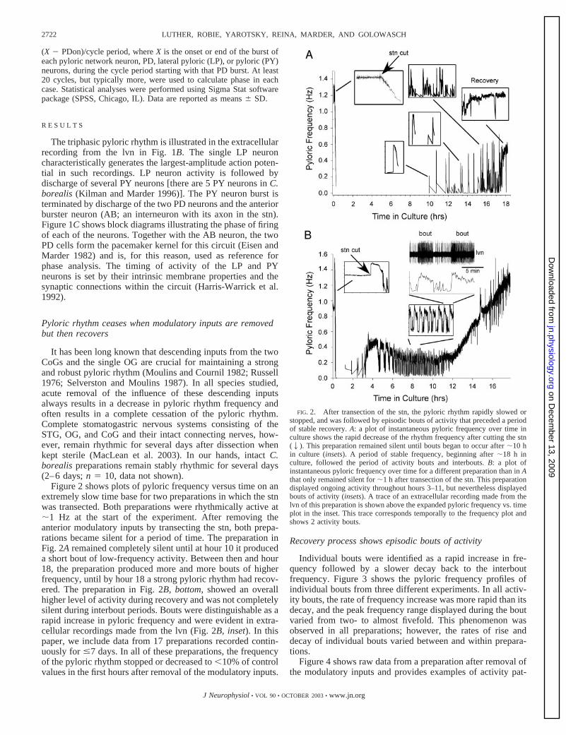
Figure 2 shows plots of pyloric frequency versus time on anextremely slow time base for two preparations in which the stnwas transected. Both preparations were rhythmically active at�1 Hz at the start of the experiment. After removing theanterior modulatory inputs by transecting the stn, both prepa-rations became silent for a period of time. The preparation inFig. 2A remained completely silent until at hour 10 it produceda short bout of low-frequency activity. Between then and hour18, the preparation produced more and more bouts of higherfrequency, until by hour 18 a strong pyloric rhythm had recov-ered. The preparation in Fig. 2B, bottom, showed an overallhigher level of activity during recovery and was not completelysilent during interbout periods. Bouts were distinguishable as arapid increase in pyloric frequency and were evident in extra-cellular recordings made from the lvn (Fig. 2B, inset). In thispaper, we include data from 17 preparations recorded contin-uously for �7 days. In all of these preparations, the frequencyof the pyloric rhythm stopped or decreased to �10% of controlvalues in the first hours after removal of the modulatory inputs.
Recovery process shows episodic bouts of activity
Individual bouts were identified as a rapid increase in fre-quency followed by a slower decay back to the interboutfrequency. Figure 3 shows the pyloric frequency profiles ofindividual bouts from three different experiments. In all activ-ity bouts, the rate of frequency increase was more rapid than itsdecay, and the peak frequency range displayed during the boutvaried from two- to almost fivefold. This phenomenon wasobserved in all preparations; however, the rates of rise anddecay of individual bouts varied between and within prepara-tions.
Figure 4 shows raw data from a preparation after removal ofthe modulatory inputs and provides examples of activity pat-
FIG. 2. After transection of the stn, the pyloric rhythm rapidly slowed orstopped, and was followed by episodic bouts of activity that preceded a periodof stable recovery. A: a plot of instantaneous pyloric frequency over time inculture shows the rapid decrease of the rhythm frequency after cutting the stn(2). This preparation remained silent until bouts began to occur after �10 hin culture (insets). A period of stable frequency, beginning after �18 h inculture, followed the period of activity bouts and interbouts. B: a plot ofinstantaneous pyloric frequency over time for a different preparation than in Athat only remained silent for �1 h after transection of the stn. This preparationdisplayed ongoing activity throughout hours 3–11, but nevertheless displayedbouts of activity (insets). A trace of an extracellular recording made from thelvn of this preparation is shown above the expanded pyloric frequency vs. timeplot in the inset. This trace corresponds temporally to the frequency plot andshows 2 activity bouts.
2722 LUTHER, ROBIE, YAROTSKY, REINA, MARDER, AND GOLOWASCH
J Neurophysiol • VOL 90 • OCTOBER 2003 • www.jn.org
on Decem
ber 13, 2009 jn.physiology.org
Dow
nloaded from
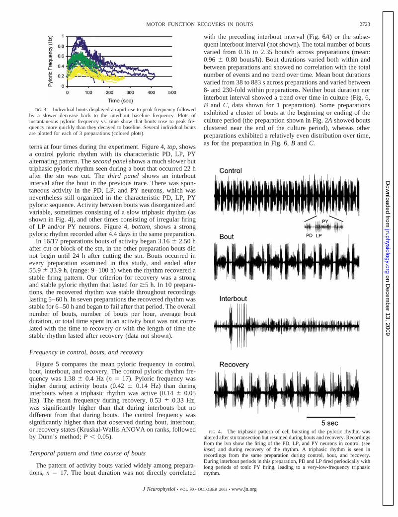
terns at four times during the experiment. Figure 4, top, showsa control pyloric rhythm with its characteristic PD, LP, PYalternating pattern. The second panel shows a much slower buttriphasic pyloric rhythm seen during a bout that occurred 22 hafter the stn was cut. The third panel shows an interboutinterval after the bout in the previous trace. There was spon-taneous activity in the PD, LP, and PY neurons, which wasnevertheless still organized in the characteristic PD, LP, PYpyloric sequence. Activity between bouts was disorganized andvariable, sometimes consisting of a slow triphasic rhythm (asshown in Fig. 4), and other times consisting of irregular firingof LP and/or PY neurons. Figure 4, bottom, shows a strongpyloric rhythm recorded after 4.4 days in the same preparation.
In 16/17 preparations bouts of activity began 3.16 � 2.50 hafter cut or block of the stn, in the other preparation bouts didnot begin until 24 h after cutting the stn. Bouts occurred inevery preparation examined in this study, and ended after55.9 � 33.9 h, (range: 9–100 h) when the rhythm recovered astable firing pattern. Our criterion for recovery was a strongand stable pyloric rhythm that lasted for �5 h. In 10 prepara-tions, the recovered rhythm was stable throughout recordingslasting 5–60 h. In seven preparations the recovered rhythm wasstable for 6–50 h and began to fail after that period. The overallnumber of bouts, number of bouts per hour, average boutduration, or total time spent in an activity bout was not corre-lated with the time to recovery or with the length of time thestable rhythm lasted after recovery (data not shown).
Frequency in control, bouts, and recovery
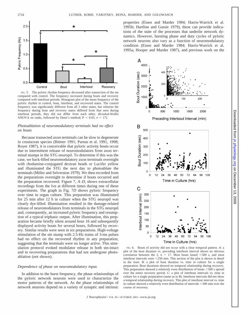
Figure 5 compares the mean pyloric frequency in control,bout, interbout, and recovery. The control pyloric rhythm fre-quency was 1.38 � 0.4 Hz (n � 17). Pyloric frequency washigher during activity bouts (0.42 � 0.14 Hz) than duringinterbouts when a triphasic rhythm was active (0.14 � 0.05Hz). The mean frequency during recovery, 0.53 � 0.33 Hz,was significantly higher than that during interbouts but nodifferent from that during bouts. The control frequency wassignificantly higher than that observed during bout, interbout,or recovery states (Kruskal-Wallis ANOVA on ranks, followedby Dunn’s method; P � 0.05).
Temporal pattern and time course of bouts
The pattern of activity bouts varied widely among prepara-tions, n � 17. The bout duration was not directly correlated
with the preceding interbout interval (Fig. 6A) or the subse-quent interbout interval (not shown). The total number of boutsvaried from 0.16 to 2.35 bouts/h across preparations (mean:0.96 � 0.80 bouts/h). Bout durations varied both within andbetween preparations and showed no correlation with the totalnumber of events and no trend over time. Mean bout durationsvaried from 38 to 883 s across preparations and varied between8- and 230-fold within preparations. Neither bout duration norinterbout interval showed a trend over time in culture (Fig. 6,B and C, data shown for 1 preparation). Some preparationsexhibited a cluster of bouts at the beginning or ending of theculture period (the preparation shown in Fig. 2A showed boutsclustered near the end of the culture period), whereas otherpreparations exhibited a relatively even distribution over time,as for the preparation in Fig. 6, B and C.
FIG. 4. The triphasic pattern of cell bursting of the pyloric rhythm wasaltered after stn transection but resumed during bouts and recovery. Recordingsfrom the lvn show the firing of the PD, LP, and PY neurons in control (seeinset) and during recovery of the rhythm. A triphasic rhythm is seen inrecordings from the same preparation during control, bout, and recovery.During interbout periods in this preparation, PD and LP fired periodically withlong periods of tonic PY firing, leading to a very-low-frequency triphasicrhythm.
FIG. 3. Individual bouts displayed a rapid rise to peak frequency followedby a slower decrease back to the interbout baseline frequency. Plots ofinstantaneous pyloric frequency vs. time show that bouts rose to peak fre-quency more quickly than they decayed to baseline. Several individual boutsare plotted for each of 3 preparations (colored plots).
2723MOTOR FUNCTION RECOVERS IN BOUTS
J Neurophysiol • VOL 90 • OCTOBER 2003 • www.jn.org
on Decem
ber 13, 2009 jn.physiology.org
Dow
nloaded from
Photoablation of neuromodulatory terminals had no effecton bouts
Because transected axon terminals can be slow to degeneratein crustacean species (Bittner 1991; Parnas et al. 1991, 1998;Royer 1987), it is conceivable that pyloric activity bouts occurdue to intermittent release of neuromodulators from axon ter-minal stumps in the STG neuropil. To determine if this was thecase, we back-filled neuromodulatory axon terminals overnightwith rhodamine-conjugated dextran beads or Lucifer yellowand illuminated the STG the next day to photoablate theterminals (Miller and Selverston 1979). We then recorded fromthe preparations overnight to determine if bouts occurred andthe preparation recovered. Figure 7, A–D, shows extracellularrecordings from the lvn at different times during one of theseexperiments. The graph in Fig. 7D shows pyloric frequencyover time in organ culture. This preparation was illuminatedfor 25 min after 12 h in culture when the STG neuropil wasclearly dye-filled. Illumination resulted in the damage-relatedrelease of neuromodulators from terminals in the STG neuropiland, consequently, an increased pyloric frequency and resump-tion of a typical triphasic output. After illumination, this prep-aration became briefly silent around hour 16 and subsequentlydisplayed activity bouts for several hours, followed by recov-ery. Similar results were seen in six preparations. High-voltagestimulation of the stn stump with 2.5-Hz trains of 3-ms pulseshad no effect on the recovered rhythm in any preparation,suggesting that the terminals were no longer active. This stim-ulation protocol evoked modulator release in both stn-intactand in recovering preparations that had not undergone photo-ablation (not shown).
Dependence of phase on neuromodulatory input.
In addition to the burst frequency, the phase relationships ofthe pyloric network neurons were used to characterize themotor patterns of the network. As the phase relationships ofnetwork neurons depend on a variety of synaptic and intrinsic
properties (Eisen and Marder 1984; Harris-Warrick et al.1995b; Hartline and Gassie 1979), these can provide indica-tions of the state of the processes that underlie network dy-namics. However, bursting phase and duty cycles of pyloricnetwork neurons also vary as a function of neuromodulatorycondition (Eisen and Marder 1984; Harris-Warrick et al.1995a; Hooper and Marder 1987), and previous work on the
FIG. 6. Bouts of activity did not occur with a clear temporal pattern. A: aplot of the bout duration vs. preceding interbout interval shows no obviouscorrelation between the 2, n � 17. Most bouts lasted �500 s, and mostinterbout intervals were �250 min. This section of the plot is shown in detailin the inset. B: a plot of bout duration vs. time in culture for a singlepreparation. Bout durations showed no temporal relationship during recovery.This preparation showed a relatively even distribution of bouts �500 s spreadover the entire recovery period. C: a plot of interbout intervals vs. time inculture for a single preparation (same as in B). Interbout intervals did not showa temporal relationship during recovery. This plot of interbout interval vs. timein culture showed a relatively even distribution of intervals �300 min over thecourse of recovery.
FIG. 5. The pyloric rhythm frequency decreased after transection of the stncompared with control. The frequency increased during bouts and recoverycompared with interbout periods. Histogram plot of the mean frequency of thepyloric rhythm in control, bout, interbout, and recovered states. The controlfrequency was significantly different from all 3 other states, but whereas thefrequency during bout and recovery states differed from that seen duringinterbout periods, they did not differ from each other, (Kruskal-WallisANOVA on ranks, followed by Dunn’s method; P � 0.05, n � 17).
2724 LUTHER, ROBIE, YAROTSKY, REINA, MARDER, AND GOLOWASCH
J Neurophysiol • VOL 90 • OCTOBER 2003 • www.jn.org
on Decem
ber 13, 2009 jn.physiology.org
Dow
nloaded from

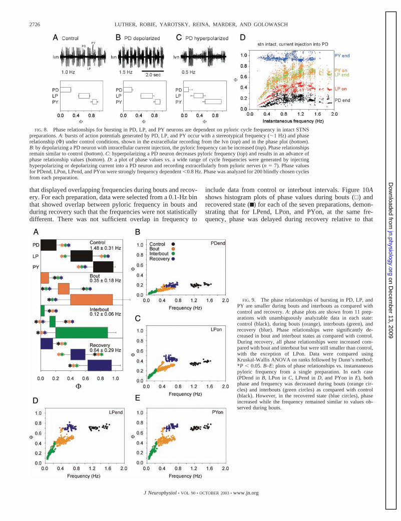
lobster, Panulirus interruptus, showed that some of the pyloricnetwork phase relationships are frequency dependent (Hooper1997a,b). To compare the control phase relationships to phaserelationships during bouts and interbouts and after recovery,the changes in frequency in these states had to be taken intoaccount. Therefore we first examined the effect of frequencyon the onset and end phase of the LP and PY neuron firing andthe end phase of PD in the control condition, by injectingcurrent into one of the two PD neurons. Figure 8, A–C, showsraw data and phase plots of the pyloric rhythm in a preparationin which the frequency was altered over a range from 0.5 to 1.5Hz by current injection into a PD neuron. This individualexample shows that at the higher frequencies there was littlechange in phase, but when the preparation was slowed, thephase of firing of the LP and PY neurons was altered. Thepooled data from seven preparations (Fig. 8D) show that atfrequencies �0.8 Hz the PD neuron terminated its burst at anearlier phase, and the LP neuron (onset and end) and the PYneuron onset phases were also advanced. The PY termination(PYend) did not show this frequency dependence because thePY neurons tend to stay active until they are inhibited by thenext PD burst. Thus PYend phase is always close to 1.
Figure 9 provides comparisons of the phase relationships ofthe PD, LP, and PY neurons in control (stn intact), bout,
interbout, and recovered states. Figure 9A shows the meanphase relationships for 11 preparations that showed clear andunambiguously analyzable pyloric rhythms in each state. Thephase of PDend, LPon, LPend, and PYon were significantlydecreased during bout and interbout states compared withcontrol. During recovery all phase relationships increased com-pared with bout and interbout but were still decreased com-pared with control except LPon. This seems unlikely to be dueentirely to increased rhythm frequency in recovery because thefrequency recovered to only 43% of control (0.64 � 0.29 vs.1.48 � 0.31 Hz). Figure 9, B–E, shows plots of phase versusinstantaneous pyloric frequency for an individual preparationfor control, bout, interbout, and recovered states (colored cir-cles). The phase relationships for control (black) and recovered(blue) states are similar despite having different frequencyranges, whereas bout (orange) and recovered (blue) have dif-ferent phase relationships but have overlapping frequencyranges. This suggests that the delay in phase seen in pyloricrhythm recovery is not only a consequence of changes inbursting frequency.
To better separate the change in phase during recovery fromthe confound arising from the fact that phase is frequencydependent at low frequencies, we compared phase relation-ships for PDend, LPon, LPend, and PYon in seven preparations
FIG. 7. Photoablation of neuromodulatory terminals in the STG neuropil did not stop bouts or recovery. A: a trace showing thetriphasic pyloric rhythm output recorded extracellularly from the lvn under control conditions with the stn intact. The stn wassubsequently cut in this preparation, and the neuromodulatory terminals were back-filled overnight with rhodamine-dextran beads.B: during illumination, damage-related release of neuromodulators caused a resumption of the pyloric rhythm as seen in thisextracellular recording from the lvn. C: after 27 h in culture, this preparation regained a triphasic pyloric rhythm output but at alower frequency than control. D: a plot of instantaneous pyloric frequency vs. time in culture shows that after blocking and cuttingthe stn (gray bar, left), the pyloric rhythm rapidly stopped. The neuromodulatory terminals were back-filled in this preparation for11 h. On illumination (gray bar, right), the pyloric output regained a typical triphasic pattern and increased in frequency due todamage-related neuromodulator release. Within 3 h after photoablation, this preparation exhibited bouts of activity (shown inexpanded extracellular recordings from the lvn) and subsequently recovered a stable triphasic pyloric output.
2725MOTOR FUNCTION RECOVERS IN BOUTS
J Neurophysiol • VOL 90 • OCTOBER 2003 • www.jn.org
on Decem
ber 13, 2009 jn.physiology.org
Dow
nloaded from
that displayed overlapping frequencies during bouts and recov-ery. For each preparation, data were selected from a 0.1-Hz binthat showed overlap between pyloric frequency in bouts andduring recovery such that the frequencies were not statisticallydifferent. There was not sufficient overlap in frequency to
include data from control or interbout intervals. Figure 10Ashows histogram plots of phase values during bouts (�) andrecovered state (■ ) for each of the seven preparations, demon-strating that for LPend, LPon, and PYon, at the same fre-quency, phase was delayed during recovery relative to that
FIG. 8. Phase relationships for bursting in PD, LP, and PY neurons are dependent on pyloric cycle frequency in intact STNSpreparations. A: bursts of action potentials generated by PD, LP, and PY occur with a stereotypical frequency (�1 Hz) and phaserelationship (�) under control conditions, shown in the extracellular recording from the lvn (top) and in the phase plot (bottom).B: by depolarizing a PD neuron with intracellular current injection, the pyloric frequency can be increased (top). Phase relationshipsremain similar to control (bottom). C: hyperpolarizing a PD neuron decreases pyloric frequency (top) and results in an advance ofphase relationship values (bottom). D: a plot of phase values vs. a wide range of cycle frequencies were generated by injectinghyperpolarizing or depolarizing current into a PD neuron and recording extracellularly from pyloric nerves (n � 7). Phase valuesfor PDend, LPon, LPend, and PYon were strongly frequency dependent �0.8 Hz. Phase was analyzed for 200 blindly chosen cyclesfrom each preparation.
FIG. 9. The phase relationships of bursting in PD, LP, andPY are smaller during bouts and interbouts as compared withcontrol and recovery. A: phase plots are shown from 11 prep-arations with unambiguously analyzable data in each state:control (black), during bouts (orange), interbouts (green), andrecovery (blue). Phase relationships were significantly de-creased in bout and interbout states as compared with control.During recovery, all phase relationships were increased com-pared with bout and interbout but were still smaller than control,with the exception of LPon. Data were compared usingKruskal-Wallis ANOVA on ranks followed by Dunn’s method;*P � 0.05. B–E: plots of phase relationships vs. instantaneouspyloric frequency from a single preparation. In each case(PDend in B, LPon in C, LPend in D, and PYon in E), bothphase and frequency was decreased during bouts (orange cir-cles) and interbouts (green circles) as compared with control(black). However, in the recovered state (blue circles), phaseincreased while the frequency remained similar to values ob-served during bouts.
2726 LUTHER, ROBIE, YAROTSKY, REINA, MARDER, AND GOLOWASCH
J Neurophysiol • VOL 90 • OCTOBER 2003 • www.jn.org
on Decem
ber 13, 2009 jn.physiology.org
Dow
nloaded from

during bouts. In each preparation, this reached statistical sig-nificance (P � 0.001), except in one case for LPend (3rdpanel). For comparison, the preparation shown in Fig. 9, B–E,
is represented by the second pair of columns from the right ineach graph in Fig. 10A, F. Mean values for each neuronalphase relationship were: PDend, 0.12 � 0.01 versus 0.14 �0.02 Hz; LPon, 0.28 � 0.02 versus 0.37 � 0.03 Hz; LPend,0.45 � 0.05 versus 0.56 � 0.05 Hz; PYon, 0.48 � 0.04 versus0.60 � 0.04 Hz for bout versus recovered, respectively. Inter-estingly, the phase relationship for PDend did not differ greatlybetween bout and recovery in most preparations; this suggeststhat the differences seen in phase for the other cells are notsimply due to changes in frequency. Figure 10B compares datafrom three additional preparations in which the PD neuronswere hyperpolarized by current injection in control and/or afterrecovery so that the phase could be compared at the samefrequency in control and after recovery. This comparisonshows that the phase in the recovered preparations were de-layed relative to the control preparations.
Blockade of glutamatergic synapses in the STG does notprevent bouts or recovery
An alteration of the pyloric network that results in compen-sation for loss of neuromodulation and resumption of outputcould arise through changes in the intrinsic properties of indi-vidual neurons and/or changes in synaptic properties of thepyloric network. Glutamate and acetylcholine are the principaltransmitters at synapses between STG neurons (Eisen andMarder 1982). Picrotoxin (PTX) blocks graded and spike-mediated glutamatergic synaptic transmission among STGneurons, therefore its application disconnects much of thenetwork (Bidaut 1980; Marder and Eisen 1984). To testwhether alterations in glutamatergic synapses were involved inthe recovery process, we applied PTX during recovery from stntransection in culture. The PTX was applied shortly beforetransection of the stn and allowed to remain until the prepara-tion recovered. In control, pyloric neuron groups burst alter-nately creating a triphasic pattern (Fig. 11A). With the stnintact, application of 10�5 M PTX disrupted but did not stopthe pyloric rhythm (Fig. 11B). In the presence of PTX, burstsin the pyloric neurons overlapped temporally before and afterrecovery (Fig. 11, C–E). The triphasic pattern of alternatebursts returned when PTX was washed off (Fig. 11F). Six ofnine preparations displayed activity bouts and recovered inPTX.
D I S C U S S I O N
Adult CPG networks must maintain the ability to producestable neuronal outputs over long time periods, in some cases,over many years. Nonetheless, they must also be responsive tothe animal’s behavioral needs in the short term. Sensory andneuromodulatory control systems enable the animal to adaptthe output of its circuits to the specific behavioral context inwhich the animal is found. At the same time, there must be astable “baseline state” to which CPG networks return aftershort-term perturbations. In this study, we use the pyloricrhythm of the adult crab as a model system in which to studythe return to a baseline state in response to long-term removalof descending neuromodulatory inputs.
Adult crabs live many years, and the animals that we used inthis study were several years old. In vivo recordings frombehaving crabs showed ongoing pyloric rhythms in the 0.2- to
FIG. 10. Frequency-controlled neuronal phase relationships after recoverycompared with phase relationships during activity bouts and during control.Data are shown as histogram plots of phase values (�) under 2 conditions, andpairs of data correspond to different experiments. �, data during bouts (A) andcontrol (B); ■ , data after recovery. Top to bottom: comparisons of phase valuesof the end of the PD burst (PDend), beginning and end of the LP burst (LPonand LPend, respectively), and the beginning of the PY burst (PYon). A:preparations during bouts and after recovery were compared for 7 preparationsthat displayed overlapping frequencies in the 2 states. In 4 preparations, thephase of PDend was similar, but in 3 preparations, PDend was significantlylarger in recovery than during bouts. In every case of the phase histograms forLPon, LPend, and PYon, except 1 (last set of LPend), phase values were largerafter recovery than during bouts. Note that the phase relationships for the samepreparation shown in Fig. 9, B–E, are shown for a frequency of 0.5 Hz in eachgraph (F). B: phase relationships are plotted for 3 preparations in which thepyloric frequency was modified by current injection into 1 PD neuron duringcontrol and/or recovery to match the frequencies between control and recovery.These 3 experiments are from different preparations as those shown in A. In allcases but 1 (PDend, middle), there is a significantly higher phase value afterrecovery as compared with control for each phase value (PDend, LPon, LPend,and PYon). Data were compared using either Student’s t-test for normallydistributed data sets or the Mann-Whitney rank-sum test for nonnormallydistributed data; ***P � 0.001.
2727MOTOR FUNCTION RECOVERS IN BOUTS
J Neurophysiol • VOL 90 • OCTOBER 2003 • www.jn.org
on Decem
ber 13, 2009 jn.physiology.org
Dow
nloaded from

0.4-Hz frequency range in unfed animals, while rhythms of1.0–1.5 Hz were routinely seen subsequent to feeding(R. Zarum and E. Marder, unpublished results). Thus in thebehaving animal, the unfed baseline state has frequencies sim-ilar to those that we obtain subsequent to recovery after re-moval of descending modulatory inputs (Golowasch et al.1999b).
Bout properties
Bouts of activity occurred in all preparations that showedrecovery in this study, but the temporal pattern of the bouts andthe overall number of bouts was highly variable between andwithin preparations. Although bouts had not been previouslyreported in decentralized STG preparations (Golowasch et al.1999b; Mizrahi et al. 2001; Thoby-Brisson and Simmers1998), in the earlier studies, recordings were not made contin-uously and the low frequency of activity bouts (on average, 1�3-min bout/h) would make them easily overlooked withoutcontinuous recordings.
The episodic bouts of activity characterized in this study insome ways resemble spontaneous activity bouts seen in devel-oping neural networks in many systems (Ben-Ari 2001; Ka-mioka et al. 1996; Katz and Shatz 1996; Murphy et al. 1992;O’Donovan 1999). Similar spontaneous bursts are also ob-served in epileptiform bursting in adult hippocampal CA3networks (Staley et al. 1998). Bouts of activity in these other
systems are thought to be important for guiding the appropriateconstruction of the developing networks (Ben-Ari 2001; Katzand Shatz 1996; O’Donovan 1999). An attractive possibility isthat bouts in the recovering pyloric network represent a similarunderlying mechanism that guides the restructuring of thepyloric network to allow a physiologically meaningful output.Nonetheless, there are some important differences betweenbouts seen in developing motor systems and the bouts we seein the recovering pyloric network. There does not seem to be arefractory period or activity-dependent depression after boutsin the pyloric network because we observed no correlationbetween bout duration and the preceding or subsequent inter-bout interval. In contrast, a refractory period is thought to be acommon factor contributing to episodic bouts in a variety ofdeveloping neural networks (O’Donovan 1999). Additionally,activity bouts did not appear to be related to recovery of phaseor frequency of the pyloric rhythm. The overall number ofactivity bouts, number of bouts per hour, overall time spent inactivity bouts, or average bout duration in a given preparationwas not correlated with the time to recovery or the percentagerecovery of phase or frequency in our study. This is in contrastto developmental changes, such as that occurring in the de-layed rectifier potassium current kinetics seen in amphibianspinal neurons, that are dependent on spontaneous activitybouts in a particular frequency range (Gu and Spitzer 1995).Nevertheless, bouts of activity were consistently associatedwith rhythm recovery and ceased when a stable rhythm wasgenerated. The discontinuous nature of the recovery processand the intermittent nature of activity bouts suggests that theunderlying mechanism itself is discontinuous or is dependenton several competing processes that must reach equilibriumbefore the rhythm can resume stably. In the future, it will beimportant to determine if activity bouts are a necessary processthat regulate pyloric rhythm recovery or if they are an epiphe-nomenon that occurs due to another underlying homeostaticprocess.
What processes might cause the bouts that we have ob-served? In developing retina and spinal cord, it is believed thatspontaneous bouts of activity are caused by excitatoryGABAergic, glutamatergic, or cholinergic synaptic contactsthrough network-dependent mechanisms, whereas the durationof the interbout is determined by the recovery from synapticdepression or depletion (Chub and O’Donovan 1998; Feller1999; Feller et al. 1996). As all chemical synapses among thepyloric network neurons are inhibitory, it is possible that anendogenous burst in any pyloric network neuron could triggera bout by a postinhibitory rebound-like mechanism. This seemsunlikely, because we observed that blockade of glutamatergicsynaptic transmission between STG neurons did not blockeither the appearance of the bouts or recovery. Additionally,we saw no evidence for a refractory period after bouts. How-ever, this does not rule out that the cholinergic STG synapsesmight trigger bouts.
We showed that both the recovery process and bouts persistafter descending modulatory inputs were photoablated. Thisindicates that activity in the presynaptic terminals of the de-scending modulatory neurons is not solely responsible for theproduction of bouts. We observed that neuronal phase relation-ships were different in recovered preparations compared withthese during bouts or control at a given frequency. Because thephase relationships are determined by the intrinsic membrane
FIG. 11. Blocking glutamatergic synaptic transmission between STG neu-rons alters the pyloric rhythm but does not block rhythm recovery. Extracel-lular recordings from the pdn, lvn, and pyn. A: control preparation. B: with thestn intact, 10�5 M picrotoxin (PTX) alters the triphasic pyloric rhythm due touncoupling of many of the neurons. C and D: after cutting the stn the pyloricrhythm and PD bursts decreased in frequency. E: after 24 h, this preparationregained a stable rhythmic output. However, the pyloric neurons exhibitedoverlapping bursts as opposed to the normal alternating triphasic burstingpattern seen in control. F: 13 h after washout of PTX the pyloric rhythm hadrecovered and displayed the typical alternating triphasic pattern.
2728 LUTHER, ROBIE, YAROTSKY, REINA, MARDER, AND GOLOWASCH
J Neurophysiol • VOL 90 • OCTOBER 2003 • www.jn.org
on Decem
ber 13, 2009 jn.physiology.org
Dow
nloaded from

properties of STG neurons and their synaptic connections, oneor many of these properties may be altered during recovery.Evidence suggests that the recovery process involves changesin the intrinsic properties of the pyloric neurons (Golowasch etal. 1999b; Mizrahi et al. 2001; Thoby-Brisson and Simmers2002) as they retune themselves in response to their alteredenvironment and level of activity (Golowasch et al. 1999a;Soto-Trevino et al. 2001). Thoby-Brisson and Simmers (2002)reported that in the lobster, Jasus lalandii, the delayed rectifierand calcium-dependent potassium currents were downregu-lated and the hyperpolarization-activated cation current wasupregulated in PD neurons after rhythm recovery. This alter-ation of membrane currents favors endogenous, regenerativebursting behavior, which PD neurons do not possess immedi-ately after stn transection. A large increase in the propensity ofthe pacemaker kernel to display bursting behavior, as sug-gested by Thoby-Brisson and Simmers (2002), could accountfor rhythm recovery. However, an alternative possibility is thatmany small changes could occur in the intrinsic properties ofmany of the pyloric neurons, or their synaptic contacts, thattogether account for rhythm recovery. Future experiments willbe needed to determine how much each of these potentialmechanisms contribute to rhythm recovery in the pyloric net-work.
A similar mechanism is believed to occur in recovery ofspontaneous activity of neurons of the vestibular nucleus com-plex after deafferentation (Darlington et al. 2002). If channeldensities are functionally linked to neuronal activity, as sug-gested by modeling studies (LeMasson et al. 1993; Liu et al.1998), it can be imagined that activity bouts arise as channeldensities are altered. An incremental change in one or moremembrane conductance may allow a temporary resumption ofthe pyloric rhythm, but further changes in membrane conduc-tances could result in the network falling silent again. Therhythm could switch on and off in this manner until theprocesses that link activity and membrane conductances reachequilibrium. If this was true, then the pattern of bouts over timeand the time to recovery would depend on the initial conditionsof the network elements. This is likely to be different for eachpreparation, explaining the variability we see in the recoveryprocess. Recent work (MacLean et al. 2003) showed thatoverexpression of A-type potassium channels subsequent tomRNA injection resulted in little change in PD neuron activityas the increase in A-type channel density was accompanied bya compensating increase in an inwardly rectifying cation chan-nel. These data add to a growing body of evidence arguing fora number of homeostatic mechanisms, both activity dependentand independent, that may act together during recovery pro-cesses to reinstate baseline activity.
Crabs must benefit greatly from having a high degree ofstability in a system that is essential to life such as the pyloricnetwork. Indeed, a highly plastic or unstable feeding-pattern-generating network would be seriously detrimental to anyanimal. The processes that maintain a stable pyloric networkmay be common to many rhythmic networks, including essen-tial systems such as those that control breathing, circulatory, ordigestive processes. The mechanisms that stabilize the pyloricnetwork seem to be highly versatile, as they can compensatefor a change as dramatic as complete removal of all neuro-modulatory input. Understanding these processes in the pyloricnetwork will help elucidate mechanisms that maintain stability
in neural networks in general during learning, physical growth,and development and in disease.
We thank Dr. William Miller for help with data analysis in the early stagesof this work and Dr. Dirk Bucher for invaluable help with Spike 2 analysis. Weespecially thank Dr. Farzan Nadim for generous support of J. Golowasch, J.Yarotsky, and C. Reina during part of this project.
D I S C L O S U R E S
This research was supported by National Institutes of Health Grants R01MH-64711 to J. Golowach, R37 MH-46742 to E. Marder, and T32 NS-07292to J. A. Luther and an undergraduate summer IGERT 9972756 NationalScience Foundation grant to A. A. Robie.
Present address for C. Reina: Department of Pharmacology, University ofPennsylvania, 3620 Hamilton Walk, Philadelphia, PA 19104.
REFERENCES
Ben-Ari Y. Developing networks play a similar melody. Trends Neurosci 24:353–360, 2001.
Ben-Ari Y, Cherubini E, Corradetti R, and Gaiarsa JL. Giant synapticpotentials in immature rat CA3 hippocampal neurons. J Physiol 416: 303–325, 1989.
Bidaut M. Pharmacological dissection of pyloric network of the lobsterstomatogastric ganglion using picrotoxin. J Neurophysiol 44: 1089–1101,1980.
Bittner GD. Long-term survival of anucleate axons and its implications fornerve regeneration. Trends Neurosci 14: 188–193, 1991.
Chub N and O’Donovan MJ. Blockade and recovery of spontaneous rhyth-mic activity after application of neurotransmitter antagonists to spinal net-works of the chick embryo. J Neurosci 18: 294–306, 1998.
Darlington CL, Dutia MB, and Smith PF. The contribution of the intrinsicexcitability of vestibular nucleus neurons to recovery from vestibular dam-age. Eur J Neurosci 15: 1719–1727, 2002.
Eisen JS and Marder E. Mechanisms underlying pattern generation in lobsterstomatogastric ganglion as determined by selective inactivation of identifiedneurons. III. Synaptic connections of electrically coupled pyloric neurons.J Neurophysiol 48: 1392–1415, 1982.
Eisen JS and Marder E. A mechanism for production of phase shifts in apattern generator. J. Neurophysiology 51: 1375–1393, 1984.
Feller MB. Spontaneous correlated activity in developing neural circuits.Neuron 22: 653–656, 1999.
Feller MB, Wellis DP, Stellwagen D, Werblin FS, and Shatz CJ. Require-ment for cholinergic synaptic transmission in the propagation of spontane-ous retinal waves. Science 272: 1182–1187, 1996.
Garaschuk O, Hanse E, and Konnerth A. Developmental profile and syn-aptic origin of early network oscillations in the CA1 region of rat neonatalhippocampus. J Physiol 507: 219–236, 1998.
Golowasch J, Abbott LF, and Marder E. Activity-dependent regulation ofpotassium currents in an identified neuron of the stomatogastric ganglion ofthe crab Cancer borealis. J Neurosci 19: RC33, 1999a.
Golowasch J, Casey M, Abbott LF, and Marder E. Network stability fromactivity-dependent regulation of neuronal conductances. Neural Comput 11:1079–1096, 1999b.
Gu X and Spitzer NC. Distinct aspects of neuronal differentiation encoded byfrequency of spontaneous Ca2 transients. Nature 375: 784–787, 1995.
Harris-Warrick RM, Coniglio LM, Barazangi N, Guckenheimer J, andGueron S. Dopamine modulation of transient potassium current evokesphase shifts in a central pattern generator network. J Neurosci 15: 342–358,1995a.
Harris-Warrick RM, Coniglio LM, Levini RM, Gueron S, and Gucken-heimer J. Dopamine modulation of two subthreshold currents producesphase shifts in activity of an identified motoneuron. J Neurophysiol 74:1404–1420, 1995b.
Harris-Warrick RM, Marder E, Selverston AI, and Moulins M. DynamicBiological Networks. The Stomatogastric Nervous System. Cambridge, MA:MIT Press, 1992.
Hartline DK and Gassie DV Jr. Pattern generation in the lobster (Panulirus)stomatogastric ganglion. I. Pyloric neuron kinetics and synaptic interactions.Biol Cybern 33: 209–222, 1979.
Him A and Dutia MB. Intrinsic excitability changes in vestibular nucleusneurons after unilateral deafferentation. Brain Res 908: 58–66, 2001.
2729MOTOR FUNCTION RECOVERS IN BOUTS
J Neurophysiol • VOL 90 • OCTOBER 2003 • www.jn.org
on Decem
ber 13, 2009 jn.physiology.org
Dow
nloaded from

Holliday J and Spitzer NC. Spontaneous calcium influx and its roles indifferentiation of spinal neurons in culture. Dev Biol 141: 13–23, 1990.
Hooper SL. Phase maintenance in the pyloric pattern of the lobster (Panulirusinterruptus) stomatogastric ganglion. J Comput Neurosci 4: 191–205,1997a.
Hooper SL. The pyloric pattern of the lobster (Panulirus interruptus) stoma-togastric ganglion comprises two phase maintaining subsets. J ComputNeurosci 4: 207–219, 1997b.
Hooper SL and Marder E. Modulation of the lobster pyloric rhythm by thepeptide proctolin. J Neurosci 7: 2097–2112, 1987.
Jones EG. Cortical and subcortical contributions to activity-dependent plas-ticity in primate somatosensory cortex. Annu Rev Neurosci 23: 1–37, 2000.
Kamioka H, Maeda E, Jimbo Y, Robinson HP, and Kawana A. Sponta-neous periodic synchronized bursting during formation of mature patterns ofconnections in cortical cultures. Neurosci Lett 206: 109–112, 1996.
Katz LC and Shatz CJ. Synaptic activity and the construction of corticalcircuits. Science 274: 1133–1138, 1996.
Kilman VL and Marder E. Ultrastructure of the stomatogastric ganglionneuropil of the crab, Cancer borealis. J Comp Neurol 374: 362–375, 1996.
Landmesser LT and O’Donovan MJ. Activation patterns of embryonic chickhind limb muscles recorded in ovo and in an isolated spinal cord preparation.J Physiol 347: 189–204, 1984a.
Landmesser LT and O’Donovan MJ. The activation patterns of embryonicchick motoneurones projecting to inappropriate muscles. J Physiol 347:205–224, 1984b.
LeMasson G, Marder E, and Abbott LF. Activity-dependent regulation ofconductances in model neurons. Science 259: 1915–1917, 1993.
Liu Z, Golowasch J, Marder E, and Abbott LF. A model neuron withactivity-dependent conductances regulated by multiple calcium sensors.J Neurosci 18: 2309–2320, 1998.
MacLean JN, Zhang Y, Johnson BR, and Harris-Warrick RM. Activity-independent homeostasis in rhythmically active neurons. Neuron 37: 109–120, 2003.
Marder E and Eisen JS. Transmitter identification of pyloric neurons: elec-trically coupled neurons use different neurotransmitters. J Neurophysiol 51:1345–1361, 1984.
Meister M, Wong RO, Baylor DA, and Shatz CJ. Synchronous bursts ofaction potentials in ganglion cells of the developing mammalian retina.Science 252: 939–943, 1991.
Miller JP and Selverston A. Rapid killing of single neurons by irradiation ofintracellularly injected dye. Science 206: 702–704, 1979.
Mizrahi A, Dickinson PS, Kloppenburg P, Fenelon V, Baro DJ, Harris-Warrick RM, Meyrand P, and Simmers J. Long-term maintenance ofchannel distribution in a central pattern generator neuron by neuromodula-tory inputs revealed by decentralization in organ culture. J Neurosci 21:7331–7339, 2001.
Moulins M and Cournil I. All-or-none control of the bursting properties ofthe pacemaker neurons of the lobster pyloric pattern generator. J Neurobiol13: 447–458, 1982.
Murphy TH, Blatter LA, Wier WG, and Baraban JM. Spontaneous syn-chronous synaptic calcium transients in cultured cortical neurons. J Neurosci12: 4834–4845, 1992.
O’Donovan MJ. The origin of spontaneous activity in developing networks ofthe vertebrate nervous system. Curr Opin Neurobiol 9: 94–104, 1999.
O’Donovan MJ, Chub N, and Wenner P. Mechanisms of spontaneousactivity in developing spinal networks. J Neurobiol 37: 131–145, 1998.
Parnas I, Dudel J, and Atwood HL. Synaptic transmission in decentralizedaxons of rock lobster. J Neurosci 11: 1309–1315, 1991.
Parnas I, Shahrabany-Baranes O, Feinstein N, Grant P, Adelsberger H,and Dudel J. Changes in the ultrastructure of surviving distal segments ofsevered axons of the rock lobster. J Exp Biol 201: 779–791, 1998.
Royer SM. Chronic effects of de-afferentation on the stomatogastric ganglionof Panulirus. In: The Crustacean Stomatogastric Nervous System, edited bySelverston AI and Moulins M. Berlin: Springer-Verlag, 1987, p. 251–257.
Russell DF. Rhythmic excitatory inputs to the lobster stomatogastric ganglion.Brain Res 101: 582–588, 1976.
Russell DF. CNS control of pattern generators in the lobster stomatogastricganglion. Brain Res 177: 598–602, 1979.
Russell DF and Hartline DK. Bursting neural networks: a reexamination.Science 200: 453–456, 1978.
Selverston AI and Moulins M. The Crustacean Stomatogastric System.Berlin: Springer-Verlag, 1987.
Soto-Trevino C, Thoroughman KA, Marder E, and Abbott LF. Activity-dependent modification of inhibitory synapses in models of rhythmic neuralnetworks. Nat Neurosci 4: 297–303, 2001.
Staley KJ, Longacher M, Bains JS, and Yee A. Presynaptic modulation ofCA3 network activity. Nat Neurosci 1: 201–209, 1998.
Thoby-Brisson M and Simmers J. Neuromodulatory inputs maintain expres-sion of a lobster motor pattern-generating network in a modulation-depen-dent state: evidence from long-term decentralization in vitro. J Neurosci 18:2212–2225, 1998.
Thoby-Brisson M and Simmers J. Transition to endogenous bursting afterlong-term decentralization requires de novo transcription in a critical timewindow. J Neurophysiol 84: 596–599, 2000.
Thoby-Brisson M and Simmers J. Long-term neuromodulatory regulation ofa motor pattern-generating network: maintenance of synaptic efficacy andoscillatory properties. J Neurophysiol 88: 2942–2953, 2002.
Vaculin S, Franek M, and Rokyta R. Dorsal rhizotomy changes the spon-taneous neuronal activity of nuclei in the medial thalamus. Physiol Res 49:279–283, 2000.
Weng HR, Lee JI, Lenz FA, Schwartz A, Vierck C, Rowland L, andDougherty PM. Functional plasticity in primate somatosensory thalamusfollowing chronic lesion of the ventral lateral spinal cord. Neuroscience 101:393–401, 2000.
Wong ROL. Retinal waves and visual system development. Annu Rev Neu-rosci 22: 29–47, 1999.
Wong ROL, Chernjavsky A, Smith SJ, and Shatz CJ. Early functionalneural networks in the developing retina. Nature 374: 716–718, 1995.
2730 LUTHER, ROBIE, YAROTSKY, REINA, MARDER, AND GOLOWASCH
J Neurophysiol • VOL 90 • OCTOBER 2003 • www.jn.org
on Decem
ber 13, 2009 jn.physiology.org
Dow
nloaded from


![How to diagnose cervicogenic dizziness...disorder characterized by episodic bouts of aural full-ness, vertigo, and hearing loss [12, 13]. Cervicogenic diz-ziness however, typically](https://img.pdfslide.us/doc/110x75/60da0a3a8accdb0b806b5ee4/how-to-diagnose-cervicogenic-dizziness-disorder-characterized-by-episodic-bouts.jpg)