Embed Size (px)
Citation preview

1
DOTTORATO IN NEUROSCIENZE DI BASE E DELLO
SVILUPPO
Tesi di dottorato
Epigenetic mechanisms in the normal brain
development
and in a neuropathological condition:
the Rett syndrome
Candidato Giuseppina Lonetti
Relatori:
Prof. Tommaso Pizzorusso
Prof. ssa Enrica Strettoi

2
Index
ABSTRACT 4
RIASSUNTO 7
Chapter 1: Plasticity in the visual cortex 13 Critical period in the development of the visual system 13 Molecular mechanisms of visual plasticity 15 Visual experience induces changes in the synaptic efficacy of the connections in the visual cortex. Several factors have been involved in OD plasticity. NMDA receptors and neurotrophins, and Brain-derived neurotrophic factor (BDNF) in particular, are two candidate molecules important in visual plasticity. 15 Molecular basis of visual plasticity: the ERK cascade 16 ERK is associated with molecular mechanisms of memory formation 18 ERK in the visual system 21 CREB transcription factor is important in synaptic plasticity 22
Chapter 2: Epigenetic modifications in pathology of nervous system 26 Chromatin remodelling in brain pathology 26 Rett syndrome: clinical features 26 Phenotypic variability in RTT 27 MeCP2: structure and function 28 The BDNF gene: a target of MeCP2 32 Mouse Models for studying RTT 34 Synaptic plasticity is affected in RTT 37 Learning is compromised in MeCP2 mutants 38 Level of anxiety in MeCP2 mutants mice 39 Disease reversibility in mouse models of RTT 40
Chapter 3:Environmental enrichment: a complex combination of multisensorial stimuli 42
Environmental enrichment in wild-type rodents 43 Effects of early enrichment on neuronal development 44 Environmental enrichment in rodent models of CNS disorders 47 Environmental enrichment in murine models of RTT 51
THESIS SCOPE AND DESIGN 53
MATERIALS AND METHODS 54
1. RESULTS I ERRORE. IL SEGNALIBRO NON È DEFINITO.
Chapter 4: Regulation of chromatin remodelling in visual cortex during critical period Errore. Il segnalibro non è definito.
Visual Experience during the critical period causes Histone H3 Phosphoaceylation Errore. Il segnalibro non è definito.

3
Chapter 5: Phosphorylation of ERK and activation of a CREB- mediated gene are differentially regulated by visual stimulation during development Errore. Il segnalibro non è definito.
2. RESULTS II ERRORE. IL SEGNALIBRO NON È DEFINITO.
Chapter 6: Effects of EE on male MeCP2y/- mice Errore. Il segnalibro non è definito. General features of EE MeCP2y/- mice Errore. Il segnalibro non è definito. EE ameliorates motor coordination and motor learning in MeCP2-/y Errore. Il segnalibro non è definito. EE increases synaptic density in MeCP2-/y mice Errore. Il segnalibro non è definito. Effects of EE on LTP Errore. Il segnalibro non è definito. Levels of BDNF in brain of MeCP2y/- mice Errore. Il segnalibro non è definito. Regulation of protein synthesis is altered in cortex and cerebellum of MeCP2y/- mice Errore. Il segnalibro non è definito.
Chapter 7: The effects of EE on female MeCP2 mutant mice Errore. Il segnalibro non è definito.
EE rescues performance deficits of female MeCP2 MeCP2+/- in the Morris water maze Errore. Il segnalibro non è definito. Wildtype-like behavior of EE female MeCP2+/- in the open field, an anxiety-related behavioral test Errore. Il segnalibro non è definito.
DISCUSSION AND CONCLUSIONS ERRORE. IL SEGNALIBRO NON È DEFINITO.
Chapter 8: Visual system: different regulation of signaling pathways during the development Errore. Il segnalibro non è definito.
Visual stimulation during the Critical Period causes ERK-Dependent Histone Acetylation and Phosphorylation Errore. Il segnalibro non è definito. Regulation of Visually Stimulated Gene Transcription in adult mice Errore. Il segnalibro non è definito.
Chapter 9: Prevention of synaptic alterations and improvement of behavioural deficits of MeCP2 null mice by early environmental stimulation Errore. Il segnalibro non è definito.
Synaptic effects of EE in MeCP2 mutants Errore. Il segnalibro non è definito. Effects of EE in MeCP2+/- mice Errore. Il segnalibro non è definito.
REFERENCES ERRORE. IL SEGNALIBRO NON È DEFINITO.

4
ABSTRACT
Regulation of gene expression occurs at different levels, from DNA to proteins, and
through various mechanisms. Chromatin remodelling and DNA methylation are
involved in the definition of transcriptional active and inactive regions of the
chromosomes; posttranslational modifications of histones cause chromatin remodelling
known as “epigenetic modifications”. These are chemical changes of the genetic
material that do not induce variability within the DNA sequences.
Epigenetic regulation of gene expression has been recently implicated in brain
development and plasticity in the normal and pathological brain. The overall aim of the
present thesis is to investigate the role of epigenetic mechanisms in the development of
the nervous system in normal and pathological conditions. We decided to develop our
investigation using a dual approach, therefore designing two main experimental
sections: in the first, we focus on the effects of epigenetic modification on the
development of the visual system, considered as a paradigmatic example of normal
nervous system development. In particular, we analyze how developmental
modifications of epigenetic mechanisms regulate visual cortical plasticity in juvenile
and adult animals.
In the second experimental section we focus on a human neurodevelopmental disorder,
the Rett syndrome, that represents an example of dysfunctional brain pathology caused
by altered regulation of epigenetic mechanisms.
The visual cortex displays various forms of anatomical and functional plasticity.
Manipulations of visual experience such as dark rearing (dr) or monocular deprivation
(MD) performed during early development cause modifications in the typical response
pattern and connectivity of visual cortical neurons. Although present to some extent in
the adult, the ability of the cortex to recover from these effects is maximal during a
critical period of postnatal development. The long-term effects of this altered
experience are likely to be mediated by signaling cascades regulating experience-
dependent gene transcription. Indeed, pharmacological inhibition of the kinase ERK

5
prevents synaptic plasticity as well as the effects of MD on ocular dominance in the
developing visual cortex (Di Cristo et al., 2001). Cancedda et al. (Cancedda et al., 2003)
showed that visual stimulation during the critical period causes a strong activation of
ERK kinase and CREB-mediated gene expression.
In our investigation we focused on the mechanisms that link visual experience
dependent activation of ERK pathway with CREB-mediated gene expression in the
visual cortex during the critical period. We found that, during the critical period, visual
experience triggered ERK-dependent histone H3 acetylation and phosphorylation and
histone H4 acetylation. We formulated the hypothesis that a developmental regulation
of the experience-dependent mechanisms relating ERK pathway to CREB-mediated
gene expression could also be involved in determining the different plasticity between
the young and adult cortex. Indeed, we found a substantial reduction of the effects of
visual stimulation on CREB phosphorylation and on CRE-mediated gene expression in
adult animals. This decrease was associated with a decline in the ability of visual
experience to drive changes in phosphorylation and histone acetylation that control gene
transcription. As opposed to CREB, visual stimulation of ERK and MSK
phosphorylation were similar in juvenile and adult animals.
Histone acetylation is regulated by acetyltransferases and deacetylases that are recruited
by chromatin by interaction with DNA protein. Loss-of-function mutations in the gene
methyl-CpG binding protein 2 (MeCP2) is causally implicated in the majority of Rett
Syndrome cases (RTT). MeCP2 binds methylated DNA recruiting histone deacetylase
and repressing transcription. Thus, RTT, the subject of the second part of the thesis, is
characterized by abnormal epigenetic regulation. RTT is an X-linked
neurodevelopmental disorder affecting 1:10000 girls worldwide. MeCP2 mouse mutants
recapitulate many features of human RTT. In particular, MeCP2 knockout are
characterized by a short life span, breathing defects, motor impairments, deficit in
synaptic plasticity and alterations in neuronal morphology. RTT does not involve
neurodegeneration or gross neuroanatomical abnormalities. However, long-term
plasticity is defective in cortices of mice lacking MeCP2; in addition, the activity-
dependent transcription of BDNF, a neurotrophin important for synaptic plasticity, is
reduced in MeCP2 null mice.
Environmental enrichment (EE) is a housing condition that facilitates enhanced
stimulation at sensory, cognitive, social and motor level. This is demonstrated by a
number of studies showing the amelioration of conditions in disorders such as

6
Parkinson’s disease, amyotrophic lateral sclerosis, fragile X syndrome, Down
syndrome. We used electron microscopy, electrophysiology, biochemical and
behavioural techniques to investigate whether early exposure of MeCP2 null mice to EE
could rescue molecular, synaptic, motor, emotional and cognitive abnormalities. We
reared female and male MeCP2 mutants in large groups in cages containing toys and
wheels since early ages (in EE from P10). We found that EE strongly ameliorated motor
coordination as assessed with rotarod tests at all the ages analyzed (P30-P60). A trend
toward a prolonged life span was also observed. Electron microscope analysis showed
increased synaptic density in the somatonsensory cortex (S1) and in the cerebellum of
EE MeCP2 knockout mice with respect to MeCP2 knockouts reared in standard
conditions. EE was also able to compensate for the LTP deficits showed by MeCP2
knockout in S1 slice preparations as well as a measurable increase of cortical BDNF
levels. We also investigated the regulation of protein synthesis in MeCP2 knockout
mice studying the phosphorylation of S6 Kinase at Ser235/236, a site that is correlated
with translational activity. We detected a striking reduction of phosphorylation of S6 at
Ser235/236 in the MeCP2 knockout in both the cortex and cerebellum.
Finally, we investigated the effect of EE on cognitive ability and anxiety-related
behaviour in young MeCP2 females that do not exhibit strong motor impairments
precluding cognitive and emotional behavioural testing. These mice show behavioural
abnormalities similar to male animals but with a later age of onset. Our findings
revealed that female MeCP2 mutant mice are impaired in the Morris water maze and
display increased anxiety-related behaviour in the open field. More importantly, EE
could completely prevent these behavioural alterations suggesting that environmental
stimulation could have positive effects also on learning memory and emotional aspects
of RTT pathology.
Extrapolating our results to humans, it is reasonable to envision that prompt
rehabilitative intervention after early diagnosis would accomplish the best results in
RTT patients.

7
RIASSUNTO
La regolazione della trascrizione genica avviene a diversi livelli, dal DNA alle proteine,
per mezzo di svariati meccanismi. Il rimodellamento della cromatina è implicato
nell’attivazione o nella repressione di sequenze all’interno dei cromosomi. Tale
rimodellamento si attua tramite metilazione del DNA e modificazioni postraduzionali
degli istoni, generalmente note come modificazioni epigenetiche. Queste sono definite
come modificazioni chimiche del materiale genetico che non generano variazioni nella
sequenza del DNA. La regolazione epigenetica è stata recentemente implicata nello
sviluppo del cervello e nella sua plasticità così come nelle sue patologie. Obbiettivo di
questa tesi è investigare il ruolo dei meccanismi epigenetici nello sviluppo del sistema
nervoso in condizioni fisiologiche e patologiche.
Abbiamo sviluppato la nostra analisi secondo due linee sperimentali. Nella prima parte
ci siamo concentrati sullo studio degli effetti delle modificazioni epigenetiche sullo
sviluppo del sistema visivo, prendendolo come paradigma di sviluppo del sistema
nervoso in condizioni normali. In questo contesto, abbiamo analizzato come i
meccanismi epigenetici siano in grado di regolare la plasticità visiva anche nell’animale
adulto. Nella seconda parte di questo studio ci siamo focalizzati su una malattia umana:
la sindrome di Rett, un esempio di patologia del cervello causata da un’alterazione della
regolazione dei meccanismi epigenetici.
Lo sviluppo della corteccia visiva è caratterizzato da aspetti di plasticità strutturale e
funzionale. Le manipolazioni dell’ingresso visivo, come l’allevamento al buio o la
deprivazione monoculare eseguite nelle prime fasi dello sviluppo, causano
modificazioni tipiche delle connessioni sinaptiche in corteccia visiva. Sebbene alcune
forme di plasticità siano presenti anche nell’adulto, l’abilità della corteccia di rispondere
a manipolazioni esterne di questo tipo è massima in una finestra temporale dello
sviluppo post-natale chiamato periodo critico. Gli effetti a lungo termine dovuti
all’esperienza visiva sono probabilmente mediati da vie di traduzione del segnale che
regolano la trascrizione genica esperienza-dipendente. Ad esempio, la chinasi ERK è
importante nella plasticità del sistema visivo: un blocco farmacologico della sua
attivazione impedisce i fenomeni di plasticità sinaptica, cosi come gli effetti della
deprivazione monoculare sulla dominanza oculare nello sviluppo della corteccia visiva
(Di Cristo et al., 2001). Cancedda et al. (Cancedda et al., 2003) hanno evidenziato

8
inoltre che la stimolazione visiva durante il periodo critico causa una forte attivazione
della chinasi ERK.
Nelle nostre analisi ci siamo concentrati sui meccanismi che correlano l’attivazione
esperienza-dipendente di specifiche vie di traduzione all’espressione genica mediata dal
fattore di trascrizione CREB in corteccia visiva, durante il periodo critico. Abbiamo
inoltre analizzato il ruolo di modificazioni epigenetiche, quali l’acetilazione e la
fosforilazione degli istoni, nello sviluppo del sistema visivo durante il periodo critico,
per cercare di capire come l’espressione genica mediata da CREB sia ad esse correlata.
Dai risultati ottenuti è emerso che l’esperienza visiva causa la fosforilazione e
l’acetilazione dell’istone H3 e l’acetilazione dell’istone H4.
Abbiamo inoltre riscontrato una sostanziale riduzione degli effetti della stimolazione
visiva sulla fosforilazione di CREB e sulla espressione genica negli animali adulti.
Questa riduzione si associa a un decremento della capacità dell’esperienza visiva di
indurre la fosforilazione e l’acetilazione degli istoni che controllano la trascrizione
genica. Invece, la stimolazione visiva causa gli stessi effetti sull’attivazioni delle
chinasi ERK e MSK, sia nell’animale piccolo che nell’adulto, suggerendo che
un’attenuazione dei meccanismi attività-dipendente di attivazione trascrizionale con lo
sviluppo possa contribuire alla riduzione di plasticità che sia ha nella corteccia adulta.
L’acetilazione degli istoni è regolata dagli enzimi acetiltrasferasi e deacetlasi che sono
reclutati dalla cromatina interagendo con altre proteine. La perdita di funzione del gene
per la proteina MeCP2, che reprime la trascrizione legando il DNA metilato e
reclutando specifiche istone deacetilasi, causa la maggior parte dei casi della sindrome
di Rett (RTT). Questa patologia è quindi caratterizzata da una anormale regolazione
epigenetica e si manifesta come un disordine neurologico dello sviluppo che colpisce
1:10000 di donne nella popolazione mondiale.
I topi mutanti MeCP2 ricapitolano molte caratteristiche della sindrome umana. In
particolare, i topi MeCP2 KO sono caratterizzati da una minore durata di vita, deficit
nella respirazione, deficit motori e alterazioni nella morfologia neuronale. La plasticità a
lungo termine è alterata nelle cortecce di topi che mancano della proteina MeCP2;
inoltre la trascrizione attività dipendente del BDNF, un fattore neurotrofico importante
nella plasticità sinaptica, è ridotta nei MeCP2 KO.
L’ambiente arricchito è una condizione di allevamento che comporta un aumento della
stimolazione sensoriale, cognitiva e motoria, come dimostrato in molti studi che
mostrano un miglioramento dei sintomi in patologie quali il Parkinson, la sclerosi

9
amiotrofica laterale, la sindrome dell’X fragile e la sindrome di Down. Abbiamo usato
metodologie morfologiche, elettrofisiologiche, molecolari e comportamentali per
indagare se l’arricchimento ambientale precoce può correggere le anormalità
molecolari, sinaptiche, motorie e cognitive. Abbiamo allevato topi maschi e femmine
mutati per MeCP2 in grandi gruppi, in grandi gabbie, con giochi e ruote, a partire
dall’età di 10 giorni (P10). L’arricchimento ambientale aumenta decisamente la
coordinazione motoria come è stato analizzato mediante il test del rotarod a tutte le età
analizzate (P30-P60). Inoltre si osserva un trend all’aumento della durata della vita.
L’analisi mediante microscopia elettronica evidenzia un aumento della densità sinaptica
nella corteccia somatensoriale e nel cervelletto nei MeCP2 KO arricchiti rispetto a
quelli allevati nelle condizioni standard. Inoltre, l’arricchimento ambientale è in grado
di compensare i deficit della long term potentiation (LTP) evidenziata con tecniche
elettrofisiologiche in vitro su fettine ottenute da topi MeCP2 KO. I livelli corticali di
BDNF risultano pure aumentati negli stessi soggetti.
Abbiamo indagato la regolazione della sintesi proteica nei topi MeCP2 KO analizzando
la fosforilazione della chinasi S6 sulla serina 235/236. Abbiamo osservato una forte
riduzione della fosforilazione di S6 sulla serina 235/236 nei topi MeCP2 KO, sia nella
corteccia che nel cervelletto. Infine, abbiamo investigato gli effetti dell’arricchimento
ambientale sulle abilità cognitive e sui comportamenti ansiosi nelle femmine MeCP2
mutate che riepilogano le caratteristiche dei pazienti con RTT che esprimono una sola
copia del normale allele. Questi topi evidenziano anormalità simili agli animali maschi
ma con una insorgenza più tardiva dei sintomi e permettono quindi lo esecuzione di test
cognitivi ed emotivi basati sul comportamento motorio. I nostri risultati evidenziano che
le femmine MeCP2 mutate hanno deficit cognitivi misurati con il test del “Morris water
maze” e mostrano un comportamento di tipo ansiogeno nel test del “open field”.
Concludendo, l’arricchimento ambientale sembra avere effetti benefici
sull’apprendimento e sugli aspetti emozionali della patologia della sindrome di Rett.
Pertanto, è presumibile che un intervento riabilitativo in età precoce attuato nelle
bambine colpite dalla Rett potrebbe produrre risultati positivi.

10
INTRODUCTION Chromatin remodelling: a role in central nervous system development and plasticity Epigenetic mechanisms are the focus of this thesis. In the following sections we will
analyze the evidences supporting an important role of epigenetic mechanisms in
nervous system development. Specifically, we will describe how epigenetic
mechanisms regulate brain function in normal conditions, such as in visual
development, and in certain pathophysiological states, and namely in Rett syndrome
(RTT), a neurodevelopmental human disease.
Epigenetic mechanisms regulate gene expression without a change in DNA sequence. In
the nucleus, the chromatin is a complex of DNA, histone and nonhistone proteins. The
histone proteins are packaged in a core octamer with DNA, in an assembly known as
nucleosome. This octamer is organized in one structure that contains two copies of each
one of the histones H2A, H2B, H3 and H4.
The chromatin is distinguished in euchromatin and heterochromatin. The euchromatin
consists in an open state of chromatin and permits gene transcription, while the
heterochromatin is a condensed state of chromatin that does not permit transcription.
DNA-histone interactions are modulated by modifications in the residues in the N-
terminal tail of histones. Histone acetyltransferases (HAT) and histone kinases cause
acetylation and phosphorylation of histones (fig1); these modifications disrupt their
interactions with DNA, so that chromatin remodelling allows the transcriptional
machinery to become accessible.
Chromatin remodelling is important in gene regulation in general and specifically in
neurons. An example is provided by genes involved in circadian rhythms: these are
daily rhythms that regulate the physiology of organisms during the day. The biological
timekeeping mechanism is known as the circadian clock that relies mainly to the
suprachiasmatic nucleus (SCN) of the hypothalamus. During a circadian cycle, the
promoters of genes in the molecular clock are regulated by acetylation of histones H3
and H4. Epigenetic mechanisms directly regulate the transcription of the mouse clock
gene period 1 (Per1) of the circadian clock. In particular, infusion in experimental
animals of trichostatin A, an inhibitor of deacetylase, causes an increase of per1 gene
transcription (Naruse et al., 2004).

11
Based on the critical role that chromatin remodelling plays in the regulation of the
transcription-permissive or silencing state of the genome, it is likely that modifications
of histones may be involved in synaptic plasticity processes.
AA
BB
Fig1: A) Structure of nucleosome: the picture shows a DNA strand wrapped around a
histone octamer composed of two copies each of the histones H2A, H2B, H3 and H4.
The amino (N) termini tails of the histones face outward from the nucleosome complex.
B) Multiple sites in the N terminus of a single histone (for example He) can be targets
for epigenetic tagging: acetylation, phosphorylation and methylation.
In diverse forms of synaptic plasticity at excitatory and inhibitory synapses, as in the
long-term potentiation (LTP) or long-term depression (LTD), the efficacy of synaptic
transmission is up-or down-regulated, respectively. Changes in gene expression and
changes of chromatin remodelling play a crucial role in these forms of plasticity. For
example, H4 acetylation at specific promoters in Aplysia is altered after LTP and LTD

12
(Guan et al., 2002). During synaptic transmission, neurotransmitters cause activation in
target neurons of two major families of receptors, namely ligand-gated ion channels and
G protein-coupled receptors. Growth factors and cytokines are released from neurons in
an activity-dependent manner, and act on target neurons through receptor-mediated
signalling. Signalling cascades in target neurons cause changes in gene expression,
regulating transcription and chromatin remodelling. The transcription factor CREB
activated by signalling-induced phosphorylation in forms of synaptic plasticity, recruits
CREB-binding protein (CBP), a coactivator with intrinsic HAT activity, causing histone
acetylation (Lonze et al., 2002). These mechanisms of epigenetic regulation can vary
during different stages of development, for example in visual development, as it will be
described in next section.
13
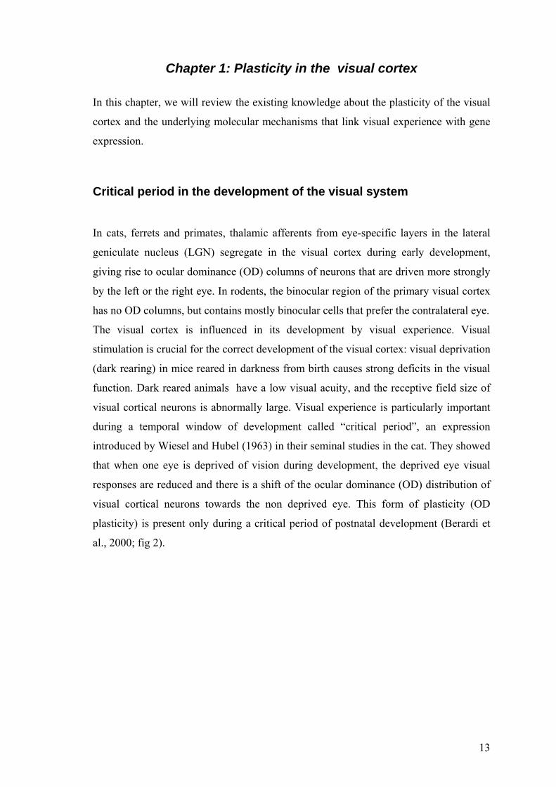
Chapter 1: Plasticity in the visual cortex
In this chapter, we will review the existing knowledge about the plasticity of the visual
cortex and the underlying molecular mechanisms that link visual experience with gene
expression.
Critical period in the development of the visual system
In cats, ferrets and primates, thalamic afferents from eye-specific layers in the lateral
geniculate nucleus (LGN) segregate in the visual cortex during early development,
giving rise to ocular dominance (OD) columns of neurons that are driven more strongly
by the left or the right eye. In rodents, the binocular region of the primary visual cortex
has no OD columns, but contains mostly binocular cells that prefer the contralateral eye.
The visual cortex is influenced in its development by visual experience. Visual
stimulation is crucial for the correct development of the visual cortex: visual deprivation
(dark rearing) in mice reared in darkness from birth causes strong deficits in the visual
function. Dark reared animals have a low visual acuity, and the receptive field size of
visual cortical neurons is abnormally large. Visual experience is particularly important
during a temporal window of development called “critical period”, an expression
introduced by Wiesel and Hubel (1963) in their seminal studies in the cat. They showed
that when one eye is deprived of vision during development, the deprived eye visual
responses are reduced and there is a shift of the ocular dominance (OD) distribution of
visual cortical neurons towards the non deprived eye. This form of plasticity (OD
plasticity) is present only during a critical period of postnatal development (Berardi et
al., 2000; fig 2).

14
A BA BA B
Fig.2: Ocular dominance (OD) plasticity in juvenile mouse visual cortex.
A) Schematic representation of the mouse visual system. The binocular region of
primary visual cortex (V1) receives an input from the contralateral retina (turquoise
projections) and additionally is innervated by ipsilateral projections (red).
Thalamocortical axons from the dorsal lateral geniculate nucleus (dLGN) arborise not
only within layer 4 (L4) but also in superficial layers(L1-3).
B) Monocular deprivation in juvenile mice during the critical period leads to strong
changes in binocular cortical responses. There is a shift of OD distribution of cortical
neurons towards the eye remained open (ipsilateral eye). Ocular dominance classes 1–7
illustrate relative responsiveness of neurons to contralateral and ipsilateral eye
stimulation (1 or 7, cells respond only to the contralateral or ipsilateral eye,
respectively; 4, equal response to both eyes). By visually-evoked potentials on
population response strength, the responses of deprived eye (compare turquoise bars)
are lower than non-deprived eye (compare red bars). Abbreviations: bV1, binocular V;
mV1, monocular V1 (Hofer et al., 2006).

15
Molecular mechanisms of visual plasticity
Visual experience induces changes in the synaptic efficacy of the connections in the
visual cortex. Several factors have been involved in OD plasticity. NMDA receptors
and neurotrophins, and Brain-derived neurotrophic factor (BDNF) in particular, are two
candidate molecules important in visual plasticity.
NMDA receptors
Based on importance that NMDA receptors have in synaptic plasticity of several brain
areas, various studies investigated their role on visual plasticity. It was found that the
pharmacological blockade of NMDA receptors prevented the effects of monocular
deprivation (Bear, M.F. et al., 1990). In addition, the subunit composition of NMDA
receptors changed in the visual cortex from a prevalent expression of the 2B subunit at
juvenile states to a prevalent expression of the 2A subunit in correlation with the critical
period closure.
BDNF and intracortical inhibition
Electrical activity and visual stimulation regulate the production and release of
neurotrophins that, in turn, modulate synaptic transmission efficacy. The role of
neurotrophins in OD plasticity, and of BDNF in particular, has been investigated.
BDNF controls the time course of the critical period by accelerating the maturation of
GABA-mediated inhibition. By overexpression of BDNF in wt mice the critical period
begins precociously. It appears that an inhibitory threshold has to be surpassed before
the critical period can start. On the contrary, if intracortical inhibition is reduced, the
onset of the critical-period is delayed. Transgenic mice, characterized by loss of 65-kDa
isoform of the GABA-synthesizing enzyme GAD (GAD65), do not show ocular
dominance shift after monocular deprivation (Hensch et al., 1998). However, normal
plasticity can be rescued in these animals at any age, if GABA-mediated transmission is
enhanced in the visual cortex by means of benzodiazepines (Fagiolini and Hensch;
2000) or by overexpression of BDNF (Hanover. et al., 1999). BDNF regulates also the
closure of critical period by prompting the maturation of a second inhibitory threshold
(Feldman, 2000). Mice that over-express BDNF post-natally in the forebrain show a

16
precocious closure of the critical period, accompanied by an accelerated development of
visual acuity.
Electrical activity and neurotrophin signalling can control visual plasticity by activation
of cascades of signalling. Recent experiments have investigated three kinases that are
necessary for the shift of ocular dominance during monocular deprivation: cAMP-
dependent protein kinase (PKA), extracellular-signal-regulated kinase (ERK) and a
Ca2+/calmodulin-dependent protein kinase II (CaMKII; Taha, et al. 2002; Di Cristo et
al., 2001; Beaver, et al. 2001). Our studies focused on the ERK cascade, further
analyzed in the next section.
Molecular basis of visual plasticity: the ERK cascade The extracellular signal regulated kinase (ERK) is a subfamily of mitogen-activated
protein kinase (MAPK) cascade. The ERK cascade is activated by extracellular stimuli
as neurotrophins and electrical activity that cause an increase in the active GTP-bound
form protein Ras. GTP-activated Ras causes the activation of the protein kinase Raf,
which in turn phosphorylates MEK (mitogen extracellular regulating kinase), a dual
specific kinase. MEK phosphorylates ERK 1 and 2 (two isoforms of ERK kinase) on a
tyrosine and threonine residue. ERKs are serine/threonine kinases that activate other
kinases or transcription factors leading to altered gene expression (Thomas GM,
Huganir RL; 2004). This cascade is important in regulating cell division and
differentiation. In the CNS, ERK cascade plays a relevant role in synaptic plasticity and
memory formation mechanisms (fig.3).

17
A BA BA BA BA BA B
Fig 3: A) The ERK/MAPK cascade is known for its crucial role in mediating the
transduction of signals from receptor tyrosine kinases (RTKs). The engagement of the
ERK/MAPK cascade in response to ligand binding to RTKs is initiated by the activation
of the small GTPase RAS. This step is accompanied by the recruitment of a protein
complex consisting of the RAS exchange factor son-of-sevenless (SOS) and the growth-
factor-receptor bound protein-2 (GRB2). Activated RAS triggers the activation of the
MAPKKK RAF. Activated RAF then phosphorylates the MAPKK MEK leading to its
activation. Subsequently, activated MEK catalyses the dual phosphorylation of the
MAPK ERK. Phosphorylated ERK translocates to the nucleus where it phosphorylates
and activates transcription factors that control the expression of genes that are required
for cell growth, differentiation and survival (Thomas and Huganir; 2004).
18
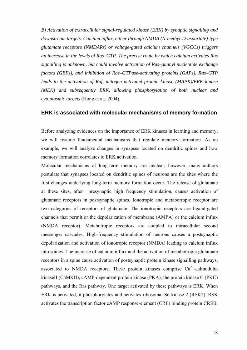
B) Activation of extracellular signal-regulated kinase (ERK) by synaptic signalling and
downstream targets. Calcium influx, either through NMDA (N-methyl-D-aspartate)-type
glutamate receptors (NMDARs) or voltage-gated calcium channels (VGCCs) triggers
an increase in the levels of Ras–GTP. The precise route by which calcium activates Ras
signalling is unknown, but could involve activation of Ras–guanyl nucleotide exchange
factors (GEFs), and inhibition of Ras–GTPase-activating proteins (GAPs). Ras–GTP
leads to the activation of Raf, mitogen activated protein kinase (MAPK)/ERK kinase
(MEK) and subsequently ERK, allowing phosphorylation of both nuclear and
cytoplasmic targets (Hong et al., 2004).
ERK is associated with molecular mechanisms of memory formation
Before analyzing evidences on the importance of ERK kinases in learning and memory,
we will resume fundamental mechanisms that regulate memory formation. As an
example, we will analyze changes in synapses located on dendritic spines and how
memory formation correlates to ERK activation.
Molecular mechanisms of long-term memory are unclear; however, many authors
postulate that synapses located on dendritic spines of neurons are the sites where the
first changes underlying long-term memory formation occur. The release of glutamate
at these sites, after presynaptic high frequency stimulation, causes activation of
glutamate receptors in postsynaptic spines. Ionotropic and metabotropic receptor are
two categories of receptors of glutamate. The ionotropic receptors are ligand-gated
channels that permit or the depolarization of membrane (AMPA) or the calcium influx
(NMDA receptor). Metabotropic receptors are coupled to intracellular second
messenger cascades. High-frequency stimulation of neurons causes a postsynaptic
depolarization and activation of ionotropic receptor (NMDA) leading to calcium influx
into spines. The increase of calcium influx and the activation of metabotropic glutamate
receptors in a spine cause activation of postsynaptic protein kinase signalling pathways,
associated to NMDA receptors. These protein kinases comprise Ca2+-calmodulin
kinaseII (CaMKII), cAMP-dependent protein kinase (PKA), the protein kinase C (PKC)
pathways, and the Ras pathway. One target activated by these pathways is ERK. When
ERK is activated, it phosphorylates and activates ribosomal S6-kinase 2 (RSK2). RSK
activates the transcription factor cAMP response-element (CRE) binding protein CREB.

19
CREB recruits a number of transcriptional co-activators for transcriptional activation of
genes important for memory consolidation (for review: Weeber et al., 2002; fig.4).
The involvement of ERK in mammalian long-term memory has been best characterized
for models of spatial learning and fear conditioning. The most common spatial learning
experiments generally assess the ability of an animal to learn and remember the location
of a hidden platform in a water maze test (Morris water maze). Training in the water
maze leads to activation of ERK in the hippocampus while systemic administration of
an inhibitor of ERK activation, SL32742, impairs the ability of animals to subsequently
remember the location of the hidden platform, also impairing performance in a maze
task (Selcher et al., 1999; Hebert et al., 1999). Another form of long-term memory, fear
conditioning, is also ERK dependent. Fear conditioning is generally studied by placing
an animal in a particular environment (or ‘context’), delivering an audible cue and then
administering a foot-shock that causes typical fear responses (such as freezing) when
the animals are re-exposed to the cue or to the context. As for the water-maze task,
training in this experiment leads to ERK activation (Schafe et al., 2000). On the
contrary, when re-presented to the cue or the context, the animals that were pre-treated
with MEK inhibitors freeze far less frequently, indicating that they have not learned to
associate the cue or context with the foot-shock. Finally, ERK activation occurs in
correlated forms of memory, such as conditioned taste aversion, a model of learning in
which animals learn to associate a new taste to an aversive reaction; this learning is also
prevented by ERK inhibition.
ERK activation is also important in fundamental types of synaptic plasticity, such as in
long- term potentiation (LTP) at hippocampal synapses or in forms of LTP in the
dentate gyrus and in the amygdale, on which it is associated to fear-dependent learning
(fig.4).

20
PKCPKC
Fig. 4: In a post synaptic spine ERK signaling can be activated by increasing
intracellular calcium via cAMP, PKA, PKC pathways. Upon the release of glutamate,
after presynaptic high frequency stimulation, NMDA-type glutamate receptors allow
Ca2+ entering into neuronal cell. As a consequence of calcium influx, phosphorylation
of CaMKII occurs. Phosphorylation of CaMKII is regulated by activation of
phosphatase 1, PP1, that leads to dephosphorylation of CaMKII. CaMKII in turn
positively regulates NMDA and AMPA receptors causing an increase of conductance.
Also, the increasing concentration of intracellular calcium can activate ERK pathway
though Ras. Ras can induce Raf to activate MEK that phosphorylate ERK.
Alternatively, the Ca2+/CaM (calmodulin) complex can activate adenylyl cyclases
resulting in the accumulation of cAMP, which in turn activates Rap1 (Ras homolog), via
protein kinase A (PKA). Rap1 may then activate B-Raf (Raf 1 homologous) which
activates MEK, which in turn activate ERK. Moreover, PKA can directly activate MEK
21
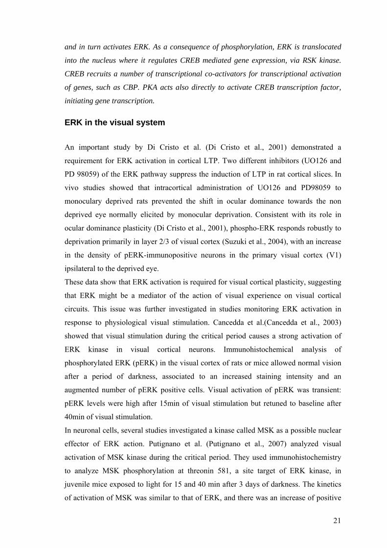
and in turn activates ERK. As a consequence of phosphorylation, ERK is translocated
into the nucleus where it regulates CREB mediated gene expression, via RSK kinase.
CREB recruits a number of transcriptional co-activators for transcriptional activation
of genes, such as CBP. PKA acts also directly to activate CREB transcription factor,
initiating gene transcription.
ERK in the visual system
An important study by Di Cristo et al. (Di Cristo et al., 2001) demonstrated a
requirement for ERK activation in cortical LTP. Two different inhibitors (UO126 and
PD 98059) of the ERK pathway suppress the induction of LTP in rat cortical slices. In
vivo studies showed that intracortical administration of UO126 and PD98059 to
monoculary deprived rats prevented the shift in ocular dominance towards the non
deprived eye normally elicited by monocular deprivation. Consistent with its role in
ocular dominance plasticity (Di Cristo et al., 2001), phospho-ERK responds robustly to
deprivation primarily in layer 2/3 of visual cortex (Suzuki et al., 2004), with an increase
in the density of pERK-immunopositive neurons in the primary visual cortex (V1)
ipsilateral to the deprived eye.
These data show that ERK activation is required for visual cortical plasticity, suggesting
that ERK might be a mediator of the action of visual experience on visual cortical
circuits. This issue was further investigated in studies monitoring ERK activation in
response to physiological visual stimulation. Cancedda et al.(Cancedda et al., 2003)
showed that visual stimulation during the critical period causes a strong activation of
ERK kinase in visual cortical neurons. Immunohistochemical analysis of
phosphorylated ERK (pERK) in the visual cortex of rats or mice allowed normal vision
after a period of darkness, associated to an increased staining intensity and an
augmented number of pERK positive cells. Visual activation of pERK was transient:
pERK levels were high after 15min of visual stimulation but retuned to baseline after
40min of visual stimulation.
In neuronal cells, several studies investigated a kinase called MSK as a possible nuclear
effector of ERK action. Putignano et al. (Putignano et al., 2007) analyzed visual
activation of MSK kinase during the critical period. They used immunohistochemistry
to analyze MSK phosphorylation at threonin 581, a site target of ERK kinase, in
juvenile mice exposed to light for 15 and 40 min after 3 days of darkness. The kinetics
of activation of MSK was similar to that of ERK, and there was an increase of positive

22
cells after 15 min of visual exposure returning to baseline after 40 min. Activation of
MSK was blocked by treating the visual cortex with UO126, an inhibitor of the ERK,
upstream kinase MEK. In slices of cortex treated by UO126, a strong reduction of MSK
positive cells was showed, with respect to the contralateral cortex used as control. In
addition, colocalization studies showed activation of MSK and ERK in the same cells.
These results demonstrated that ERK and its downstream kinase MSK are strongly
activated by visual experience in the visual cortex of developing animals.
CREB transcription factor is important in synaptic plasticity
CREB (cAMP response element binding protein) belongs to the bZIP superfamily of
transcription factors, contains a C-terminal basic domain that can bind DNA at CRE
sequence and a leucine zipper domain that facilitates dimerization; in addition, it has
two glutamine rich domains, separated by the kinase inducible domain (KID). Within
the KID resides the residue Ser-133, which when phosphorylated, is a target for the
transcriptional co-activator, CREB binding protein (CBP). CBP is a transcriptional co-
activator that interacts with multiple transcriptional regulators and facilitates the
assembly of the basic transcriptional machinery; CBP also acts as an HAT (Lonze et al.,
2002). Histone acetylation is one mechanism for the local and global control of
chromatin structure and it is crucial for facilitating gene expression. Based on the
observation that CREB phosphorylation and CRE-mediated gene expression occur in
response to mitogens, neurotrophins, and other neuronal growth factors, several studies
investigated whether CREB-mediated gene expression is both necessary and sufficient
for survival and/or degeneration of multiple neuronal subtypes. In Aplysia, long-term
facilitation, recapitulating a memory-like behaviour, CREB activation is necessary for
memory formation (Chen et al., 2003). After evidences on Aplysia, different studies
investigated CREB importance in LTP of hippocampal neurons. CREB dependent gene
expression appears to be required for spatial learning, as shown by the deficits in spatial
learning induced by intra-hippocampal infusion of CREB antisenses. Also in visual
development CREB function is crucial. Pham et al., (Pham et al., 1999) have shown that
monocular deprivation induces CRE-mediated gene expression robustly in the
monocular visual cortex of young mice (Pham et al., 1999). Mower et al., (Mower et al.,
2002; showed that CREB function is essential for ocular dominance plasticity during
monocular deprivation. Indeed, the expression in the primary visual cortex of a

23
dominant-negative form of CREB (HSV–mCREB) containing a single point mutation
preventing its activation blocked ocular dominance plasticity.
Cancedda et al. (Cancedda et al., 2003) also showed that visual stimulation during the
critical period activates CREB-mediated gene expression. These authors used a
transgenic model expressing the reporter gene lacZ, under the control of the CRE
sequences bound by CREB. They found an increase of cells positive for b-
galactosidase, the gene product of lacZ, in mice exposed to visual stimulation. Visually
induced CREB-mediated gene expression was not present in visual cortices infused with
the ERK inhibitor UO126. Which ones are the factors mediating the action of ERK on
CREB? Visual stimulation also activated the phosphorylation of CREB. A Western blot
analysis of the effect of visual stimulation was performed on phosphorylation of Ser 133
of CREB, a phosphorylation site necessary for CREB-mediated gene transcription. It
was found that visual stimulation induced an increase in CREB Ser 133 phosphorylation
after 40 min of stimulation in juvenile mice. Interestingly, CREB Ser-133 is a substrate
of MSK kinase, that acts downstream of ERK and is activated by visual experience.
In summary, ERK and CREB activation are important in learning and memory, as well
as in visual synaptic plasticity during the critical period. Indeed, the ERK pathway is
central in mediating the action of visual experience on gene transcription mediated by
CREB, a mechanism necessary for OD plasticity. It is not known however whether this
mechanism is still active in the adult organism, after the termination of the critical
period. In this research we focussed on the mechanisms that link visual experience
dependent activation of the ERK pathway to CREB-mediated gene expression in the
visual cortex during the critical period.
Several evidences indicate that epigenetic mechanisms coordinate the transcriptional
processes in neurons that participate in the formation of memories, such as behavioural
adaptation to stressful events. A model to study the underlying neurobiological
mechanisms of stress-related memory formation is the forced swim test. In this test,
animals are forced swimming and predominantly show immobility a re-test 24 h after
the first time. Forced swimming increased the phospho-acetylation of histone H3 in a
mature population of dentate granule neurons, correlated to gene expression of c-Fos in
dentate neurons, through recruitment of the ERK1 ⁄ 2 ⁄ MSK pathway (Chandramohan et
al., 2008). This suggests that, specific gene transcriptional events occur in these cells,
associated with modification of the chromatin structure. Here, we will analyze whether

24
visual experience is also able to trigger epigenetic modifications of chromatin in visual
cortical neurons of juvenile mice. In addition, we will analyze the ERK pathway and
CREB-mediated gene expression (Fig. 5) after at the end of critical period, when OD
plasticity decrease, to test the hypothesis that developmental differences of experience-
dependent activation of ERK pathway, or gene transcription regulation mechanisms are
present between juvenile and adult mice.
Kv 4.2MAP2
nucleus
trk
MEK
ERK
CREBRSK ?
Influx Ca++
gene expression
Raf-1
RasACPKA
mR
Metabotropicreceptors
rasGRF1
MSK
H3P
CBP
Ac
P
NT
phosphatases?
Visual experience
Kv 4.2MAP2
nucleus
trk
MEK
ERK
CREBRSK ?
Influx Ca++
gene expression
Raf-1
RasACPKA
mR
Metabotropicreceptors
rasGRF1
MSK
H3P
CBP
Ac
P
NT
phosphatases?
Visual experience
Fig.5: A possible mechanism of ERK pathway action in the juvenile visual cortex.
Question marks indicate the steps investigated in this thesis. Neurotrophins and
electrical activity modulate ERK through two separate pathways that converge on the
ERK kinase MEK. The neurotrophin signal converges from the tyrosine kinase
receptors (trk) to Ras, leading to activation of ERK and CREB (Ginty et al., 1994;
Pizzorusso et al., 2000). Electrical activity is translated in two different intracellular
signals: (1) an increase of calcium, caused by influx through voltage-gated channels
and NMDA receptors, and (2) an increase of cAMP attributable to activation of
metabotropic receptors (mR) or by Ca-dependent adenylate cyclase (AC). ERK

25
translocates to the nucleus and phosphorylates two Kinases, MSK and RSK, that in turn
phosphorylate CREB. CREB can interact with CBP leading toe acetylation of histone
H3, while MSK causes its phosphorylation. CREB transcription factor and histone
phosphoacetylase can active gene expression.

26
Chapter 2: Epigenetic modifications in pathology of nervous system
Chromatin remodelling in brain pathology Chromatin remodelling is an epigenetic mechanism important during the development
and normal functioning of the central nervous system. However, alterations in
epigenetic mechanisms can also damage brain development. Many neurodevelopmental
syndromes exhibit an abnormal epigenetic activation or silencing of selective genes. For
example, Rubinstein-Taybi syndrome (RTS) is a human genetic disease characterized
by mental retardation. This syndrome is caused by mutations of CREB-binding protein
(CBP), a transcriptional co-activator, that binds to the activated form of the CREB
protein and then it acts as a Histone AcetylTransferase (HAT). Abnormalities of CBP
function have been shown to underlie long-term memory deficits formation.
Alarcon et al. (Alarcón et al., 2004) studied a mouse model of RTS) that shows
abnormalities in long-term memory and in the late phase long-term potentation (L-
LTP). In these mice that showed a truncated form of CBP, chromatin acetylation, some
forms of memory and the late phase of hippocampal long-term potentiation (L-LTP)
were impaired. By inhibiting HAT, the molecular counterpart of histone acetylation
function of CBP, memory defects in these mice are reversed, underling the importance
of histone acetylation (Alarcón et al., 2004).
In this thesis, we will focus on a neurodevelopmental disorder, the RTT, characterized
by abnormal epigenetic regulation, as a model of neuropathology of nervous system
caused by improper chromatin remodelling.
Rett syndrome: clinical features
RTT is a progressive neurodevelopmental disorder that predominantly affects girls. The
incidence in the population is of 1/10,000 on female births. No effective
pharmacological treatment is as yet available for this disorder. Patients with RTT appear
to develop normally up to 6–18 months age, but they show phenotypes of illness with a
developmental regression between the ages of 1 to 3 years (Chahrour and 2007). Before
the illness, the child with RTT achieves appropriate milestones as a normal child,

27
including the ability to walk and some patients even say a few words. Unfortunately, by
the second year of life, there a child with RTT exhibits a deceleration of head growth
with microcephaly, accompanied by general growth retardation and weight loss. During
the time course of the disease, the syndrome is characterized by loss of use of patient’s
hand with development of stereotypic hand movements. In addition, patients manifest
loss of language, irritability and autistic features such as expressionless face, lack of
eye-to-eye contact and indifference to the surrounding environment. The clinical
features of this syndrome become more and more dramatic: girls evidence seizures,
breathing anomalies and deficits in motor coordination. Despite a normal appetite,
patients continue to lose weight and many suffer from osteopenia, scoliosis and rigidity.
At advanced states, most girls with RTT lose mobility and are often wheelchair-bound
during the teenage years (Chahrour and Zoghbi 2007). Finally, the disease becomes
stationary and some patients can survive up to the sixth or seventh decade of life in a
debilitated physical condition. In the next paragraphs, we will describe the main
concepts of molecular mechanisms that are correlated to RTT, focusing on the
importance of animal models for the study of this disease (Chahrour, Zoghbi ; 2007).
Phenotypic variability in RTT
Given that RTT patients are for the large majority females, early reports postulated an
X-linked dominant mode of inheritance, with lethality in males. However, in the
majority of the cases this syndrome is sporadic; so it was very difficult to map the
disease locus by traditional linkage analysis. Using the report of familial cases, an Xq28
candidate region on chromosome X has been postulated as responsible for the disease
(Amir et al., 1999). By screening candidate genes in patients with RTT, it has been
found that in many cases (in more than 95% of cases) the disease is caused by mutations
in the X-linked gene encoding methyl-CpG binding protein 2 (MeCP2).
In most of cases, these mutations are associated with a loss of MeCP2 function, due to
the introduction of premature stop codons throughout the gene that cause a null allele.
In other cases, hypomorphic alleles may also present causing truncated forms of MeCP2
that conserve partial function. By the clinical analysis of affects patients, it appears
difficult to consider RTT as one illness with a defined clinical picture.
There is a wide of spectrum of atypical forms of RTT that deviate from the classical
clinical of RTT. Milder variants exist, such as the ‘‘forme fruste’’ that has a later age of
28
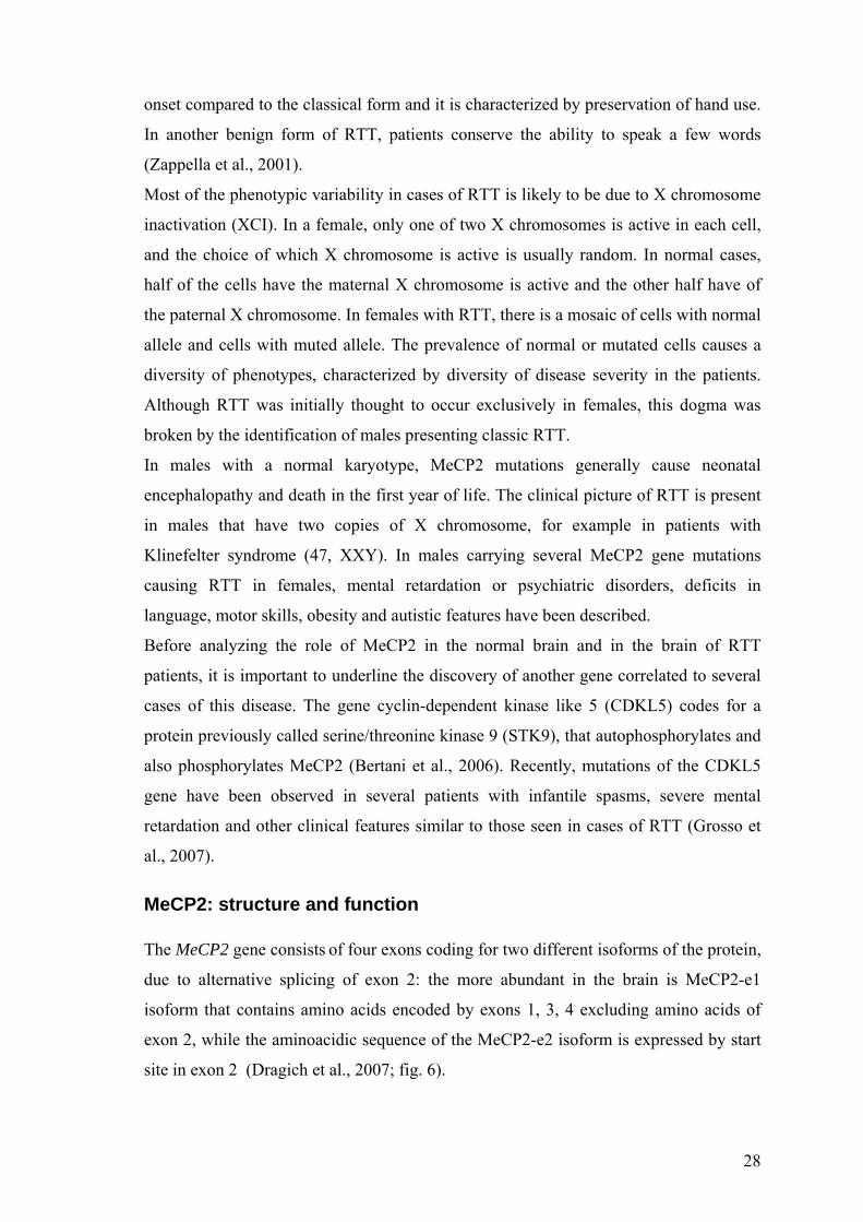
onset compared to the classical form and it is characterized by preservation of hand use.
In another benign form of RTT, patients conserve the ability to speak a few words
(Zappella et al., 2001).
Most of the phenotypic variability in cases of RTT is likely to be due to X chromosome
inactivation (XCI). In a female, only one of two X chromosomes is active in each cell,
and the choice of which X chromosome is active is usually random. In normal cases,
half of the cells have the maternal X chromosome is active and the other half have of
the paternal X chromosome. In females with RTT, there is a mosaic of cells with normal
allele and cells with muted allele. The prevalence of normal or mutated cells causes a
diversity of phenotypes, characterized by diversity of disease severity in the patients.
Although RTT was initially thought to occur exclusively in females, this dogma was
broken by the identification of males presenting classic RTT.
In males with a normal karyotype, MeCP2 mutations generally cause neonatal
encephalopathy and death in the first year of life. The clinical picture of RTT is present
in males that have two copies of X chromosome, for example in patients with
Klinefelter syndrome (47, XXY). In males carrying several MeCP2 gene mutations
causing RTT in females, mental retardation or psychiatric disorders, deficits in
language, motor skills, obesity and autistic features have been described.
Before analyzing the role of MeCP2 in the normal brain and in the brain of RTT
patients, it is important to underline the discovery of another gene correlated to several
cases of this disease. The gene cyclin-dependent kinase like 5 (CDKL5) codes for a
protein previously called serine/threonine kinase 9 (STK9), that autophosphorylates and
also phosphorylates MeCP2 (Bertani et al., 2006). Recently, mutations of the CDKL5
gene have been observed in several patients with infantile spasms, severe mental
retardation and other clinical features similar to those seen in cases of RTT (Grosso et
al., 2007).
MeCP2: structure and function The MeCP2 gene consists of four exons coding for two different isoforms of the protein,
due to alternative splicing of exon 2: the more abundant in the brain is MeCP2-e1
isoform that contains amino acids encoded by exons 1, 3, 4 excluding amino acids of
exon 2, while the aminoacidic sequence of the MeCP2-e2 isoform is expressed by start
site in exon 2 (Dragich et al., 2007; fig. 6).

29
Fig6: two isoforms of MeCP2 are due to alternative splicing of exon 2 (Chahrour et al.,
2007).
In addition, MeCP2 has a large, highly conserved 3′-untranslated region that contains
multiple polyadenylation sites, which can be alternatively used to generate four
different transcripts. Expression of different MECP2 transcripts is regulated during
different stages of development. The longest transcript is the most abundant in the brain,
with large expression during embryonic development, a decline after birth and a
subsequent increase in expression levels in the adult age.
The MeCP2 gene codes for a 80Kda protein (MeCP2), member of a family of methyl-
CpG- binding proteins, able to bind methylated DNA and playing the role of
transcriptional repressors. The MeCP2 protein contains four functional domains: (1) a
methyl-CpG-binding domain (MBD) which binds to 5-methyl cytosine of DNA; (2) a
transcriptional repression domain (TRD) which interacts with histone deacetylase and
transcriptional corepressor SIN 3A; (3) the nuclear localization signal, which may be
responsible for the transport of MeCP2 into the nucleus and (4) the C-terminal segment
which facilitates its binding to the nucleosome core (Matijevic et al., 2009).
MeCP2 is more abundant in the brain tissue where it is highly expressed in neurons and
very scarce in glial cells. MeCP2 is localized to cell nuclei, in pericentric
heterochromatin (fig7). Initially, MeCP2 protein is expressed in the spinal cord and in
Cajal-Retzius (C-R) cortical neurons; subsequently, its levels increase in the midbrain,
thalamus, cerebellum, and deep cortical neurons. Expression in basal ganglia,
hypothalamus, hippocampus, and superficial cortical layers appears later, and the
number of MeCP2-positive neurons in the cerebral cortex continues to increase until 10
years of age (Zoghbi; 2003; fig8).
.

30
5µm5µm
Fig7: nuclear localization of MeCP2 (green signal). Immunofluorescence experiments
on neuronal cultures from mice expressing GFP show that MeCP2 is localized in
pericentric heterochromatin (chromocenters).
Fig.8: spatial and temporal distribution of MeCP2 during human development. Wg:
weeks of gestation (Zoghbi; 2003).
The function of MeCP2 as a transcriptional repressor was first inferred from
experiments in vitro showing that MeCP2 specifically inhibited gene transcription from
methylated promoters. MeCP2 was also shown to recruit additional co-repressor
complexes, which remodel chromatin into a repressive state that does not permit gene

31
transcription. Deficiency or absence of Mecp2 leads to disorganization of chromatin
structure ultimately causing abnormal transcription (fig.9). Nevertheless, recent studies
by Chahrour et al., (Chahrour et al., 2008) on gene expression in the hypothalamus raise
the interesting issue on whether MeCP2 can function as both an activator and a
repressor of transcription. Indeed, Chahrour et al. (Chahrour et al., 2008) examined gene
expression patterns in the hypothalamus of mice that either lack or over-express
MeCP2. In both models, MeCP2 dysfunction induced changes in the expression levels
of thousands of genes in the hypothalamus; 85% of genes were up-regulated in mice
that over-express MeCP2 and only 377 genes were down-regulated in mice with loss of
MeCP2. These data suggest that, unexpectedly, the majority of genes (~85%) analyzed
in the hypothalamus are activated by MeCP2. In addition, they show that MeCP2 is
associated with the transcriptional activator CREB1, on the promoters of activated
target genes, but not at repressed sites, suggesting that MeCP2 can associate to CREB
activator, permitting activation of gene expression.
Fig.9: A) Transcription is generally suppressed in a promoter region containing
methylated CpGs that are bound by MeCP2. MeCP2 binds methylated DNA and
recruits a chromatin-remodelling complex that contains SIN3A, a transcriptional co-
repressor, a BRM chromatin remodelling protein and a histone deacetilases (HDACs).
This complex leads to chromatin condensation owing to histone deacetylation, which

32
results in a limited accessibility of the transcriptional machinery to promoter regions.
When MeCP2 is not bound to methylated DNA the complex is not recruited.
The lack of MeCP2 binding to DNA could be due to the activity of CDKL5, which is
thought to bind and contributes to the phosphorylation of MeCP2, resulting in the
inability of MeCP2 to bind methylated DNA; in each of these cases, histones remain
acetylated and the DNA at the promoter remains in a open conformation, allowing
transcription factors to bind DNA and initiate transcription. B) MeCP2 is also a
chromatin condensing protein and can repress gene expression independently of DNA
methylation. When MeCP2 is absent, gene expression is activated (Bienvenu and
Chelly 2006).
The BDNF gene: a target of MeCP2 MeCP2 regulates Brain-derived neurotrophic factor (BDNF) gene expression. BDNF
gene encodes a neurotrophic, factor essential for neuronal survival, differentiation and
synaptic plasticity. Rat BDNF gene is under the control of four promoters, one of which,
promoter III (that corresponds to mouse BDNF promoter IV), is activated by calcium
influx through L-type voltage-sensitive calcium channels conferring to BDNF
expression its regulation by electrical activity (fig. 10). Chen et al. (Chen et al., 2003)
investigated in wild-type cultured neurons that in the absence of neuronal activity,
MeCP2 is bound to the rat BDNF promoter III and cause its transcriptional repression.
Indeed, MeCP2 exhibits three major site of phosphorylation: one of them is S421, than,
when phosphorylated, controls the regulation of BDNF transcription. Through a
CaMKII dependent mechanism, phosphorylation of S421 on MeCP2 is an event
inducible upon membrane depolarization. Phosphorylated MeCP2 is released from the
promoter of the BDNF gene, thus allowing transcription (Chen et al., 2003). The
unphosphorylated form of MeCP2 cannot be released from the BDNF promoter in
response to membrane depolarization. Zhou et al. (Zhou et al., 2006) demonstrated that
in neurons expressing an S421A mutant form of MeCP2, the neuronal activity-
dependent transcription of the mouse BDNF promoter IV was impaired. So, while the
results obtained in cortical culture studies provide evidence that MeCP2 binds the
BDNF promoter and active BDNF transcription in an activity-dependent manner; one
would predict that, when all the neurons are silent, the MeCP2 mutant brain should
express more BDNF than the wild-type brain, such as at the basal level. Yet, when all

33
the neurons are active, the MeCP2 mutant brain should express the same amount of
BDNF as the wild-type brain, consequently to the highly induced level. However, many
in vivo data are in contrast with this hypothesis. The studies of Chang and colleagues
(Chang et al., 2006) investigated the role of BDNF in mouse models of RTT pathology.
In spite of predictions of an increase of BDNF level, these author found that BDNF
level were actually lower in extracts of brain from symptomatic knockout MeCP2 mice.
No difference was detectable between mutant and wild-type mice at the pre-
symptomatic stage (2 weeks of age). And yet, at an age when mice become
symptomatic (6–8 weeks of age), they found that the BDNF protein level in MeCP2
mutant brains was 69% of the wild-type level. They generated a conditional BDNF
knockout mouse, characterized by diminution of brain weight, by hind limb clasping
phenotype (feature common with MeCP2 mutants) and by reduction of the size of CA2
neurons and olfactory glomeruli. These experiments suggest that BDNF may mediate
part of the late RTT phenotype. In addition, deletion of BDNF in MeCP2 knockout
mice causes an earlier onset of locomotors dysfunction and a reduced lifespan, while
over-expression of BDNF improved locomotors function and extended their lifespan
(Chang et al., 2006). These results obtained in vivo, demonstrate the occurrence of
interactions between MeCP2 and BDNF and a correlation between altered BDNF levels
and neurological impairment in MeCP2 null mice. The discrepancy observed in BDNF
levels between data obtained in cell culture and results in vivo can be explained taking
into account that MeCP2 knockout mice and wt brains are at different activity states,
MeCP2 mice being hypoactive. Given that BDNF expression depends on neuronal
activity and that MeCP2 deficiency can reduce neuronal activity, the resultant effect
would be an indirect effect of MeCP2 removal upon decreasing BDNF protein level.
Chahrour et al. (Chahrour et al., 2009) studied gene expression in the hypothalamus of
MeCP2 mutants. Interestingly, they found that almost equal numbers of genes were up-
and down regulated by MeCP2 deletion raising the possibility that MeCP2 might act not
only as a repressor of gene transcription but also activating gene expression. Indeed,
they found that MeCP2 interacted with CREB in activating gene transcription. With
respect to BDNF, the authors found BDNF was up-regulated in mice overexpressing
MeCP2, while it was down-regulated in MeCP2 knockout animals. These results
suggest that MeCP2 can be an activator on the BDNF promoter, giving a possible
explanation of data reporting a decrease of BDNF levels in MeC2 knockout brains.

34
Fig.10: Structure of the mouse BDNF gene. Exons are evidenced in green by roman
numbers, introns are indicated in grey. Each of six alternative promoters controls the
expression of a unique mRNA that consists of one unique exon (numbered I through VI)
that is directly spliced to a common exon (exon VIII), which contains the entire BDNF
coding region (orange). The diversity of BDNF transcripts is made even greater,
because the second exon can be spliced from three alternative splice sites. Moreover,
transcripts initiated at exon VI can sometimes include an additional exon (exon VII).
Finally, two alternative polyadenylation sites can be utilized within the 3-UTR. The
regulatory element of the BDNF promoter IV is regulated by calcium -dependent
electrical activity. In this promoter, a functional sequence CRE has been identified that
binds CREB, which, in turn, binds CBP. CBP and the HDAC deacetylase enzyme
regulate acetylation of histones. MeCP2 can also bind the BDNF promoter regulating
gene expression. Calcium response factor (CaRF) and upstream stimulatory factors
(USFs) can bind to two additional calcium-responsive elements, identified within BDNF
promoter IV, in close proximity to CRE sequence (Flavell and Greenberg; 2008).
Mouse Models for studying RTT
Three mouse models of RTT have been generated (fig.11):
1) Mice with total deletion of MeCP2
2) Mice with a truncated form of MeCP2
3) Mice with over-expression of MeCP2

35
1) Chen et al. and Guy et al. generated mice with fully deleted MeCP2 sequences
(MeCP2 knockout mice). These MeCP2 conditional knockouts, lacking either exon 3 or
both exon 3 and 4 (Chen et al., 2001; Guy et al., 2001; fig.12) have a total deletion of
the MeCP2 gene. Female Mecp2+/_ mice (carrying one copy of the normal allele) have
behavioural abnormalities such as the males, but with a later age of onset. MeCP2
knockout males are normal until 3–6 weeks of age, when they develop a stiff,
uncoordinated gait, hypo-activity, tremor, hind limb clasping, and irregular breathing.
Symptoms worsen and ultimately lead to weight loss and death by 10 weeks. These
mice have deficits in respiratory rhythm due to alterations in bulbar post-inspiratory
discharges and to abnormal function of the pons leading to respiratory dysrhythmia and
apnoea. The brains of MeCP2 null mice appear smaller in size and weight than brains of
wild-type animals, although they lack major structural abnormalities, except for smaller,
more densely packed neurons. Recently, another MeCP2 null mouse model was
generated in which exon 3 and part of exon 4 were deleted. These mice are hypoactive
and have learning deficits and increased anxiety (Pelka et al., 2006).
2) Shahbazian et al. (Shahbazian et al., 2002) generated mice with a truncating mutation
of MeCP2, derived by the insertion of a stop codon into the MeCP2 gene at the position
corresponding to the amino acid 308, resulting in a hypomorphic allele that eliminates
the C-terminal region of the protein, similar to the C-terminal deletions found in RTT
patient (MeCP2308/Y mice). MeCP2308/Y mice have a phenotype less severe than
knockout mice and appear normal until six weeks, when they start to exhibit
stereotypes, hypoactivity, tremor, seizures, social behaviour abnormalities, increased
anxiety-related behaviour and learning and memory deficits. These symptoms are
similar to clinical aspects of illness in human patients. Female mice heterozygous for
the MeCP2 truncated gene display milder and more variable features.
3) The last model is the mouse with over-expression of human MeCP2 (MeCP2Tg mice)
under the control of its endogenous promoter. These mice present progressive
neurological abnormalities with the onset of phenotypes around ten weeks of age.
Initially, MeCP2Tg mice have increased synaptic plasticity and improvement in motor
and contextual learning abilities. However, at 20 weeks of age, transgenic mice become

36
hypoactive and are aggressive, undergo seizures and motor abnormalities, and die by 1
year of age (Collins et al., 2004).
Fig.11: table of mouse models s (Chahrour et al., 2007).
Fig.12: phenotypes of mice with the MeCP2-null mutation (Guy et al., 2001). Left and
middle panels show normal spreading of hind limbs in wild-type and six-week mutant
males, respectively. The right panel show typical limb-clasping phenotype present in
mutant males aged 7 weeks.

37
Synaptic plasticity is affected in RTT
Anatomically, the brains of RTT patients and MeCP2 null mice appear without gross
morphological abnormalities. However, reductions in brain size and of the whole CNS
have been described in human RTT patients and animal models of the disease. A
reduction of 12%–34% of brain weight and volume has been reported for the prefrontal,
posterior frontal, and anterior temporal regions of patients with RTT (Armstrong; 2005).
However, RTT is not a neurodegenerative process; there are no signs of degeneration,
atrophy or inflammation (Reiss, et al., 1993). The reduction of brain size in RTT
patients is accompanied by neurons that are abnormally small and densely packed, with
markedly shortened and simplified dendritic arbors. In the RTT frontal cortex, dendritic
spines appear sparse and short; in addition, reduced dendritic branching of layers III and
V pyramidal neurons in the frontal, temporal and motor regions, and of layers II and IV
of the subiculum have been described (Armstrong; 2005). Recently, it has been
suggested that excitatory synaptic activity may be particularly affected in brains with
RTT. Synapses that use the excitatory amino acid neurotransmitter glutamate play an
important role in circuits of the cerebral cortex, basal ganglia and brainstem responsible
for movement and breathing. Several neurochemical studies evidenced that glutamate-
mediated neurotransmission is disrupted in RTT. The analysis of cerebrospinal fluid in
RTT girls demonstrated elevations in glutamate (Hamberger et al., 1992). It appears that
the abnormal development of glutamate synapses is responsible for cortical hyper-
excitability and seizures.
In MeCP2 mutant mice, it has been found that pyramidal neurons in S1 slices are less
active at 2 weeks of age. The reduced cortical activity is progressive; the reduction is 4
fold when animals begin to exhibits symptoms. The decrease of spontaneous activity is
accompanied by reduced spontaneous excitatory synaptic inputs and by an increase of
inhibition (Dani et al., 2005). In MeCP2 null mice, it has been shown that hippocampal
glutamatergic neurons display a strong reduction in the synaptic response, affecting
excitatory synapses (Chao et al., 2007). Synaptic plasticity is also impaired: LTP is
reduced in cortical slices of MeCP2 null mice (Asaka et al., 2006) while cortical and
hippocampal slices obtained from Mecp2308/Ymice (with a truncated form of MeCP2)
show a reduction of LTP (Moretti et al., 2006). In contrast, there is an increase of LTP

38
in hippocampal slices from Mecp2Tg1mice, characterized by a doubling of MeCP2
(Collins et al., 2004).
Learning is compromised in MeCP2 mutants
Pelka et al. (Pelka et al., 2006) demonstrated deficits in cerebellar learning in rotarod
test in MeCP2-null mice. The rotarod test measures motor coordination and balancing
ability by a rotating rod that progressively accelerates. Pelka et al. (Pelka et al., 2006)
showed that successive trials on the rotarod for the MeCP2-null mice revealed
significant deficits in cerebellar learning, as mutant mice failed to show improvements
in their ability to stay on the rotarod, compared to wild-type littermates.
Hippocampus dependent learning is also compromised in male mice with a truncated
form of MeCP2. Moretti et al. (Moretti et al., 2006) studied mice with a truncated form
of MeCP2 (MeCP2 308/Y mice) finding deficits in learning in two tasks, respectively
implying hippocampus-dependent spatial learning and contextual fear conditioning.
Spatial learning was detected in ‘Morris water maze’ test. In this test, mice are trained
to find a hidden platform in a circular pool of water. Each mouse is given training trials
every day, for several consecutive days. The time necessary to the mouse to locate the
platform after training is recorded; when the platform is removed, the mouse that learnt
the platform position spent significantly longer time in the maze quadrant in which the
platform was previously located. Moretti et al. (Moretti et al., 2006) found that MeCP2 308/Y mice took significantly longer than control littermates in locating a submerged
hidden platform and spent similar amounts of time in all sectors of the pool, while wt
animals spent more time where the platform was previously located.
Contextual fear conditioning was also impaired in MeCP2 308/Y mice; a reduction in the
amount of freezing in MeCP2308/Y mice in a contextual fear conditioning test has been
described. A detailed table of learning deficits in mouse models and patients with RTT
is showed in fig.13.

39
Fig13: deficits in mouse models and in patients with RTT (Stearns et al., 2007).
Level of anxiety in MeCP2 mutants mice Several studies have demonstrated an increase of anxiety in MeCP2308/Y mice.
Behavioural tests, such as open field and elevated plus maze, revealed anxiety–like
behaviour in these mutants. This behaviour was correlated at an increase into circulation
levels of corticosterone, a glucocorticoid generally released by the adrenal cortex in
response to stress. In these mice, the high corticosterone level is accompanied by an
increase of Crh gene expression. Crh encodes for the Corticotropin-releasing hormone

40
(CRH), a neuropeptide important for the regulation of the response to stress (McGill et
al., 2006).
Disease reversibility in mouse models of RTT Recently, it has been shown that upon introduction of a copy of the MECP2 transgene
into MeCP2 null mice (so that MeCP2 protein levels become normal), neurological
abnormalities of null mice are rescued. Indeed, two groups recently demonstrated
disease reversibility in mouse models of RTT (Guy et al., 2007; Giacometti et al.,
2007). Guy et al. generated a mouse model in which endogenous MeCP2 is silenced
causing similar features of MeCP2 null mutation; however MeCP2 transcription
activation was inducible by a tamoxifen-dependent mechanism. Tamoxifen injections
caused activation of MeCP2 and led to either rapid death or complete phenotypic rescue
of the adult null mice. The associated toxicity was caused by MeCP2 over-expression
and not by tamoxifen injections. Using smaller and repeated doses of tamoxifen, the
toxicity was eliminated and the late-onset neurological phenotype of adult mutant mice
was reversed. These data provide evidence that MeCP2-deficient neurons are not
permanently damaged and MeCP2 loss of function is reversible, rescuing neurological
deficits. In an independent study, Giacometti et al. (Giacometti et al., 2007)
demonstrated partial disease rescue, by postnatal reactivation of MeCP2 in mutant
animals. The authors show that the expression of a MeCP2 transgene in postmitotic
neurons of Mecp2-null mutant mice can cause a phenotypical rescue of the symptoms.
They constructed a conditionally active Mecp2 ‘‘rescue transgene’’ that was activated
between P0 and P30. The most efficient symptomatic rescues (with significantly
prolonged life span and delayed behavioural deterioration) were obtained early at 1 and
2 weeks of age, in correlation to expression of MeCP2 in most neurons. However, at 4
weeks of age, the induction of MeCP2 transgene was less efficient in rescuing
symptoms.
The partial rescue in mice obtained by Giacometti (Giacometti et al., 2007) was
probably due to the use of non-endogenous promoters, since this was not seen by Guy
et al., (Guy et al., 2007) who showed how the rescue was more effective when MeCP2
was activated under the control of endogenous promoters. These results indicate that it
is difficult to obtain a total rescue of symptoms in RTT mouse models. In addition,
MeCP2 levels must be normal to regulate gene transcription for proper
neurodevelopment.

41
Different studies demonstrated that MeCP2 overexpression can also be detrimental.
Indeed, a mouse model that over-expresses MeCP2 initially presents deficits in
cerebellar learning and synaptic plasticity and afterwards develops seizures, spasticity
up to death at approximately 1 year.
In summary, available MeCP2 mutants show clear pathological alterations at
anatomical, functional and behavioural level reminiscent of RTT. In addition, the
absence of MeCP2 seems to strongly impair plasticity of brain circuits. Different studies
such as those of Giacometti et al. (Giacometti et al., 2007) or Guy et al. (Guy et al.,
2007) analyzed a genetic therapeutic approach for this syndrome. However, a successful
genetic approach proved to be difficult because in vivo MeCP2 levels must be tightly
regulated and subtle perturbations in protein levels result in deleterious neurological
consequences.
Up to now, it was not known whether environmental stimulation is capable of
overcoming at least part of the described deficits of mouse models of RTT. Recently, it
has been observed that multisensory treatments can help RTT patients. Clinical
behavioural treatments like music therapy accompanied by movement programs can
ameliorate communications in patient affected from RTT (Wigram et al., 2005).
In this conceptual framework, the second section of this thesis will analyze gene–
environment interactions in the development and plasticity in RTT, by investigating
whether a complex multisensory stimulation, elicited by rearing MeCP2 mutant mice in
an enriched environment, can mitigate the phenotype of these mutants.
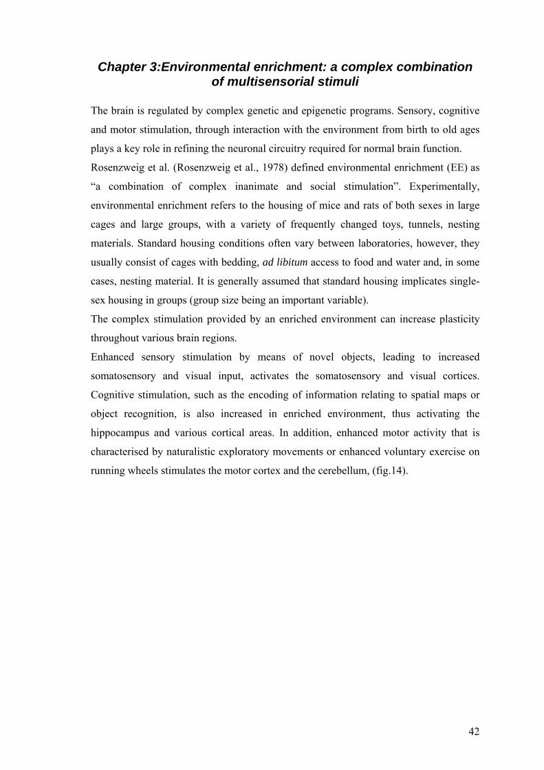
42
Chapter 3:Environmental enrichment: a complex combination of multisensorial stimuli
The brain is regulated by complex genetic and epigenetic programs. Sensory, cognitive
and motor stimulation, through interaction with the environment from birth to old ages
plays a key role in refining the neuronal circuitry required for normal brain function.
Rosenzweig et al. (Rosenzweig et al., 1978) defined environmental enrichment (EE) as
“a combination of complex inanimate and social stimulation”. Experimentally,
environmental enrichment refers to the housing of mice and rats of both sexes in large
cages and large groups, with a variety of frequently changed toys, tunnels, nesting
materials. Standard housing conditions often vary between laboratories, however, they
usually consist of cages with bedding, ad libitum access to food and water and, in some
cases, nesting material. It is generally assumed that standard housing implicates single-
sex housing in groups (group size being an important variable).
The complex stimulation provided by an enriched environment can increase plasticity
throughout various brain regions.
Enhanced sensory stimulation by means of novel objects, leading to increased
somatosensory and visual input, activates the somatosensory and visual cortices.
Cognitive stimulation, such as the encoding of information relating to spatial maps or
object recognition, is also increased in enriched environment, thus activating the
hippocampus and various cortical areas. In addition, enhanced motor activity that is
characterised by naturalistic exploratory movements or enhanced voluntary exercise on
running wheels stimulates the motor cortex and the cerebellum, (fig.14).

43
A BA B
Fig.14: A) Enriched housing conditions (Nithianantharajah and Hannan; 2006).
Environmental enrichment causes an enhanced sensory, cognitive and motor
stimulation on different brain regions. B) Standard housing conditions.
Environmental enrichment in wild-type rodents EE increases synaptic plasticity: anatomical and behavioural evidences
Many studies reported positive effects of gene–environment interactions in the
development and plasticity of the normal brain in wild-type rodents.
Early studies investigated the effects of EE on brain anatomy; animals exposed to EE
showed an increase of cortical weight and thickness, correlated to an increase in nerve
cell size, in dendritic arborizations and in the number and length of dendrites (Diamond,
2001; Rosenzweig e Bennett, 1996). More recently, it has been shown that the exposure
to EE increases hippocampal neurogenesis, (Bruel-Jungerman, E et al., 2005), due to
mechanisms involving vascular endothelial growth factor (VEGF; During, M.et al.,
2006) and it reduces naturally occurring cell death.
One interesting effect of EE is its capacity to act at the behavioural level. The exposure
of animals to a more complex environment (EE) improves spatial learning and memory,
as shown by the studies of Leggio et al.( Leggio et al.,2005) on adult rats, enriched for
three months after weaning. These animals learnt the location of the hidden platform of
Morris water maze faster than control animals, suggesting that rearing in an enriched
environment increases the abilities to solve spatial problems. In addition, it has been

44
shown that, EE decreases anxiety (Benaroya-Milshtein et al., 2004) and reduces
cognitive decline in aged animals (Bennett et al., 2006).
Molecular factors modulated by EE
At molecular level, EE causes alterations in the expression of genes involved in
synaptic function and cellular plasticity. Neurotrophins, such as brain-derived
neurotrophic factor (BDNF) and nerve growth factor (NGF), known as regulators of
structural and functional plasticity, increase after EE (Ickes, et al., 2000).
The expression of synaptic proteins, including the presynaptic vesicle protein
synaptophysin and postsynaptic density-95 protein (PSD-95), increase in enriched
animals in correlation to increased synaptogenesis (Frick et al., 2003) also. In addition,
enrichment is correlated to alterations in the expression of NMDA (N-methyl-d-
aspartate) and AMPA (α-amino-3-hydroxy-5-methyl-4-isoxazole propionic acid)
receptor subunits (Naka et al., 2005), suggesting an increase of synaptic plasticity,
including long-term potentiation (LTP; Duffy et al., 2001).
Finally, it is worth noticing several studies indicating that the effects of EE might be
mediated by modifications induced through chromatin remodelling. Recently, Fischer et
al. (Fischer et al., 2007) examined the beneficial effects of EE in the p25 transgenic
mouse. The p25 protein has been implicated in neurodegenerative disorders such as
Alzheimer’s disease. Fischer et al. (Fischer et al., 2007) reported that the induction of
the p25 transgene in adult mice causes learning and memory impairments and
neurodegeneration. However, if mice with anatomical and functional deficits were
introduced into an enriched environment for 4 weeks, the ability to form new memories
was rescued. The authors detected an increase in histone acetylation in the hippocampus
and in the cortex of wild-type enriched mice, suggesting that the effects of EE in the
p25 transgenic might be mediated, at least in part, by histone acetylation (Pizzorusso et
al., 2007). Interestingly, the administration of an histone deacetylation inhibitor that
promotes histone acetylation, rescues learning deficits in these mice, mimicking the
action of EE.
Effects of early enrichment on neuronal development
Differently from the large amount of data obtained about adult animals, the effect of EE
during development has been analyzed only very recently. One source of precocious

45
sensory experience for the pup is the mother. Maternal cares have important effects on
the development of stress responses in offspring in their adult age and increase after EE
(Sale et al., 2004). In addition, recent studies suggest that postnatal development of the
visual system can also be regulated by early environment. We will describe in detail
these two paradigms that explain how EE can act early in life.
Maternal cares
In rodents, there are natural variations in care levels of mothers. It is possible to
distinguish mothers with high or low maternal cares, according to the frequency with
which they perform characteristic maternal behaviours towards their pups, and namely
licking/grooming (LG) and arched back nursing (ABN).
Two experimental protocols reveal the influence of maternal experience on the
development of stress responses in offspring: ‘handling’ and maternal separation.
‘Handling’ is a treatment consisting in a brief (3-15 min) period of daily maternal
separation of rat and mouse pups, during the first two postnatal weeks. On the contrary,
maternal separation consists in longer periods (3-6 h) of daily separation from the
mother. It has been shown that postnatal handling increases the levels of maternal care
received by handled pups (Francis et Meaney 1999), reducing stress responses in adult
offspring, at both behavioural and endocrine level. On the contrary, maternal separation
enhances the responses of adult offspring to stress associated with adult cognitive
deficits (Oitzl et al., 2000). Studies on animal models have also shown that the offspring
of mothers that had received high levels of maternal cares in adult ages exhibit a
decreased of level stress, with increased spatial memory and enhanced BDNF-mRNA in
the hippocampus.
Recently, different studies have investigated molecular mechanisms involved in
response to stress by maternal cares. Central corticotropin-releasing factor (CRF)
system regulates the activation of behavioural, emotional, autonomic and endocrine
responses to stress. CRF is produced by two neuronal regions: the paraventricular
nucleus of the hypothalamus and the central nucleus of the amygdale. CRF acts on the
adrenal glands, eliciting the release of glucocorticoids, which ultimately mediate the
organism response to stress. The difference in stress response between the offspring
with high and those with low maternal cares is correlated to the difference in DNA
methylation of glucocorticoid receptor (GR) gene promoter, at hippocampal level
(Weaver et al., 2004). DNA methylation is an epigenetic modification that regulates

46
gene expression suppressing transcription. In offspring with extensive maternal cares,
there is a decrease of methylation of the glucocorticoid receptor (GR) gene, associated
with an active chromatin structure and an increased transcriptional activity, resulting in
augmented GR gene expression. GR enhances glucocorticoid negative feedback
sensitivity and decreases hypothalamic corticotrophin release factor (CRF) mRNA
levels, reducing the response to stress (Meaney et Szyf ; 2005).
Different studies investigated the consequences of EE on maternal cares. Sale et al.
showed that substantial differences in maternal care behaviour occur between EE and
standard conditions. The time spent in the nest by enriched mothers is higher with
respect to that of SC mothers. Maternal behaviour is critical for normal growth of
newborns and is associated to hormonal regulation of growth and with development of
neuroendocrine and behavioural responses to stress (Sale et al., 2004). Different levels
of maternal cares received by pups in different environmental conditions could explain
the earliest effects of EE on the development of the visual system. Indeed, eye-opening
is significantly accelerated of about 2 days in EE with respect to SC mice. Several data
showed that experiences acquired at early ages can be essential in promoting and
regulating neural development: the complex stimulation of enriched environment can be
crucial for the developmental plasticity of the visual system (Sale et al., 2004).
Early EE accelerates visual development
EE animals show a pronounced acceleration of visual acuity maturation, tested by
means of electrophysiological recordings of visual evoked potentials (VEPs) in the
visual cortex and by behavioural tests (Cancedda et al., 2004). At P25, the visual acuity
of EE mice is higher than that of SC mice by more than a factor of 2. In EE mice, visual
acuity development is accelerated significantly by 6 days as compared to SC mice. It is
interesting to underline that EE leads to an acceleration of visual development in a
temporal window, the critical period, characterized by high experience-dependent
plasticity. EE regulates the critical period time course, as suggested by the precocious
closure of critical period for ocular dominance plasticity in response to monocular
deprivation (Medini et al., 2008). EE effects on visual development are correlated to an
increase of BDNF in enriched animals that, in turn, stimulates the inhibitory
GABAergic system. A precocious increase of BDNF levels is detected in pups at P7 and
correlates to an increase of the GABA biosynthetic enzymes (GAD65-67). BDNF and
GAD65-67 increased levels are associated to precocious and robust CRE-mediated gene

47
expression in the developing visual system of EE mice, as shown using transgenic mice
carrying the lacZ reporter gene under the control of the CRE promoter (Sale et al.,
2004).
Environmental enrichment in rodent models of CNS disorders
Many studies have investigated the role of enriched environment in the dysfunctional
brain (fig 15). We will briefly describe the effects of EE on various diseases underlying
how EE could be a positive modulator of the phenotype of these models. The
comparison between murine models of diseases reared in standard and EE conditions
could represent a valuable tool to unravel molecular factors regulating the manifestation
and the severity of symptoms.

48
Fig. 15: Influence of environmental enrichment on CNS disorders (Nithianantharajah
and Hannan; 2006)
Huntington’s disease
Huntington’s disease (HD) is an autosomal dominant brain disorder, characterized by
degeneration of the cerebral cortex and of the striatum. HD causes movement disorders
(including chorea), cognitive deficits (dementia) and psychiatric symptoms. In this
disease, an expanded CAG translates into an expanded polyglutamine tract in the N-
terminal region of the huntingtin protein, causing neuronal dysfunction and death.
Transgenic HD mice have been generated, in which the CAG repeats that causes an
extended polyglutamine expansion are stably expressed. These mice develop

49
neuropathology, as well as motor and cognitive symptoms similar to those seen in
clinical HD and, in addition, exhibit intracellular inclusions due to pathological protein
aggregation. These are also found in the brains of patients with HD. Exposure to EE of
transgenic HD mice delays the onset of motor symptoms and prevents associated
cerebral atrophy. Behaviourally, EE causes clasping onset delay and rescues motor
deficit on rotarod test in these transgenics. At molecular level, EE reduces the diameter
of protein aggregates in the cortex and rescues striatal and hippocampal BDNF protein
deficits (van Dellen et al., 2000).
Alzheimer's disease
Alzheimer's disease (AD) is a progressive neurodegenerative disorder of the CNS,
causing the most common form of dementia. The illness is characterised by an initial
impairment of cognitive function and subsequently by memory loss, abstraction and
language deficits. The disease is characterized by two pathological hallmarks, senile
plaques and neurofibrillary tangles (NFTs). Plaques are extracellular deposits of
amyloid that consist of Aβ peptide, derived from proteolysis of the amyloid precursor
protein (APP). NFTs are intraneuronal aggregations of hyperphosphorylated forms of
the microtubule-associated protein tau. Mutations in APP, presenellin1, presenellin2
genes cause familial AD. For studying this disease, mouse models have been generated
s, the most common of which being the one carrying a mutation of APP. Current
literature shows that EE is important for ameliorating cognitive decline in transgenic
models of familial AD. However, there are controversies about EE influence on Aβ
peptide deposition that causes toxicity. Lazarov et al. (Lazarov et al., 2005)
demonstrated that Aβ levels and amyloid deposition are reduced by EE in a transgenic
mouse over-expressing APP. On the contrary, Jankowsky et al. (Jankowsky et al., 2005)
analyzed the effects of EE on transgenic mice coexpressing a variant of APP and a
variant of presenilin. These mice show cognitive deficits in several hippocampal-
dependent spatial memory tasks. EE ameliorates these deficits, but unfortunately these
effects are accompanied by increased Aβ levels.
Berardi et al. (Berardi et al., 2007) explored the possibility that Environmental
Enrichment (EE) may reduce the disease progression in a comprehensive mouse model
for AD like neurodegeneration, the AD11 mice. They found that in AD11 mice exposed
to EE from 2 to 7 months of age, performance in visual recognition memory and spatial
memory tests was significantly better than in non EE AD11 mice, and indistinguishable

50
from that of wild-type mice of the same age. Exposure to EE from 2 to 7 months
significantly reduced the appearance of AD neuropathological hallmarks, such as Aβ
levels (Berardi et al., 2007).
Parkinson’s disease
Parkinson’s disease (PD) is caused by a combination of genetic and environmental
factors. The illness is characterized by muscular rigidity, a characteristic tremor, and
impairments in cognitive function, accompanied by dementia in the late stage. At
molecular level, the disease shows loss of dopaminergic neurons in the substantia nigra,
accompanied by the formation of cytoplasmic inclusions called Lewy body. The
treatment in rodent animals of 1-methyl-4-phenyl-1,2,3,6-tetrahydropyridine (MPTP), a
major neurotoxin, causes key pathogenic features of human PD. It has been shown that
an enriched environment provides significant protection against both the locomotor and
cellular effects of MPTP-induced neurodegeneration in rodents, through a molecular
mechanisms correlated to downregulation of dopamine transporter (DAT), required for
MPTP-induced dopaminergic neurotoxicity (Bezard et al., 2006).
Amyotrophic lateral sclerosis
Amyotrophic lateral sclerosis (ALS) is the most common form of motor neuron disease.
ALS is characterized by the degeneration of motor neurons in the cortex, brainstem and
spinal cord. The most used mouse model of ALS over-expresses the mutant human form
of superoxide dismutase-1 (SOD1), a well-known anti-oxidant enzyme. Several studies
investigated the importance of EE on the onset and progression of disease symptoms in
transgenic ALS mice (with a SOD1 mutation). EE significantly improved motor
performance in these mice (Nithianantharajah and. Hannan; 2006)
Disorders of nervous system development: Fragile X syndrome and Down syndrome
Fragile X syndrome, the most frequent form of hereditary mental retardation, is due to a
mutation of the fragile X mental retardation gene FMR1 on the X chromosome. In
mouse models, deletion of , FMR1 gene causes abnormal cognitive and neuronal
features. Enrichment of FMR1 knockouts rescues behavioural abnormalities, such as an
exploratory behaviour and increases dendritic branching and spine number, usually
affected in KO mice.
Down syndrome is the most significant genetic cause of mental retardation and involves
trisomy of chromosome 21. The most commonly used murine model of DS is the

51
Ts65Dn (TS) mouse, trisomic for the region of the mouse chromosome 16 orthologous
to human chromosome 21. TS mice show an alteration of neural morphology,
reductions in size and number of neurons of different cerebral areas, neuronal
degeneration and modifications in synaptic number and size At behavioural level, TS
mice have reduced capacity of attention and alterations in learning and memory. EE
improves spatial learning in female Ts65Dn mice compared to standard conditions
(Dierssen et al., 2003).
Environmental enrichment in murine models of RTT
Recently, two studies have analyzed the effects of EE on MeCP2 mutant mice. The first
is the study of Kondo et al., (Kondo et al., 2008) on the effects of EE starting from 4
weeks of age on hemizygous males and heterozygous females null for the MeCP2 gene
(Mecp2tm1Tam-null mutant mice; Pelka et al., 2006). EE improved motor coordination in
heterozygous females but not in males. In addition, the authors detected a significant
effect of EE on hippocampal BDNF levels, but not in the cortex and cerebellum, of
female mice; male mice showed identical deficits of cerebellar BDNF, irrespective of
their housing environment.
The second study of EE in MeCP2 mutants is that by Nag et al. (Nag et al. 2009), who
analyzed the effects of EE at weaning on MeCP2 null mice. These −/0 mice have a
deletion of exon 3 of the MeCP2 gene, resulting in a loss of functional MeCP2 protein.
Locomotor activity in mutant mice rised in SC is reduced; on the contrary this is
significantly improved. in EE −/0 mice. Given that −/0 mice have regional specific
deficits in the brain, Nag et al. analyzed volumes of the whole brain, cerebellum and
motor cortex that resulted unchanged after EE, while the ventricular volume of −/0 mice
was reduced.
These results suggest that the brain volume (and presumably that of both the gray and
white matter) increased after EE, supporting the idea that structural changes in neurons
and dendrites might occur after EE. These studies also suggest that symptomatic
alleviation of RTT may be achieved by enhanced sensory, cognitive and motor
stimulation. However, the effects observed in these studies are quite limited. And yet, it
is possible that the relatively weak rescue of molecular and behaviour deficits obtained
in MeCP2 mutants by EE in these studies can be attributed to the late age of initiation of

52
EE (4 weeks), when the onset of symptoms likely prevents the animals from taking full
advantage of the environmental stimulation.

53
THESIS SCOPE AND DESIGN
The general aim of the present thesis was to investigate the role of epigenetic
mechanisms in the development of the nervous system in normal and pathological
conditions. The experiments performed can be divided into two main experimental
sections. In the first we focused on developmental plasticity of the visual cortex, a
paradigmatic example of nervous system development in normal conditions. This model
was used to analyze how epigenetic mechanisms regulate plasticity during development
and in the adult. In the second set of experiments, we focused on a human neurological
disorder, the Rett Syndrome (RTT), an example of brain dysfunction due to altered
regulation of epigenetic mechanisms.
Experience-dependent induction of post-translational modifications of histones in visual cortex We investigated whether posttranslational modifications of histones regulate plasticity
during the critical period and in adult mice after visual stimulation. By analysis of
western blots, we focused on acetylation and phosphorylation of histones that regulate
chromatin remodelling and control activation of transcription. Different regulations of
histone modifications were analyzed during the critical period and in the adult age, in
correlation with CREB mediated gene expression.
Environmental enrichment early in life in mice mutants for MeCP2 We investigated whether an early enhanced sensory-motor stimulation could moderate
the phenotype of MeCP2 mutant mice, an animal model of human RTT. To explore this
possibility, we used a protocol of strong early enrichment from P10, on mouse mutants
reared in either enriched or standard condition. The assessment of the effects of
enrichment was performed using a multidisciplinary approach. We designed two series
of experiments that employed male or female mice with mutations for MeCP2, for
analyzing different aspects of the anatomical, physiological, behavioural and molecular
alterations present in these mutants.

54
MATERIALS AND METHODS
Animal Treatment for the analysis of histone modifications and CREB
phosphorylation
Mice (P27–P28 or P80–P100) were sacrificed in darkness (dr) or at various time points
upon light exposure after 3 days of dr. A group of animals was implanted, in one cortex,
with osmotic minipumps (ALZET 1007D, 0.5 ml/hr) connected to a cannula (gauge 30,
model 1007D; Alzet, Palo Alto, CA) via a PVC tubing, at a cerebral location 4 mm
anterior and 3 mm lateral to lambda. Minipumps were filled with 250 mMUO126
(Promega) or vehicle (DMSO 0.5% in saline). Another group of mice received an
intraperitoneal injection (100 ml/kg i.p.) of SL327 (Bristol-Myers Squibb, Princeton,
New Jersey, USA) before light exposure, while control mice received the same injection
of vehicle (DMSO). The effectiveness of ERK inhibition was controlled by
immunohistochemistry for pERK.
Animal treatment and animal housing of MeCP2 mutants mice
Experimental subjects were derived from heterozygous Mecp2tm1–1Jae knock-out females
(MeCP2+/-) (Chen et al., 2001). Females were originally crossed to C57BL6 for two
generations, followed by breeding amongst offspring of the same generation with
breeder changes, and were maintained on a mixed background. Mixed background
reduced mortality and was necessary to obtain the high numbers of mice required by
extensive analysis. Age-matched littermates were used in all experiments to control for
possible effects of genetic background unrelated to the MeCP2 mutation (Wolfer et al.,
2002). Mutant animals and their corresponding wild-type littermates were housed under
standard or enriched conditions starting from P10. Weight of male mutant mice and WT
littermates was recorded every two days starting at P18.
The standard environment consisted of standard laboratory cages (26 × 42 × 18 cm)
housing two or three mice. The enriched environment consisted of a large cage (44 × 62
× 28 cm) containing various types of nesting material, a running wheel, and differently
shaped objects (tunnels, shelters, stairs) that were repositioned once per day to maintain
novelty and completely substituted with others once a week. Every cage housed at least
two mothers, 10 pups, and two additional filler females (Cancedda et al., 2004).
Animals were maintained in a 12-h light–dark cycle. All behavioural tests were

55
conducted between 08:00 and 18:00 h during the light phase. Food and water were
available ad libitum in all boxes. All experiments were carried out in accordance with
the European Communities Council Directive of 24 November 1986 (86/609/EEC) and
were approved by the Italian Ministry of Health.
Electron microscopy
Male P52 WT and MeCP2 mutant mice were anaesthetized with an intraperitoneal
injection of chloral hydrate and transcardially perfused with ice cold 2%
paraformaldehyde and 2,5% glutaraldehyde in 0.1 M phosphate buffer (PB; pH 7.4).
After perfusion, brains were removed from the skull, postfixed in the same solution
overnight at 4°C and then washed several times in 0.1M PB. Slices (500μm) were cut
through the primary somatosensory (S1) cerebral cortex and the cerebellar cortex using
a vibratome (Leica VT 1000 S, Leica Microsystems; Nussloch, Germany), postfixed
with 1% osmium tetroxide (in 0.1M cacodylate buffer) for 1 hr, dehydrated in a graded
series of ethanol (30-100%) and infiltrated with Epon-Araldite. The slices were
subsequently flat embedded in rubber molds. Ultrathin serial sections (70nm) were cut
with an ultramicrotome (Leica Ultracut, Germany), collected on pioloform-coated
single slot grids and counterstained with uranyl acetate and lead citrate. The sections
were observed with a JEM-1010 electron microscope (Jeol, Japan) equipped with a
side-mounted CCD camera (Mega View III; Soft Imaging System GmbH, Muenster,
Germany). The density of excitatory synapses was evaluated using the double dissector,
an unbiased stereological method (Sterio, 1984). Two adjacent serial sections were
photographed, the first of which was designated as the reference section and the second
as the look-up section. In each mouse, 25 pairs of nonoverlapping electron-micrographs
were taken at a magnification of X20,000 in the third layer of the somatosensory cortex
and in the molecular layer of the cerebellum. The criterion that allowed the
identification of a presynaptic terminal was the presence of 2-3 vesicles within the
profile, adjacent to the presynaptic membrane. The presence of a postsynaptic density
was used to identify a postsynaptic profile. Within an unbiased counting frame of area
28 μm2, the number of synapses that were present in the reference section but absent in
the look-up section were manually counted by a naïve observer.

56
Electrophysiology
Male aged P45-P50 belonging to the WT and MeCP2 mutant strains were used. Mice
were quickly anaesthetized by inhalation of 0.1 cc isoflurane and then decapitated. The
brain was rapidly removed and immersed in ice-cold dissection buffer (composition in
mM: NaCl, 132.8; KCl, 3.1; HEPES, 10; NaHCO3, 4; ascorbic acid, 1; myo inositol,
0.5; sodium piruvate, 2; K2HPO4, 1; glucose, 5; MgCl2, 2; and CaCl2, 1), bubbled with
oxygen. The somatosensory cortex was rapidly removed and 340 μm-thick coronal
slices were cut using a Leica VT 1000 S microslicer (Leica Microsystems; Nussloch,
Germany). Slices were allowed to recover for 1 h at 30°C in a submersion chamber
containing oxygenated artificial cerebrospinal fluid (ACSF), and then moved to room
temperature until use. ACSF contained 132.80 mM NaCl, 3.10 mM KCl, 1.00 mM
K2HPO4, 10.00 mM HEPES, 1.00 mM ascorbic acid, 0.50 mM myo inositol, 2.00 mM
sodium piruvate, 4.00 mM NaHCO3, 1 mM MgCl2, 2 mM CaCl2, and 5.00mM
glucose, saturated in oxygen. Slices were transferred into an interface recording
chamber and perfused with 31°C ACSF at a rate of 1.5 ml/min. Extracellular glass
electrodes (filled with ACSF; 1.0 MΩ) placed in layer 2/3 were used to monitor field
excitatory postsynaptic potentials (fEPSPs) evoked by a stimulating electrode
(concentric bipolar tungsten) placed in layer 5-6 at a location underlying the recording
electrode. The magnitude of responses was monitored by measuring the peak amplitude
of the field potential. Test stimuli consisted of a 100 μs pulses given every 30 s. Signal
was filtered (0.1-3 kHz), amplified (1000x), and captured at 10 kHz on a PC using Lab
view software (National instruments, USA). After measuring the input-output curve,
stimulation intensity was adjusted to evoke a response 50% of the maximal value. After
a 20 minute stable baseline, LTP was induced using theta burst stimulation (TBS). TBS
protocol consists in 2 events, 15 sec apart, each event consisting of 10 trains delivered
every 200 ms consisting of 4 pulses at 100 Hz. Field potential amplitudes were recorded
every 30 s for 45 minutes following the cessation of the stimulation protocol. The data
were normalized, averaged, and reported as means ±SEM.
RT-PCR analysis
The samples were lysed and RNA was extracted using Tripure (Roche) and
retrotranscribed using the Qunatitect kit (Qiagen). mRNA was quantified by means of
qPCR using Taqman inventoried assays by Applied Biosystems. Data were analyzed
using the ΔΔCt method using GAPDH to normalize mRNA levels.

57
Western Blotting
Experimental protocol for analysis of acetylation histones and phosphorylation CREB
A total of 136 C57BL6/J mice were killed by decapitation, and brains were removed
rapidly and frozen on dry ice. Cortices were then homogenized in a hypotonic lysis
buffer containing 10 mM Tris (pH7.5), 1 mM EDTA, 2.5 mM sodium pyrophosphate,
1mMbglycerophosphate,1mMsodiumorthovanadate,1mMphenylmethylsulfonylfluoride,
10 mg/ml Aprotinin, 10 mg/ml Leupeptin (Sigma, Italy), and 1%Igepal CA-630.
Histones were extracted from the nuclear fraction by the addition of five volumes of 0.2
MHCl and 10% glycerol, and the insoluble fraction was pelleted by centrifugation
(18,000 3 g; 30 min; 4°Ċ). Histones in the acid supernatant were precipitated with ten
volumes of ice-cold acetone followed by centrifugation (18,000 3 g; 30min; 4°C). The
histone pellet was then resuspended in 9M urea. Protein concentration was determined
by Biorad assay (Biorad, Italy). Each sample, consisting of pooled cortices of 1–3
animals, was boiled, and 30 mg/lane was loaded into 12% acrylamide gels using the
Precast Gel System (Biorad, Italy). Samples were blotted onto nitrocellulose membrane
(Amersham, Bucks, UK), blocked in 4% nonfat dry milk in Tris-buffered saline for 1
hr, and then probed with the following antibodies: AcH3, H3, AcH4, H4, pCREB
(Upstate, NY), and CREB (Cell Signaling, Beverly, MA). All antibodies were diluted in
TTBS and 2% milk or 2% BSA and incubated overnight at 4C. Blots were then rinsed
for 20 min in TTBS, incubated in HRP-conjugated anti-mouse or anti-rabbit (1:3000
Biorad, Italy, in 2% milk or 2% BSA and TTBS), rinsed, incubated in enhanced
chemiluminescent substrate (Biorad, Italy), and exposed to film (Hyperfilm, Amersham
Biosciences, Europe). Films were scanned, and densitometry was obtained through
Metamorph Offline software. To minimize variability, each sample was loaded in
parallel in two lanes and two gels were run simultaneously on the same apparatus. For
each gel, the corresponding filters obtained after blotting were cut in two parts to obtain
a complete series of samples for each filter. One of the two filters was reacted with an
antibody for the modified protein (AcH3, AcH4, or pCREB) and the other with an
antibody insensitive to the target protein modifications (H3, H4, or CREB). The
densitometric quantification of the band corresponding to the modified protein was then
normalized to the value obtained for the total amount of protein from the same gel. Each
sample was analyzed four times using this procedure.

58
Experimental protocol for the analysis of BDNF level and phosphorylation of S6 Mice were killed by decapitation, their brains were removed rapidly and frozen on dry-
ice. Cortices were then homogenized in a lysis buffer containing: 137 mM NaCl, 20
mM Tris (pH 8), 1% Igepal CA-630, 1mM sodium pyrophosphate, 0,5 mM sodium
orthovanadate, 10% glycerol, 10 μg/ml Aprotinin, 1 μg/ml Leupeptin (Sigma, Italy).
Tissue homogenates were centrifuged at 13000x g for 30 min at 4°C. The supernatant
(cytoplasmic fraction) was aspirated and transferred to a fresh tube, and protein
concentration was determined in each sample by the Bradford method (BioRad
Laboratories). Each sample, consisting of pooled cortices of one animal, was boiled,
and 80 μg/lane of protein was loaded into 12% acrylamide gels using the Precast Gel
System (Biorad, Italy). Samples were blotted onto nitrocellulose membrane
(Amersham, Bucks, UK), blocked in 4% nonfat
dry milk o 4% BSA in Tris-buffered saline for 2 hr, and then probed with polyclonal
anti-BDNF (1: 200; Santa Cruz Biotechnology, Santa Cruz, CA, USA) or with
polyclonal S6 on Ser 235/ 236 (Cell signalling: 1:1000) and β-tubulin (1: 4000; Sigma,
St Louis, MO, USA) antibodies. As a positive control, one lane of each gel was loaded
with human recombinant BDNF (2 ng, Promega, Madison, WI, USA). This protocol
was previously used in our laboratory to analyze BDNF levels through western blotting
and it was able to detect an approximately 50% reduction in BDNF expression in
heterozygous BDNF knockout mice (Rossi et al., 2006). All antibodies were diluted in
TTBS and 2% milk or 2% BSA and incubated overnight at 4°C. Blots were then rinsed
for 20 min in TTBS, incubated in HRP-conjugated anti-mouse or anti-rabbit IgG
(1:3000 Biorad, Italy, in 2% milk or 2% BSA and TTBS) rinsed, incubated in enhanced
chemiluminescent substrate (Biorad, Italy), and exposed to film (Hyperfilm, Amersham
Biosciences, Europe). The film signals were scanned and band optical densities were
measured with Image J software.
Immunohistochemistry
A total of 102 C57BL6/J mice were employed. Transcardial perfusion was executed
with ice-cold 4% paraformaldehyde in 0.1 M TBS and sodium orthovanadate 1 mM (pH
7.4) (TBSV, an inhibitor of tyrosine phosphatases). Brains were quickly removed and
cryoprotected in 30% sucrose overnight; 35 μm thick coronal sections were cut on
microtome and processed for pERK, pMSK and pH3 immunohistochemistry. Free-
floating sections were subjected to a 1–2 hr block (TBSV containing 10% BSA and

59
0.3% Triton X-100) followed by incubation with pERK antibody (1:1000, Sigma; in
TBSV containing 5% BSA and 0.1% Triton), pMSK1 antibody (1:50 MBL; in PBS
containing 5% BSA and 0.1% Triton), or pH3 antibody (1:750 Cell Signaling). The
reaction was completed with biotinylated secondary antibodies (1:200, Vector
Laboratories, Burlingame, CA) followed by Extravidin-Cy3 (1:500, Sigma). Some
sections were counterstained with Neurotrace (Molecular Probes). For double
immunostaining, pERK was added after the end of pMSK and pH3
immunohistochemistry. pERK was revealed using Alexa 488 labeled secondary
antibodies (Molecular Probes 1:400). Slices were coded and, for each animal, confocal
images (Olympus FV-300) of at least six representative fields (706 x 706 mm) of the
binocular part of the primary visual cortex were acquired by an operator blind to the
treatment. Images were analyzed through Metamorph software to count positive
neurons and to measure their fluorescence intensity. The code was broken only at the
end of the analysis process. A total of 20,350 cells were counted for pMSK, 48,353 cells
for pH3, 4,284 cells for pERK, and 1,054 cells for LacZ. X-gal histochemistry was
performed as described (Cancedda et al.,2003) on adult CRE-LacZ transgenic mice
(Impey et al., 1996).
Rotating rod test (rotarod)
Male mice were placed on a drum at the baseline speed of 4 rpm (Letica Inst, Spain).
During the 10 min observation period, the speed of rotation increased linearly to 40
rpm. The time spent on the rotarod was recorded by an automated unit, which stopped
as the mouse fell. Each trial ended when mouse fell from the apparatus or when 10 min
had elapsed. For developmental studies, mice were administered with two trials with an
interval of 5 min. Two training trials were considered one session. The training sessions
were performed every five days commencing from P30. In a different group of naïve
animals, motor learning was assessed by conducting a rotarod test for five consecutive
times with an interval of 5 min in the same day.
Morris water maze
Spatial learning was examined in a water maze task with a hidden platform. Maze
consists in a round pool (60 cm radius) filled with room temperature water. The
platform, a transparent square (10 cm side) made of plexiglass, is hidden 1 cm below
the surface of the water, made opaque with nontoxic white paint, and remains at a fixed

60
position. The test was conducted over nine consecutive days with two trials per day with
an inter trial interval of 30 minutes. The two opposite starting points were varied daily
in a pseudorandom way. Latency to find the platform, swim path length and swim speed
were recorded by an automated video tracking system (Noldus Ethovision XT,
Wageningen, The Netherlands). At the end of each trial the animal were kept on the
platform for 20s. If the animal had not found the platform after a maximum of 60 s, the
animal was placed on
the platform by the operator and the trial was interrupted. On the ninth day, after the
two daily trials, a probe trial was performed with the platform removed from the pool.
The probe trial lasted 60 s during which the track of the animal was recorded. In the
recording software the arena was subdivided in four quadrants, and the time spent in
each of the quadrant was automatically computed to assess to existence of a preferential
exploration of the quadrant where the target platform had been located.
Open field test
Mice were placed into a square arena with white plastic floor (60 x 60 cm) surrounded
by black walls for 5 min. Animal position was continuously recorded by a video
tracking system (Noldus Ethovision XT, Wageningen, The Netherlands). In the
recording software interface, an area corresponding to the center of the arena (a central
square 30 x 30 cm), and an area corresponding to the corners (a quarter of a circle of 8
cm radius with center in the arena corner) were defined. Total movement of the animal
and time spent in the center or in the corner areas were automatically computed.
Statistical analysis
All statistical analysis were performed by using Sigma Stat analysis software (Systat
Software Inc., San Jose, CA, USA). To compare two groups, we used Student t-test or
Mann-Whitney rank sum test (if normality test and/or equal variance test failed). The
significance of factorial effects and differences between samples was estimated by
means of ANOVA/MANOVA and post hoc Bonferroni t-test or Tukey test.
Genotyping
Mouse genomic DNA is taken to P10, and by PCR, using the following primers, WT
and mutant alleles were distinguished. The first primer, the 5' primer -
5’CACCACAGAAGTACTATGATC) was combined with either 3' primer: 3' -ATG
CTGACA AGC TTT CTT CTA to amplify the null allele and yield a PCR product of
250 bp.

61
In the next section I will report the results obtained during my PhD project. I have
performed all the molecular techniques such as immunochemistry, western blot, PCR
(for genotyping the MeCP2 mice) and RT-PCR. I have also been responsible for
keeping the MeCP2 mice colony and for rearing animals in enriched conditions. I also
performed all the behavioural analyses.
The electron microscopy results have been obtained by Dr. Giustetto and colleagues of
the University of Turin after that I had perfused the animals in Pisa. The
electrophysiological experiments on LTP were performed by Dr. Angelucci on animals
that I had genotyped and maintained in the different rearing conditions.
The first part of results obtained about epigenetic modifications in visual development,
is integrated in the more extensive set of data published by Putignano et al (Putignano et
al., 2007), of which I am a coauthor. The second part of the thesis addressing the effects
of environmental enrichment on MeCP2 mutant mice is under revision of Journal of
Neuroscience (Prevention of synaptic alterations and improvement of behavioural
deficits of MeCP2 null mice by early environmental stimulation; Authors: Lonetti G,
Angelucci A, Morando L, Boggio E, Giustetto M, Pizzorusso T).