Embed Size (px)
Citation preview
REVIEW Open Access
Epidrugs: novel epigenetic regulators thatopen a new window for targetingosteoblast differentiationMahsa Ghorbaninejad1,2,3†, Maliheh Khademi-Shirvan2,3†, Samaneh Hosseini2,4*
and Mohamadreza Baghaban Eslaminejad2*
Abstract
Efficient osteogenic differentiation of mesenchymal stem cells (MSCs) is a critical step in the treatment of bonedefects and skeletal disorders, which present challenges for cell-based therapy and regenerative medicine. Thus, it isnecessary to understand the regulatory agents involved in osteogenesis. Epigenetic mechanisms are considered tobe the primary mediators that regulate gene expression during MSC differentiation. In recent years, epigeneticenzyme inhibitors have been used as epidrugs in cancer therapy. A number of studies mentioned the role ofepigenetic inhibitors in the regulation of gene expression patterns related to osteogenic differentiation. This reviewattempts to provide an overview of the key regulatory agents of osteogenesis: transcription factors, signalingpathways, and, especially, epigenetic mechanisms. In addition, we propose to introduce epigenetic enzymeinhibitors (epidrugs) and their applications as future therapeutic approaches for bone defect regeneration.
Keywords: Epigenetic, Epidrug, Mesenchymal stem cell, Osteoblast
BackgroundMesenchymal stem cells (MSCs) are considered to be potenttools for regenerative medicine, tissue engineering, and cell-based therapy because of their tri-lineage differentiation, self-renewal potential, low immunogenicity, decreased risk fortumorigenicity, capability for expansion, and ease of accessi-bility [1]. During bone homeostasis and bone fracture repair,MSCs differentiate into osteoblasts that ultimately result inbone formation and regeneration. However, the applicationof MSCs in cell-based therapy may present challenges, suchas unexpected differentiation of MSCs in different in vivosituations and low survival of MSCs after transplantation.
Thus, it is necessary to have a better understanding of theosteogenesis process and its exact regulatory mechanism [2,3]. The process of osteogenic differentiation of MSCs islargely controlled by successive changes in the pattern ofgene expression related to osteogenesis. Osteoblastdifferentiation of MSCs is highly modulated by cross-talkbetween genes, transcription factors, signaling pathways, andepigenetic mechanisms [4]. Epigenetic modulators alterchromatin architecture and change accessibility of genes totranscription factors and other regulators. These changes arelargely responsible for the up- and downregulation of specificgenes during a cell’s lifespan [5]. Disruption of epigeneticregulation is associated with human diseases, including thoseresponsible for abnormalities in development, cancer, andneuropsychiatric disorders. Hence, an epigenetic study of thegene expressions involved in the osteogenic differentiationpathway would be beneficial in order to understand theprocess of osteogenesis. Major epigenetic mechanisms
© The Author(s). 2020 Open Access This article is licensed under a Creative Commons Attribution 4.0 International License,which permits use, sharing, adaptation, distribution and reproduction in any medium or format, as long as you giveappropriate credit to the original author(s) and the source, provide a link to the Creative Commons licence, and indicate ifchanges were made. The images or other third party material in this article are included in the article's Creative Commonslicence, unless indicated otherwise in a credit line to the material. If material is not included in the article's Creative Commonslicence and your intended use is not permitted by statutory regulation or exceeds the permitted use, you will need to obtainpermission directly from the copyright holder. To view a copy of this licence, visit http://creativecommons.org/licenses/by/4.0/.The Creative Commons Public Domain Dedication waiver (http://creativecommons.org/publicdomain/zero/1.0/) applies to thedata made available in this article, unless otherwise stated in a credit line to the data.
* Correspondence: [email protected];[email protected]†Mahsa Ghorbaninejad and Maliheh Khademi-Shirvan contributed equally tothis work.2Department of Stem Cells and Developmental Biology, Cell ScienceResearch Center, Royan Institute for Stem Cell Biology and Technology,ACECR, Tehran, IranFull list of author information is available at the end of the article
Ghorbaninejad et al. Stem Cell Research & Therapy (2020) 11:456 https://doi.org/10.1186/s13287-020-01966-3

include DNA methylation, histone modifications, chromatinremodeling, and miRNA [6]. One attractive strategy that cantarget epigenetic change is the application of chemical modi-fiers as epigenetic enzyme inhibitors, termed epidrugs. Theseepidrugs may represent a step forward in cancer therapy andtreatment of other diseases in which epigenetic regulationplays a key role [7]. The effects of epidrugs have been con-firmed in relevant biological models. Several chemical modi-fiers have been studied in the induction of epigeneticregulation in MSC differentiation toward the osteoblastlineage [8]. In this article, we review the genes, transcriptionfactors, signaling pathways, and, particularly, epigeneticmechanisms that govern the osteogenic process. We discussthe role of epigenetic changes and their modifiers as one ofthe most promising and expanding fields in MSC differenti-ation. Finally, the impact of epidrugs as attractive candidatesfor the regulation of epigenetic mechanisms in osteoblast dif-ferentiation of MSCs is discussed.
Regulation of osteogenesis by genes andsignaling pathwaysOsteoblastogenesis has three important phases thatare characterized by sequential expression of specificosteoblastic markers: proliferation, matrix maturation,and mineralization. Major genes involved in theosteoblast differentiation process include alkalinephosphatase (ALP), type 1 collagen (COL1A1), osteo-pontin (OPN), bone sialoprotein (BSP), and osteocal-cin (OCN) [9]. ALP, BSP, and COL1A1 are typicallyexpressed during the early stage of osteoblast differ-entiation, while OCN is expressed during the latestage and is associated with mineralization. Runt-related transcription factor 2 (Runx2) and osterix(Osx) are two important transcription factors thatpromote osteoblastogenesis [10].Runx2, the most important transcription factor, is
essential for bone formation and activation of osteo-genic genes [11]. Runx2 acts as a master regulatorupstream of the genes and transcription factors thatare involved in osteogenesis. It binds to a specific siteof the OCN, COL1A1, ALP, and OPN gene pro-moters [12]. Runx2 also regulates Osx expression, an-other major transcription factor for osteogenicdifferentiation [13]. Similarly, Osx, known as Sp7, isexpressed by osteoblasts. Osx is essential for boneformation. Osx activates COL1A1 by interaction withits promoter [14, 15].In addition to specific genes in the osteogenesis
process, there are several known signaling pathways thatplay critical roles during osteoblast differentiation. MSCsproliferate and differentiate in response to these signal-ing pathways during different stages of osteogenesis. Ac-tivation of specific genes mediated by molecularsignaling pathways regulates MSC differentiation [16].
Hedgehog (Hh), Notch, Wnt, parathyroid hormone(PTH), fibroblast growth factor (FGF), and the trans-forming growth factor-β (TGF-β) super family signalingare the most well-known signaling pathway regulators ofosteogenesis.
Hh, an important secreted molecule of the hedgehogfamily, regulates cell functions during bone formation.The Hh receptor is expressed on the surface of osteo-blasts. Shh, Ihh, and Dhh are members of the Hh familyin mammals, and Shh and Ihh are vital for bone devel-opment. Family members of the Hh protein bind to itsreceptor, Ptc, which inhibits Smo, a key transducer inHh signaling, and leads to activation of the GLI family(Gli1-3) transcription factors. Gli translocate into thenucleus and activate a series of Hh target genes (Fig. 1a).Results of studies based on rodent mesenchymal celllines have demonstrated that Hh increased the expres-sion of osteoblastic markers and mineralization [17].In addition, Notch signaling has a key role in cellular
development and tissue morphogenesis. Notch signalingrequires cell-cell interaction between Notch receptorsand Delta or Jagged family ligands present on the surfaceof the neighboring cells. Notch receptors activate bybinding with ligands, which results in cleavage byADAM/TACE metalloprotease, followed by cleavagewith γ-secretase. This leads to release and nuclear trans-location of NICD, where NICD interacts with the CSLfamily transcription factor and MAML co-activator. Thiscomplex targets HES and the HEY family of transcrip-tion factors, which control the expressions of othergenes such as Runx2 (Fig. 1b). Conflicting results havebeen reported in terms of Notch signaling and regula-tion of osteoblast differentiation. Activation of Notchduring the early stages of osteogenesis suppresses differ-entiation of osteoblasts from osteoprogenitors, while ac-tivation in the late stages exerts anabolic effects andcauses excessive production of osteocytes from osteo-blasts, resulting in bone formation [18]. Wnt signaling isanother signaling pathway in development and skeletalpattern. Canonical Wnt signaling is activated by theWnt1/3a protein when it binds to the G protein-coupledreceptor and co-receptors at the cell surface, namedFZD, and the low-density LRP5 or LRP6 complex. In theabsence of Wnt, β-catenin is phosphorylated by a com-plex that contains GSK3. The ligand-receptor complexesinhibit GSK3, which prevents β-catenin degradation. β-catenin accumulates in the cytosol and translocates inthe nucleus to regulate target gene transcription. In theWnt-PCP pathway, Wnt binding to FZD activates Rho/Rack GTPase and the JNK signaling cascade (Fig. 1c).Wnt proteins regulate a wide range of cellular activities,including growth, cell fate determination, polarity, differ-entiation, migration, and proliferation [19].
Ghorbaninejad et al. Stem Cell Research & Therapy (2020) 11:456 Page 2 of 14

Fig. 1 (See legend on next page.)
Ghorbaninejad et al. Stem Cell Research & Therapy (2020) 11:456 Page 3 of 14

Binding of PTH and PTHrP to PTHR leads to activa-tion of the cAMP/protein kinase A (PKA) signalingpathway. Subsequently, PKA phosphorylates CREB,which regulates transcription of PTH target genes dur-ing osteogenesis (Fig. 1d). PTH may have either cata-bolic or anabolic effects on bone formation, dependingon its route of administration. Continuous exposure ofPTH results in bone resorption by activation of osteo-clasts [20], whereas intermittent subcutaneous infusionslead to bone augmentation [21]. FGF signaling plays animportant role during skeletal development, and it con-trols endochondral and intramembranous ossification[22]. Following ligand-receptor binding, intrinsic tyro-sine residues of FGFR undergo dimerization and phos-phorylation and, in turn, activate several signalingpathways such as MAP/ERK, PLCγ/PKCα, and PI3K-AKT (Fig. 1e) [22]. According to the literature, FGF2and FGF18 are the most important regulators in pro-moting osteogenic differentiation, and they can enhancestabilization and expression of Runx2 [23].The TGF-β superfamily consists of over 40 members,
including TGF-βs, Activins, Nodal, bone morphogeneticproteins (BMPs), and growth differentiation factors(GDFs). TGF-β and BMP signaling are of utmost im-portance in both bone formation during skeletal devel-opment and maintenance of postnatal bone [24]. BMPfamily members are secreted by most skeletal cell typessuch as osteoprogenitor cells, osteoblasts, and boneextracellular matrix [25]. TGF-β is a multifunctionalgrowth factor that has a complex role in bone remodel-ing. Although three isoforms of TGF-β (β1–β3) are syn-thesized by the majority of cells, platelets, endothelialcells, bone, and cartilage are the main sources of TGF-β[26]. In the Smad-dependent signaling pathway, BMP/TGF-β bind to their specific type II receptor, which re-cruits and phosphorylates a type I receptor. The type Ireceptor then phosphorylates and activates R-Smads.Smad1, Smad5, and Smad8 are BMP activated, whereasSmad 2 and 3 are TGF-β activated. R-Smads form a
complex with co-Smad (Smad 4) and enter the nucleusto regulate transcription of target genes. BMPs can alsotransduce signals to Smad-independent signaling path-ways, notably via MAP kinases (Fig. 1f). Of note, all ofthe mentioned signaling pathways interact with eachother to modulate osteogenesis [27–31].The interplay of molecular signals and genes enables
MSCs to begin the differentiation process. The role ofepigenetic modification in precise control of specifictranscriptional programs and signaling pathways in theosteogenesis process has been less explored.
Regulation of osteogenic differentiation byepigenetic mechanismsEpigenetics are heritable changes in the patterns of geneexpression that do not directly alter DNA nucleotide se-quences. Epigenetic marks include DNA methylation,histone modifications, microRNA (miRNA), and chro-matin remodeling in diverse biological processes. Theresults from a growing number of studies have indicatedthat these mechanisms participate in all aspects of osteo-genesis [32]. Below, we provide a detailed description ofepigenetic marks and their impact on osteogenesis.
DNA methylationDNA methylation is an epigenetic process that causeschanges to chromatin that are associated with gene re-pression. In DNA methylation, a methyl group is addedto the 5′ position of cytosine in a CpG dinucleotide [33].Methylation is mediated by a number of DNA methyltransferases (DNMT) that consist of maintenance en-zyme DNMT1 and de novo methyl transferasesDNMT3A/3B. Methylation of DNA can be removed bydemethylases, which include GADD45A, the TET family,and OCT4 [34].DNA methylation associated with transcriptional silen-
cing participates in biological processes such as imprint-ing of specific genes, X chromosome inactivation, andcell type-specific gene expressions [35]. According to
(See figure on previous page.)Fig. 1 Osteogenesis regulating signaling pathways. a Hedgehog signaling pathway: Hh, an important secreted molecule of the hedgehog family,regulates cell functions during bone formation. Hh, hedgehog; Shh, Sonic hedgehog; Ihh, Indian hedgehog, Dhh; Desert hedgehog; Ptc, patched;Smo, smoothened. b Notch signaling pathway: Notch signaling has a key role in cellular development and tissue morphogenesis. Runx2, runt-related transcription factor 2; ADAM, a disintegrin and metalloprotease; TACE, tumor necrosis factor-α converting enzyme; NICD, Notchintracellular domain; CSL, C protein binding factor 1/suppressor of Hairless/Lag-1; MAML, mastermind-like, HES, hairy and enhancer of split; HEY,HES-related with YRPW motif. c Wnt signaling pathway: Wnt signaling is another signaling pathway in development and skeletal pattern. Thenoncanonical signaling pathway plays a role in regulating the osteoblast lineage. FZD, frizzled; LRP5 or LRP6, lipoprotein receptor-related protein5 or 6; GSK3, glycogen synthase kinase 3; JNK, c-Jun N-terminal kinase. d PTH/ PTHrP signaling pathway: PTH may have either catabolic oranabolic effects on bone formation, depending on its route of administration. PTH, parathyroid hormone; PTHrP, PTH-related peptide; PTHR, PTH/PTHrP receptor; PKA, protein kinase A; CREB, cAMP response element binding protein. e FGF signaling pathway: FGF signaling plays an importantrole during skeletal development and it controls endochondral and intramembranous ossification. FGF, fibroblast growth factor. f TGF-β/BMPsignaling: TGF-β and BMP signaling are of utmost importance in both bone formation during skeletal development and maintenance of postnatalbone. TGF-β, transforming growth factor-β; BMPs, bone morphogenetic proteins; R-Smad, receptor-regulated Smad; co-Smad,common-mediator Smad
Ghorbaninejad et al. Stem Cell Research & Therapy (2020) 11:456 Page 4 of 14

numerous researches, DNA methylation plays a key rolein osteogenesis of MSCs [36]. Delgado-Calle et al. haveshown that an alteration in ALP gene expression wasregulated by DNA methylation of its promoter regionduring osteogenesis in such a way that hypomethylationof the ALP promoter region in osteoblasts resulted inupregulation of the ALP gene. In contrast, reduced ALPexpression in mature osteocytes resulted from hyperme-thylation of its promoter [37, 38]. Sepulveda et al. re-ported that DNA methylation processes control Sp7gene expression during osteogenesis. They found thatSp7 gene silencing was mediated by DNMT1/3a. In con-trast, activation of the Sp7 gene accompanied by DNAdemethylation was mediated by SWI/SNF- and Tet1/Tet2-containing complexes [39]. Wakitani et al. reportedthat DNA methylation in a new genomic region was re-lated to Runx2 expression; methylation of Runx2-DMRdecreased during osteogenic differentiation in mice andcanines [40]. Hypomethylation in promoters of otherspecific osteogenic genes, such as ROR2, Dlx5, Runx2,OCN, and Osx, occurs during osteogenic differentiationand results in increased mRNA expression [41, 42]. Arecent study mentioned the pivotal role of DNAdemethylases in the maintenance of MSCs and bonehomeostasis. The results indicated that demethylasesTET1 and TET2, through demethylation of the P2rX7promoter and exosome releasing control, promoteRunx2 signaling such that deletion of Tet1 and Tet2 in amouse model led to an osteoporotic phenotype and im-paired BMMSC differentiation [43]. These findings indi-cate that DNA methylation regulates gene expressionduring osteogenesis in a time-dependent manner.
Histone modificationsHistone modifications are covalent post-translationalmodifications on the histone proteins. These modifica-tions change the architecture of chromatin, which gener-ally occurs at chemically unstable residues (i.e., lysine,arginine, serine, threonine, tyrosine, and histidine) onthe histone tail regions. Histone tails are modified byacetylation, methylation, ubiquitination, phosphoryl-ation, sumoylation, and ADP-ribosylation [44, 45].Among these, most studies have focused on the role ofhistone acetylation and methylation during osteogenesis.Histone acetylation is mediated by two classes of en-
zymes, histone acetyltransferases (HATs) and histonedeacetylases (HDACs). Acetylation of lysine residues onhistones neutralizes the partial electric charge of lysine,which relaxes the chromatin structure that correlateswith transcriptional activation. In contrast, histone dea-cetylation compacts the chromatin structure and inducestranscriptional repression [46].Many studies have reported that histone acetylation
regulates expression of osteo-specific genes. According
to Shen et al., H3 and H4 acetylation was associated withfluctuations of OCN gene expression during osteogen-esis. In the proliferative phase, acetylation of H3 and H4decreased, which resulted in inactivation of OCN. Incontrast, OCN was active in mature osteoblasts with in-creasing H4 acetylation [4]. Subsequently, Tan et al. per-formed ChIP-on-chip analysis on the full humangenome promoter to study the role of histone modifica-tion during osteogenic differentiation of MSCs. Theirfindings revealed that H3K9Ac decreased whileH3K9Me2 increased globally at the gene promoters dur-ing MSC osteogenic differentiation [47]. Another studyreported that p300/CBP-associated factor (PCAF), anHAT, was required for osteogenic differentiation. PCAF,via histone acetylation of Runx2, promotes osteoblastdifferentiation. PCAF stimulates H3K9 acetylation at thepromoters of the BMP signaling genes that controlosteogenesis [48]. The Runx2/P57 P1 promoter regionwas accompanied by enhancement of activating histonemarks, H3ac and H3K27ac, during osteogenesis ofWharton’s Jelly MSCs (WJ-MSCs) [49]. In addition toHATs, HDACs are widely involved in osteogenic differ-entiation of MSCs. Gordon et al. have reported down-regulation of HDAC1, HDAC2, and HDAC3 duringosteogenesis [50]. A study by Simic et al. showed thatSirtuin 1 (SIRT1), an HDAC, deacetylated β-catenin pro-tein at lysine 49 and 345 (K49 and K345), which led toaccumulation of β-catenin and eventually promotedosteogenesis [51].Histone methylation is another histone modification
that regulates gene expression during osteogenesis. His-tone methyltransferases (HMTs) transfer up to threemethyl groups to histone proteins, commonly at lysineresidues, in contrast to histone demethylases (HDMs)that remove the methyl groups. These modifications ac-tivate or repress transcription depending on the locationof the methylation [52]. The impact of histone methyla-tion during MSC osteogenic differentiation has been thefocus of a number of studies. Hassan et al. reported thatHOXA10, a key gene for embryonic patterning of skel-etal elements, was involved in differentiation of MSCs tothe osteogenic lineage through activation of the Runx2,ALP, OCN, and BSP genes. They found that these effectswere mediated by hyperacetylation of whole chromatinand H3K4 hypermethylation of these genes [53]. Higa-shihori et al. reported that H3K9 mono- and di-methyltransferase G9a was involved in osteogenesisregulation by twist gene repression [54]. HDMs havebeen shown to play an important role in osteo-differentiation of MSCs isolated from patients withoculo-facio-cardio-dental (OFCD) syndrome. Patientswith OFCD have the BCL-6 corepressor (BCOR) muta-tion, which is characterized by canine teeth with longroots and craniofacial defects. MSCs obtained from these
Ghorbaninejad et al. Stem Cell Research & Therapy (2020) 11:456 Page 5 of 14

subjects had H3K4 and H3K36 hypermethylation at pro-moter of the AP2a gene. AP2a is an essential transcrip-tion factor for craniofacial development [55].Zhang et al. demonstrated that JMJD3 (KDM6B), an
H3K27me3 demethylase, was necessary for both intra-membranous and endochondral ossification. JMJD3might act as a co-activator of Runx2, and their cooper-ation promoted osteoblast differentiation and bone for-mation in mice [56].H3K27me3 (repressive mark) has an important func-
tion in osteogenesis regulation. This epigenetic mark isstimulated by enhancer of zeste homology 2 (EZH2), anHMT, and is removed by lysine demethylase 6A(KDM6A). Ye et al. reported that the HDMs, KDM4Band KDM6B, regulated DLX and HOX gene expressionsby removing H3K9me3 and H3K27me3, respectively,which indicated their pivotal role in osteogenic commit-ment of MSCs [57]. EZH2 and KDM6A also act as epi-genetic switches to determine the cell fate in an osteo-adipo lineage. EZH2 and KDM6A influence the levels ofH3K27me3 on the promoters of master regulatory genes.Enhanced expression of EZH2 in MSC stimulates adipo-genesis in vitro and inhibits the osteogenic differenti-ation potential both in vitro and in vivo, whereasKDM6A acts in the opposite manner [8]. Therefore,both histone acetylation and methylation are importantin osteogenic differentiation because they control osteo-genic gene expressions. The histone modification statusdetermines the fate of MSCs; therefore, we could changethe cellular fate by changing gene expressions via artifi-cial intervention.
Chromatin remodelingChromatin remodeling is the dynamic modification ofchromatin by which nucleosomes move to a new loca-tion on the genomic DNA, which induces chromatin re-construction to access DNA and control gene expression[58]. Reconstructed chromatin supplies required energyfrom ATP hydrolysis [59].ATP-dependent chromatin remodelers are categorized
into four distinct families: switch/sucrose-non-fermenting(SWI/SNF), imitation switch (ISWI), chromodomain-helicase-DNA-binding (CHD), and inositol requiring 80(INO80) [60]. Other major chromatin remodeling proteinsinclude the polycomb group (PcG) and trithorax group(TrxG) [61]. The PcG complex is involved in regulation ofosteogenic differentiation of MSCs. PcG proteins formmulti-protein complexes, termed polycomb-repressivecomplexes (PRCs). PRC2 contains EZH2, embryonic de-velopment protein (Eed), and suppressor of zeste12(Suz12). Wei et al. investigated cyclin dependent kinase 1(CDK1)-dependent phosphorylation of EZH2. They ob-served disruption of EZH2 binding with the other PRC2components. Repression of EZH2 methyltransferase
activity caused enhanced osteogenic differentiation ofMSCs [62]. The role of CHD7 in osteogenesis was alsosupported by Yi et al. Binding of CHD7 to the enhancerregion of sp7 was necessary for osteogenesis of humanMSCs [59].
MicroRNAsmiRNAs are endogenous small non-coding RNAs ~ 20nucleotides long that post-transcriptionally regulate geneexpression. miRNAs perform their actions through inter-action with sites of antisense complementarity in 3′ un-translated regions (UTRs) of specific target mRNAs.They cause degradation of the target mRNAs or transla-tional suppression depending on the degree of miRNA-mRNA complementarity [63, 64]. In humans, miRNAstarget and regulate approximately more than 60% ofgenes responsible for protein production [65]. They con-tribute to the regulation of other epigenetic mechanismssuch as histone acetylation and methylation [66, 67].miRNAs act as positive or negative regulators of osteo-differentiation processes. The role of miRNAs in modu-lation of osteoblast differentiation of MSCs has been re-ported (Table 1).Collectively, the abovementioned studies illustrated
that epigenetic factors regulate genes, transcription fac-tors, and signaling pathways. Precise knowledge of epi-genetic patterns in MSCs before and after differentiationis necessary to improve efficient differentiation of MSCs.These findings enable us to discover regulation of osteo-genesis via epigenetic modifications. In addition, theyprovide clues to discover novel treatments for bone de-fects by targeting specific regulators involved in osteo-genesis and can help us to better control the fate ofMSCs in cell-based therapy and advances in regenerativemedicine and tissue engineering.
Epigenetic modifiers, known as epidrugsThe results of various experiments show that growthfactors, peptides, small molecule, and epidrugs, as chem-ical compounds, play important roles in targeted osteo-genesis. These compounds affect genes and transcriptionfactors, and can induce MSC differentiation to osteo-blasts. Some have been assessed in the in vitro studiesand in vivo (animal models). However, only a few ofthese components have reached the clinical trial stage[81]. Table 2 lists some of the osteogenic componentsthat induce osteogenic commitment of MSCs.Epigenetics is defined as reversible heritable changes
in gene expression without concomitant changes to thegenomic sequence that engages in the biological processof mammalian development, and cellular differentiationand maintenance of tissue- and cell type-specific func-tions [95]. Extensive studies of cancers have revealedthat human cancer cells harbor both genetic mutations
Ghorbaninejad et al. Stem Cell Research & Therapy (2020) 11:456 Page 6 of 14
and epigenetic aberrations [96, 97]. Accordingly, becauseof the pivotal reversible nature of epigenetic modifica-tions, attention has been given to the application ofepigenetic-based drugs (epidrugs). Epidrugs are drugsthat target epigenetic marks, which are responsible for
epigenetic alterations, such as DNMT and HDAC ormirRNAs. These drugs inhibit or activate disease-associated epigenetic proteins and lead to improvement,treatment, or prevention of diseases [95, 96]. Epidrugs,alone or with other anticancer drugs, have been used to
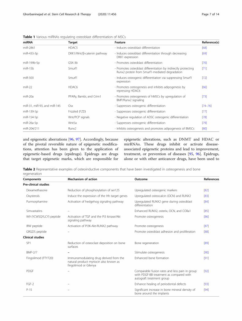
Table 1 Various miRNAs regulating osteoblast differentiation of MSCs
miRNA Target Feature Reference(s)
miR-2861 HDAC5 - Induces osteoblast differentiation [68]
miR-433-3p DKK1/Wnt/β-catenin pathway - Induces osteoblast differentiation through decreasingDKK1 expression
[69]
miR-199b-5p GSK-3b - Promotes osteoblast differentiation [70]
miR-15b Smurf1 - Promotes osteoblast differentiation by indirectly protectingRunx2 protein from Smurf1-mediated degradation
[71]
miR-503 Smurf1 - Induces osteogenic differentiation via suppressing Smurf1expression
[72]
miR-22 HDAC6 - Promotes osteogenesis and inhibits adipogenesis byrepressing HDAC6
[66]
miR-20a PPARγ, Bambi, and Crim1 - Promotes osteogenesis of hMSCs by upregulation ofBMP/Runx2 signaling
[73]
miR-31, miR-93, and miR-145 Osx - Suppresses osteogenic differentiation [74–76]
miR-139-5p Frizzled (FZD) - Suppresses osteogenic differentiation [77]
miR-154-5p Wnt/PCP signals - Negative regulation of ADSC osteogenic differentiation [78]
miR-26a-5p Wnt5a - Suppresses osteogenic differentiation [79]
miR-204/211 Runx2 - Inhibits osteogenesis and promotes adipogenesis of BMSCs [80]
Table 2 Representative examples of osteoinductive components that have been investigated in osteogenesis and boneregeneration
Components Mechanism of action Outcome References
Pre-clinical studies
Dexamethasone Reduction of phosphorylation of ser125 Upregulated osteogenic markers [82]
Oxysterols Induce the expression of the Hh target genes Upregulated osteocalcin (OCN) and RUNX2 [83]
Purmorphamine Activation of hedgehog signaling pathway Upregulated RUNX2 gene during osteoblastdifferentiation
[84]
Simvastatins – Enhanced RUNX2, osterix, OCN, and COlla1 [85]
W9 (YCWSQYLCY) peptide Activation of TGF and the PI3 kinase/Aktsignaling pathway
Promote osteogenesis [86]
IRW peptide Activation of PI3K-Akt-RUNX2 pathway Promote osteogenesis [87]
GRGDS peptide – Promote osteoblast adhesion and proliferation [88]
Clinical studies
SP1 Reduction of osteoclast deposition on bonesurfaces
Bone regeneration [89]
BMP-2/7 – Stimulate osteogenesis [90]
Fingolimod (FTY720) Immunomodulating drug derived from thenatural product myriocin also known asfingolimod or Gilenya
Enhanced bone formation [91]
PDGF – Comparable fusion rates and less pain in groupwith PDGF-BB treatment as compared withautograft treatment group
[92]
FGF-2 – Enhance healing of periodontal defects [93]
P-15 – Significant increase in bone mineral density ofbone around the implants
[94]
Ghorbaninejad et al. Stem Cell Research & Therapy (2020) 11:456 Page 7 of 14
treat cancers where epigenetic regulation has a key role[98]. For example, 5-azacitidine (Vidaza) is an FDA-approved drug for myelodysplastic syndrome (MDS) andis one of the DNMT inhibitors (DNMTi). This drug hasimproved the quality of life for patients with MDS [99].5-Aza-2′-deoxycytidine (5-Aza-dC, decitabine, Daco-gen), the deoxy form of 5-azacitidine, has been used totreat ovarian cancer. It has passed a phase I clinical trialfor hematopoietic malignancies [100, 101]. Dysregulationof epigenetic modifications plays a role in other condi-tions, including inflammation, obesity, type 2 diabetes,dyslipidemia, cardiovascular diseases, neurological disor-ders, and metabolic disorders [102]. Epidrugs that havethe capability to improve the epigenetic imbalances inthese disorders are potentially ideal candidates for futuretreatments.HDACs modulate gene expression. Histone deacety-
lase inhibitors (HDACi) alter gene expression and arebeing considered as favorable drugs for the treatment ofmalignancy. HDACi are divided into several classes:hydroxamic acids, cyclic peptides, short-chain fatty acids,and epoxides. These inhibitors arrest cell growth and in-duce differentiation or apoptosis of cancer cells bothin vitro and in vivo [103]. Several HDACi such as val-proic acid (VPA), suberoylanilide hydroxamic acid(SAHA, vorinostat), and entinostat have been used totreat MDS, cutaneous T cell lymphoma, and breast can-cer, respectively [104–106]. In addition to HDACi, theEZH2 inhibitors EPZ-6438, GSK126, GSK343, andUNC1999 have also been used in cancer therapy [96].miRNAs are another important aspect of epigenetic
regulation in cancers. The expression profiles of miR-NAs deregulate during oncogenesis. Hence, Epi-miRsopen up new horizons for novel cancer therapies [107].The results of a number of studies indicate that miRNAexpression is modulated by other epigenetic processessuch as DNA methylation and histone modification incancer. For example, miR-31/-34/-145/-21/-125b/-181a/-141 are downregulated in various cancers. Theepigenetic drug 5-aza-2′-deoxycytidine has been used tomodulate their expressions [96].Despite promising reports on the potential application
of epidrugs as therapeutic strategies, there is significantconcern that surrounds the unwanted adverse and off-target effects. Hematologic complications, such as bleed-ing and anemia, can occur following treatment withDNMTi [108]. In mouse RMS cells, 5-Aza-dC promotesmetastasis via reactivation of the pro-metastatic Ezringene [109]. Unfavorable adverse effects are a concernwith HDACi. Most HDACi are non-selective and targetmultiple classes or isozymes of HDACs (pan-HDACi),and result in toxic adverse effects. The most commoncomplaints associated with SAHA are anemia, increasedblood urea, anorexia, hyperglycemia, thrombocytopenia,
fatigue, and nausea. However, the toxic effects of SAHAwere dose-dependent and SAHA was more efficient at alower dose [110]. Usual adverse effects of anotherHDACi, VPA, included hepatic toxicity, gastrointestinaldisorders, blood dyscrasias, and hyperammonemia.These adverse effects were mild, dose-dependent, andreversible. However, valproate-induced hyperammone-mic encephalopathy (VHE) is a rare, concerning adverseeffect [111]. Also, commonly reported adverse effects ofentinostat include fatigue and gastrointestinal,hematologic, and metabolic disorders, which appear tobe concentration-dependent [112]. The therapeutic ap-proaches in the base of miRNAs as epidrugs, as well asthe mentioned epidrugs, suffer from off-target problems.A single miRNA can target multiple genes and cause un-wanted effects [113].In order to reduce the unwanted adverse effects, it is
necessary to precisely recognize epidrug targets in orderto design selective and class or isoform-specific enzymeinhibitors for specific cells. Also, the use of a suitabledrug delivery system with controlled local, sustained re-lease and evaluation of the lowest effective dose mightovercome this issue.
Epidrugs as regulators of osteogenesisMSCs are of tremendous interest to stem cell-basedtransplantation therapy; newly emerging research hasimplicated the important role of epigenetic mechanismsin regulating mesodermal lineage determination and dif-ferentiation. Evidence indicates that the induction ofMSC differentiation toward osteoblasts is supported byepigenetic regulations [114].There is growing evidence that pertains to the effect of
epidrugs on bone formation as well as cancer clinicaltherapy. A number of studies have highlighted the roleof DNMTi during osteogenesis. Chen et al. reported therole of 5-Aza-dC as a lineage determinator between adi-pogenesis and osteoblastogenesis. They found that itpromotes osteoblastogenesis by demethylation and up-regulation of the Wnt10a gene and osteoblastogenicmarkers ALP, OSX, Twist1, and Dlx5 in 3T3-L1 preadi-pocytes and the ST2 MSC line [115]. Lee et al. examinedthe effect of 5-Aza-dC on C2C12 cells. They observedthat 5-Aza-dC demethylated promoters of the Dlx5 andOsx genes in a dose- and time-dependent manner. Theexpression levels of the ALP and OCN genes increasedafter 5-Aza-dC treatment [116]. Similarly, pretreatmentof mouse bone marrow MSCs with 5-azacytidine in-creased expressions of DLX5, Runx2, COL1A1, Osx, andOCN, and accelerated osteogenesis. Hypomethylation ofthe DLX5 promoter occurred after pretreatment with 5-azacytidine as evidenced by the bisulfite sequencingtechnique [117].
Ghorbaninejad et al. Stem Cell Research & Therapy (2020) 11:456 Page 8 of 14

In addition to DNMTi, some studies have focused onHDACi and their role during differentiation of MSCs toan osteoblast lineage. VPA is a short-chain branchedfatty acid prescribed for treatment of epilepsy andneurological disorders. Cho et al. reported that treat-ment of human adipose tissue-derived stromal cells(hADSCs) with VPA as an HDAC inhibitor increasedthe expressions of OSX, OPN, Runx2, and BMP2 duringosteogenesis, which indicates a positive role for VPA inosteogenesis [118]. Paino et al. assessed the impact ofVPA on human dental pulp stem cells. They reportedthat VPA significantly enhanced matrix mineralizationby enhancing OPN and bone sialoprotein (BSP) expres-sions, but had a negative effect on late-stage marker ofdifferentiation (OCN), which indicated that VPA did notimpact the terminal step of osteogenesis. This effect wasstrongly related to inhibition of HDAC2 and has impliedthat HDAC2 is a fundamental enzyme for osteogenesis[103]. La Noce et al. demonstrated that treatment of hu-man dental pulp stem cells with VPA caused well-organized bone tissue formation after subcutaneous im-plantation of the cells into immunodeficient mice, des-pite a decrease in OCN expression [119].SAHA is another HDACi that is clinically used to treat
cancer. In a study, the treatment of C57BL/6J mice withSAHA caused decreased mineralization and OPN,COL1A1, and OCN gene expression, and eventually ledto bone loss in rodents [120]. In contrast, other re-searches showed that optimum dose of SAHA had nonegative effects on bone formation and could promoteosteogenesis via upregulation of Runx2 and BMP-2-dependent ALP activity in both human bone marrowMSCs and an osteoporotic mice model [121, 122]. Thesecontradicting results for SAHA in osteogenesis indicatethat it has a dose-dependent function during osteo-differentiation.Trichostatin A (TSA), a widely applied HDACi, has
been assessed in bone studies. Boer et al. reported thatALP expression and bone formation were dramaticallyenhanced by TSA-treated human MSCs in an ex vivocultured mouse calvaria but implantation of these cellsinto immune-deficient mice did not show any significantresults [123]. TSA also promoted rat adipose-derivedstem cell (ADSC) osteogenic differentiation by hyperace-tylation of the Runx2 promoter in a BMP signaling-dependent manner [124]. Hu et al. reported that TSApotentiated BMP9-induced ALP activity, OCN, OPN,and matrix mineralization in mouse MSCs. Also, find-ings from a fetal limb explant culture showed the rolefor TSA in endochondral bone formation [125].Sodium butyrate (NaBu) is a short-chain fatty acid that
plays a critical function in the homeostasis of the gastro-intestinal tract; NaBu inhibits HDACs. It has beenshown that NaBu modulates osteogenic differentiation
in hMSCs by an ERK-dependent Runx2 activation [126].NaBu, by increasing H3K9ac and decreasing H3K9meonto the Runx2 promoter, has been shown to enhancerat ADSC osteogenesis [127]. Ali et al. assessed the effectof abexinostat, an HDACi, on osteoblast differentiationof human MSCs. They observed that abexinostat en-hanced the expression levels of the ALP and Osx genesthrough the WNT signaling pathway [128].Besides the effect of HDACi during osteoblast differ-
entiation, several studies were conducted to assess inhib-ition of the EZH2 epigenetic factor, a novel therapeutictarget for improvement of bone formation. Inhibition ofEZH2-mediated 3-deazaneplanocin A (DZNep) pro-moted osteoblast differentiation of hMSCs [8]. Jing et al.sought to determine a correlation to inhibition of EZH2via DZNep during osteoporosis. They reported thatknockdown of EZH2 via DZNep decreased the level ofH3K27me3 on the promoters of Wnt genes and recov-ered mouse osteoporotic BMSCs in vitro and in vivo[129]. GSK126, another EZH2 inhibitor, showed similarresults and enhanced osteogenic commitment of humanadipose-derived MSCs (hAMSCs). The results indicatedthat GSK126 decreased H3K27me3 levels by inhibitionof EZH2, which led to promotion of ALP and Runx2 ex-pressions [130]. More recently, Khanban et al. have re-ported the impact of the G9A inhibitor A366 onosteogenic potential of BMSCs. They found that Runx2,ALP, COL1A1, Osx, and OCN gene expression levelsand osteogenesis decreased in BMSCs derived fromA366-treated rats [131]. Table 3 lists some epidrugs andtheir targets involved in osteogenesis. Epidrugs can beconsidered to be suitable choices for targeting epigeneticaberrations in the treatment of bone defects and efficientdifferentiation of MSCs for application in regenerativemedicine. Figure 2 shows the impact of epidrugs on thepromotion of osteogenesis.The survival of MSCs is a significant point to be
made about the use of epidrugs in promotion of MSCdifferentiation. The results of studies have shown thatthe viability of MSCs was affected by various doses ofepidrugs; increased doses had a negative impact onMSC survival [117, 119, 132]. An evaluation of the ef-fect of VPA and NaBu on MSCs showed a dose-dependent reduction in MSC proliferation. Inaddition, the proliferation of MSCs from diverse ori-gins (umbilical cord blood, adipose tissue, and bonemarrow) has different sensitivities to epidrugs [133].In a recent study, Salami et al. have reported that, inaddition to the concentration of VPA, the mediumcontext could have a key role in the effects of VPAon Wharton’s Jelly MSCs [134]. These findings high-light the importance of determining a suitable dose ofepidrugs with minimal cytotoxicity in different cellsand contexts before their use in vitro and in vivo.
Ghorbaninejad et al. Stem Cell Research & Therapy (2020) 11:456 Page 9 of 14

Table 3 Various epidrugs targeting osteogenesis
Epidrug Alternate name Target(s) Impact on osteogenesis References
5-Azacytidine Vidaza DNMT Upregulation of osteogenic gene markers (ALP, Dlx5, Runx2,Col1a1, osterix, and osteocalcin)
[117]
5-Aza-2′-deoxycytidine Decitabine DNMT Upregulation of Wnt10a, a key factor determining the fate ofthe mesenchymal lineage toward osteoblastsIncreased Dlx5 and Osx genes
[115, 116]
Valproic acid – HDAC Enhanced BMP2 expressionEnhanced matrix mineralizationNegative effect on OCN
[103, 118]
Vorinostat SAHA HDAC Inhibition of immature osteoblastsInduction of osteogenic markers (RUNX2, BMP-2, and ALP)
[120–122]
Trichostatin A TSA HDAC Promotion of osteogenic differentiation via increased in osteocalcin,osteopontin, and ALP
[123–125]
Sodium butyrate – HDAC Promotion of osteogenic differentiation via RUNX2 [126, 127]
Abexinostat – HDAC Increased OSX and ALP [128]
GSK126 – HMT Acceleration of osteogenic differentiation by regulation of Bglap,Sparc, Spp1, and Ibsp genes
[130]
3-Deazaneplanocin A DZNep HMT Upregulation of Wnt1, Wnt6, and Wnt10a osteogenic genes [129]
Fig. 2 Schematic presentation of epidrugs’ effect on the promotion of osteogenesis. After treatment of MSCs with epidrugs, there is a decreasein the epigenetic marks responsible for the silencing genes (DNA methylation, histone methylation) and an increase in gene-activating epigeneticmarks (histone acetylation) in osteogenic genes, which result in promotion of osteogenesis
Ghorbaninejad et al. Stem Cell Research & Therapy (2020) 11:456 Page 10 of 14

Future trends and concluding remarksMSC characteristics include their ability to be isolatedfrom various sources, multipotent properties, immuno-suppressive capacity, no risk of tumorigenesis, and easyexpansion in vitro. These characteristics make themideal candidates for regenerative medicine and tissue en-gineering. However, the use of these cells for stem cell-based therapy still requires a rigorous understanding ofthe mechanisms that occur during in vitro and in vivoMSC differentiation. Osteogenesis is controlled by nu-merous genes, transcription factors, signaling pathways,and epigenetic mechanisms. Nevertheless, detailedknowledge of epigenetic regulation of osteoblast differ-entiation is obscure. Hence, a novel insight into preven-tion and treatment of disease-associated epigeneticdefects such as cancer would include the identificationand targeting of epigenetic modification enzymes by epi-drugs. Recently, the use of epidrugs to treat various can-cers has been studied. Some have been approved by theFDA, and several are in phase I/II studies. The role ofsome epigenetic enzyme inhibitors has been addressedin osteoblast differentiation of MSCs, and the resultshave shown that some of these epidrugs improve osteo-genesis and bone formation via alterations in epigeneticarrangements. According to the emerging knowledge onthe role of these epigenetic modifiers in osteogenesis,epidrugs can be potentially used to improve in vitro andin vivo osteogenic differentiation with the goal to en-hance the capability of MSCs to be used in tissue engin-eering and regenerative medicine. Epidrugs have theability to be used as treatment for metabolic bone disor-ders such as osteoarthritis and osteoporosis; however,cytotoxicity is the main problem for their clinical use. Inorder to overcome the adverse effects of an epidrug, thefirst step would be to identify which epigenetic markershould be targeted in a certain disease and use its classor isoform-specific enzyme inhibitors. Furthermore, add-itional research is needed to determine the optimal doseand discover the appropriate epidrug transfer systemsprior to their clinical applications in bone tissue engin-eering. Currently, a gap exists between our knowledgeand clinical applications, and a more thorough investiga-tion in this context would be essential prior to the use ofepidrugs as therapy. It is, therefore, necessary that fur-ther research should be conducted to fully understandthe epigenetic changes that occur with bone disordersand to recognize epigenetic patterns in genes responsiblefor their repair and rejuvenation.
AcknowledgementsWe are grateful to Zeynab Ghorbaninejad for preparing the schematicfigures.
Authors’ contributionsMG, MKS, SH, and MBE designed the concept. MG and MKS searched theliterature. MG and MK wrote the manuscript and created the figure and
table. SH and MBE revised the manuscript. The authors read and approvedthe final manuscript.
FundingFunding information is not applicable to this article.
Availability of data and materialsData sharing is not applicable to this article as no datasets were generatedor analyzed during the current study.
Ethics approval and consent to participateNot applicable.
Consent for publicationNot applicable.
Competing interestsThe authors declare that they have no competing interests.
Author details1Basic and Molecular Epidemiology of Gastrointestinal Disorders ResearchCenter, Research Institute for Gastroenterology and Liver Diseases, ShahidBeheshti University of Medical Sciences, Tehran, Iran. 2Department of StemCells and Developmental Biology, Cell Science Research Center, RoyanInstitute for Stem Cell Biology and Technology, ACECR, Tehran, Iran.3Department of Genetics, Reproductive Biomedicine Research Center, RoyanInstitute for Reproductive Biomedicine, ACECR, Tehran, Iran. 4Department ofCell Engineering, Cell Science Research Center, Royan Institute for Stem CellBiology and Technology, ACECR, Tehran, Iran.
Received: 29 July 2020 Accepted: 5 October 2020
References1. Hosseini S, Taghiyar L, Safari F, Eslaminejad MB. Regenerative medicine
applications of mesenchymal stem cells. Cell Biol Translational Med. 2018;2:115–41 Springer.
2. Garg P, Mazur MM, Buck AC, Wandtke ME, Liu J, Ebraheim NA. Prospectivereview of mesenchymal stem cells differentiation into osteoblasts. OrthopSurg. 2017;9(1):13–9.
3. Hosseini S, Naderi-Manesh H, Vali H, Eslaminejad MB, Sayahpour FA,Sheibani S, et al. Contribution of osteocalcin-mimetic peptide enhancesosteogenic activity and extracellular matrix mineralization of humanosteoblast-like cells. Colloids Surf B: Biointerfaces. 2019;173:662–71.
4. Shen J, Hovhannisyan H, Lian JB, Montecino MA, Stein GS, Stein JL, et al.Transcriptional induction of the osteocalcin gene during osteoblastdifferentiation involves acetylation of histones h3 and h4. Mol Endocrinol.2003;17(4):743–56.
5. Reik W. Stability and flexibility of epigenetic gene regulation in mammaliandevelopment. Nature. 2007;447(7143):425–32.
6. Han JW, Yoon Y-S. Epigenetic landscape of pluripotent stem cells. AntioxidRedox Signal. 2012;17(2):205–23.
7. Fragale A, Romagnoli G, Licursi V, Buoncervello M, Del Vecchio G, Giuliani C,et al. Antitumor effects of Epidrug/IFNα combination driven by modulatedgene signatures in both colorectal cancer and dendritic cells. CancerImmunol Res. 2017;5(7):604–16.
8. Hemming S, Cakouros D, Isenmann S, Cooper L, Menicanin D, Zannettino A,et al. EZH2 and KDM6A act as an epigenetic switch to regulatemesenchymal stem cell lineage specification. Stem Cells. 2014;32(3):802–15.
9. Jensen ED, Gopalakrishnan R, Westendorf JJ. Regulation of gene expressionin osteoblasts. Biofactors. 2010;36(1):25–32.
10. Javed A, Chen H, Ghori FY. Genetic and transcriptional control of boneformation. Oral Maxillofac Surg Clin. 2010;22(3):283–93.
11. Bruderer M, Richards R, Alini M, Stoddart M. Role and regulation of RUNX2in osteogenesis. Eur Cell Mater. 2014;28(28):269–86.
12. Ducy P, Zhang R, Geoffroy V, Ridall AL, Karsenty G. Osf2/Cbfa1: atranscriptional activator of osteoblast differentiation. Cell. 1997;89(5):747–54.
13. Kanno T, Takahashi T, Tsujisawa T, Ariyoshi W, Nishihara T. Mechanical stress-mediated Runx2 activation is dependent on Ras/ERK1/2 MAPK signaling inosteoblasts. J Cell Biochem. 2007;101(5):1266–77.
Ghorbaninejad et al. Stem Cell Research & Therapy (2020) 11:456 Page 11 of 14

14. Ortuño MJ, Susperregui AR, Artigas N, Rosa JL, Ventura F. Osterix inducesCol1a1 gene expression through binding to Sp1 sites in the bone enhancerand proximal promoter regions. Bone. 2013;52(2):548–56.
15. Nakashima K, Zhou X, Kunkel G, Zhang Z, Deng JM, Behringer RR, et al. Thenovel zinc finger-containing transcription factor osterix is required forosteoblast differentiation and bone formation. Cell. 2002;108(1):17–29.
16. Nishimura R, Hata K, Ikeda F, Ichida F, Shimoyama A, Matsubara T, et al.Signal transduction and transcriptional regulation during mesenchymal celldifferentiation. J Bone Miner Metab. 2008;26(3):203.
17. Plaisant M, Fontaine C, Cousin W, Rochet N, Dani C, Peraldi P. Activation ofhedgehog signaling inhibits osteoblast differentiation of humanmesenchymal stem cells. Stem Cells. 2009;27(3):703–13.
18. Ji Y, Ke Y, Gao S. Intermittent activation of notch signaling promotes boneformation. Am J Transl Res. 2017;9(6):2933.
19. Kohn AD, Moon RT. Wnt and calcium signaling: β-catenin-independentpathways. Cell Calcium. 2005;38(3–4):439–46.
20. McSheehy P, Chambers T. Osteoblastic cells mediate osteoclasticresponsiveness to parathyroid hormone. Endocrinology. 1986;118(2):824–28.
21. Datta NS, Pettway GJ, Chen C, Koh AJ, McCauley LK. Cyclin D1 as a targetfor the proliferative effects of PTH and PTHrP in early osteoblastic cells. JBone Miner Res. 2007;22(7):951–64.
22. Senarath-Yapa K, McArdle A, Renda A, Longaker MT, Quarto N. Adipose-derived stem cells: a review of signaling networks governing cell fate andregenerative potential in the context of craniofacial and long bone skeletalrepair. Int J Mol Sci. 2014;15(6):9314–30.
23. Liu Z, Xu J, Colvin JS, Ornitz DM. Coordination of chondrogenesis andosteogenesis by fibroblast growth factor 18. Genes Dev. 2002;16(7):859–69.
24. Oshimori N, Fuchs E. The harmonies played by TGF-β in stem cell biology.Cell Stem Cell. 2012;11(6):751–64.
25. Fujii M, Takeda K, Imamura T, Aoki H, Sampath TK, Enomoto S, et al. Roles ofbone morphogenetic protein type I receptors and Smad proteins inosteoblast and chondroblast differentiation. Mol Biol Cell. 1999;10(11):3801–13.
26. Carreira A, Lojudice F, Halcsik E, Navarro R, Sogayar M, Granjeiro J. Bonemorphogenetic proteins: facts, challenges, and future perspectives. J DentRes. 2014;93(4):335–45.
27. Long F, Chung U-I, Ohba S, McMahon J, Kronenberg HM, McMahon AP. Ihhsignaling is directly required for the osteoblast lineage in the endochondralskeleton. Development. 2004;131(6):1309–18.
28. De Jong D, Steegenga W, Hendriks J, Van Zoelen E, Olijve W, Dechering K.Regulation of Notch signaling genes during BMP2-induced differentiation ofosteoblast precursor cells. Biochem Biophys Res Commun. 2004;320(1):100–7.
29. Qiu T, Wu X, Zhang F, Clemens TL, Wan M, Cao X. TGF-β type II receptorphosphorylates PTH receptor to integrate bone remodelling signalling. NatCell Biol. 2010;12(3):224–34.
30. Jiang T, Ge S, Shim YH, Zhang C, Cao D. Bone morphogenetic protein isrequired for fibroblast growth factor 2-dependent later-stage osteoblasticdifferentiation in cranial suture cells. Int J Clin Exp Pathol. 2015;8(3):2946.
31. Kamiya N, Kobayashi T, Mochida Y, Yu PB, Yamauchi M, Kronenberg HM,et al. Wnt inhibitors Dkk1 and Sost are downstream targets of BMPsignaling through the type IA receptor (BMPRIA) in osteoblasts. J BoneMiner Res. 2010;25(2):200–10.
32. Mortada I, Mortada R. Epigenetic changes in mesenchymal stem cellsdifferentiation. Eur J Med Genet. 2018;61(2):114–8.
33. Daniunaite K, Serenaite I, Misgirdaite R, Gordevicius J, Unguryte A, Fleury-Cappellesso S, et al. Epigenetic regulation of human adipose-derived stemcells differentiation. Mol Cell Biochem. 2015;410(1–2):111–20.
34. Jones PA. Functions of DNA methylation: islands, start sites, gene bodiesand beyond. Nat Rev Genet. 2012;13(7):484–92.
35. Chen T, Ueda Y, Dodge JE, Wang Z, Li E. Establishment and maintenance ofgenomic methylation patterns in mouse embryonic stem cells by Dnmt3aand Dnmt3b. Mol Cell Biol. 2003;23(16):5594–605.
36. Arnsdorf EJ, Tummala P, Castillo AB, Zhang F, Jacobs CR. The epigeneticmechanism of mechanically induced osteogenic differentiation. J Biomech.2010;43(15):2881–6.
37. Delgado-Calle J, Sañudo C, Sánchez-Verde L, García-Renedo RJ, ArozamenaJ, Riancho JA. Epigenetic regulation of alkaline phosphatase in human cellsof the osteoblastic lineage. Bone. 2011;49(4):830–8.
38. Zhang R-P, Shao J-Z, Xiang L-X. GADD45A protein plays an essential role inactive DNA demethylation during terminal osteogenic differentiation ofadipose-derived mesenchymal stem cells. J Biol Chem. 2011;286(47):41083–94.
39. Sepulveda H, Villagra A, Montecino M. Tet-mediated DNA demethylation isrequired for SWI/SNF-dependent chromatin remodeling and histone-modifying activities that trigger expression of the Sp7 osteoblast mastergene during mesenchymal lineage commitment. Mol Cell Biol. 2017;37(20):e00177–17.
40. Wakitani S, Yokoi D, Hidaka Y, Nishino K. The differentially DNA-methylatedregion responsible for expression of runt-related transcription factor 2. J VetMed Sci. 2017;79(2):230–7.
41. Chen JR, Zhang J, Lazarenko OP, Kang P, Blackburn ML, Ronis MJ, et al.Inhibition of fetal bone development through epigenetic down-regulationof HoxA10 in obese rats fed high-fat diet. FASEB J. 2012;26(3):1131–41.
42. Tarfiei G, Noruzinia M, Soleimani M, Kaviani S, Maymand MM, Hagh MF,et al. ROR2 promoter methylation change in osteoblastic differentiation ofmesenchymal stem cells. Cell J (Yakhteh). 2011;13(1):11.
43. Yang R, Yu T, Kou X, Gao X, Chen C, Liu D, et al. Tet1 and Tet2 maintainmesenchymal stem cell homeostasis via demethylation of the P2rX7promoter. Nat Commun. 2018;9(1):1–14.
44. Kouzarides T. Chromatin modifications and their function. Cell. 2007;128(4):693–705.
45. Huang H, Sabari BR, Garcia BA, Allis CD, Zhao Y. SnapShot: histonemodifications. Cell. 2014;159(2):458 e1.
46. Kuo MH, Allis CD. Roles of histone acetyltransferases and deacetylases ingene regulation. Bioessays. 1998;20(8):615–26.
47. Tan J, Lu J, Huang W, Dong Z, Kong C, Li L, et al. Genome-wide analysis ofhistone H3 lysine9 modifications in human mesenchymal stem cellosteogenic differentiation. PLoS One. 2009;4(8):e6792.
48. Zhang P, Liu Y, Jin C, Zhang M, Lv L, Zhang X, et al. Histone H3K9acetyltransferase PCAF is essential for osteogenic differentiation throughbone morphogenetic protein signaling and may be involved inosteoporosis. Stem Cells. 2016;34(9):2332–41.
49. Sepulveda H, Aguilar R, Prieto CP, Bustos F, Aedo S, Lattus J, et al.Epigenetic signatures at the RUNX2-P1 and Sp7 gene promoters controlosteogenic lineage commitment of umbilical cord-derived mesenchymalstem cells. J Cell Physiol. 2017;232(9):2519–27.
50. Gordon JA, Hassan MQ, Koss M, Montecino M, Selleri L, Van Wijnen AJ, et al.Epigenetic regulation of early osteogenesis and mineralized tissueformation by a HOXA10-PBX1-associated complex. Cells Tissues Organs.2011;194(2–4):146–50.
51. Simic P, Zainabadi K, Bell E, Sykes DB, Saez B, Lotinun S, et al. SIRT1regulates differentiation of mesenchymal stem cells by deacetylating β-catenin. EMBO Mol Med. 2013;5(3):430–40.
52. Avgustinova A, Benitah SA. Epigenetic control of adult stem cell function.Nat Rev Mol Cell Biol. 2016;17(10):643.
53. Hassan MQ, Tare R, Lee SH, Mandeville M, Weiner B, Montecino M, et al.HOXA10 controls osteoblastogenesis by directly activating bone regulatoryand phenotypic genes. Mol Cell Biol. 2007;27(9):3337–52.
54. Higashihori N, Lehnertz B, Sampaio A, Underhill T, Rossi F, Richman J.Methyltransferase G9A regulates osteogenesis via twist gene repression. JDent Res. 2017;96(10):1136–44.
55. Fan Z, Yamaza T, Lee JS, Yu J, Wang S, Fan G, et al. BCOR regulatesmesenchymal stem cell function by epigenetic mechanisms. Nat Cell Biol.2009;11(8):1002–9.
56. Zhang F, Xu L, Xu L, Xu Q, Karsenty G, Chen CD. Histone demethylaseJMJD3 is required for osteoblast differentiation in mice. Sci Rep. 2015;5:13418.
57. Ye L, Fan Z, Yu B, Chang J, Al Hezaimi K, Zhou X, et al. Histonedemethylases KDM4B and KDM6B promotes osteogenic differentiation ofhuman MSCs. Cell Stem Cell. 2012;11(1):50–61.
58. Ehrenhofer-Murray AE. Chromatin dynamics at DNA replication, transcriptionand repair. Eur J Biochem. 2004;271(12):2335–49.
59. Chen Y, Wang M, Chen D, Wang J, Kang N. Chromatin remodeling enzymeCHD7 is necessary for osteogenesis of human mesenchymal stem cells.Biochem Biophys Res Commun. 2016;478(4):1588–93.
60. Zhang P, Torres K, Liu X, Liu C-g, E Pollock R. An overview of chromatin-regulating proteins in cells. Curr Protein Peptide Sci. 2016;17(5):401–10.
61. Im G-I, Shin K-J. Epigenetic approaches to regeneration of bone andcartilage from stem cells. Expert Opin Biol Ther. 2015;15(2):181–93.
62. Wei Y, Chen Y-H, Li L-Y, Lang J, Yeh S-P, Shi B, et al. CDK1-dependentphosphorylation of EZH2 suppresses methylation of H3K27 and promotesosteogenic differentiation of human mesenchymal stem cells. Nat Cell Biol.2011;13(1):87–94.
Ghorbaninejad et al. Stem Cell Research & Therapy (2020) 11:456 Page 12 of 14

63. He L, Hannon GJ. MicroRNAs: small RNAs with a big role in gene regulation.Nat Rev Genet. 2004;5(7):522.
64. Ambros V. The functions of animal microRNAs. Nature. 2004;431(7006):350.65. Friedman RC, Farh KK-H, Burge CB, Bartel DP. Most mammalian mRNAs are
conserved targets of microRNAs. Genome Res. 2009;19(1):92–105.66. Huang S, Wang S, Bian C, Yang Z, Zhou H, Zeng Y, et al. Upregulation of
miR-22 promotes osteogenic differentiation and inhibits adipogenicdifferentiation of human adipose tissue-derived mesenchymal stem cells byrepressing HDAC6 protein expression. Stem Cells Dev. 2012;21(13):2531–40.
67. Huszar JM, Payne CJ. MIR146A inhibits JMJD3 expression and osteogenicdifferentiation in human mesenchymal stem cells. FEBS Lett. 2014;588(9):1850–6.
68. Li H, Xie H, Liu W, Hu R, Huang B, Tan Y-F, et al. A novel microRNAtargeting HDAC5 regulates osteoblast differentiation in mice andcontributes to primary osteoporosis in humans. J Clin Invest. 2009;119(12):3666–77.
69. Tang X, Lin J, Wang G, Lu J. MicroRNA-433-3p promotes osteoblastdifferentiation through targeting DKK1 expression. PLoS One. 2017;12(6):e0179860.
70. Zhao R, Li Y, Lin Z, Wan J, Xu C, Zeng Y, et al. miR-199b-5p modulatesBMSC osteogenesis via suppressing GSK-3β/β-catenin signaling pathway.Biochem Biophys Res Commun. 2016;477(4):749–54.
71. Vimalraj S, Partridge NC, Selvamurugan N. A positive role of microRNA-15bon regulation of osteoblast differentiation. J Cell Physiol. 2014;229(9):1236–44.
72. Sun Y, Xu J, Xu L, Zhang J, Chan K, Pan X, et al. MiR-503 promotes boneformation in distraction osteogenesis through suppressing Smurf1expression. Sci Rep. 2017;7(1):409.
73. Zhang J-F, Fu W-M, He M-L, Xie W-D, Lv Q, Wan G, et al. MiRNA-20apromotes osteogenic differentiation of human mesenchymal stem cells byco-regulating BMP signaling. RNA Biol. 2011;8(5):829–38.
74. Baglìo SR, Devescovi V, Granchi D, Baldini N. MicroRNA expression profilingof human bone marrow mesenchymal stem cells during osteogenicdifferentiation reveals Osterix regulation by miR-31. Gene. 2013;527(1):321–31.
75. Yang L, Cheng P, Chen C, He HB, Xie GQ, Zhou HD, et al. miR-93/Sp7function loop mediates osteoblast mineralization. J Bone Miner Res. 2012;27(7):1598–606.
76. Jia J, Tian Q, Ling S, Liu Y, Yang S, Shao Z. miR-145 suppresses osteogenicdifferentiation by targeting Sp7. FEBS Lett. 2013;587(18):3027–31.
77. Long H, Sun B, Cheng L, Zhao S, Zhu Y, Zhao R, et al. miR-139-5p repressesBMSC osteogenesis via targeting Wnt/β-catenin signaling pathway. DNACell Biol. 2017;36(8):715–24.
78. Li J, Hu C, Han L, Liu L, Jing W, Tang W, et al. MiR-154-5p regulatesosteogenic differentiation of adipose-derived mesenchymal stem cellsunder tensile stress through the Wnt/PCP pathway by targeting Wnt11.Bone. 2015;78:130–41.
79. Li S, Hu C, Li J, Liu L, Jing W, Tang W, et al. Effect of miR-26a-5p on theWnt/Ca 2+ pathway and osteogenic differentiation of mouse adipose-derived mesenchymal stem cells. Calcif Tissue Int. 2016;99(2):174–86.
80. Huang J, Zhao L, Xing L, Chen D. MicroRNA-204 regulates Runx2 proteinexpression and mesenchymal progenitor cell differentiation. Stem Cells.2010;28(2):357–64.
81. Wang Y, Zhu G, Li N, Song J, Wang L, Shi X. Small molecules and theircontrolled release that induce the osteogenic/chondrogenic commitmentof stem cells. Biotechnol Adv. 2015;33(8):1626–40.
82. Phillips JE, Gersbach CA, Wojtowicz AM, García AJ. Glucocorticoid-inducedosteogenesis is negatively regulated by Runx2/Cbfa1 serinephosphorylation. J Cell Sci. 2006;119(3):581–91.
83. Jang WG, Kim EJ, Bae I-H, Lee K-N, Kim YD, Kim D-K, et al. Metformininduces osteoblast differentiation via orphan nuclear receptor SHP-mediated transactivation of Runx2. Bone. 2011;48(4):885–93.
84. Oliveira F, Bellesini L, Defino H, da Silva HC, Beloti M, Rosa A. Hedgehogsignaling and osteoblast gene expression are regulated by purmorphaminein human mesenchymal stem cells. J Cell Biochem. 2012;113(1):204–8.
85. Pagkalos J, Cha JM, Kang Y, Heliotis M, Tsiridis E, Mantalaris A. Simvastatininduces osteogenic differentiation of murine embryonic stem cells. J BoneMiner Res. 2010;25(11):2470–8.
86. Otsuki Y, Ii M, Moriwaki K, Okada M, Ueda K, Asahi M. W9 peptide enhancedosteogenic differentiation of human adipose-derived stem cells. BiochemBiophys Res Commun. 2018;495(1):904–10.
87. Shang N, Bhullar KS, Hubbard BP, Wu J. Tripeptide IRW initiatesdifferentiation in osteoblasts via the RUNX2 pathway. Biochim Biophys ActaGen Subj. 2019;1863(6):1138–46.
88. Kim G-H, Park S-W, Lee K, Yun K-D, Kim H-S, Oh G-J, et al. Evaluation ofosteoblast-like cell viability and differentiation on the Gly-Arg-Gly-Asp-Serpeptide immobilized titanium dioxide nanotube via chemical grafting. JNanosci Nanotechnol. 2016;16(2):1396–9.
89. Segar CE, Ogle ME, Botchwey EA. Regulation of angiogenesis and boneregeneration with natural and synthetic small molecules. Curr Pharm Des.2013;19(19):3403–19.
90. Cipitria A, Reichert JC, Epari DR, Saifzadeh S, Berner A, Schell H, et al.Polycaprolactone scaffold and reduced rhBMP-7 dose for the regenerationof critical-sized defects in sheep tibiae. Biomaterials. 2013;34(38):9960–8.
91. Aronin CEP, Sefcik LS, Tholpady SS, Tholpady A, Sadik KW, Macdonald TL,et al. FTY720 promotes local microvascular network formation andregeneration of cranial bone defects. Tissue Eng A. 2010;16(6):1801–9.
92. DiGiovanni CW, Lin SS, Baumhauer JF, Daniels T, Younger A, Glazebrook M,et al. Recombinant human platelet-derived growth factor-BB and beta-tricalcium phosphate (rhPDGF-BB/β-TCP): an alternative to autogenousbone graft. JBJS. 2013;95(13):1184–92.
93. Cochran DL, Oh T-J, Mills MP, Clem D, McClain P, Schallhorn R, et al. Arandomized clinical trial evaluating rh-FGF-2/β-TCP in periodontal defects. JDent Res. 2016;95(5):523–30.
94. Emam H, Beheiri G, Elsalanty M, Sharawy M. Microcomputed tomographicand histologic analysis of anorganic bone matrix coupled with cell-bindingpeptide suspended in sodium hyaluronate carrier after sinus augmentation:a clinical study. Int J Oral Maxillofac Implants. 2011;26(3):561–70.
95. Sharma S, Kelly TK, Jones PA. Epigenetics in cancer. Carcinogenesis. 2010;31(1):27–36.
96. Salarinia R, Sahebkar A, Peyvandi M, Reza Mirzaei H, Reza Jaafari M, MatbouRiahi M, et al. Epi-drugs and Epi-miRs: moving beyond current cancertherapies. Curr Cancer Drug Targets. 2016;16(9):773–88.
97. Stegemann R, Buchner DA. Transgenerational inheritance of metabolicdisease. Semin Cell Dev Biol. 2015;43:131–40. .
98. Ivanov M, Barragan I, Ingelman-Sundberg M. Epigenetic mechanisms ofimportance for drug treatment. Trends Pharmacol Sci. 2014;35(8):384–96.
99. Kornblith AB, Herndon JE, Silverman LR, Demakos EP, Odchimar-Reissig R,Holland JF, et al. Impact of azacytidine on the quality of life of patients withmyelodysplastic syndrome treated in a randomized phase III trial: a Cancerand Leukemia Group B study. J Clin Oncol. 2002;20(10):2441–52.
100. Issa JP, Garcia-Manero G, Giles FJ, Mannari R, Thomas D, Faderl S, et al.Phase 1 study of low-dose prolonged exposure schedules of thehypomethylating agent 5-aza-2′-deoxycytidine (decitabine) inhematopoietic malignancies. Blood. 2004;103(5):1635–40.
101. Matei D, Fang F, Shen C, Schilder J, Arnold A, Zeng Y, et al. Epigeneticresensitization to platinum in ovarian cancer. Cancer Res. 2012;72(9):2197–205.
102. Heerboth S, Lapinska K, Snyder N, Leary M, Rollinson S, Sarkar S. Use ofepigenetic drugs in disease: an overview. Genet Epigenetics. 2014;6:GEG.S12270.
103. Paino F, La Noce M, Tirino V, Naddeo P, Desiderio V, Pirozzi G, et al. Histonedeacetylase inhibition with valproic acid downregulates osteocalcin geneexpression in human dental pulp stem cells and osteoblasts: evidence forHDAC2 involvement. Stem Cells. 2014;32(1):279–89.
104. Candelaria M, Herrera A, Labardini J, González-Fierro A, Trejo-Becerril C, Taja-Chayeb L, et al. Hydralazine and magnesium valproate as epigenetictreatment for myelodysplastic syndrome. Preliminary results of a phase-IItrial. Ann Hematol. 2011;90(4):379–87.
105. Duvic M, Vu J. Vorinostat: a new oral histone deacetylase inhibitor approvedfor cutaneous T-cell lymphoma. Expert Opin Investig Drugs. 2007;16(7):1111–20.
106. Yardley DA, Ismail-Khan RR, Melichar B, Lichinitser M, Munster PN, Klein PM,et al. Randomized phase II, double-blind, placebo-controlled study ofexemestane with or without entinostat in postmenopausal women withlocally recurrent or metastatic estrogen receptor-positive breast cancerprogressing on treatment with a nonsteroidal aromatase inhibitor. J ClinOncol. 2013;31(17):2128.
107. D'Souza W, Saranath D. Clinical implications of epigenetic regulation in oralcancer. Oral Oncol. 2015;51(12):1061–8.
108. Giri AK, Aittokallio T. DNMT inhibitors increase methylation in the cancergenome. Front Pharmacol. 2019;10:385.
109. Yu Y, Zeng P, Xiong J, Liu Z, Berger SL, Merlino G. Epigenetic drugs canstimulate metastasis through enhanced expression of the pro-metastaticEzrin gene. PLoS One. 2010;5(9):e12710.
Ghorbaninejad et al. Stem Cell Research & Therapy (2020) 11:456 Page 13 of 14
110. Blumenschein GR, Kies MS, Papadimitrakopoulou VA, Lu C, Kumar AJ, RickerJL, et al. Phase II trial of the histone deacetylase inhibitor vorinostat(Zolinza™, suberoylanilide hydroxamic acid, SAHA) in patients with recurrentand/or metastatic head and neck cancer. Investig New Drugs. 2008;26(1):81–7.
111. Guo X, Wei J, Gao L, Xing B, Xu Z. Hyperammonemic coma aftercraniotomy: hepatic encephalopathy from upper gastrointestinalhemorrhage or valproate side effect?: case report and literature review.Medicine. 2017;96(15):e6588.
112. Connolly RM, Rudek MA, Piekarz R. Entinostat: a promising treatment optionfor patients with advanced breast cancer. Future Oncol. 2017;13(13):1137–48.
113. Li Z, Rana TM. Therapeutic targeting of microRNAs: current status and futurechallenges. Nat Rev Drug Discov. 2014;13(8):622–38.
114. Eslaminejad MB, Fani N, Shahhoseini M. Epigenetic regulation of osteogenicand chondrogenic differentiation of mesenchymal stem cells in culture. CellJ (Yakhteh). 2013;15(1):1.
115. Chen YS, Wu R, Yang X, Kou S, MacDougald OA, Yu L, et al. Inhibiting DNAmethylation switches adipogenesis to osteoblastogenesis by activatingWnt10a. Sci Rep. 2016;6:25283.
116. Lee JY, Lee YM, Kim MJ, Choi JY, et al. Methylation of the mouse Dlx5 andOsx gene promoters regulates cell type-specific gene expression. Mol Cells.2006;22:182–8.
117. Zhou GS, Zhang XL, Wu JP, Zhang RP, Xiang LX, Dai LC, et al. 5-Azacytidinefacilitates osteogenic gene expression and differentiation of mesenchymalstem cells by alteration in DNA methylation. Cytotechnology. 2009;60(1–3):11.
118. Cho HH, Park HT, Kim YJ, Bae YC, Suh KT, Jung JS. Induction of osteogenicdifferentiation of human mesenchymal stem cells by histone deacetylaseinhibitors. J Cell Biochem. 2005;96(3):533–42.
119. La Noce M, Mele L, Laino L, Iolascon G, Pieretti G, Papaccio G, et al.Cytoplasmic interactions between the glucocorticoid receptor and hdac2regulate osteocalcin expression in vpa-treated MSCs. Cells. 2019;8(3):217.
120. McGee-Lawrence ME, McCleary-Wheeler AL, Secreto FJ, Razidlo DF, ZhangM, Stensgard BA, et al. Suberoylanilide hydroxamic acid (SAHA; vorinostat)causes bone loss by inhibiting immature osteoblasts. Bone. 2011;48(5):1117–26.
121. Xu S, De Veirman K, Evans H, Santini GC, Broek IV, Leleu X, et al. Effect ofthe HDAC inhibitor vorinostat on the osteogenic differentiation ofmesenchymal stem cells in vitro and bone formation in vivo. ActaPharmacol Sin. 2013;34(5):699–709.
122. Lee ZH, Kim HJ, Ryoo HM. A novel osteogenic activity of suberoylanilidehydroxamic acid is synergized by BMP-2. J Bone Metab. 2015;22(2):51–6.
123. Boer JD, Licht R, Bongers M, Klundert TVD, Arends R, Blitterswijk CV.Inhibition of histone acetylation as a tool in bone tissue engineering. TissueEng. 2006;12(10):2927–37.
124. Hu X, Zhang X, Dai L, Zhu J, Jia Z, Wang W, et al. Histone deacetylaseinhibitor trichostatin A promotes the osteogenic differentiation of ratadipose-derived stem cells by altering the epigenetic modifications onRunx2 promoter in a BMP signaling-dependent manner. Stem Cells Dev.2013;22(2):248–55.
125. Hu N, Wang C, Liang X, Yin L, Luo X, Liu B, et al. Inhibition of histonedeacetylases potentiates BMP9-induced osteogenic signaling in mousemesenchymal stem cells. Cell Physiol Biochem. 2013;32(2):486–98.
126. Chen TH, Chen WM, Hsu KH, Kuo CD, Hung SC. Sodium butyrate activatesERK to regulate differentiation of mesenchymal stem cells. Biochem BiophysRes Commun. 2007;355(4):913–8.
127. Hu X, Fu Y, Zhang X, Dai L, Zhu J, Bi Z, et al. Histone deacetylase inhibitorsodium butyrate promotes the osteogenic differentiation of rat adipose-derived stem cells. Develop Growth Differ. 2014;56(3):206–13.
128. Ali D, Hamam R, Alfayez M, Kassem M, Aldahmash A, Alajez NM. Epigeneticlibrary screen identifies abexinostat as novel regulator of adipocytic andosteoblastic differentiation of human skeletal (mesenchymal) stem cells.Stem Cells Transl Med. 2016;5(8):1036–47.
129. Jing H, Liao L, An Y, Su X, Liu S, Shuai Y, et al. Suppression of EZH2 preventsthe shift of osteoporotic MSC fate to adipocyte and enhances boneformation during osteoporosis. Mol Ther. 2016;24(2):217–29.
130. Dudakovic A, Camilleri ET, Xu F, Riester SM, McGee-Lawrence ME, BradleyEW, et al. Epigenetic control of skeletal development by the histonemethyltransferase Ezh2. J Biol Chem. 2015;290(46):27604–17.
131. Khanban H, Fattahi E, Talkhabi M. In vivo administration of G9a inhibitorA366 decreases osteogenic potential of bone marrow-derivedmesenchymal stem cells. EXCLI J. 2019;18:300.
132. Fan X, Li L, Ye Z, Zhou Y, Tan WS. Regulation of osteogenesis of humanamniotic mesenchymal stem cells by sodium butyrate. Cell Biol Int. 2018;42(4):457–69.
133. Lee S, Park JR, Seo MS, Roh KH, Park SB, Hwang JW, et al. Histonedeacetylase inhibitors decrease proliferation potential and multilineagedifferentiation capability of human mesenchymal stem cells. Cell Prolif.2009;42(6):711–20.
134. Salami H, Mowal SJ, Moukhah R, Hajebrahimi Z, Hosseini SA, Edalat H.Evaluating the differential effects of valproic acid on Wharton’s jellymesenchymal stem cells. Adv Pharm Bull. 2019;9(3):497.
Publisher’s NoteSpringer Nature remains neutral with regard to jurisdictional claims inpublished maps and institutional affiliations.
Ghorbaninejad et al. Stem Cell Research & Therapy (2020) 11:456 Page 14 of 14