Embed Size (px)
Citation preview
GLOBAL WATER PATHOGEN PROJECTPART THREE. SPECIFIC EXCRETED PATHOGENS: ENVIRONMENTAL ANDEPIDEMIOLOGY ASPECTS
ADENOVIRUSES
Annika AllardUmeå UniversityUmeå, Sweden
Apostolos VantarakisUniversity of PatrasPátrai, Greece

Copyright:
This publication is available in Open Access under the Attribution-ShareAlike 3.0 IGO (CC-BY-SA 3.0 IGO)license (http://creativecommons.org/licenses/by-sa/3.0/igo). By using the content of this publication, the usersa c c e p t t o b e b o u n d b y t h e t e r m s o f u s e o f t h e U N E S C O O p e n A c c e s s R e p o s i t o r y(ht tp : / /www.unesco.org/openaccess / terms-use-ccbysa-en) .
Disclaimer:The designations employed and the presentation of material throughout this publication do not imply theexpression of any opinion whatsoever on the part of UNESCO concerning the legal status of any country,territory, city or area or of its authorities, or concerning the delimitation of its frontiers or boundaries. Theideas and opinions expressed in this publication are those of the authors; they are not necessarily those ofUNESCO and do not commit the Organization.
Citation:Allard, A. and Vantarakis, A. 2017. Adenoviruses. In: J.B. Rose and B. Jiménez-Cisneros, (eds) Global WaterPathogens Project. http://www.waterpathogens.org (J.S Meschke, and R. Girones (eds) Part 3Viruses) http://www.waterpathogens.org/book/adenoviruses Michigan State University, E. Lansing, MI,UNESCO.Acknowledgements: K.R.L. Young, Project Design editor; Website Design (http://www.agroknow.com)
Published: January 14, 2015, 5:19 pm, Updated: August 22, 2017, 3:26 pm
Adenoviruses
3
Summary
Adenoviruses are associated with numerous diseaseoutbreaks, particularly those involving daycares, schools,children's camps, hospitals, military settings, and otherhealth care centres. Today, fifty-two different serotypes ofadenovirus have been identified. However, several typeshave also recently been identified with moleculartechniques and designated as genotypes. All serotypes andgenotypes have been divided into seven species (A to G)that infect humans. Of all the known adenovirus types, onethird are associated with human disease as gastroenteritis,respiratory infections, eye infections, acute hemorrhagiccystitis, and meningoencephalitis, while two thirds ofinfections are asymptomatic.
Children and immunocompromised individuals are moreseverely impacted by adenovirus infections. Adenoviruseshave a worldwide distribution, where infections occurthroughout the year causing 5 to 10% of all febrile illnessesin infants and young children. Most individuals haveserologic evidence of a prior adenoviral infection by the ageof 10. Although many adenovirus types are found in stoolfrom patients with diarrhea, only enteric adenoviruses (type40, 41 and 52) have been shown to be the causative agentsof gastrointestinal disease. Enteric adenoviruses replicatein the gut to 1011 virions per gram of stool, but many othernon-enteric viral particles (i.e. types 1,2,5,3 and 7) maypersist for months and even up to years in stool after aninfection. These phenomena may explain why adenovirusesare ubiquitous in the environment where contamination ofhuman faeces or sewage occur in such areas of increasedpopulation densities.
Adenoviruses have been detected in various watersworldwide including wastewater, river water, drinkingwater, ocean, and swimming pools. Adenoviruses typicallyoutnumber the enteroviruses, when both are detected insurface waters. Evidence has shown that adenovirusessurvive longer in water than enteroviruses and the hepatitisA virus; this may be due to their double-stranded DNA. Amajority of the results are based on PCR technology andindicate only the presence of nucleic acids and do notinclude infectivity data.
Adenoviruses are frequently detected in highconcentrations in wastewater and wastewater-contaminated waters, so their use as potential indicatorsfor the presence of human faeces has been suggested as acomplement to bacterial indicators. It is well documentedthat bacteria behave differently to viruses in aquaticenvironments, particularly with regard to their survivaltimes. In this respect, adenoviruses are good candidates forindicators of virus contamination with long term survivalproperties, high resistance to ultraviolet light, and a DNAgenome which simplifies and decreases the cost ofmolecular detection procedures compared to the costlyRNA processing.
In recent years, there have been several studies analyzingground water samples showing the presence of
adenoviruses with no correlation between the presence ofviruses and bacterial indicators (i.e. Escherichia coli).These results may be useful for risk assessment studiessince ground water is a common source of drinking waterand subsequently connected to the interest of public health.Adenoviruses are more often found in ground water thanother viral types, and they have been abundant in untreatedriver water and finished water after conventional watertreatment plant processes. The different treatmentprocesses (conventional and alternative) for reducingconcentrations of adenoviruses in water, have shownvariable efficiency. In addition, environmental conditionshave been reported to present variable effect on adenovirusstability.
In summary, the knowledge of adenovirus importance hasincreased significantly during the last decades, andadenovirus is now playing an important role in both clinicalsettings and in the environmental context.
AdenovirusBoth respiratory and enteric human adenoviruses
(HAdV) are found in water, cause waterborne diseaseandare present at a higher frequency in sewage than otherenteric viruses (Pina et al., 1998). These viruses have beensuggested to be preferred candidates as index organismsfor viral pathogens because they fit most criteria for anidea l i nd i ca to r such a s r e s i s t ance t o manychemical/physical agents and to UV light (Katayama et al.,2008; Gerba et al., 2002; Girones et al., 2014).
The first adenovirus strains were isolated in 1953 fromlymph nodes and got their name from adénos, the Greekword for gland(Rowe et al., 1953).The following year itbecame clear that the virus caused acute respiratoryinfection. Thus, two basic properties of adenoviruses weredetected; their ability to cause acute respiratory infectionand the propensity to persist in lymphoid tissue. Today,fifty-two different serotypes of adenovirus have beenidentified; however, several types have also in recent yearsbeen identified with molecular techniques. All types havebeen divided into seven species (A to G) that infect humans.This species breakdown is affected by the receptor bindingstructure.Adenoviruses are a family of viruses that globallyinfects most children already before the age of five.Highfever for a week is often seen but approximately everysecond adenovirus infection is non-symptomatic.Adenoviruses rarely cause life-threatening infections,except in immunodeficient individuals. It is now knownhowever that environmental occurrence and transmission isimportant.
1.0 Epidemiology of the Disease andPathogens
1.1 Global Burden of Disease
Adenoviruses have a worldwide distribution, andinfections occur throughout the year (Fox et al., 1977).Adenoviruses cause 5 to 10% of all febrile illnesses in
Adenoviruses
4
infants and young children and most individuals in NorthAmerica have serologic evidence of a prior adenoviralinfection by the age of five. (Fox et al., 1977). Bothrespiratory and enteric adenovirus infections are prevalentin daycare centers and in households with young childrenbut nosocomial transmission has also been documented(Liu et al., 2014; Tran et al., 2010).Adenovirus species Frepresenting human adenovirus type 40 and 41 has beenfound to be associated with acute gastroenteritis and isresponsible for 1 to 20% of the cases of diarrheal diseaseglobally in both outpatients and hospitalized children(Uhnoo et al., 1984; Brandt et al., 1984; Li et al., 2004).Type 40 and 41 primarily affect young children less than 2years of age and the infection occur throughout the year.The clinical characteristics include watery diarrheaaccompanied by vomiting, low-grade fever, and milddehydration.The burden of adenovirus infections isparticularly important in immunocompromised hosts (Lion,2014), where a variety of clinical syndromes can occur andreactivation of endogenous adenovirus plays a role indiseases in these patients.
1.1.1 Global distribution
Human adenovirus (HAdV) infections are common andubiquitous with a worldwide distribution.Even if most HAdVspecies appear to circulate globally, predominant typesdiffer between countries or geographic regions, and theychange over time (Lin et al., 2004; Ishiko et al., 2008;Ampuero et al., 2012). Transmission of new strains acrosscontinents may occur and lead to replacement of hithertodominant HAdV types (Kajon et al., 2010).The prevalence ofadenovirus respiratory infections in children in North andSouth America ranges from 2 to 14% and may be higherduring outbreaks (Fox et al., 1977; Videla et al., 1998).Many (30-70%) respiratory illnesses in unvaccinated newmilitary recruits in USA are adenovirus associated as well(Russell et al., 2006). About 5 to 15% of acute diarrhealinfections in all continents are due to enteric adenoviruses(Dey et al., 2011; Uhnoo et al., 1984; Kim et al., 1990;Barnes et al., 1998; Van et al. 1992; Moore et al., 2000).
Children are especially susceptible but elderly are alsoagain susceptible to the enteric types 40 and 41 due towaning immunity with increasing age. Due tocontamination by human faecesor sewage into theenvironment HAdVs seem to be more common indeveloping countries with poor sanitary conditions (Dey etal., 2011; Filho et al., 2007; Moyo et al., 2014).However,the incidence of all HAdV infections is higher incrowded closed settings, such as day care centers,boarding schools, geriatric facilities, military trainingcamps, and hospitals. Intrafamiliar infections are alsocommon and HAdVs are frequent in immunocompromisedpatients. Every serotype can cause a unique infection,which means theoretically that it is possible to acquiremore than 50 adenovirus infections to get immunity. Types53-68 are genetically typed and antibody specificity has notyet been investigated or confirmed against these recenttypes.The adenoviruses most commonly reported to beassociated with human disease globally are HAdVC1,-C2, -C5, -B3, -B7, -B21, -E4, and -F41 (Guo et al., 2012;
Yliharsila et al., 2013; Tabain et al., 2012; Barrero et al.,2012).
1.1.2 Symptomatology
HAdVare most frequently associated with upperrespiratory tract syndromes, such as pharyngitis or rhinitisbut can also cause pneumonia. Less commonly,adenoviruses cause gastrointestinal, ophthalmologic,genitourinary, and neurologic diseases. Most adenoviraldiseases are self-limiting, although fatal infections canoccur in immuno-compromised hosts and occasionally inhealthy children and adults, especially children withgastrointestinal infections in the developing world. Thenumber of diarrheal deaths caused by adenovirus (HAdV-40and HAdV-41) was estimated in 2011 to be 3-4% of alldiarrheal deaths among children under 5 years of age inthe world (Lanata et al. 2013). Patients who haveundergone organ or hematopoietic cell transplantationtogether with AIDS patients and children with severecombined immunodeficiency represent a new group ofpatients showing a spectrum of adenovirus infections fromasymptomatic shedding to fatal disseminated disease.Adenovirus infections in solid organ transplant recipientsmay range from asymptomatic to severe and disseminated,with prolonged viral shedding and significant morbidity andmortality, including associated graft dysfunction andrejection (Taniguchi et al., 2012). Adenovirus morefrequently affects paediatric than adult solid organtransplant recipients. Despite the frequency with whichthey are found in stool or urine specimens, adenovirusesare an uncommon cause of morbidity or mortality in HIV-infected patients.
1.2 Taxonomic Classification of the Agents
1.2.1 Physical description of the agent
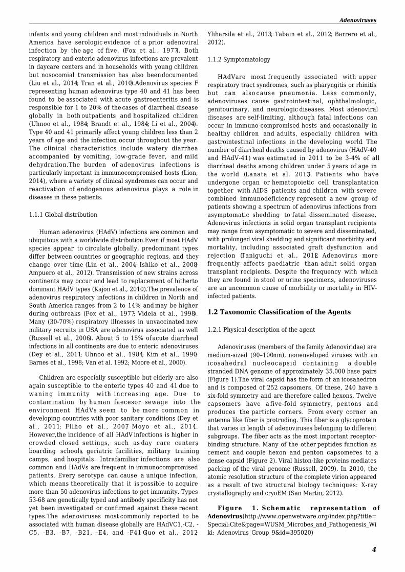
Adenoviruses (members of the family Adenoviridae) aremedium-sized (90–100nm), nonenveloped viruses with anicosahedral nucleocapsid containing a doublestranded DNA genome of approximately 35,000 base pairs(Figure 1).The viral capsid has the form of an icosahedronand is composed of 252 capsomers. Of these, 240 have asix-fold symmetry and are therefore called hexons. Twelvecapsomers have a five-fold symmetry, pentons andproduces the particle corners. From every corner anantenna like fiber is protruding. This fiber is a glycoproteinthat varies in length of adenoviruses belonging to differentsubgroups. The fiber acts as the most important receptor-binding structure. Many of the other peptides function ascement and couple hexon and penton capsomeres to adense capsid (Figure 2). Viral histon-like proteins mediatespacking of the viral genome (Russell, 2009). In 2010, theatomic resolution structure of the complete virion appearedas a result of two structural biology techniques: X-raycrystallography and cryoEM (San Martin, 2012).
Figure 1. Schematic representat ion ofAdenovirus(http://www.openwetware.org/index.php?title=Special:Cite&page=WUSM_Microbes_and_Pathogenesis_Wiki:_Adenovirus_Group_9&id=395020)
Adenoviruses
5
F i g u r e 1 . S c h e m a t i c r e p r e s e n t a t i o n o fAdenovirus(http://www.openwetware.org/index.php?title=Special:Cite&page=WUSM_Microbes_and_Pathogenesis_Wiki:_Adenovirus_Group_9&id=395020)
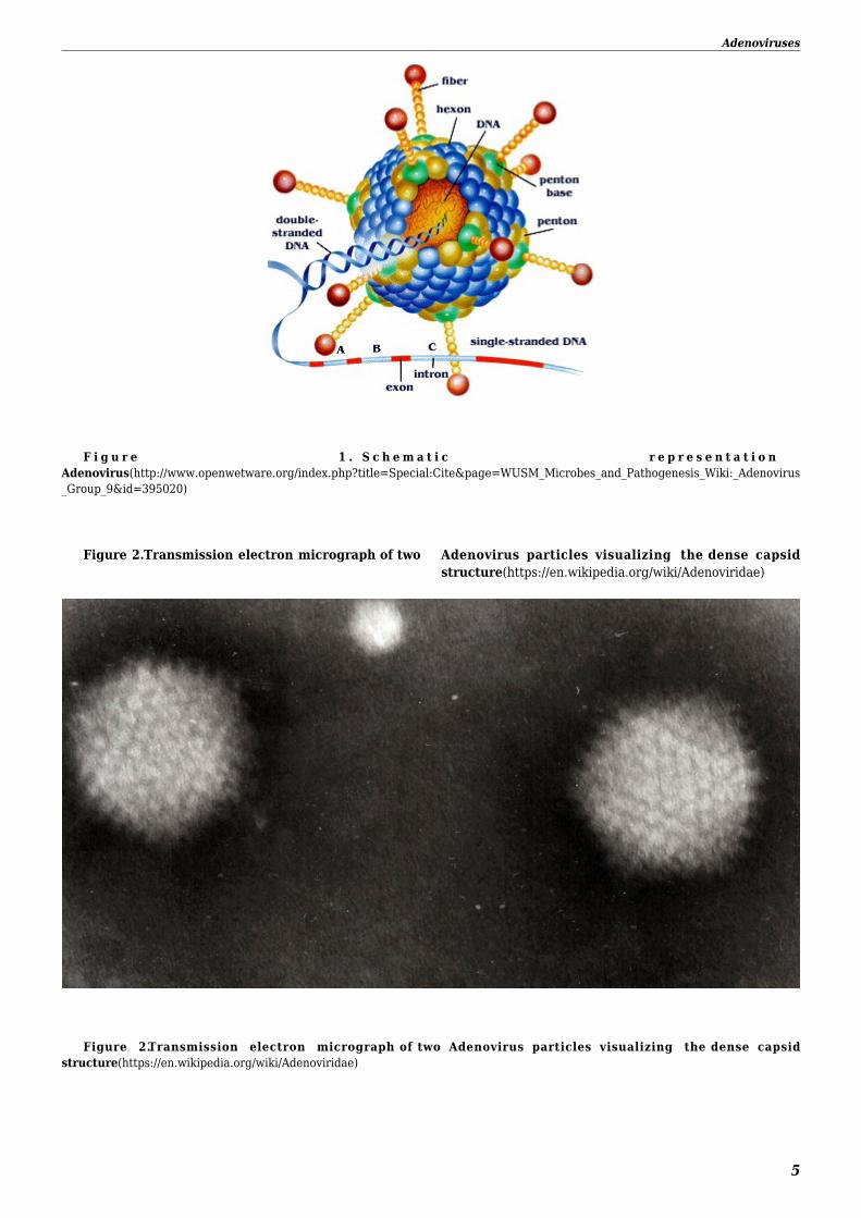
Figure 2.Transmission electron micrograph of two Adenovirus particles visualizing the dense capsidstructure(https://en.wikipedia.org/wiki/Adenoviridae)
Figure 2.Transmission electron micrograph of two Adenovirus particles visualizing the dense capsidstructure(https://en.wikipedia.org/wiki/Adenoviridae)
Adenoviruses
6
1.2.2 Taxonomy
Human adenoviruses (HAdVs) are classified in thegenus Mastadenovirus, which contains seven known HAdVspecies HAdV-A to HAdV-G. Traditionally, the HAdV specieswere classified by hemagglutination and serumneutralization reactions into different serotypes (Rosen,1960; Kjellen and Pereira, 1968). Today, there have beenreported 68 HAdV types (Brister et al., 2013). Thediscovery and division of the HAdV types 52–68 are basedon the genomic sequencing and bioinformatic analysis andthen differ from the pre-existing 51 HAdV serotypes thathave been identified by traditional serological methods incomposition and pathogenicity (Gao et al., 2014). Serotype52 constitutes a new HAdV species (G) and has beenreported after genomic sequencing and phylogeneticanalysis of an isolate in the U.S. (Jones et al., 2007). NewHAdV types have since been identified by several authorsbased on genomic data, including several emerging andrecombinant viruses (Matsushima et al., 2012). A primateadenovirus from New World monkeys was detected whichcrossed the species barrier to infect humans (Kohl et al.,2012). The majority of new HAdV types are homologousrecombination within the same subgenus, and as a result,certain new serotypes acquire different pathogenicities.Recombination is a common evolutionary way for HAdV;however, the mechanism of recombination and the potentialhazards to human beings remain unknown (Ghebremedhin,2014).
1.2.3 Tissue Tropism/Cellular receptors/Latency
In the past few years, not just one but multipleadenovirus receptors have been identified, describingdifferent HAdV species affinity for individual tissues.However, members of the largest species, species D, showgreat variability in their tropisms, with growth in tissuesranging from ocular to gastrointestinal and respiratorytissues (Pauly et al., 2014). The basis of tissue tropism isstill not well established. Adenoviral keratoconjunctivitis,which is a major cause of ocular morbidity, is mostcommonly caused by representatives of species D,including types 8, 19, and 37, but also by humanadenoviruses -E4, -C5, -B3, -B7, -B11, and -B14 (Lynch etal., 2011). Gastrointestinal manifestations are mainlyassociated with HAdV-F40 and -F41, but HAdV-G52 anddifferent members of species D, including some of the mostrecently identified types (types 65 and 67), have also beenobserved (Matsushima et al., 2012; 2013). Respiratory tractinvolvement has been associated mainly with humanadenoviruses -B3, -B7, -B16, -B21, and -E4 and variousmembers of species C (Metzgar et al., 2010). Theseexamples indicate that certain adenoviruses have strongtropisms for specific tissues, but the same clinicalmanifestations can be caused by other human adenovirustypes and species, thus requiring diagnostic screeningmethods with broad specificity.What is known today is thatthe fiber protein from species A, C and F interacts with acellular protein that belongs to the superfamilyimmunoglobulins. This receptor is called CAR, coxsackieadenovirus receptor because it is also binds tocoxsackievirus. Other types of adenoviruses utilize
alternative cellular receptors such as CD46, desmoglein 2,sialic acid, or heparan sulfate (Arnberg et al., 2012).
Currentlyavailable evidence indicates that HAdVs canpersist in a latent state in a variety of susceptible cellsfollowing primary infection. Latency is characterized byexpression of viral proteins by the host cell withoutreplication of a complete virus. A latent form of adenovirusinfection was shown to persist in tonsillar lymphocytes innearly 80% of children investigated, and the number ofadenoviral genomes per lymphoid cell apparently declineswith age (Garnett et al., 2009). Moreover, latent HAdVinfections were described to occur in intestinal Tlymphocytes and in lung epithelial cells, where they seemto play a role in the pathogenesis of obstructive airwaydisease (Hogg, 2001).
This related to the reactivation of endogenousadenovirus infections.
1.3 Transmission
1.3.1 Routes of transmission
Infections are typically transmitted by exposure toinfected individuals via inhalation of aerosolized droplets ordirect conjunctival inoculation, as well as by fecal-oralspread, including contact with recreational marine water,swimming pools freshwater or tap water (Wyn-Jones et al.,2011; Love et al., 2014; Bofill-Mas et al., 2010; vanHeerden et al., 2005; Artieda et al., 2009). Fomitetransmission can occur through exposure to infected tissue,airflow filters, or environmental surfaces (Russell et al.,2006; Soller et al., 2010). The stability of the virus at lowpH is a matter of debate, but human adenoviruses areresistant to gastric and biliary secretions and can thereforebe detected at high levels in feces (Matthes-Martin et al.,2013). Human adenoviruses are infrequently found in theurine of immonocompetent individuals but are present inthe urine of immunocompromised patients an especially inAIDS patients (Echavarria, 2008; De Jong et al., 1983;Horwitz et al., 1984). However, this secretion is probablynegligible in the context of disease transmission.
1.3.2 Reservoirs
Human adenoviruses are ubiquitous in the environmentwhere contamination by human faeces or sewage hasoccurred. Human adenoviruses typically are not pathogenicto animals and animal adenoviruses are only pathogenic tothe species of origin. However, asymptomatic infectionswith human adenovirus type 12 have been documented insimian species, and antibodies to canine, bovine and simianadenoviruses have been found in humans (AWWAssociation, 2006). Adenoviruses have a broad range ofvertebrate hosts and are known to cause respiratoryinfections in horses, cattle, pigs, sheep, and goats. The fowladenoviruses are associated with many disease conditionsin domestic fowl, but have also been found in wild birds,bats and reptiles (http://www.ictvonline.org/).
Adenoviruses
7
1.3.3 Incubation and duration periods
Following human adenovirus transmission, theincubation period ranges from 2 days to 2 weeks,depending on the viral type and mechanism of acquisition(Langley, 2005; Bhumbra and Wroblewski, 2010).Respiratory symptoms caused by adenoviruses are mostcommonly seen amongst children under the age of two andfor this selected group the incubation period before theonset of symptoms is generally 4-8 days, median 5.6 days(Lessler et al., 2009). For intestinal infections theincubation period is 3 to 10 days.Fever, coryza, cough, andsore throat, usually lasting 3-5 days, are typical symptomsof adenoviral upper respiratory disease andmost adenoviralinfections last from a few days to a week (Tabain et al.,2012). However, severe respiratory infections may lastlonger and cause lingering symptoms, such as a cough.Pneumonia can last anywhere from 2 to 4 weeks andpinkeye can persist for another several days to a week.More severe keratoconjunctivitis can last for several weeks(Pihos, 2013)and entericadenoviruses can cause diarrheathat lasts up to 2 weeks which is longer than other viraldiarrhea episodes (Uhnoo et al., 1984).
1.3.4 Period of communicability and shedding levels
The contagiousness of adenovirus is facilitated by veryhigh levels of viral particles (100,000-1,000,000/mL) in thesputum or oral secretions of infected adults. InAlthoughmany adenovirus types are found in stool from patients withdiarrhoea, only enteric adenoviruses (type 40/41/52) havebeen shown to be the causative agents of gastrointestinaldisease. A close comparison of stools from diarrheal andcontrol patients has shown that whereas non-enteric HAdVparticles are shed in stools from both diarrheal and controlpatients, enteric HAdV particles are found almostexclusively in diarrheal stools. As shown by several studies,up to 50% of healthy children carry non-enteric HAdVwhereas enteric HAdV is rarely present in healthychildren(Allard et al.,1992). Enteric adenoviruses replicatein the gut to 1.0x10¹¹virions per gram of stool and entericHAdV carriage is about 8 times higher than non-entericHAdV carriage in diarrheal stool samples. Moreover, assecretions of non-enteric adenoviral particles may persistfor months, the presence of these adenoviruses in stoolsalong with other viruses is common. HAdV 1, 2, 3 and 5 isreported to be excreted intermittently for up to 906 days(Fox et al., 1977). Quantitative Microbial Risk Assessment(QMRA) studies have been applied to drinking andrecreational water (Chigor et al., 2014; Kundu et al., 2013)and a QMRA model associated with inhaling bioaerosolsthat are contaminated with HAdV at risk-prone workplaceshave been presented by Carducci et al. (2016). Riskmanagement and dose response models can be found atQMRAwiki (http://qmrawiki.canr.msu.edu/).
1.4 Population and Individual Control Measures
1.4.1 Vaccines and therapy
1.4.1.1 Vaccines
Vaccination and infection control measures have beenapplied in certain settings to prevent adenovirus infectionsand live oral enteric-coated vaccines directed againstadenovirus serotypes 4 and 7 had been used for years inmilitary recruits in United States since the 1971 (Top et al.,1971). In the 1999, the manufacturer of the vaccinesstopped production and subsequently new outbreaks ofadenovirus serotypes 4 and 7 disease in training campsoccurred, including several fatalities, underscoring thecontinued need for the vaccines (Potter et al., 2012). Inaddition adenovirus serotype 14, a subtype B2 adenovirus,emerged in military recruit training sites and became thepredominant strain (Binn et al., 2007; Metzgar et al., 2007).However, in 2011, a new l ive, oraladenovirusvaccineagainst adenovirus serotypes 4 and 7 was approvedfor use in United States military personnel aged 17 through5 0 y e a r s ( h t t p : / / w w w . f d a . g o v /downloads/BiologicsBloodVaccines/Vaccines/ApprovedProducts/UCM247515.pdf. USA TPharmaceut. 2011). During thetwo years following reintroduction of the vaccine, UnitedStates military trainees had a 100-fold decline inadenovirus disease burden. It was also a marked decline inthe incidence of disease caused by adenovirus serotypesother than 4 and 7, including adenovirus serotype 14(Radin et al., 2014). These data suggest that the emergenceof adenovirus 14 in military recruits during the non-vaccination period was related to the discontinuation of theadenovirus serotypes 4 and 7 vaccine program, sinceheterotypic antibodies to adenovirus 14 were developedfollowing adenovirus 7 immunization. Vaccines againstadenoviruses have not been used in a large scale outsidethe United States.
Fecal shedding of adenovirus type 4 and type 7 livevaccine oral strain viruses was evaluated in a safety andimmunogenicity study of 58 subjects (30 vaccine recipientsa n d 2 8 p l a c e b o r e c i p i e n t s(http://www.fda.gov/downloads/BiologicsBloodVaccines/Vaccines/ApprovedProducts/UCM247515.pdf. USATPharmaceut. 2011). Stool or rectal swabs and throatswabs were collected on Day 0, 7, 14, 21, 28 and 56.Vaccine virus strains were shed in the stool as early as day7 following vaccination. Eight of 30 vaccine recipients(27%) tested positive at least once for adenovirus type 4fecal shedding; 18 of 30 vaccine recipients (60%) testedpositive for adenovirus type 7 fecal shedding. Noadenovirus shedding was detectable in any subject by 28days following vaccination. Vaccine strain virus was notdetected in the throat of any subject.
1.4.1.2 Antiviral agents
Antiviral therapy is general ly reserved forimmunocompromised hosts and patients with severeadenovirus disease. There have been no controlled trialsdemonstrating benefit for any antiviral agent in humanadenoviral disease. The antiviral agent most commonlyused for adenoviral infection is cidofovir, which is currentlyapproved for the treatment of cytomegalovirus (CMV)infections. This agent appears more active against
Adenoviruses
8
adenovirus in vitro than other antiviral drugs such asganciclovir (Doan et al., 2007). Antiviral drugs as ribavirinor vidarabine have no consistent activity against adenovirusin vitro (Sabroe et al., 1995).
1.4.1.3 Immunotherapy
Treatment with donor lymphocytes stimulated in vitrowith adenovirus can reduce the viral load and successfultreatment of adenovirus disease in immuno-compromisedpatients has been reported (Feuchtinger et al., 2006). T-cellrecovery with reconstitution of human adenovirus specificimmune responses is essential for effective clearance ofinvasive infections, and the initial treatment step shouldtherefore include reduction of immunosuppressivetreatment whenever possible (Matthes-Martin et al., 2012).
1.4.1.4 The use of adenovirus vectors for gene therapy
There has been considerable interest and effort indeveloping adenovirus vectors for gene therapy.Adenoviruses have several advantages over some otherviral vectors such as retroviruses and adeno-associatedvirus. Adenovirus vectors can infect a variety of cell types,including non-dividing as well as dividing cells, and can beprepared readily in large quantities in tissue culture(Vetrini and Ng, 2010; Chai et al., 2012)
1.4.1.5 Adenovirus-based vaccines against infectious pathogens
Another promising application for adenoviruses is theiruse as vaccine vectors (Gaydos and Gaydos, 1995). Asalready mentioned, live, oral, enteric-coated, adenovirusserotypes 4 and 7 vaccines were safely used for years inmilitary training camps to prevent epidemics of acuteadenoviral respiratory disease. These live, oral viralvaccines have an advantage in the ability to generate bothmucosal and systemic immunity. Thus, replication-competent as well as replication-defective adenovirusvectors are attractive candidates for immunization againstother infectious pathogens (Tatsis and Ertl, 2004).
1.4.2 Hygiene measures
Adenoviruses can stay viable for prolonged periods onenvironmental surfaces such as sinks hand towels but alsoat medical instruments and the virus isresistant to manycommon disinfectants. To prevent healthcare-associatedoutbreaks of adenovirus infections, such as epidemickeratoconjunctivitis (EKC), health care providers shouldstrictly follow infection control practices, including contactand droplet precautions, use proper disinfectants andpromptly respond to and report clusters of cases (Threlkeldet al., 1993).Because of the stability adenoviruses can alsocause significant nosocomial infections other then EKC(Swartling et al., 2015) and hand washing does not reliablyremove adenoviruses from contaminated fingers (Buehleret al., 1984; Faden et al., 2005) Decontamination ofenvironmental surfaces and instruments may be difficultsince adenoviruses are very resistant to lipid disinfectants
but are inactivated by chlorine or formaldehyde(Flomenberg, 2009). They can be inactivated by contactwith 1:5 dilution of bleach for 1 minute or 2 minutescontact with alcohol-based hand gels (Robinson andEchavarria, 2007).Adenoviruses can be physicallyinactivatedby heat (Flomenberg, 2009) by heating to 56°Cfor 30 min or 60°C for 2 min. In laboratory environmentsautoclaving for 30 minutes at 121°C or 250°F (15 lbs persquare inch of steam pressure) is recommended (Robinsonand Echavarria, 2007). In a home environment, where high-concentrated chlorine use not is recommended based onenvironmental grounds, surface disinfection with 10%bleach (1:10 dilution household bleach, such as clorox) canbe applied allowing a contact time of 15 minutes. Forstainless steel surfaces, bleach disinfection should befollowed with 70% ethanol wipedown to avoid corrosion.Liquid waste may be treated by exposing to bleach (finalvolume 10%) for 15 minutes before disposing into sink.
2.0 Environmental Occurrence andPersistence
Waterborne viruses can be introduced and remaininfectious in source waters used for recreational anddrinking water, these pathways ultimately may result inillness in some portion of the exposed population(Kokkinoset al., 2011a; Gibson, 2014; Kotwal and Cannon, 2014).
2.1 Detection Methods
Adenoviruses (HAdV) like other viruses need to beconcentrated from environmental samples and especiallyfrom water due to typically lower concentrations and theirsmall size. Several investigators have used differentmethods to detect adenoviruses in various environmentalsamples including water (Ahmed et al., 2015; Calgua et al.,2013; Ogorzaly et al., 2013; Puig et al., 1994; Sassoubre etal., 2012; Thompson et al., 2003) and air (Ziros et al., 2011;Verani et al., 2014), based on PCR procedures. Cell culturestechniques and cell culture techniques combined with PCRhave also been used (Grabow et al., 1992; Reynolds et al.,1996; Chapron et al., 2000; Ko et al., 2003; 2005). Also,recently, new metagenomics methods have been used (Awet al., 2014).
2.2 Data on Occurrence in the Environment
2.2.1 Raw sewage, sludge and treated wastewater
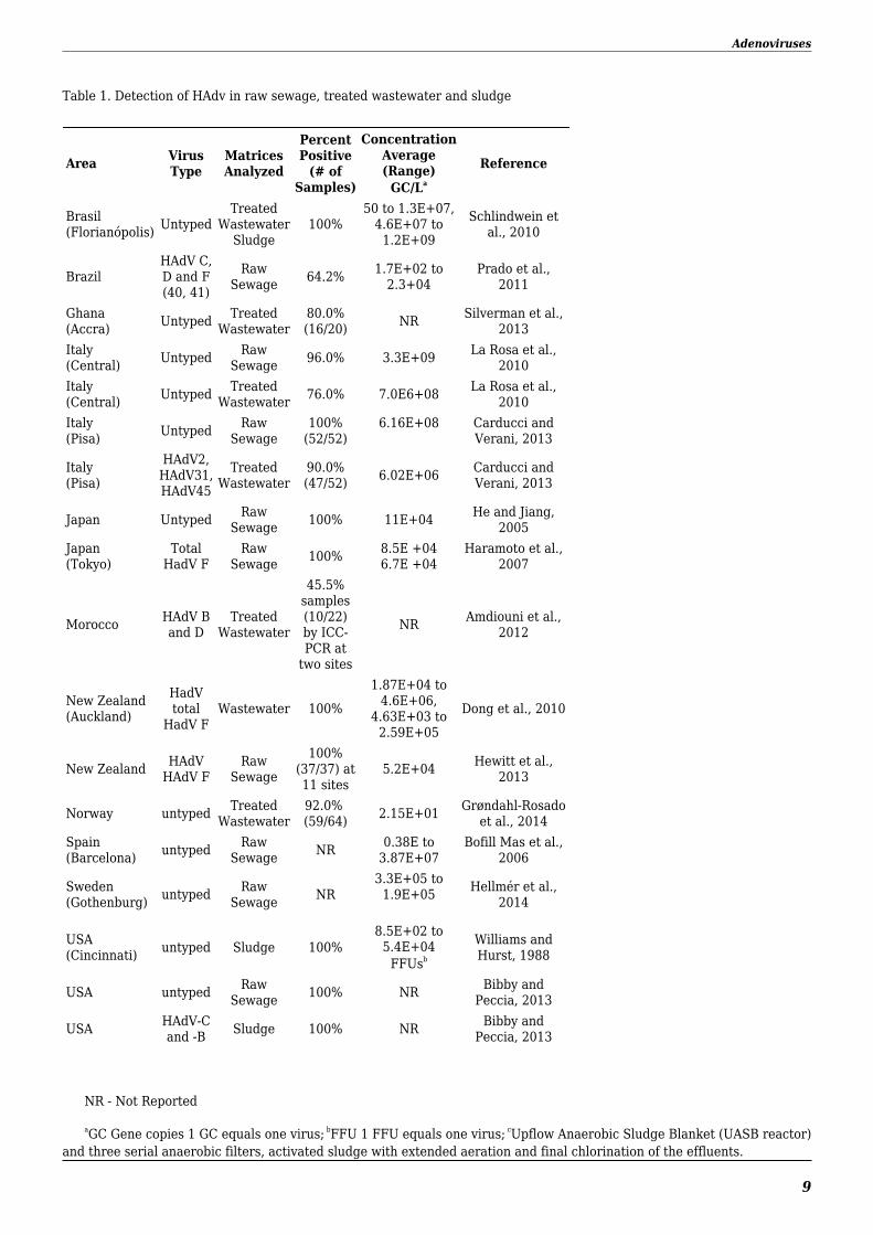
Adenoviruses of several types have been detected indomestic sewage and sludge in various countries. Thirteenstudies from 11 different countries have been reviewed inTable 1. Adenoviruses have also shown to be resistant todisinfection and thus have been detected in treatedwastewater effluents as well, with sometimes highdetection rates. In several cases, more than 80% of sewagehas been found positive for adenoviruses (Prado et al.2011,Amdiouni et al. 2012, Wong et al., 2013).
Adenoviruses
9
Table 1. Detection of HAdv in raw sewage, treated wastewater and sludge
Area VirusType
MatricesAnalyzed
PercentPositive
(# ofSamples)
ConcentrationAverage(Range)
GC/La
Reference
Brasil(Florianópolis) Untyped
TreatedWastewater
Sludge100%
50 to 1.3E+07,4.6E+07 to
1.2E+09Schlindwein et
al., 2010
BrazilHAdV C,D and F(40, 41)
RawSewage 64.2% 1.7Ε+02 to
2.3+04Prado et al.,
2011
Ghana(Accra) Untyped Treated
Wastewater80.0%(16/20) NR Silverman et al.,
2013Italy(Central) Untyped Raw
Sewage 96.0% 3.3Ε+09 La Rosa et al.,2010
Italy(Central) Untyped Treated
Wastewater 76.0% 7.0Ε6+08 La Rosa et al.,2010
Italy(Pisa) Untyped Raw
Sewage100%
(52/52)6.16E+08
Carducci andVerani, 2013
Italy(Pisa)
HAdV2,HAdV31,HAdV45
TreatedWastewater
90.0%(47/52) 6.02E+06 Carducci and
Verani, 2013
Japan Untyped RawSewage 100% 11Ε+04 He and Jiang,
2005Japan(Tokyo)
TotalHadV F
RawSewage 100% 8.5Ε +04
6.7Ε +04Haramoto et al.,
2007
Morocco HAdV Band D
TreatedWastewater
45.5%samples(10/22)by ICC-PCR at
two sites
NR Amdiouni et al.,2012
New Zealand(Auckland)
HadVtotal
HadV FWastewater 100%
1.87E+04 to4.6E+06,
4.63E+03 to2.59E+05
Dong et al., 2010
New Zealand HAdVΗΑdV F
RawSewage
100%(37/37) at11 sites
5.2E+04 Hewitt et al.,2013
Norway untyped TreatedWastewater
92.0% (59/64) 2.15Ε+01 Grøndahl-Rosado
et al., 2014Spain(Barcelona) untyped Raw
Sewage NR 0.38Ε to3.87E+07
Bofill Mas et al.,2006
Sweden(Gothenburg) untyped Raw
Sewage NR3.3Ε+05 to
1.9E+05
Hellmér et al.,2014
USA(Cincinnati) untyped Sludge 100%
8.5E+02 to5.4E+04
FFUsb
Williams andHurst, 1988
USA untyped RawSewage 100% NR Bibby and
Peccia, 2013
USA HAdV-Cand -B Sludge 100% NR Bibby and
Peccia, 2013
NR - Not Reported
aGC Gene copies 1 GC equals one virus; bFFU 1 FFU equals one virus; cUpflow Anaerobic Sludge Blanket (UASB reactor)and three serial anaerobic filters, activated sludge with extended aeration and final chlorination of the effluents.
Adenoviruses
10
Since adenoviruses are frequently detected in highconcentrations in treated wastewater and wastewater-contaminated waters, their use as potential indicators forthe presence of human sewage has been affirmed. Also, acorrelation has been noticed between the presence of HAdVwith other viruses in sewage such as human polyomavirusor norovirus (La Rosa et al. 2010; Kokkinos et al. 2011b;Hewitt et al., 2013).
Quantification of adenoviruses show usually highadenovirus concentrations in raw sewage, combined seweroverflows, primary-treated effluent, secondary-treatedeffluent, and chlorinated effluent from wastewatertreatment plants (Fong et al., 2010). Average adenovirusDNA concentrations in sewage and CSOs could be morethan 106virus particles/liter. Various adenovirus types (41,12, 40, 2, 3) have been isolated from raw sewage, sludgeand primary effluents. Surface water samples impacted bywastewater show virus concentrations and may not besuitable for full-body recreational activities (Kokkinos et al.,2011a). High concentrations of adenovirus in these watersmay be due to inefficient removal during wastewatertreatment and to the high persistence of these viruses inthe environment (Fong et al., 2010; Rodriguez et al., 2012).
2.2.2 Surface waters
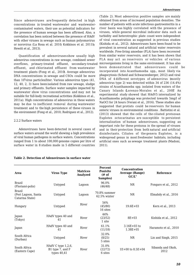
Adenoviruses have been detected in several cases ofsurface waters around the world showing a high prevalenceof viral human pathogens in surface waters. Concentrationsranged from 1 to about 100,000 genome copies per litre ofsurface water in 8 studies made in 5 different countries
(Table 2). Most adenovirus positive samples are mainlyobtained from areas of increased population densities. Thenumber of patients with acute infectious gastroenteritis in ariver basin was highly correlated with the presence ofviruses, while general microbial indicator data such asturbidity and heterotrophic plate count were independentof viral concentration as suggested in previous studies(Kishida et al., 2012). Adenoviruses have been found to beprevalent in several natural and artificial water reservoirsworldwide. Free-living amoebae (FLA) have been recoveredfrom similar water reservoirs, and it has been shown thatFLA may act as reservoirs or vehicles of variousmicroorganisms living in the same environment. It has alsobeen demonstrated that adenoviruses could beincorporated into Acanthamoeba spp., most likely viaphagocytosis (Scheid and Schwarzenberger, 2012) and viralDNA of 4 different serotypes of adenovirus (mostlyadenovirus type 2) was detected within 34 of 236 (14.4%)strains of Acanthamoeba spp. isolated from waters of theCanary Islands (Lorenzo-Morales et al., 2007). Anexperimental study showed that HAdV5 internalized byAcanthamoeba polyphaga was protected towards 5mg/l ofNaOCl for 24 hours (Verani et al., 2016). These studies alsosuggested that protists could be reservoirs for humanenteric viruses in environmental conditions . Battistini et al.(2013) showed that the cells of the ciliated protozoanEuplotes octocarinatus are susceptible to persistentinternalization of human adenoviruses, suggesting animportant role for these protozoa in the spread of virusesand in their protection from both natural and artificialdisinfectants. Ciliates of the genera Euplotes, is awidespread genus in most freshwater habitats, includingartificial ones such as sewage treatment plants (Madoni,2011).
Table 2. Detection of Adenoviruses in surface water
Area Virus Type MatricesAnalyzed
PercentPosivite
(# ofSamples)
ConcentrationAverage (Range)
GC/LaReference
Brazil(Florianó-polis) Untyped Lagoon 96.0%
(46/48) NR Fongaro et al., 2012
Brazil(Peri Lagoon, SantaCatarina State)
Untyped Lagoon 70.8% summer62.5% winter NR Elmahdy et al., 2016
Hungary Untyped River
56%(45/80)16 sites
19.6E+03 Kern et al., 2013
Japan(Tokyo)
HAdV types 40 and41 River
44%(23/52)1 site
8E+03 Kishida et al., 2012
Japan HAdV types 40 and41 River 61.1%
(11/18)3.16E+03 to
1.38E+05 Haramoto et al., 2010
South Africa(Durban) Untyped River
32.0%(8/25)5 sites
NR
Lin and Singh, 2015
South Africa(Eastern Cape)
HAdV C type 1,2,6,B1 type 7, and F
types 40,41River
31.0%(22/72)6 sites
1E+00 to 8.5E+04 Sibanda and Okoh,2012
Adenoviruses
11
South Africa(Eastern Cape)
HAdV types 21,40 and 41 River
35.0%(25/72)6 sites
1.2E+01to 4.71E+03 Chigor et al., 2012
Taiwan HadV type 41 River30.8%(4/13)
13 sites6.10E+02 to8.51E+02 Huang et al., 2015
Taiwan HAdV types2,5,31,41 River 34.3%
(99/288) 2.8E+03 Tao et al., 2015
USA HAdV40 andHAdV41 Surface water 48.3%
(14/29) NR Chapron et al., 2000
USA(Michigan) HAdV F types Lake
24.1%(14/58)2 sites
<500 Xagoraraki et al.,2007
NR - Not Reported
aGC Gene copies 1 GC equals one virus
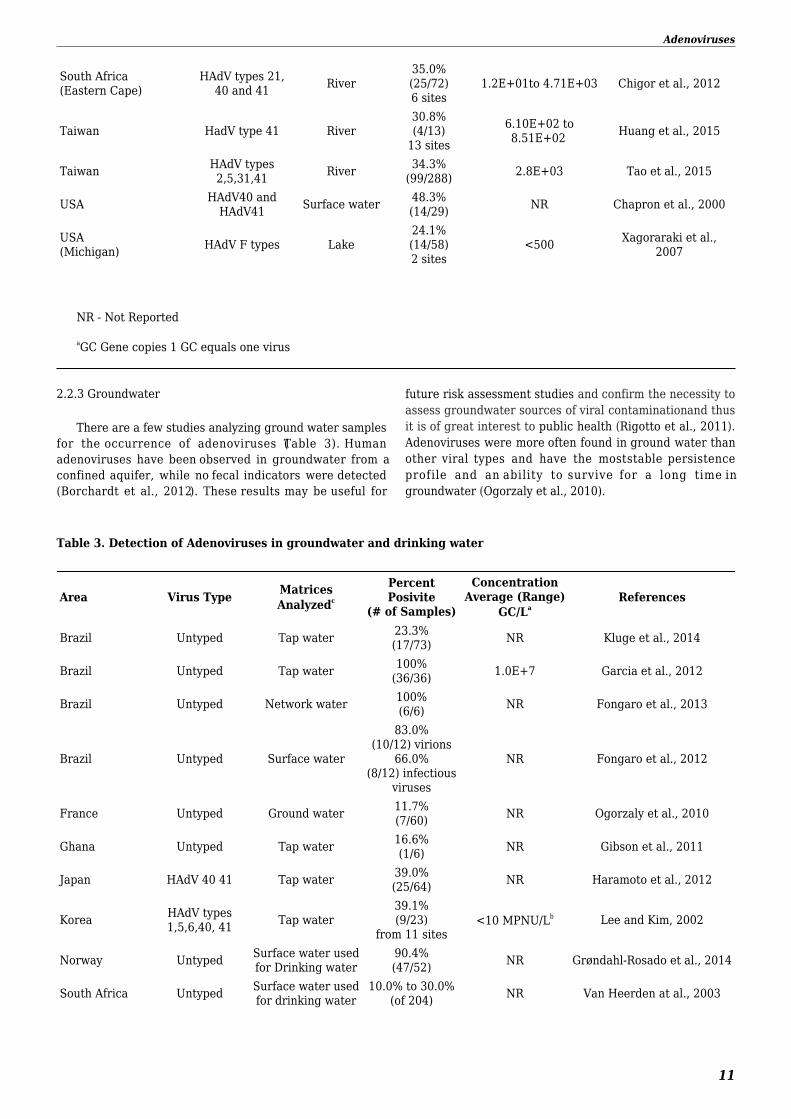
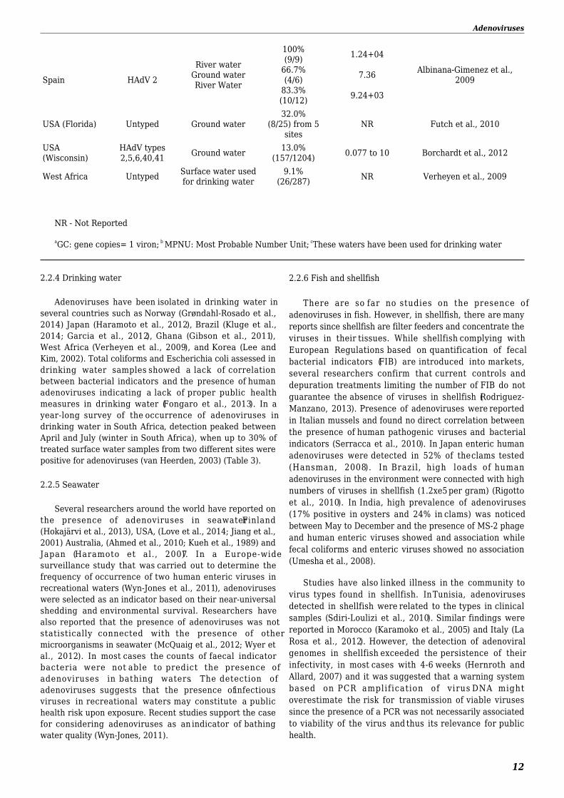
2.2.3 Groundwater
There are a few studies analyzing ground water samplesfor the occurrence of adenoviruses (Table 3). Humanadenoviruses have been observed in groundwater from aconfined aquifer, while no fecal indicators were detected(Borchardt et al., 2012). These results may be useful for
future risk assessment studies and confirm the necessity toassess groundwater sources of viral contaminationand thusit is of great interest to public health (Rigotto et al., 2011).Adenoviruses were more often found in ground water thanother viral types and have the most stable persistenceprofile and an ability to survive for a long time ingroundwater (Ogorzaly et al., 2010).
Table 3. Detection of Adenoviruses in groundwater and drinking water
Area Virus Type MatricesAnalyzedc
PercentPosivite
(# of Samples)
ConcentrationAverage (Range)
GC/LaReferences
Brazil Untyped Tap water 23.3%(17/73) NR Kluge et al., 2014
Brazil Untyped Tap water 100%(36/36) 1.0E+7 Garcia et al., 2012
Brazil Untyped Network water 100%(6/6) NR Fongaro et al., 2013
Brazil Untyped Surface water
83.0%(10/12) virions
66.0%(8/12) infectious
viruses
NR Fongaro et al., 2012
France Untyped Ground water 11.7%(7/60) NR Ogorzaly et al., 2010
Ghana Untyped Tap water 16.6%(1/6) NR Gibson et al., 2011
Japan HAdV 40 41 Tap water 39.0%(25/64) NR Haramoto et al., 2012
Korea HAdV types1,5,6,40, 41 Tap water
39.1%(9/23)
from 11 sites<10 MPNU/Lb Lee and Kim, 2002
Norway Untyped Surface water usedfor Drinking water
90.4%(47/52) NR Grøndahl-Rosado et al., 2014
South Africa Untyped Surface water usedfor drinking water
10.0% to 30.0%(of 204) NR Van Heerden at al., 2003
Adenoviruses
12
Spain
HAdV 2
River waterGround waterRiver Water
100%(9/9)
66.7%(4/6)
83.3%(10/12)
1.24+04
7.36
9.24+03
Albinana-Gimenez et al.,2009
USA (Florida) Untyped Ground water32.0%
(8/25) from 5sites
NR Futch et al., 2010
USA(Wisconsin)
HAdV types2,5,6,40,41 Ground water 13.0%
(157/1204) 0.077 to 10 Borchardt et al., 2012
West Africa Untyped Surface water usedfor drinking water
9.1%(26/287) NR Verheyen et al., 2009
NR - Not Reported
aGC: gene copies= 1 viron; b MPNU: Most Probable Number Unit; cThese waters have been used for drinking water
2.2.4 Drinking water
Adenoviruses have been isolated in drinking water inseveral countries such as Norway (Grøndahl-Rosado et al.,2014) Japan (Haramoto et al., 2012), Brazil (Kluge et al.,2014; Garcia et al., 2012), Ghana (Gibson et al., 2011),West Africa (Verheyen et al., 2009), and Korea (Lee andKim, 2002). Total coliforms and Escherichia coli assessed indrinking water samples showed a lack of correlationbetween bacterial indicators and the presence of humanadenoviruses indicating a lack of proper public healthmeasures in drinking water (Fongaro et al., 2013). In ayear-long survey of the occurrence of adenoviruses indrinking water in South Africa, detection peaked betweenApril and July (winter in South Africa), when up to 30% oftreated surface water samples from two different sites werepositive for adenoviruses (van Heerden, 2003) (Table 3).
2.2.5 Seawater
Several researchers around the world have reported onthe presence of adenoviruses in seawater Finland(Hokajärvi et al., 2013), USA, (Love et al., 2014; Jiang et al.,2001) Australia, (Ahmed et al., 2010; Kueh et al., 1989) andJapan (Haramoto et al., 2007). In a Europe-widesurveillance study that was carried out to determine thefrequency of occurrence of two human enteric viruses inrecreational waters (Wyn-Jones et al., 2011), adenoviruseswere selected as an indicator based on their near-universalshedding and environmental survival. Researchers havealso reported that the presence of adenoviruses was notstatistically connected with the presence of othermicroorganisms in seawater (McQuaig et al., 2012; Wyer etal., 2012). In most cases the counts of faecal indicatorbacteria were not able to predict the presence ofadenoviruses in bathing waters. The detection ofadenoviruses suggests that the presence of infectiousviruses in recreational waters may constitute a publichealth risk upon exposure. Recent studies support the casefor considering adenoviruses as an indicator of bathingwater quality (Wyn-Jones, 2011).
2.2.6 Fish and shellfish
There are so far no studies on the presence ofadenoviruses in fish. However, in shellfish, there are manyreports since shellfish are filter feeders and concentrate theviruses in their tissues. While shellfish complying withEuropean Regulations based on quantification of fecalbacterial indicators (FIB) are introduced into markets,several researchers confirm that current controls anddepuration treatments limiting the number of FIB do notguarantee the absence of viruses in shellfish (Rodriguez-Manzano, 2013). Presence of adenoviruses were reportedin Italian mussels and found no direct correlation betweenthe presence of human pathogenic viruses and bacterialindicators (Serracca et al., 2010). In Japan enteric humanadenoviruses were detected in 52% of the clams tested(Hansman, 2008). In Brazil, high loads of humanadenoviruses in the environment were connected with highnumbers of viruses in shellfish (1.2xe5 per gram) (Rigottoet al., 2010). In India, high prevalence of adenoviruses(17% positive in oysters and 24% in clams) was noticedbetween May to December and the presence of MS-2 phageand human enteric viruses showed and association whilefecal coliforms and enteric viruses showed no association(Umesha et al., 2008).
Studies have also linked illness in the community tovirus types found in shellfish. In Tunisia, adenovirusesdetected in shellfish were related to the types in clinicalsamples (Sdiri-Loulizi et al., 2010). Similar findings werereported in Morocco (Karamoko et al., 2005) and Italy (LaRosa et al., 2012). However, the detection of adenoviralgenomes in shellfish exceeded the persistence of theirinfectivity, in most cases with 4-6 weeks (Hernroth andAllard, 2007) and it was suggested that a warning systembased on PCR amplification of virus DNA mightoverestimate the risk for transmission of viable virusessince the presence of a PCR was not necessarily associatedto viability of the virus and thus its relevance for publichealth.
Adenoviruses
13
2.2.7 Air
Many enteric viruses are present in wastewater and dueto their small s ize, they may be transmitted indroplets.Εxposure to virus contaminated droplets in severallabour places mainly wastewater treatment plants (WWTP)and its consequence on workers' health have beendocumented. Very few studies have investigated thepresence and concentrations of airborne virus in WWTP.Masclaux et al. (2014) detected that adenovirus waspresent in 100% of summer WWTP AIR samples and 97% ofwinter AIR samples. Concentrations of potentiallypathogenic viral particles in WWTP air are non-negligibleand could partly explain the work-related gastrointestinalsymptoms often reported in employees in this sector.Concentrations reported were2.27×106genomeequivalent/m3. Different concentrations have been observedalso according to the plant areas: 2455 GC/m3 at thesewage entrance, 1622 GC/m3 at the biological oxidationtank, 933 GC/m3 at the sludge treatment and 589 GC/m3 atthe side-entrance manhole (Carducci et al., 2016).
Wan et al (2012) evaluated the distributions of airborneadenovirus in public areas in the pediatric department ofChildren's Hospital in northern Taiwan and high detectionrates of airborne adenovirus DNA were obtained in Marchwith average 278.9CG/m3. Tseng et al. (2010) investigatedthe possibility of aerosol transmission of viruses in childrenin the pediatrics department of a medical center in Taipei,Taiwan. HAdV were detected in the aerosol of air samples(36% of the samples). In toilets of healthcare settings andoffices HAdV were found in over 60% of samples atconcentrations respectively of 6.6 107 GC/mL and 1.2107GC/mL in water, of 5.8x10⁶GC/ m3 and 3.9x10⁴GC/m3inair and of 483 GC/cm2 and 91 GC/cm2 on surfaces (Verani etal., 2014). HAdV was detected in apartments with averagein winter 2106 GC/m3 and in summer 173 GC/m3 (Moon etal., 2014).
2.3 Persistence
Data on adenoviruses have shown that their genomewas more stable compared with genomes of other virusesor bacteria (El-Senousy et al., 2014). The presence ofadenoviral DNA in groundwater may be misleading in termof health risk, especially in the absence of information ontheir infective status (Ogorzaly et al., 2010). According to alinear regression model in a year-long study ingroundwater, infectivity reductions of HAdV2 ranged from0.0076 log10(10)/day (at 4°C) to 0.0279 log10(10)/day (20°C).No adenoviral genome degradation was observed at 4°C,and the reduction was estimated at 0.0036 log10(10)/day at20°C (Ogorzaly et al., 2010). Adenoviruses could not bedetected on produce after 24h of irrigation with watercontaminated with viruses (Ward and Irving, 1987).Adenoviruses were found to be stable in biosolids for atleast 60 days with no degradation (Wei et al., 2010). In astudy of health risks associated with land applied biosolidsno notable decline in HAdV numbers detected by PCR wasobserved during 6 months (T90 ≥ 180 days) (Schwarz et al.,2014).
Temperature is considered as the major factordetermining virus persistence in the environment. Thepersistence of infectious particles, encapsidated genomesand free nucleic acids of HAdV 41 was evaluated indrinking water and surface water at 4°C, 20°C and 37°C byPrevost et al. (2016). The infectivity of HAdV 41 persistedfor at least 25 days, whatever the water temperature, andfor more than 70 days at 4°C and 20°C, in both drinkingand surface water. Encapsidated genomes persisted beyond70 days, whatever the water temperature. Free nucleicacids (i.e. without capsid) could persist for at least 16 daysin drinking and surface water (Prevost et al., 2016).
In a review (Bertrand et al., 2012), virus persistence wasfound to be lower at temperatures ≥ 50°C than at temperatures <50°C, but there was also a significant temperature–matrix effect.Virus inactivation appeared to occur faster in complex than insimple matrices. The virus genome was shown to be moreresistant than infectious virus (Bertrand et al., 2012; Fongaro etal., 2013). Enriquez et al. (1995) studied the survival of poliovirustype 1, HAV and the enteric adenoviruses (types 40 and 41) in tapwater at various temperatures for up to 60 days. Overall, theyfound that the enteric adenoviruses were more stable than theother viral types at both 4°C and 15°C and at room temperature.However, in primary and secondary sewage the entericadenoviruses did not survive significantly longer than poliovirus 1at either 4°C or 15°C. El-Senousy et al. (2014) also showed thatHAdV40 was more persistent than both rotavirus and hepatitis Ain ground water. Inspiked experiments with HAdV40 in groundwater 0.05 log10, 0.5 log10 and 1.5 log10 reduction of copy numbersat 4°C, 20°C and 35°C were shown. Infectious units reductionafter 12 weeks were 0.5 log10, 1.5 log10 and 3.0 log10 at the sametemperatures. Charles et al. (2009) also showed in spikedexperiments with HAdV2 that these genomes were stable ≥ 2years in ground water with a retaining infectivity ≥ 1 year. Bofill-Mas et al. (2006)studied the stability of HAdV during 300 days insewage samples at 20°C resulting in T90of 60.9 days and aT99of132.3 days. By cloning experiments the adenovirus types 40, 41,31, 34, 35, 11 and 12 were detected over 105 days showing nosignificant differences in stability between the different typespresent during the experiment (Bofill-Mas et al., 2006).
3.0 Reductions by Sanitation Management
Adenoviruses have been detected in raw sewagethroughout the world and are associated with a number ofhuman illnesses but their occurrence and pathogenicityhave not been well studied (Jiang, 2006; Mena and Gerba,2009; Jacob et al., 2015). The continued monitoring oftreated wastewater by other researchers revealed theaverage concentration of adenoviruses in treatedwastewater (see Table 1). No correlation between thepresence of viruses and Escherichia coli has been found(Maunula et al., 2012). Moreover, the possible treatedsewage reuse in agriculture or elsewhere must beconsidered with concern (Crabtree et al., 1997; Kokkinos etal., 2010).
3.1 Wastewater Treatment
Adenoviruses
14
3.1.1 Pit Latrines, Vault Toilets, Dry Toilets
There were no data on adenovirus reductions in vaulttoilets, dry toilets.
3.1.2 Manure-based Composting
In the composting process of dairy manure HAdV type41 was more stable than RNA viruses in composts withcenter temperatures between 65°C and 70°C. There was noreduction in DNA after one day, and about a 2.1 log10loss at5 and 7 days (Wei et al., 2009). In the same experiment,more than a 4 log10reduction of adenoviral infectivity wasnoted after one day in compost. According to NorthAmerican regulatory bodies total viral inactivation isexpected to occur if compost particles maintaintemperatures greater than 55°C for at least 3 days. Thiscould either be the result of t ime–temperaturerequirements in ensuring that the time–temperaturecriteria are met by all compost particles (Wichuk andMcCartney, 2007).
3.1.3 Septic tanks
No log10reduction data on adenoviruses in septic tanksare available.
3.1.4 Waste stabilization ponds
Wastewater treatment ponds (lagoons) are one of themost common types of technologies used for wastewatermanagement worldwide, especially in small cities andtowns. They are particularly well-suited for systems wherethe effluent is reused for irrigation.Human adenoviruses inwastewater treatment plants have been used as anindicator of the effectiveness of different treatmentprocesses. HAdV concentrations in wastewater were morevariable in small and medium-sized WTP than in large-sizedplants. HAdv concentrations were detected in influent ofmost WTP with a reduced median concentration in effluent.Highest culturable HAdV concentrations in effluent werefrom a medium-sized WWTP. No matter of treatment type,adenoviruses are likely to be present in non-disinfectedeffluent, with associated human health risks dependent onconcentration and receiving water usage (Sidhu and Toze,2009; Hewitt et al., 2011).Sheludchenko et al. (2016)showed a log10reduction of adenoviruses of a 1.2 log10andJurzik et al. (2015) showed no reduction.On average,inwaste stabilization ponds,one log10reduction of viruses wasachieved for every 14.5–20.9 days of retention, but the 95thpercentile value of the data analyzed was 54 days. (Verbylaand Mihelcic, 2015).
3.1.5 Wetlands
Wetlands, open-water cells can be used to promotesunlight disinfection and remove pathogenic viruses fromwastewater. Sunlight inactivation is an important mode ofdisinfection for viruses in surface waters and rates foradenovirus inactivation are reported (Silverman et al.,2015).Rachmadi et al. (2016) showed an adenovirus
removal of 1-2 log10 in wetlands during a 9-month period. Aweak negative correlation between adenoviruslog10concentration and water temperature as well as pHwas observed (Rachmadi et al., 2016). HAdV type 3 showedinactivation rates with one log10reduction times of morethan 33 days in constructed reedbeds (Sidhu et al., 2010).
3.1.6 Aerated lagoons
The removal was found to be zero for HAdVduring thewastewater treatment in aerated lagoons (O'Hara andRubin, 2005; Jurzik et al., 2015).
3.1.7 Wastewater Treatment Facilities
3.1.7.1 Combined sewer overflows
Concentrations of adenovirus DNA in the combinedsewer overflows discharges (CFOs) were not significantlydifferent from adenovirus DNA concentrations in rawsewage showing an average concentration of 5.35x10⁵
viruses/liter.Removal of adenoviruses was lower than 2log10 at the treatment plant(Fong et al., 2010). Hata et al.(2014) reported thatrainfall events can introduce largeamount of viral contaminants including adenoviruses intosurface water by intermittent discharges from combinedsewer overflows (CSOs).
3.1.7.2 By primary /preliminary treatment
Adenoviruses showed high stability in urban sewage(Silvia Bofill-Mas et al., 2006) and they were detectedfrequently in high concentration in raw sewage influentsbut were also detected less frequently in plant effluent andat much lower concentrations with removals shown to be2-3 log10 by primary sewage treatment.In this studya t90 of60.9 days for HAdVwas reported.In another study, theaverage log10removals were 2.2–3.5 for adenoviruses whenusing sand filters in a cold temperature climate (Kauppinenet al., 2014).
3.1.7.3 By Secondary treatment and membrane bioreactors
In a one-year quantitative survey in six differentJapanese activated sludge waste-waterplants adenovirusamong other enteric viruses were observed. Adenovirusesweredetected at the highest positive ratio among the testedviruses; almost 100% in bothsecondary-treated wastewaterand in effluent after chlorination. The reductionofadenovirus was 2 log10 and was also found to be fairlyconstant and independent ofthe concentration of viruses inthe influent (Katayama et al., 2008).No removals ofadenovirus across disinfection was seen in membranebioreactors (MBR) and low median log10removals of 0.24was detected in conventional secondary plants with a fewremovals of individual samples near or above the analyticalvariability of 1.2 log10genomic copies per liter (Francy et al.,2012).
Also, the environmental monitoring carried out at awastewater treatment plant in Italy, showed the presence
Adenoviruses
15
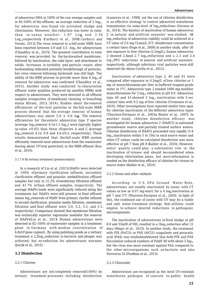
of adenovirus DNA in 100% of the raw sewage samples andin 85-100% of the effluent, an average reduction of 2 log10
for adenovirus was found via activated sludge andchlorination. Moreover, this reduction was lower in rainythan i n sunny wea ther : 1 .67 Log 1 0 and 2 .34Log10respectively (Carducci et al., 2008; Carducci andVerani, 2013).The virus removal in a full-scale MBR havebeen reported between 3.9 and 5.5 –log10 for adenoviruses(Chaudhry et al., 2015). The greatest contribution to totalremoval was provided by the backwashed membrane,followed by inactivation, the cake layer, and attachment tosolids. Increases in turbidity and particle counts afterbackwashing indicated potential breakthrough of particles,but virus removal following backwash was still high. Theability of the MBR process to provide more than 4 log10 ofremoval for adenovirus was confirmed (Chaudhry et al.,2015). Another study was conducted to characterizeeffluent water qualities produced by satellite MBRs withrespect to adenoviruses. They were detected in all effluentsamples irrespective of membrane cleaning or breachingstatus (Hirani, 2013; 2014). Studies about the removalefficiencies of the viral particles in the full-scale MBRprocess showed that the average removal of humanadenoviruses was about 5.0 ± 0.6 log10. The removalefficiencies for the enteric adenovirus type F species(average log10removal 6.5±1.3 log10) were typically higher(p-value <0.05) than those of species A and C (averagelog10removal 4.1± 0.9 and 4.6 ±0.5, respectively). Theseresults demonstrated that the full-scale MBR systemefficiently removed most adenoviruses from the wastewaterleaving about 103viral particles/L in the MBR effluent (Kuoet al., 2010).
3.1.7.4 By tertiary treatment (postsecondary)
In a research of Liu et al. (2013),HAdVs were detectedin 100% of primary clarification influent, secondaryclarification effluent and granular mediafiltration effluentsamples but only in 31.2% of membrane filtration effluentand 41.7% of final effluent samples, respectively. Theaverage HAdVs loads were significantly reduced along thetreatments but HAdVs were still present in final effluent(mean log10removals of HAdV from primary clarifer influentto second clarification, granular media filtration, membranefiltration and final effluent were 3.0, 3.2, 5.2, and 5.1,respectively). Comparison showed that membrane filtrationwas technically superior togranular mediafor the removalof HAdVs(Liu et al., 2013). Human adenoviruses weredetected in 82–100% of wastewater samples in a treatmentplant in Germany with median concentration of6.8x103gene copies/L. By using polishing ponds as a tertiarytreatment a 2.2log10reduction in bacteria and phages wasachieved, but no reduction for adenoviruses was seen(Jurzik et al., 2015).
3.2 Disinfection
3.2.1 Chlorine
Adenoviruses are not completely removed(100%) bytertiary treatment processes including disinfection
(Lazarova et al., 1999), yet the use of chlorine disinfectionis an effective strategy to control adenoviral waterbornetransmission via some level of log10reductions (Girones etal., 2014). The kinetics of inactivation of human adenovirus2 in natural and artificial seawater was studied. A4log10reduction of adenovirus viability could be achieved at aCT value of 2.6 mg Cl2min/L (CT; disinfectant concentrationx contact time) (Page et al., 2009).In another study, after 30min exposure to free chlorine (2.5mg/L), human adenovirus2 showed 2.6and 2.7 log10reductions and a 2.3 and 2.4log10PFU reductions in natural and artificial seawater,respectively, although infectious viral particles were stillobserved (de Abreu Corrêa et al., 2012).
Inactivation of adenovirus type 2, 40 and 41 werecompared after exposure to 0.2mg/L of free chlorine or 1mg of monochloramine per liter in buffered reagent-gradewater at 5°C. Adenovirus type 2 needed 1600 mg-min/litermonochloramine for 3 log10 reduction at pH 8.0. Adenovirustype 40 and 41 showed 3 log10 inactivation with 5 seccontact time with 0.2 mg of free chlorine (Cromeans et al.,2010). Other investigations have reported similar time spanfor chlorine inactivation of the different adenovirus types(Thurston-Enriquez et al., 2003a; Baxter et al., 2007). Inanother study, chlorine disinfection efficacy wasinvestigated for human adenovirus 2 (HAdV2) in untreatedgroundwater source and partially treated surface waters.Chlorine disinfection of HAdV2 proceeded very rapidly (3-4log10 inactivation within 5 to 10s) in each source water andwhen CT values could be calculated disinfection was moreeffective at pH 7 than pH 8 (Kahler et al., 2010). However,water quality could play a substantial role in theinactivation of viruses and should be considered whendeveloping chlorination plans, but more information isneeded on the disinfection efficacy of chlorine for viruses insource water (Kahler et al., 2010).
3.2.2 Ozone and other oxidants
According to U.S. EPA Ground Water Rule,adenoviruses are readily inactivated by ozone with CTvalues as low as 0.07 mg-min/L for a 4 log10inactivation atpH 7 and 5°C (Thurston-Enriques et al., 2005). In light ofthis, the combined use of ozone with UV may be a viableand safe water treatment strategy that utilities couldemploy to achieve desired reductions in pathogenicmicroorganisms.
The inactivation of adenoviruses in fecal sludge at pH8.9 and 55mM of NH3 resulted in a 3log10reduction after 21days (Magri et al., 2015). In another study, the treatmentwith PIX (FeCl3) or PAX (AlCl3) coagulants and peraceticacid (PAA) was studiedandshowed that both PIX and PAXflocculation reduced numbers of HAdV 40 with about 2 log10
but the virus was more resistant against PAA compared tothe other microorganisms such as bacteria and alsoNorovirus GI (Pradhan et al., 2013).
3.2.3 Ultraviolet
Adenoviruses are recognized as the most UV-resistantwaterborne pathogen of concern to public health
Adenoviruses
16
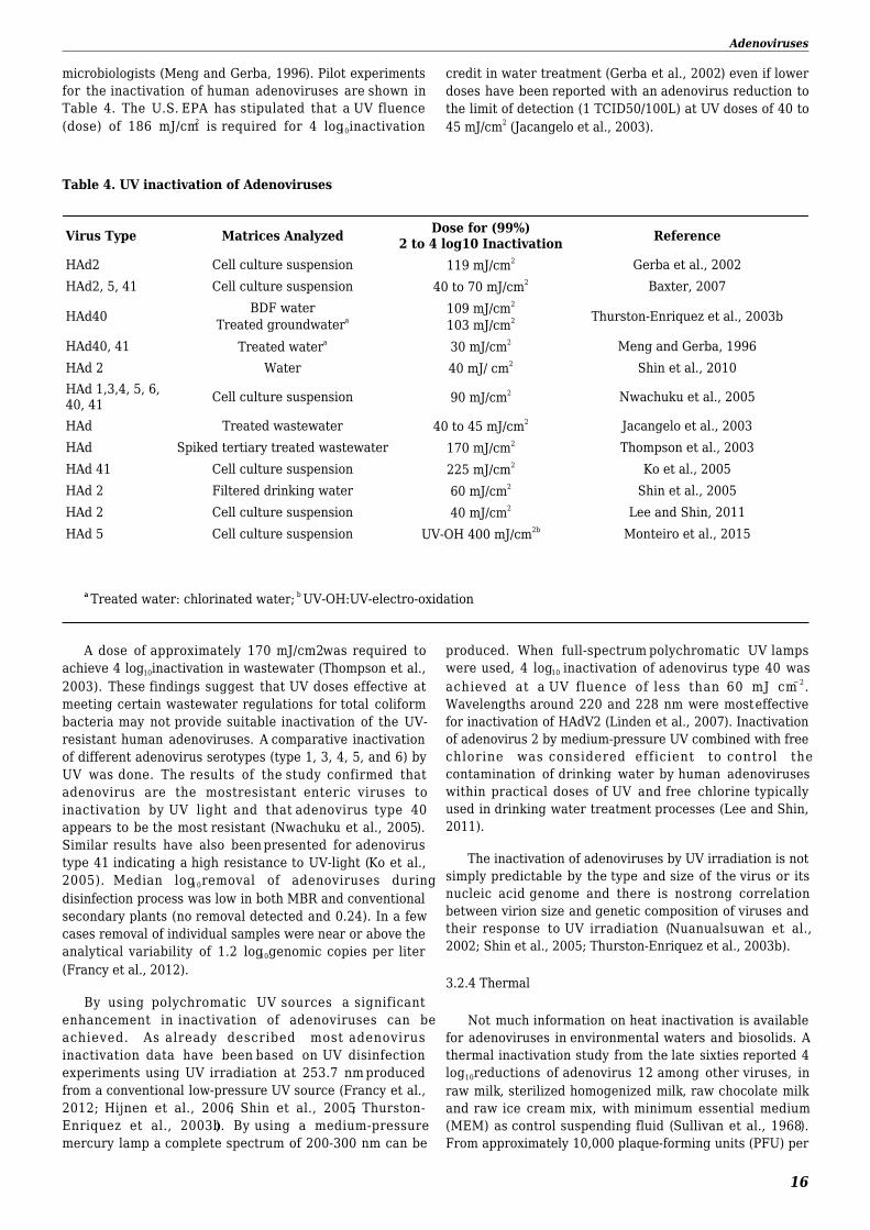
microbiologists (Meng and Gerba, 1996). Pilot experimentsfor the inactivation of human adenoviruses are shown inTable 4. The U.S. EPA has stipulated that a UV fluence(dose) of 186 mJ/cm2 is required for 4 log10inactivation
credit in water treatment (Gerba et al., 2002) even if lowerdoses have been reported with an adenovirus reduction tothe limit of detection (1 TCID50/100L) at UV doses of 40 to45 mJ/cm2 (Jacangelo et al., 2003).
Table 4. UV inactivation of Adenoviruses
Virus Type Matrices Analyzed Dose for (99%)2 to 4 log10 Inactivation Reference
HAd2 Cell culture suspension 119 mJ/cm2 Gerba et al., 2002HAd2, 5, 41 Cell culture suspension 40 to 70 mJ/cm2 Baxter, 2007
HAd40 BDF waterTreated groundwatera
109 mJ/cm2
103 mJ/cm2 Thurston-Enriquez et al., 2003b
HAd40, 41 Treated watera 30 mJ/cm2 Meng and Gerba, 1996HAd 2 Water 40 mJ/ cm2 Shin et al., 2010HAd 1,3,4, 5, 6,40, 41 Cell culture suspension 90 mJ/cm2 Nwachuku et al., 2005
HAd Treated wastewater 40 to 45 mJ/cm2 Jacangelo et al., 2003HAd Spiked tertiary treated wastewater 170 mJ/cm2 Thompson et al., 2003HAd 41 Cell culture suspension 225 mJ/cm2 Ko et al., 2005HAd 2 Filtered drinking water 60 mJ/cm2 Shin et al., 2005HAd 2 Cell culture suspension 40 mJ/cm2 Lee and Shin, 2011HAd 5 Cell culture suspension UV-OH 400 mJ/cm2b Monteiro et al., 2015
a Treated water: chlorinated water; b UV-OH:UV-electro-oxidation
A dose of approximately 170 mJ/cm2was required toachieve 4 log10inactivation in wastewater (Thompson et al.,2003). These findings suggest that UV doses effective atmeeting certain wastewater regulations for total coliformbacteria may not provide suitable inactivation of the UV-resistant human adenoviruses. A comparative inactivationof different adenovirus serotypes (type 1, 3, 4, 5, and 6) byUV was done. The results of the study confirmed thatadenovirus are the most resistant enteric viruses toinactivation by UV light and that adenovirus type 40appears to be the most resistant (Nwachuku et al., 2005).Similar results have also been presented for adenovirustype 41 indicating a high resistance to UV-light (Ko et al.,2005). Median log10removal of adenoviruses duringdisinfection process was low in both MBR and conventionalsecondary plants (no removal detected and 0.24). In a fewcases removal of individual samples were near or above theanalytical variability of 1.2 log10genomic copies per liter(Francy et al., 2012).
By using polychromatic UV sources a significantenhancement in inactivation of adenoviruses can beachieved. As already described most adenovirusinactivation data have been based on UV disinfectionexperiments using UV irradiation at 253.7 nm producedfrom a conventional low-pressure UV source (Francy et al.,2012; Hijnen et al., 2006; Shin et al., 2005; Thurston-Enriquez et al., 2003b). By using a medium-pressuremercury lamp a complete spectrum of 200-300 nm can be
produced. When full-spectrum polychromatic UV lampswere used, 4 log10 inactivation of adenovirus type 40 wasachieved at a UV fluence of less than 60 mJ cm− 2.Wavelengths around 220 and 228 nm were most effectivefor inactivation of HAdV2 (Linden et al., 2007). Inactivationof adenovirus 2 by medium-pressure UV combined with freechlorine was considered efficient to control thecontamination of drinking water by human adenoviruseswithin practical doses of UV and free chlorine typicallyused in drinking water treatment processes (Lee and Shin,2011).
The inactivation of adenoviruses by UV irradiation is notsimply predictable by the type and size of the virus or itsnucleic acid genome and there is no strong correlationbetween virion size and genetic composition of viruses andtheir response to UV irradiation (Nuanualsuwan et al.,2002; Shin et al., 2005; Thurston-Enriquez et al., 2003b).
3.2.4 Thermal
Not much information on heat inactivation is availablefor adenoviruses in environmental waters and biosolids. Athermal inactivation study from the late sixties reported 4log10reductions of adenovirus 12 among other viruses, inraw milk, sterilized homogenized milk, raw chocolate milkand raw ice cream mix, with minimum essential medium(MEM) as control suspending fluid (Sullivan et al., 1968).From approximately 10,000 plaque-forming units (PFU) per

Adenoviruses
17
ml of each suspending medium, inactivation curves at 40°C- 60°C were asymptotic to the base line, showing a tailingeffect indicating that small amounts of adenovirusessurvived, even at the higher temperature. At 65°C, the
inactivation curves approached first order reactions,indicating that temperatures near pasteurization standardswere effective in inactivating adenoviruses.
Adenoviruses
18
ReferencesAhmed, W., Goonetilleke, A. and Gardner, T. (2010). Human and bovine adenoviruses for the detection of source-specificfecal pollution in coastal waters in Australia. Water Res. 44, Englandpp. 4662–73.
Ahmed, W., Harwood, V.J, Gyawali, P., Sidhu, J.P and Toze, S. (2015). Comparison of concentration methods forquantitative detection of sewage-associated viral markers in environmental waters. Appl Environ Microbiol. 81, pp. 2042–9.
Albinana-Gimenez, N., Miagostovich, M.P, Calgua, B., Huguet, J.M, Matia, L. and Girones, R. (2009). Analysis ofadenoviruses and polyomaviruses quantified by qPCR as indicators of water quality in source and drinking-water treatmentplants. Water Res. 43, pp. 2011-9.
Allard, A., Albinsson, B. and Wadell, G. (1992). Detection of adenoviruses in stools from healthy persons and patients withdiarrhea by two-step polymerase chain reaction. J Med Virol. 37, pp. 149–57.
Amdiouni, H., Faouzi, A., Fariat, N., Hassar, M., Soukri, A. and Nourlil, J. (2012). Detection and molecular identification ofhuman adenoviruses and enteroviruses in wastewater from Morocco. Lett Appl Microbiol. 54, pp. 359–66.
Ampuero, J.S, Ocana, V., Gomez, J., Gamero, M.E, Garcia, J., Halsey, E.S et al. (2012). Adenovirus respiratory tractinfections in Peru. PLoS One. 7, pp. e46898.
Arnberg, N. (2012). Adenovirus receptors: implications for targeting of viral vectors. Trends Pharmacol Sci. 33, pp. 442–8.
Artieda, J., Pineiro, L., Gonzalez, M., Munoz, M., Basterrechea, M., Iturzaeta, A. et al. (2009). A swimming pool-relatedoutbreak of pharyngoconjunctival fever in children due to adenovirus type 4, Gipuzkoa, Spain, 2008. Euro Surveill. 14,
Association, AWater Work (2006). Waterborne Pathogens. Adenoviruses. 2nd edition, Denver Colorado, American WaterWorks Association
Aw, T.G, Howe, A. and Rose, J.B (2014). Metagenomic approaches for direct and cell culture evaluation of the virologicalquality of wastewater. J Virol Methods. 210C, 2014 Elsevier B.Vpp. 15–21.
Barnes, G.L, Uren, E., Stevens, K.B and Bishop, R.F (1998). Etiology of acute gastroenteritis in hospitalized children inMelbourne, Australia, from April 1980 to March 1993. J Clin Microbiol. 36, pp. 133–8.
Barrero, P.R, Valinotto, L.E, Tittarelli, E. and Mistchenko, A.S (2012). Molecular typing of adenoviruses in pediatricrespiratory infections in Buenos Aires, Argentina (1999-2010). J Clin Virol. 53, Netherlands, 2011 Elsevier B.Vpp. 145–50.
Battistini, R., Marcucci, E., Verani, M., Di Giuseppe, G., Dini, F. and Carducci, A. (2013). Ciliate-adenovirus interactions inexperimental co-cultures of Euplotes octocarinatus and in wastewater environment. Eur J Protistol. 49, Germany, 2012Elsevier GmbHpp. 381–8.
Baxter, CS, Hofmann, R, Templeton, MR, Brown, M and Andrews, RC (2007). Inactivation of adenovirus types 2,5, and 41in drinking water by UV light, free chlorine, and monochloramine. J Environmental engineering. 133, pp. 95-103.
Bertrand, I., Schijven, J.F, Sanchez, G., Wyn-Jones, P., Ottoson, J., Morin, T. et al. (2012). The impact of temperature on theinactivation of enteric viruses in food and water: a review. J Appl Microbiol. 112, pp. 1059–74.
Bhumbra, N. and Wroblewski, M.E (2010). Adenovirus. Pediatr Rev. 31, United Statespp. 173–4.
Bibby, K. and Peccia, J. (2013). Prevalence of respiratory adenovirus species B and C in sewage sludge. Environ SciProcess Impacts. 15, pp. 336–8.
Binn, L.N, Sanchez, J.L and Gaydos, J.C (2007). Emergence of adenovirus type 14 in US military recruits–a new challenge. JInfect Dis. 196, United Statespp. 1436–7.
Adenoviruses
19
Bofill-Mas, S., Albinana-Gimenez, N., Clemente-Casares, P., Hundesa, A., Rodriguez-Manzano, J., Allard, A. et al.(2006). Quantification and stability of human adenoviruses and polyomavirus JCPyV in wastewater matrices. Appl EnvironMicrobiol. 72, pp. 7894–6.
Bofill-Mas, S, Albiñana-Gimenez, N, Clemente-Casares, P, Hundesa, A, Rodriguez-Manzano, J, Allard, A et al.(2006). Quantification and stability of human adenoviruses and polyomavirus JCPyV in wastewater matrices. Applied andEnvironmental Microbiology. 72, pp. 7894–7896.
Bofill-Mas, S, Calgua, B, Clemente-Casares, P, La Rosa, G, Iaconelli, M, Michele, M et al. (2010). Quantification of humanadenovirus in european recreational Waters.. Food Environ Virol. 2, pp. 101–109.
Borchardt, M.A, Spencer, S.K, Kieke, B.A, Lambertini, E. and Loge, F.J (2012). Viruses in nondisinfected drinking waterfrom municipal wells and community incidence of acute gastrointestinal illness. Environ Health Perspect. 120, pp. 1272–9.
Brandt, C.D, Rodriguez, W.J, Kim, H.W, Arrobio, J.O, Jeffries, B.C and Parrott, R.H (1984). Rapid presumptive recognitionof diarrhea-associated adenoviruses. J Clin Microbiol. 20, pp. 1008–9.
Brister, P.D.JRodney,,, Heim, M.D.Albert, Jones, P.D.Morris S, Kajon, P.D.Adriana et al. (2013). Human AdenovirusGenotype Classification. , Word Press
Buehler, J.W, Finton, R.J, Goodman, R.A, Choi, K., Hierholzer, J.C, Sikes, R.K et al. (1984). Epidemic keratoconjunctivitis:report of an outbreak in an ophthalmology practice and recommendations for prevention. Infect Control. 5, pp. 390–4.
CS, B, R, H, MR, T, M, B and RC, A (2007). Inactivation of adenovirus types 2, 5, qnd 41 in drinking water by UV light, freechlorine, and monochloramine. Journal of environmental engineering. 133, pp. 95–103.
Calgua, B., Rodriguez-Manzano, J., Hundesa, A., Sunen, E., Calvo, M., Bofill-Mas, S. et al. (2013). New methods for theconcentration of viruses from urban sewage using quantitative PCR. J Virol Methods. 187, Netherlands, 2012 ElsevierB.Vpp. 215–21.
Carducci, A, Donzelli, G, Cioni, L and Verani, M (2016). Quantitative Microbial Risk Assessment in Occupational SettingsApplied to the Airborne Human Adenovirus Infection. Int. J. Environ. Res. Public Health. 13, pp. 733-742.
Carducci, A., Morici, P., Pizzi, F., Battistini, R., Rovini, E. and Verani, M. (2008). Study of the viral removal efficiency in aurban wastewater treatment plant. Water Sci Technol. 58, pp. 893–7.
Carducci, A. and Verani, M. (2013). Effects of bacterial, chemical, physical and meteorological variables on virus removalby a wastewater treatment plant. Food Environ Virol. 5, pp. 69–76.
Chai, L., Liu, S., Mao, Q., Wang, D., Li, X., Zheng, X. et al. (2012). A novel conditionally replicating adenoviral vector withdual expression of IL-24 and arresten inserted in E1 and the region between E4 and fiber for improved melanoma therapy.Cancer Gene Ther. 19, Englandpp. 247–54.
Chapron, C.D, Ballester, N.A, Fontaine, J.H, Frades, C.N and Margolin, A.B (2000). Detection of astroviruses,enteroviruses, and adenovirus types 40 and 41 in surface waters collected and evaluated by the information collection ruleand an integrated cell culture-nested PCR procedure. Appl Environ Microbiol. 66, pp. 2520–5.
Charles, K.J, Shore, J., Sellwood, J., Laverick, M., Hart, A. and Pedley, S. (2009). Assessment of the stability of humanviruses and coliphage in groundwater by PCR and infectivity methods. J Appl Microbiol. 106, pp. 1827–37.
Chaudhry, R.M, Nelson, K.L and Drewes, J.E (2015). Mechanisms of pathogenic virus removal in a full-scale membranebioreactor. Environ Sci Technol. 49, pp. 2815–22.
Chigor, V.N and Okoh, A.I (2012). Quantitative detection and characterization of human adenoviruses in the Buffalo Riverin the Eastern Cape Province of South Africa. Food Environ Virol. 4, pp. 198–208.
Chigor, V.N, Sibanda, T. and Okoh, A.I (2014). Assessment of the risks for human health of adenoviruses, hepatitis A virus,rotaviruses and enteroviruses in the Buffalo River and three source water dams in the Eastern Cape. Food Environ Virol. 6,
Adenoviruses
20
pp. 87-98.
A. Correa, deAbreu, Carratala, A., Barardi, C.R, Calvo, M., Girones, R. and Bofill-Mas, S. (2012). Comparative inactivationof murine norovirus, human adenovirus, and human JC polyomavirus by chlorine in seawater. Appl Environ Microbiol. 78,pp. 6450–7.
Crabtree, K.D, Gerba, C.P, Rose, J.B and Haas, C.N (1997). Waterborne adenovirus: A risk assessment. Wat Sci Technol.35, pp. 1-6.
Cromeans, T.L, Kahler, A.M and Hill, V.R (2010). Inactivation of adenoviruses, enteroviruses, and murine norovirus inwater by free chlorine and monochloramine. Appl Environ Microbiol. 76, pp. 1028–33.
Dey, R.S, Ghosh, S., Chawla-Sarkar, M., Panchalingam, S., Nataro, J.P, Sur, D. et al. (2011). Circulation of a novel patternof infections by enteric adenovirus serotype 41 among children below 5 years of age in Kolkata, India. J Clin Microbiol. 49,pp. 500–5.
Doan, M.L, Mallory, G.B, Kaplan, S.L, Dishop, M.K, Schecter, M.G, McKenzie, E.D et al. (2007). Treatment of adenoviruspneumonia with cidofovir in pediatric lung transplant recipients. J Heart Lung Transplant. 26, pp. 883–9.
Dong, Y, Kim, J and Lewis, GD (2010). Evaluation of methodology for detection of human adenoviruses in wastewater,drinking water, stream water and recreational waters. Journal of Applied Microbiology. 108, pp. 800-809.
El-Senousy, W.M, Osman, G.A and Melegy, A.A (2014). Survival of Adenovirus, Rotavirus, Hepatitis A Virus, PathogenicBacteria and Bacterial Indicators in Ground Water. World Applied Sciences Journal. 29, pp. 337-348.
Elmahdy, EM, Fongaro, G, Schissi, CD, Petrucio, MM and Barardi, CRM (2016). Enteric Viruses in Surface Water andSediment Samples From the Catchment Area of Peri Lagoon, Santa Catarina State, Brazil. Journal of Water Health.14(1), pp. 142-154.
Enriquez, CE, Hurst, CJ and Gerba, CP (1995). Survival of the enteric adenoviruses 40 and 41 in tap, sea, and waste water.Water Research. 29, pp. 2548–2553.
Faden, H., Wynn, R.J, Campagna, L. and Ryan, R.M (2005). Outbreak of adenovirus type 30 in a neonatal intensive careunit. J Pediatr. 146, pp. 523–7.
Feuchtinger, T., Matthes-Martin, S., Richard, C., Lion, T., Fuhrer, M., Hamprecht, K. et al. (2006). Safe adoptive transfer ofvirus-specific T-cell immunity for the treatment of systemic adenovirus infection after allogeneic stem cell transplantation.Br J Haematol. 134, pp. 64–76.
Filho, E.P, Faria, N.R da Cost, Fialho, A.M, de Assis, R.S, Almeida, M.M, Rocha, M. et al. (2007). Adenoviruses associatedwith acute gastroenteritis in hospitalized and community children up to 5 years old in Rio de Janeiro and Salvador, Brazil. JMed Microbiol. 56, Englandpp. 313–9.
Flomenberg, P (2009). Adenovirus infections. Medicine. 37(12),
Fong, T.T, Phanikumar, M.S, Xagoraraki, I. and Rose, J.B (2010). Quantitative detection of human adenoviruses inwastewater and combined sewer overflows influencing a Michigan river. Appl Environ Microbiol. 76, pp. 715–23.
Fongaro, G., Nascimento, M.A, Rigotto, C., Ritterbusch, G., da Silva, A.D, Esteves, P.A et al. (2013). Evaluation andmolecular characterization of human adenovirus in drinking water supplies: viral integrity and viability assays. Virol J. 10,pp. 166.
Fongaro, G., Nascimento, M.A, Viancelli, A., Tonetta, D., Petrucio, M.M and Barardi, C.R (2012). Surveillance of humanviral contamination and physicochemical profiles in a surface water lagoon. Water Sci Technol. 66, Englandpp. 2682–7.
Fox, J.P, Hall, C.E and Cooney, M.K (1977). The Seattle Virus Watch. VII. Observations of adenovirus infections. Am JEpidemiol. 105, pp. 362–86.
Adenoviruses
21
Francy, D.S, Stelzer, E.A, Bushon, R.N, Brady, A.M, Williston, A.G, Riddell, K.R et al. (2012). Comparative effectiveness ofmembrane bioreactors, conventional secondary treatment, and chlorine and UV disinfection to remove microorganismsfrom municipal wastewaters. Water Res. 46, England, Published by Elsevier Ltd.pp. 4164–78.
Futch, J.C, Griffin, D.W and Lipp, E.K (2010). Human enteric viruses in groundwater indicate offshore transport of humansewage to coral reefs of the Upper Florida Keys. Environ Microbiol. 12, pp. 964–74.
Gao, W.J, Jin, Y. and Duan, Z.J (2014). [Research progress in human adenovirus]. Bing Du Xue Bao. 30, pp. 193–200.
Garcia, L.A, Viancelli, A., Rigotto, C., Pilotto, M.R, Esteves, P.A, Kunz, A. et al. (2012). Surveillance of human and swineadenovirus, human norovirus and swine circovirus in water samples in Santa Catarina, Brazil. J Water Health. 10, pp.445–52.
Garnett, C.T, Talekar, G., Mahr, J.A, Huang, W., Zhang, Y., Ornelles, D.A et al. (2009). Latent species C adenoviruses inhuman tonsil tissues. J Virol. 83, pp. 2417–28.
Gaydos, C.A and Gaydos, J.C (1995). Adenovirus vaccines in the U.S. military. Mil Med. 160, pp. 300–4.
Gerba, C.P, Gramos, D.M and Nwachuku, N. (2002). Comparative inactivation of enteroviruses and adenovirus 2 by UVlight. Appl Environ Microbiol. 68, pp. 5167–9.
Ghebremedhin, B. (2014). Human adenovirus: Viral pathogen with increasing importance. Eur J Microbiol Immunol (Bp). 4,pp. 26–33.
Gibson, KE (2014). Viral pathogens in water: occurrence, public health impact, and available control strategies. Currentopinion in virology. 4, pp. 50–57.
Gibson, K.E, Opryszko, M.C, Schissler, J.T, Guo, Y. and Schwab, K.J (2011). Evaluation of human enteric viruses in surfacewater and drinking water resources in southern Ghana. Am J Trop Med Hyg. 84, pp. 20–9.
Girones, R., Carratala, A., Calgua, B., Calvo, M., Rodriguez-Manzano, J. and Emerson, S. (2014). Chlorine inactivation ofhepatitis E virus and human adenovirus 2 in water. J Water Health. 12, pp. 436–42.
Grabow, W.O, Puttergill, D.L and Bosch, A. (1992). Propagation of adenovirus types 40 and 41 in the PLC/PRF/5 primaryliver carcinoma cell line. J Virol Methods. 37, pp. 201–7.
Grondahl-Rosado, R.C, Yarovitsyna, E., Trettenes, E., Myrmel, M. and Robertson, L.J (2014). A One Year Study on theConcentrations of Norovirus and Enteric Adenoviruses in Wastewater and A Surface Drinking Water Source in Norway.Food Environ Virol.
Guo, L., Gonzalez, R., Zhou, H., Wu, C., Vernet, G., Wang, Z. et al. (2012). Detection of three human adenovirus species inadults with acute respiratory infection in China. Eur J Clin Microbiol Infect Dis. 31, pp. 1051–8.
Hansman, G.S, Oka, T., Li, T.C, Nishio, O., Noda, M. and Takeda, N. (2008). Detection of human enteric viruses in Japaneseclams. J Food Prot. 71, pp. 1689–95.
Haramoto, E., Katayama, H., Oguma, K. and Ohgaki, S. (2007). Quantitative analysis of human enteric adenoviruses inaquatic environments. J Appl Microbiol. 103, Englandpp. 2153–9.
Haramoto, E., Kitajima, M., Katayama, H., Ohgaki, S., Oguma, K., Kishida, N. et al. (2010). Real-time PCR detection ofadenoviruses, polyomaviruses, and torque teno viruses in river water in Japan Quantitative analysis of human entericadenoviruses in aquatic environments Occurrence of viruses and protozoa in drinking water sources of J. Water Res. 44,England United States, 2009 Elsevier Ltdpp. 1747–52.
Haramoto, E., Kitajima, M., Kishida, N., Katayama, H., Asami, M. and Akiba, M. (2012). Occurrence of viruses and protozoain drinking water sources of Japan and their relationship to indicator microorganisms. Food Environ Virol. 4, pp. 93–101.
Hata, A., Katayama, H., Kojima, K., Sano, S., Kasuga, I., Kitajima, M. et al. (2014). Effects of rainfall events on the
Adenoviruses
22
occurrence and detection efficiency of viruses in river water impacted by combined sewer overflows. Sci Total Environ.468-469, Netherlands, 2013.pp. 757–63.
He, JWen and Jiang, S (2005). Quantification of enterococci and human adenoviruses in environmental samples by real-time PCR. Applied and Environmental Microbiology. 71, pp. 2250–2255.
Hellmer, M., Paxeus, N., Magnius, L., Enache, L., Arnholm, B., Johansson, A. et al. (2014). Detection of pathogenic virusesin sewage provided early warnings of hepatitis A virus and norovirus outbreaks. Appl Environ Microbiol. 80, pp. 6771–81.
Hernroth, B. and Allard, A. (2007). The persistence of infectious adenovirus (type 35) in mussels (Mytilus edulis) andoysters (Ostrea edulis). Int J Food Microbiol. 113, pp. 296–302.
Hewitt, J., Greening, G.E, Leonard, M. and Lewis, G.D (2013). Evaluation of human adenovirus and human polyomavirus asindicators of human sewage contamination in the aquatic environment. Water Res. 47, England, 2013 Elsevier Ltdpp.6750–61.
Hewitt, J., Leonard, M., Greening, G.E and Lewis, G.D (2011). Influence of wastewater treatment process and thepopulation size on human virus profiles in wastewater. Water Res. 45, England, 2011 Elsevier Ltdpp. 6267–76.
Hijnen, W.A, Beerendonk, E.F and Medema, G.J (2006). Inactivation credit of UV radiation for viruses, bacteria andprotozoan (oo)cysts in water: a review. Water Res. 40, Englandpp. 3–22.
Hirani, Z.M, Bukhari, Z., Oppenheimer, J., Jjemba, P., LeChevallier, M.W and Jacangelo, J.G (2014). Impact of MBRcleaning and breaching on passage of selected microorganisms and subsequent inactivation by free chlorine. Water Res.57, England, 2014 Elsevier Ltdpp. 313–24.
Hirani, Z.M, Bukhari, Z., Oppenheimer, J., Jjemba, P., LeChevallier, M.W and Jacangelo, J.G (2013). Characterization ofeffluent water qualities from satellite membrane bioreactor facilities. Water Res. 47, England, 2013 Elsevier Ltdpp.5065–75.
Hogg, J.C (2001). Role of latent viral infections in chronic obstructive pulmonary disease and asthma. Am J Respir CritCare Med. 164, pp. S71–5.
Hokajarvi, A.M, Pitkanen, T., Siljanen, H.M, Nakari, U.M, Torvinen, E., Siitonen, A. et al. (2013). Occurrence ofthermotolerant Campylobacter spp. and adenoviruses in Finnish bathing waters and purified sewage effluents. J WaterHealth. 11, pp. 120–34.
Horwitz, MS, Valderrama, G, Hatcher, V, Korn, R, deJong, P and Spigland, I (1984). Characterization of adenovirus isolatesfrom AIDS patients. Ann. N.Y. Acad. Sci.. 437, pp. 161-174.
Huang, Z.M, Hsu, B.M, Kao, P.M, Chang, T.Y, Hsu, T.K, Ho, Y.N et al. (2015). Prevalence, quantification, and typing ofhuman adenoviruses detected in river water in Taiwan. Environ Sci Pollut Res Int. 22, pp. 8359–66.
Ishiko, H., Shimada, Y., Konno, T., Hayashi, A., Ohguchi, T., Tagawa, Y. et al. (2008). Novel human adenovirus causingnosocomial epidemic keratoconjunctivitis. J Clin Microbiol. 46, pp. 2002–8.
J, W, Y, J, T, S and KE, K (2009). Survival of human adenovirus 41 in landapplied manure and biosolids.. Food andenvironmental virology. 1, pp. 148–154.
Jacangelo, J.G, Loughran, P., Petrik, B., Simpson, D. and McIlroy, C. (2003). Removal of enteric viruses and selectedmicrobial indicators by UV irradiation of secondary effluent. Water Sci Technol. 47, pp. 193–8.
Jacob, P., Henry, A., Meheut, G., Charni-Ben-Tabassi, N., Ingrand, V. and Helmi, K. (2015). Health risk assessment relatedto waterborne pathogens from the river to the tap. Int J Environ Res Public Health. 12, pp. 2967–83.
Jiang, S.C (2006). Human adenoviruses in water: occurrence and health implications: a critical review. Environ SciTechnol. 40, pp. 7132–40.
Adenoviruses
23
Jiang, S., Noble, R. and Chu, W. (2001). Human adenoviruses and coliphages in urban runoff-impacted coastal waters ofSouthern California. Appl Environ Microbiol. 67, pp. 179–84.
Jones, M.S, Harrach, B., Ganac, R.D, Gozum, M.M, Cruz, W.PDela, Riedel, B. et al. (2007). New adenovirus species found ina patient presenting with gastroenteritis. J Virol. 81, pp. 5978–84.
Jr, FPWilliams and C, H (1988). Detection of environmental viruses in sludge: Enhancement of enterovirus plaque assaytiters with 5-iodo-2�-deoxyuridine and comparison to adenovirus and coliphage titers.. Water Research. 22, pp. 847–851.
KM, W and D, MC (2007). A review of the effectiveness of current time-temperature regulations on pathogen inactivationduring composting.. Journal of environmental engineering and science. 6, pp. 573–586.
Kahler, A.M, Cromeans, T.L, Roberts, J.M and Hill, V.R (2010). Effects of source water quality on chlorine inactivation ofadenovirus, coxsackievirus, echovirus, and murine norovirus. Appl Environ Microbiol. 76, pp. 5159–64.
Kajon, A.E, Lu, X., Erdman, D.D, Louie, J., Schnurr, D., George, K.S et al. (2010). Molecular epidemiology and brief historyof emerging adenovirus 14-associated respiratory disease in the United States. J Infect Dis. 202, pp. 93–103.
Karamoko, Y., Ibenyassine, K., Aitmhand, R., Idaomar, M. and Ennaji, M.M (2005). Adenovirus detection in shellfish andurban sewage in Morocco (Casablanca region) by the polymerase chain reaction. J Virol Methods. 126, Netherlandspp.135–7.
Katayama, H., Haramoto, E., Oguma, K., Yamashita, H., Tajima, A., Nakajima, H. et al. (2008). One-year monthlyquantitative survey of noroviruses, enteroviruses, and adenoviruses in wastewater collected from six plants in Japan. WaterRes. 42, Englandpp. 1441–8.
Kauppinen, A., Martikainen, K., Matikka, V., Veijalainen, A.M, Pitkanen, T., Heinonen-Tanski, H. et al. (2014). Sand filtersfor removal of microbes and nutrients from wastewater during a one-year pilot study in a cold temperate climate. J EnvironManage. 133, England, 2013 Elsevier Ltdpp. 206–13.
Kern, A., Kadar, M., Szomor, K., Berencsi, G., Kapusinszky, B. and Vargha, M. (2013). Detection of enteric viruses inHungarian surface waters: first steps towards environmental surveillance. J Water Health. 11, pp. 772–82.
Kim, K.H, Yang, J.M, Joo, S.I, Cho, Y.G, Glass, R.I and Cho, Y.J (1990). Importance of rotavirus and adenovirus types 40 and41 in acute gastroenteritis in Korean children. J Clin Microbiol. 28, pp. 2279–84.
Kishida, N., Morita, H., Haramoto, E., Asami, M. and Akiba, M. (2012). One-year weekly survey of noroviruses and entericadenoviruses in the Tone River water in Tokyo metropolitan area, Japan. Water Res. 46, England, 2012 Elsevier Ltdpp.2905–10.
Kjellen, L. and Pereira, H.G (1968). Role of adenovirus antigens in the induction of virus neutralizing antibody. J Gen Virol.2, pp. 177–85.
Kluge, M., Fleck, J.D, Soliman, M.C, Luz, R.B, Fabres, R.B, Comerlato, J. et al. (2014). Human adenovirus (HAdV), humanenterovirus (hEV), and genogroup A rotavirus (GARV) in tap water in southern Brazil. J Water Health. 12, pp. 526–32.
Ko, G., Cromeans, T.L and Sobsey, M.D (2005). UV inactivation of adenovirus type 41 measured by cell culture mRNA RT-PCR. Water Res. 39, pp. 3643–9.
Ko, G, Cromeans, TL and Sobsey, MD (2003). Detection of infectious adenovirus in cell culture by mRNA reversetranscription-PCR.. Applied and environmental microbiology. 69, pp. 7377-7384.
Kohl, C., Vidovszky, M.Z, Muhldorfer, K., Dabrowski, P.W, Radonic, A., Nitsche, A. et al. (2012). Genome analysis of batadenovirus 2: indications of interspecies transmission. J Virol. 86, pp. 1888–92.
Kokkinos, P., Ziros, P., Meri, D., Filippidou, S., Kolla, S., Galanis, A. et al. (2011). Environmental surveillance. Anadditional/alternative approach for virological surveillance in Greece?. Int J Environ Res Public Health. 8, pp. 1914-22.
Adenoviruses
24
Kokkinos, P.A, Ziros, P.G, Mpalasopoulou, A., Galanis, A. and Vantarakis, A. (2011). Molecular detection of multiple viraltargets in untreated urban sewage from Greece. Virol J. 8, Englandpp. 195.
Kotwal, G. and Cannon, J.L (2014). Environmental persistence and transfer of enteric viruses. Curr Opin Virol. 4,Netherlands, 2014 Elsevier B.Vpp. 37–43.
Kueh, C.S and Grohmann, G.S (1989). Recovery of viruses and bacteria in waters off Bondi beach: a pilot study. Med JAust. 151, pp. 632–8.
Kundu, A, McBride, G and Wuertz, S (2013). Adenovirus-associated health risks for recreational activities in a multi-usecoastal watershed based on site-specific quantitative microbial risk assessment. Water Res. 47(16), pp. 6309-6325.
Kuo, D.H, Simmons, F.J, Blair, S., Hart, E., Rose, J.B and Xagoraraki, I. (2010). Assessment of human adenovirus removal ina full-scale membrane bioreactor treating municipal wastewater. Water Res. 44, England, 2009. Published by ElsevierLtd.pp. 1520–30.
L, J, IA, H and M, W (2015). Investigating the reduction of human adenovirus (HAdV) and human polyomavirus (HPyV) in asewage treatment plant with a polishing pond as a tertriary treatment. Water, Air and Soil pollution. 226, pp. 226 articlenumber.
La Rosa, G., Fratini, M., V. Vennarucci, S, Guercio, A., Purpari, G. and Muscillo, M. (2012). GIV noroviruses and otherenteric viruses in bivalves: a preliminary study. New Microbiol. 35, pp. 27-34.
La Rosa, G., Pourshaban, M., Iaconelli, M. and Muscillo, M. (2010). Quantitative real-time PCR of enteric viruses in influentand effluent samples from wastewater treatment plants in Italy. Ann Ist Super Sanita. 46, pp. 266-73.
Lanata, C.F, Fischer-Walker, C.L, Olascoaga, A.C, Torres, C.X, Aryee, M.J and Black, R.E (2013). Global causes of diarrhealdisease mortality in children \textless5 years of age: a systematic review. PLoS One. 8, pp. e72788.
Langley, J.M (2005). Adenoviruses. Pediatr Rev. 26, United Statespp. 244–9.
Lazarova, V, Savoye, P, Janex, ML, III, ERBlatchley and M, P (1999). Advanced wastewater disinfection Technologies: Stateof the art and perspectives.. Water science and technology. 40, pp. 203–213.
Lee, J.K and Shin, G.A (2011). Inactivation of human adenovirus by sequential disinfection with an alternative UVtechnology and free chlorine. J Water Health. 9, pp. 53–8.
Lee, S.H and Kim, S.J (2002). Detection of infectious enteroviruses and adenoviruses in tap water in urban areas in Korea.Water Res. 36, Englandpp. 248–56.
Lessler, J., Reich, N.G, Brookmeyer, R., Perl, T.M, Nelson, K.E and Cummings, D.A (2009). Incubation periods of acuterespiratory viral infections: a systematic review. Lancet Infect Dis. 9, pp. 291–300.
Li, L., Shimizu, H., Doan, L.T, Tung, P.G, Okitsu, S., Nishio, O. et al. (2004). Characterizations of adenovirus type 41isolates from children with acute gastroenteritis in Japan, Vietnam, and Korea. J Clin Microbiol. 42, pp. 4032–9.
Lin, J. and Singh, A. (2015). Detection of human enteric viruses in Umgeni River, Durban, South Africa. J Water Health. 13,pp. 1098–112.
Lin, K.H, Lin, Y.C, Chen, H.L, Ke, G.M, Chiang, C.J, Hwang, K.P et al. (2004). A two decade survey of respiratoryadenovirus in Taiwan: the reemergence of adenovirus types 7 and 4. J Med Virol. 73, pp. 274–9.
Linden, K.G, Thurston, J., Schaefer, R. and Malley, Jr., J.P (2007). Enhanced UV inactivation of adenoviruses underpolychromatic UV lamps. Appl Environ Microbiol. 73, pp. 7571–4.
Lion, T. (2014). Adenovirus infections in immunocompetent and immunocompromised patients. Clin Microbiol Rev. 27, pp.441–62.
Adenoviruses
25
Liu, L., Qian, Y., Zhang, Y., Deng, J., Jia, L. and Dong, H. (2014). Adenoviruses associated with acute diarrhea in children inBeijing, China. PLoS One. 9, United Statespp. e88791.
Liu, P., Herzegh, O., Fernandez, M., Hooper, S., Shu, W., Sobolik, J. et al. (2013). Assessment of human adenovirus removalby qPCR in an advanced water reclamation plant in Georgia, USA. J Appl Microbiol. 115, pp. 310–8.
Lorenzo-Morales, J., Coronado-Alvarez, N., Martinez-Carretero, E., Maciver, S.K and Valladares, B. (2007). Detection offour adenovirus serotypes within water-isolated strains of Acanthamoeba in the Canary Islands, Spain. Am J Trop Med Hyg.77, United Statespp. 753–6.
Love, D.C, Rodriguez, R.A, Gibbons, C.D, Griffith, J.F, Yu, Q., Stewart, J.R et al. (2014). Human viruses and viral indicatorsin marine water at two recreational beaches in Southern California, USA. J Water Health. 12, pp. 136–50.
Lynch, J.P, Fishbein, M. and Echavarria, M. (2011). Adenovirus. Semin Respir Crit Care Med. 32, pp. 494–511.
Madoni, P (2011). Protozoa in wastewater treatment processes: A minireview. Italian Journal of Zoology. 78, pp. 3–11.
Magri, M.E, Fidjeland, J., Jonsson, H., Albihn, A. and Vinneras, B. (2015). Inactivation of adenovirus, reovirus andbacteriophages in fecal sludge by pH and ammonia. Sci Total Environ. 520, Netherlands, 2015 Elsevier B.Vpp. 213–21.
Masclaux, F.G, Hotz, P., Gashi, D., Savova-Bianchi, D. and Oppliger, A. (2014). Assessment of airborne virus contaminationin wastewater treatment plants. Environ Res. 133, United States, 2014 Elsevier Incpp. 260–5.
Matsushima, Y., Shimizu, H., Kano, A., Nakajima, E., Ishimaru, Y., Dey, S.K et al. (2013). Genome sequence of a novel virusof the species human adenovirus d associated with acute gastroenteritis. Genome Announc. 1,
Matsushima, Y., Shimizu, H., Kano, A., Nakajima, E., Ishimaru, Y., Dey, S.K et al. (2012). Novel human adenovirus strain,Bangladesh. Emerg Infect Dis. 18, pp. 846–8.
Matthes-Martin, S., Boztug, H. and Lion, T. (2013). Diagnosis and treatment of adenovirus infection inimmunocompromised patients. Expert Rev Anti Infect Ther. 11, pp. 1017–28.
Matthes-Martin, S., Feuchtinger, T., Shaw, P.J, Engelhard, D., Hirsch, H.H, Cordonnier, C. et al. (2012). Europeanguidelines for diagnosis and treatment of adenovirus infection in leukemia and stem cell transplantation: summary ofECIL-4 (2011). Transpl Infect Dis. 14, pp. 555–63.
Maunula, L., Soderberg, K., Vahtera, H., Vuorilehto, V.P, von Bonsdorff, C.H, Valtari, M. et al. (2012). Presence of humannoro- and adenoviruses in river and treated wastewater, a longitudinal study and method comparison. J Water Health. 10,pp. 87–99.
McQuaig, S., Griffith, J. and Harwood, V.J (2012). Association of fecal indicator bacteria with human viruses and microbialsource tracking markers at coastal beaches impacted by nonpoint source pollution. Appl Environ Microbiol. 78, pp.6423–32.
Mena, K.D and Gerba, C.P (2009). Waterborne adenovirus. Rev Environ Contam Toxicol. 198, pp. 133–67.
Meng, QS and Gerba, CP (1996). Comparative inactivation of enteric adenoviruses, poliovirus and coliphages by ultravioletirradiation. Water Research. 30(11), pp. 2665-2668.
Metzgar, D., Gibbins, C., Hudson, N.R and Jones, M.S (2010). Evaluation of multiplex type-specific real-time PCR assaysusing the LightCycler and joint biological agent identification and diagnostic system platforms for detection andquantitation of adult human respiratory adenoviruses. J Clin Microbiol. 48, pp. 1397–403.
Metzgar, D., Osuna, M., Kajon, A.E, Hawksworth, A.W, Irvine, M. and Russell, K.L (2007). Abrupt emergence of diversespecies B adenoviruses at US military recruit training centers. J Infect Dis. 196, pp. 1465–73.
Monteiro, GS, Staggemeier, R, Klauck, CR, Bernardes, AM, Rodrigues, MA and Spilki, FR (2015). Degradation andinactivation of adenovirus in water by photo-electro-oxidation. Braz J Biol. 75(4 Suppl 2), pp. S37-42.
Adenoviruses
26
Moon, K.W, Huh, E.H and Jeong, H.C (2014). Seasonal evaluation of bioaerosols from indoor air of residential apartmentswithin the metropolitan area in South Korea. Environ Monit Assess. 186, pp. 2111–20.
Moore, P.L, Steele, A.D and Alexander, J.J (2000). Relevance of commercial diagnostic tests to detection of entericadenovirus infections in South Africa. J Clin Microbiol. 38, pp. 1661–3.
Moyo, S.J, Hanevik, K., Blomberg, B., Kommedal, O., Nordbo, S.A, Maselle, S. et al. (2014). Prevalence and molecularcharacterisation of human adenovirus in diarrhoeic children in Tanzania; a case control study. BMC Infect Dis. 14, pp. 666.
Nuanualsuwan, S., Mariam, T., Himathongkham, S. and Cliver, D.O (2002). Ultraviolet inactivation of feline calicivirus,human enteric viruses and coliphages. Photochem Photobiol. 76, pp. 406–10.
Nwachuku, N., Gerba, C.P, Oswald, A. and Mashadi, F.D (2005). Comparative inactivation of adenovirus serotypes by UVlight disinfection. Appl Environ Microbiol. 71, pp. 5633–6.
O'Hara, R.E and Rubin, R. (2005). Reducing bioaerosol dispersion from wastewater treatment and its land application: areview and analysis. J Environ Health. 68, pp. 24–9.
Ogorzaly, L., Bertrand, I., Paris, M., Maul, A. and Gantzer, C. (2010). Occurrence, survival, and persistence of humanadenoviruses and F-specific RNA phages in raw groundwater. Appl Environ Microbiol. 76, pp. 8019–25.
Ogorzaly, L., Bonot, S., Moualij, B.E, Zorzi, W. and Cauchie, H.M (2013). Development of a quantitative immunocapturereal-time PCR assay for detecting structurally intact adenoviral particles in water. J Virol Methods. 194, Netherlands, 2013Elsevier B.Vpp. 235–41.
P, K, S, F, K, K and A, V (2010). Molecular typing of enterovirus, adenovirus, and hepatitis A viruses in untreated andtreated sewage of a biological treatment plant in Greece.. Food and environmetal virology. 2, pp. 89–96.
Page, M.A, Shisler, J.L and Marinas, B.J (2009). Kinetics of adenovirus type 2 inactivation with free chlorine. Water Res. 43,Englandpp. 2916–26.
Pauly, M., Hoppe, E., Mugisha, L., Petrzelkova, K., Akoua-Koffi, C., Couacy-Hymann, E. et al. (2014). High prevalence anddiversity of species D adenoviruses (HAdV-D) in human populations of four Sub-Saharan countries. Virol J. 11, pp. 25.
Pihos, AM (2013). Epidemic keratoconjunctivitis: A review of current concepts in management. Journal of Optometry. 06,pp. 69–74.
Pina, S., Puig, M., Lucena, F., Jofre, J. and Girones, R. (1998). Viral pollution in the environment and in shellfish: humanadenovirus detection by PCR as an index of human viruses. Appl Environ Microbiol. 64, pp. 3376–82.
Potter, R.N, Cantrell, J.A, Mallak, C.T and Gaydos, J.C (2012). Adenovirus-associated deaths in US military duringpostvaccination period, 1999-2010. Emerg Infect Dis. 18, pp. 507–9.
Pradhan, S.K, Kauppinen, A., Martikainen, K., Pitkanen, T., Kusnetsov, J., Miettinen, I.T et al. (2013). Microbial reduction inwastewater treatment using Fe(3+) and Al(3+) coagulants and PAA disinfectant. J Water Health. 11, pp. 581–9.
Prado, T., Silva, D.M, Guilayn, W.C, Rose, T.L, Gaspar, A.M and Miagostovich, M.P (2011). Quantification and molecularcharacterization of enteric viruses detected in effluents from two hospital wastewater treatment plants. Water Res. 45,England, A 2010 Elsevier Ltdpp. 1287–97.
Prevost, B., Goulet, M., Lucas, F.S, Joyeux, M., Moulin, L. and Wurtzer, S. (2016). Viral persistence in surface and drinkingwater: Suitability of PCR pre-treatment with intercalating dyes. Water Res. 91, England, 2016 Elsevier Ltdpp. 68–76.
Puig, M., Jofre, J., Lucena, F., Allard, A., Wadell, G. and Girones, R. (1994). Detection of adenoviruses and enteroviruses inpolluted waters by nested PCR amplification. Appl Environ Microbiol. 60, pp. 2963–70.
Rachmadi, A.T, Kitajima, M., Pepper, I.L and Gerba, C.P (2016). Enteric and indicator virus removal by surface flowwetlands. Sci Total Environ. 542, Netherlands, 2015 Elsevier B.Vpp. 976–82.
Adenoviruses
27
Radin, J.M, Hawksworth, A.W, Blair, P.J, Faix, D.J, Raman, R., Russell, K.L et al. (2014). Dramatic decline of respiratoryillness among US military recruits after the renewed use of adenovirus vaccines. Clin Infect Dis. 59, pp. 962–8.
Reynolds, K.A, Gerba, C.P and Pepper, I.L (1996). Detection of infectious enteroviruses by an integrated cell culture-PCRprocedure. Appl Environ Microbiol. 62, pp. 1424–7.
Rigotto, C., Hanley, K., Rochelle, P.A, De Leon, R., Barardi, C.R and Yates, M.V (2011). Survival of adenovirus types 2 and41 in surface and ground waters measured by a plaque assay. Environ Sci Technol. 45, pp. 4145–50.
Rigotto, C., Victoria, M., Moresco, V., Kolesnikovas, C.K, Correa, A.A, Souza, D.S et al. (2010). Assessment of adenovirus,hepatitis A virus and rotavirus presence in environmental samples in Florianopolis, South Brazil. J Appl Microbiol. 109,England, 2010 The Authors. Journal of Applied Microbiology 2010 The Society for Applied Microbiology.pp. 1979–87.
Robinson, C, Echavarria, M, Murray, PJ, Baron, EJ, Jorgensen, J, Pfaller, M et al. (2007). Adenoviruses. Manual of ClinicalMicrobiology . , ASM Pres
Rodriguez, R.A, Gundy, P.M, Rijal, G.K and Gerba, C.P (2012). The impact of combined sewage overflows on the viralcontamination of receiving waters. Food Environ Virol. 4, pp. 34–40.
Rodriguez-Manzano, J., Hundesa, A., Calgua, B., Carratala, A., C. de Motes, M, Rusiñol, M. et al. (2013). Adenovirus andNorovirus Contaminants in Commercially Distributed Shellfish. Food Environ Virol.
Rosen, L. (1960). A hemagglutination-inhibition technique for typing adenoviruses. Am J Hyg. 71, pp. 120–8.
Rowe, W.P, Huebner, R.J, Gilmore, L.K, Parrott, R.H and Ward, T.G (1953). Isolation of a cytopathogenic agent from humanadenoids undergoing spontaneous degeneration in tissue culture. Proc Soc Exp Biol Med. 84, pp. 570–3.
Russell, K.L, Broderick, M.P, Franklin, S.E, Blyn, L.B, Freed, N.E, Moradi, E. et al. (2006). Transmission dynamics andprospective environmental sampling of adenovirus in a military recruit setting. J Infect Dis. 194, pp. 877–85.
Russell, W.C (2009). Adenoviruses: update on structure and function. J Gen Virol. 90, pp. 1–20.
Sabroe, I., McHale, J., Tait, D.R, Lynn, W.A, Ward, K.N and Shaunak, S. (1995). Treatment of adenoviral pneumonitis withintravenous ribavirin and immunoglobulin. Thorax. 50, pp. 1219–20.
San Martin, C. (2012). Latest insights on adenovirus structure and assembly. Viruses. 4, pp. 847–77.
Sassoubre, L.M, Love, D.C, Silverman, A.I, Nelson, K.L and Boehm, A.B (2012). Comparison of enterovirus and adenovirusconcentration and enumeration methods in seawater from Southern California, USA and Baja Malibu, Mexico. J WaterHealth. 10, pp. 419–30.
Scheid, P. and Schwarzenberger, R. (2012). Acanthamoeba spp. as vehicle and reservoir of adenoviruses. Parasitol Res.111, pp. 479–85.
Schlindwein, AD, Rigotto, C, Simões, CM and Barardi, CR (2010). Detection of enteric viruses in sewage sludge and treatedwastewater effluent.. Water Science \&\ Technology. 61, pp. 537–544.
Schwarz, K.R, Sidhu, J.P, Pritchard, D.L, Li, Y. and Toze, S. (2014). Decay of enteric microorganisms in biosolids-amendedsoil under wheat (Triticum aestivum) cultivation. Water Res. 59, England, Crown 2014. Published by Elsevier Ltdpp.185–97.
Sdiri-Loulizi, K., Hassine, M., Aouni, Z., Gharbi-Khelifi, H., Chouchane, S., Sakly, N. et al. (2010). Detection and molecularcharacterization of enteric viruses in environmental samples in Monastir, Tunisia between January 2003 and April 2007. JAppl Microbiol. 109, England, 2010 The Authors. Journal compilation 2010 The Society for Applied Microbiology.pp.1093–104.
Serracca, L., Verani, M., Battistini, R., Rossini, I., Carducci, A. and Ercolini, C. (2010). Evaluation of Adenovirus and E. colias indicators for human enteric viruses presence in mussels produced in La Spezia Gulf (Italy). Lett Appl Microbiol. 50,
Adenoviruses
28
Englandpp. 462–7.
Sheludchenko, M., Padovan, A., Katouli, M. and Stratton, H. (2016). Removal of Fecal Indicators, Pathogenic Bacteria,Adenovirus, Cryptosporidium and Giardia (oo)cysts in Waste Stabilization Ponds in Northern and Eastern Australia. Int JEnviron Res Public Health. 13,
Shin, G, Linden, KG and Faubert, G (2010). Reactivation of Giardia lamblia cysts after exposure to polychromatic UV light.Lett. Appl. Microbiol. 51, pp. 395–399.
Shin, GA, Linden, KG and Sobsey, MD (2005). Low pressure ultraviolet inactivation of pathogenic enteric viruses andbacteriophages. Journal of Environmental Engineering and Science. 4(S1), pp. S7-S11.
Sibanda, T. and Okoh, A.I (2012). Assessment of the incidence of enteric adenovirus species and serotypes in surfacewaters in the eastern cape province of South Africa: Tyume River as a case study. ScientificWorldJournal. 2012, pp.949216.
Sidhu, J.P and Toze, S.G (2009). Human pathogens and their indicators in biosolids: a literature review. Environ Int. 35,United Statespp. 187–201.
Sidhu, J.P, Toze, S., Hodgers, L., Shackelton, M., Barry, K., Page, D. et al. (2010). Pathogen inactivation during passage ofstormwater through a constructed reedbed and aquifer transfer, storage and recovery. Water Sci Technol. 62, pp. 1190–7.
Silverman, A.I, Akrong, M.O, Amoah, P., Drechsel, P. and Nelson, K.L (2013). Quantification of human norovirus GII,human adenovirus, and fecal indicator organisms in wastewater used for irrigation in Accra, Ghana. J Water Health. 11, pp.473-88.
Silverman, A.I, Nguyen, M.T, Schilling, I.E, Wenk, J. and Nelson, K.L (2015). Sunlight inactivation of viruses in open-waterunit process treatment wetlands: modeling endogenous and exogenous inactivation rates. Environ Sci Technol. 49, pp.2757–66.
Soller, J.A, Bartrand, T., Ashbolt, N.J, Ravenscroft, J. and Wade, T.J (2010). Estimating the primary etiologic agents inrecreational freshwaters impacted by human sources of faecal contamination. Water Res. 44, pp. 4736–47.
Sullivan, R and Read, RB (1968). Method of recovery of viruses from milk and milk products. Journal of Dairy Science.51(11), pp. 1748-1751.
Swartling, L, Allard, A, Törlen, J, Ljungman, P, Mattsson, J and Sparrelid, E (2015). Prolonged outbreak of adenovirus A31in allogenic stem cell transplant recipients. Transpl Infect Dis. 17(6),
Söderström, A, Osterberg, P, Lindqvist, A, Jönsson, B, Lindberg, A, S Ulander, B et al. (2008). A large Escherichia coli O157outbreak in Sweden associated with locally produced lettuce.. Foodborne pathogens and disease. 5, pp. 339–49.
Tabain, I., Ljubin-Sternak, S., Cepin-Bogovic, J., Markovinovic, L., Knezovic, I. and Mlinaric-Galinovic, G.(2012). Adenovirus respiratory infections in hospitalized children: clinical findings in relation to species and serotypes.Pediatr Infect Dis J. 31, pp. 680–4.
Taniguchi, K., Yoshihara, S., Tamaki, H., Fujimoto, T., Ikegame, K., Kaida, K. et al. (2012). Incidence and treatmentstrategy for disseminated adenovirus disease after haploidentical stem cell transplantation. Ann Hematol. 91, pp. 1305–12.
Tao, C.W, Hsu, B.M, Kao, P.M, Huang, W.C, Hsu, T.K, Ho, Y.N et al. (2015). Seasonal difference of human adenoviruses ina subtropical river basin based on 1-year monthly survey. Environ Sci Pollut Res Int.
Tatsis, N. and Ertl, H.C (2004). Adenoviruses as vaccine vectors. Mol Ther. 10, United Statespp. 616–29.
Thompson, S.S, Jackson, J.L, Suva-Castillo, M., Yanko, W.A, Z. Jack, E, Kuo, J. et al. (2003). Detection of infectious humanadenoviruses in tertiary-treated and ultraviolet-disinfected wastewater. Water Environ Res. 75, pp. 163–70.
Threlkeld, AB, Froggat, JW3rd, Schein, OD and Forman, MS (1993). Efficiancy of a disinfectant wipe method for the
Adenoviruses
29
removal of adenovirus 8 from tonometer tips. Ophthalmology. 100(12), pp. 1841-1845.
Thurston-Enriquez, J.A, Haas, C.N, Jacangelo, J. and Gerba, C.P (2005). Inactivation of enteric adenovirus and felinecalicivirus by ozone. Water Res. 39, pp. 3650–6.
Thurston-Enriquez, J.A, Haas, C.N, Jacangelo, J. and Gerba, C.P (0). Chlorine inactivation of adenovirus type 40 and felinecalicivirus. Appl Environ Microbiol. 69, pp. 3979–85.
Thurston-Enriquez, J.A, Haas, C.N, Jacangelo, J., Riley, K. and Gerba, C.P (0). Inactivation of feline calicivirus andadenovirus type 40 by UV radiation. Appl Environ Microbiol. 69, pp. 577–82.
Top, Jr., F.H, Dudding, B.A, Russell, P.K and Buescher, E.L (1971). Control of respiratory disease in recruits with types 4and 7 adenovirus vaccines. Am J Epidemiol. 94, pp. 142–6.
Tran, A., Talmud, D., Lejeune, B., Jovenin, N., Renois, F., Payan, C. et al. (2010). Prevalence of rotavirus, adenovirus,norovirus, and astrovirus infections and coinfections among hospitalized children in northern France. J Clin Microbiol. 48,pp. 1943-6.
Tseng, C.C, Chang, L.Y and Li, C.S (2010). Detection of airborne viruses in a pediatrics department measured using real-time qPCR coupled to an air-sampling filter method. J Environ Health. 73, pp. 22–8.
Uhnoo, I., Wadell, G., Svensson, L. and Johansson, M.E (1984). Importance of enteric adenoviruses 40 and 41 in acutegastroenteritis in infants and young children. J Clin Microbiol. 20, pp. 365–72.
Umesha, K.R, Bhavani, N.C, Venugopal, M.N, Karunasagar, I. and Krohne, G. (2008). Prevalence of human pathogenicenteric viruses in bivalve molluscan shellfish and cultured shrimp in south west coast of India. Int J Food Microbiol. 122,Netherlandspp. 279–86.
Van Heerden, J., Ehlers, M.M and Grabow, W.O (2005). Detection and risk assessment of adenoviruses in swimming poolwater. J Appl Microbiol. 99, Englandpp. 1256–64.
Van Heerden, J., Ehlers, M.M, Van Zyl, W.B and Grabow, W.O (2003). Incidence of adenoviruses in raw and treated water.Water Res. 37, pp. 3704–8.
Van, R., Wun, C.C, O'Ryan, M.L, Matson, D.O, Jackson, L. and Pickering, L.K (1992). Outbreaks of human entericadenovirus types 40 and 41 in Houston day care centers. J Pediatr. 120, pp. 516–21.
Verani, M, Bigazzi, R and Carducci, A (2014). Viral contamination of aerosol and surfaces through toilet use in health careand other settings.. American journal of infection control. 42, pp. 758–762.
Verbyla, M.E and Mihelcic, J.R (2015). A review of virus removal in wastewater treatment pond systems. Water Res. 71,England, 2014 Elsevier Ltdpp. 107–24.
Verheyen, J., Timmen-Wego, M., Laudien, R., Boussaad, I., Sen, S., Koc, A. et al. (2009). Detection of adenoviruses androtaviruses in drinking water sources used in rural areas of Benin, West Africa. Appl Environ Microbiol. 75, pp. 2798–801.
Vetrini, F. and Ng, P. (2010). Gene therapy with helper-dependent adenoviral vectors: current advances and futureperspectives. Viruses. 2, pp. 1886–917.
Videla, C., Carballal, G., Misirlian, A. and Aguilar, M. (1998). Acute lower respiratory infections due to respiratory syncytialvirus and adenovirus among hospitalized children from Argentina. Clin Diagn Virol. 10, pp. 17–23.
WM, E-S, GA, O and AA, M (2014). Survival of adenovirus, rotavirus, hepatitis A virus, pathogenic bacteria and bacterialindicators in ground water.. World applied sciences journal. 29, pp. 337–348.
Wan, G.H, Huang, C.G, Huang, Y.C, Huang, J.P, Yang, S.L, Lin, T.Y et al. (2012). Surveillance of airborne adenovirus andMycoplasma pneumoniae in a hospital pediatric department. PLoS One. 7, pp. e33974.
Adenoviruses
30
Ward, BK and Irving, LG (1987). Virus survival on vegetables spray-irrigated with wastewater.. Water Research. 21, pp.57–63.
Wei, J., Jin, Y., Sims, J.T and Kniel, K.E (2010). Fate of human enteric viruses during dairy manure-based composting. JFood Prot. 73, pp. 1543–7.
Wong, M.V, Hashsham, S.A, Gulari, E., Rouillard, J.M, Aw, T.G and Rose, J.B (2013). Detection and characterization ofhuman pathogenic viruses circulating in community wastewater using multi target microarrays and polymerase chainreaction. J Water Health. 11, pp. 659–70.
Wyer, M.D, Wyn-Jones, A.P, Kay, D., Au-Yeung, H.K, Girones, R., Lopez-Pila, J. et al. (2012). Relationships between humanadenoviruses and faecal indicator organisms in European recreational waters. Water Res. 46, England, 2012 ElsevierLtdpp. 4130–41.
Wyn-Jones, A.P, Carducci, A., Cook, N., D'Agostino, M., Divizia, M., Fleischer, J. et al. (2011). Surveillance of adenovirusesand noroviruses in European recreational waters. Water Res. 45, England, A 2010 Elsevier Ltdpp. 1025–38.
Xagoraraki, I., Kuo, D.H, Wong, K., Wong, M. and Rose, J.B (2007). Occurrence of human adenoviruses at two recreationalbeaches of the great lakes. Appl Environ Microbiol. 73, pp. 7874–81.
Yliharsila, M., Harju, E., Arppe, R., Hattara, L., Holsa, J., Saviranta, P. et al. (2013). Genotyping of clinically relevanthuman adenoviruses by array-in-well hybridization assay. Clin Microbiol Infect. 19, pp. 551–7.
Ziros, P.G, Kokkinos, P.A, Legaki, E. and Vantarakis, A. (2011). Development of an optimized method for the detection ofairborne viruses with real-time PCR analysis. Virol J. 8, pp. 369.