Embed Size (px)
Citation preview
RESEARCH ARTICLE Open Access
Epidemiology and genetic diversity ofcirculating dengue viruses in Medellin,Colombia: a fever surveillance studyJacqueline Kyungah Lim1* , Mabel Carabali1,2, Erwin Camacho3, Diana Carolina Velez4, Andrea Trujillo4,Jorge Egurrola4, Kang-Sung Lee1, Ivan Dario Velez4 and Jorge E. Osorio5
Abstract
Background: Dengue fever is a major public health problem in Colombia. A fever surveillance study was conducted forevaluation of the clinical, epidemiological, and molecular patterns of dengue, prior to Chikungunya and Zika epidemics.
Methods: In November 2011–February 2014, a passive facility-based surveillance was implemented in Santa CruzHospital, Medellin, and enrolled eligible febrile patients between 1 and 65 years-of-age. Acute and convalescent bloodsamples were collected 10–21 days apart and tested for dengue using IgM/IgG ELISA. RNA was extracted for serotypingusing RT-PCR on acute samples and genotyping was performed by sequencing.
Results: Among 537 febrile patients enrolled during the study period, 29% (n = 155) were identified to be dengue-positive. Only 7% of dengue cases were hospitalized, but dengue-positive patients were 2.6 times more likely to behospitalized, compared to non-dengue cases, based on a logistic regression. From those tested with RT-PCR (n = 173), 17were dengue-confirmed based on PCR and/or virus isolation showing mostly DENV-3 (n = 9) and DENV-4 (n = 7) with 1DENV-1. Genotyping results showed that: DENV-1 isolate belongs to the genotype V or American/African genotype;DENV-3 isolates belong to genotype III; and DENV-4 isolates belong to the II genotype and specifically to the IIb sub-genotype or linage.
Conclusions: Our surveillance documented considerable dengue burden in Santa Cruz comuna during non-epidemicyears, and genetic diversity of circulating DENV isolates, captured prior to Chikungunya epidemic in 2014 and Zikaepidemic in 2015. Our study findings underscore the need for continued surveillance and monitoring of dengue andother arboviruses and serve as epidemiological and molecular evidence base for future studies to assess changes in DENVtransmission in Medellin, given emerging and re-emerging arboviral diseases in the region.
Keywords: Dengue, Colombia, Surveillance, Genotyping
BackgroundDengue infection, caused by dengue viruses (DENV 1–4)and transmitted by Aedes mosquitoes, is a major publichealth problem in tropical and sub-tropical countries, in-cluding Colombia [1]. Clinical presentations of dengue
can range from dengue fever (DF); high fever, rash, andmuscle and joint pain to severe dengue with plasma leak-age, bleeding, or organ failure [2–4]. DF and severe den-gue are major causes of mortality and morbidity with: 390million DENV infections; 500,000 of severe dengue casesrequiring hospitalization; and approximately 20,000 deathsestimated annually worldwide [2, 4].An effective and safe vaccine against dengue is needed.
Recently, the first dengue vaccine (Dengvaxia®, by Sanofi
© The Author(s). 2020 Open Access This article is licensed under a Creative Commons Attribution 4.0 International License,which permits use, sharing, adaptation, distribution and reproduction in any medium or format, as long as you giveappropriate credit to the original author(s) and the source, provide a link to the Creative Commons licence, and indicate ifchanges were made. The images or other third party material in this article are included in the article's Creative Commonslicence, unless indicated otherwise in a credit line to the material. If material is not included in the article's Creative Commonslicence and your intended use is not permitted by statutory regulation or exceeds the permitted use, you will need to obtainpermission directly from the copyright holder. To view a copy of this licence, visit http://creativecommons.org/licenses/by/4.0/.The Creative Commons Public Domain Dedication waiver (http://creativecommons.org/publicdomain/zero/1.0/) applies to thedata made available in this article, unless otherwise stated in a credit line to the data.
* Correspondence: [email protected] Vaccine Initiative, International Vaccine Institute, SNU Research Park,1 Gwanak-ro, Gwanak-gu, Seoul 08826, Republic of KoreaFull list of author information is available at the end of the article
Lim et al. BMC Infectious Diseases (2020) 20:466 https://doi.org/10.1186/s12879-020-05172-7
Pasteur) was licensed in multiple countries in Asia andLatin America. However, this vaccine has variable effi-cacy and has a restricted indication in dengue-exposedsubjects only from 9 years and above, due to increasedrisk of severe dengue in seronegative subjects [4, 5].In Colombia, dengue is hyper-endemic with circula-
tion of all four serotypes, and there has been a signifi-cant increase in the number of cases of DF/severedengue in the last 10 years, with epidemics occurringevery 3–4 years [1, 6]. Colombia experienced an out-break in 2016 with 103,822 dengue cases reported [7]since a peak observed in 2013 with 65,464 lab-confirmedcases among 127,000 clinical cases [8].There is a well-established national dengue surveil-
lance system. However, most of existing data are focusedon hospitalized cases, even though outpatient dengue ac-counts for the greatest burden of disease, and data ondengue among adults are relatively scarce compared towhat is available for children [1, 9]. To understand epi-demiology and genetic diversity of circulating DENVs, a
health facility-based fever surveillance was launched in acatchment area population of approximately 100,000residents in Medellin, Colombia.
MethodsStudy site and populationThe metropolitan area of Medellin in the State of Antio-quia is the second largest city in Colombia. Dengue isendemic in Medellin with a reported annual incidence of161–745/100,000 and a recent epidemic in 2016 [7].With all 4 serotypes in circulation in Antioquia, theprevalent serotypes were DENV-2 and DENV-1 in2000–2010 [6].Population size for the catchment area was determined
using an adjusted incidence of dengue, augmented to ac-count for the level of under-reporting as previously doc-umented in the region [10]. The overall sample size ofthe catchment area population was 45,193 and, consider-ing for 20% loss follow-up, the population size of thecatchment area was 54,232.
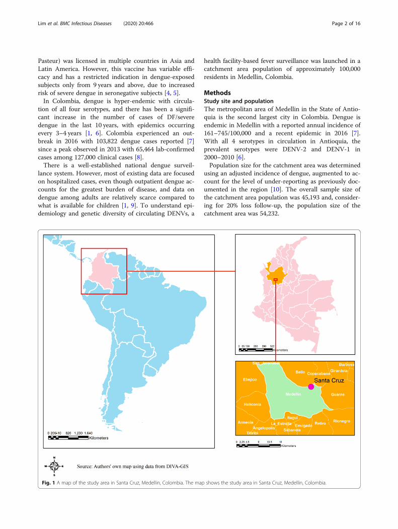
Fig. 1 A map of the study area in Santa Cruz, Medellin, Colombia. The map shows the study area in Santa Cruz, Medellin, Colombia.
Lim et al. BMC Infectious Diseases (2020) 20:466 Page 2 of 16
Santa Cruz comuna, one of the 16 sub-districts inMedellin with a population size of 107,869, was selectedto be the catchment area for the surveillance. Approxi-mately, 87% of the population are Mestizo and White,with 12% Afro-Colombians. In terms of socio-economiclevel, approximately 96% of the households belong tothe socioeconomic stratum 2 (low). In Santa Cruzcomuna, there are three basic health centers and SantaCruz Hospital (SCH), a 48-bed medium-sized secondarycare facility (Fig. 1).
Study designIn the passive fever surveillance implemented at SCH,eligible subjects among febrile outpatients and hospital-ized patients were enrolled and tested for dengue. Eligi-bility for dengue screening was based on the age (1–65years), presence of fever (body temperature ≥ 37.5 °C) orhistory of fever for ≤7 days of duration, without localiz-ing signs (i.e. fever caused by a localized infection orwith other known/confirmed etiology), being resident ofthe Santa Cruz comuna, and not being a participant inany dengue vaccine clinical trials during the studyperiod. Exclusion of infants < 1 year-of-age was based onconsideration of the presence of maternal antibodies andchallenges of infantile bleeding.If eligible and agree to participate, the patient was re-
ferred to the study physician (Fig. 2). After collection ofan acute sample of blood of 7–10mL at enrollment, astudy physician/nurse completed the surveillance casereport form based on physical examination, to collectmedical history and laboratory results. Then, a follow-upvisit took place at SCH for collection of a convalescentblood sample and clinical data, between 10 and 14 daysafter visit 1. If the patient was not able to visit the hos-pital, a home visit was made within 21 days after visit 1.
Laboratory analysesAt enrollment, subjects were tested for the detection ofDENV NS1 antigen by rapid test [Standard Diagnostics(SD), Yongin-Si, Korea]. The RDT result was used as aninitial screening tool for further testing. Blood sampleswere tested using dengue IgM/IgG ELISA (SD DengueIgM & IgG Capture ELISA®, Standard Diagnostics,Yongin-Si, Korea) in Programa de Estudio y Control deEnfermedades Tropicales (PECET) in Universidad deAntioquia. Samples showing a test result ≥ the definedcut-off were considered to have presence of detectableanti-dengue IgM and IgG antibodies, which were inter-preted as primary or recent/past infection.Additionally, those acute serum samples which met at
least one of the following eligibility criteria, underwentfurther molecular analyses at PECET and University ofWisconsin, Madison: (i) NS1 test positive on RDT inacute phase, (ii) IgM anti-DENV positive in acute and/or
convalescent phase, and (iii) IgG anti-DENV positive inacute phase. Also, RT-PCR was performed on a smallnumber of acute sera of the samples that stayed sero-negative between acute and convalescence phase on IgMcapture ELISA.The samples with negative results on RT-PCR and
sero-negative results on paired IgM and IgG ELISA re-sults were classified as non-dengue. A positive IgM orIgG serology in a single serum collected after day 5 ofsymptoms onset were diagnostic criteria of probabledengue [11]. Sero-conversion of anti-dengue IgM fromnegative in the acute phase to positive in the convales-cent phase and/or virus detection (RT-PCR) in the acuteserum specimen were considered laboratory-confirmeddengue. Confirmed- and probable-dengue were groupedtogether as dengue-positive cases in this analysis. Sero-conversion on IgM ELISA with sero-positive results onIgG ELISA was classified as secondary dengue infections.When the test shows sero-conversion of anti-dengueIgG from negative in the acute phase to positive in theconvalescent phase, it was considered as either currentor recent exposure with DENV.
Virus isolation and serotypingViral isolation assays were conducted by inoculating thepositive serum samples, in addition to a subset of sam-ples which tested negative by RT-PCR but NS1 positiveon RDT, into C6/36 cells monolayers. After 10 days ofinoculation, supernatants were collected for RT-PCRtests and unattached cells were fixed for Immunofluores-cence assays. For virus identification in the supernatants,RNA extractions (Zymo Viral RNA Kit – Zymo Re-search), Reverse Transcriptions (Superscript III FirstStrand cDNA Synthesis kit - Invitrogen), and multiplexPCR (One Taq 2X Master Mix – New England Biolabs),were conducted using serotype-specific primers de-scribed previously [12, 13]. Due to low band intensity ornegative results obtained with these procedures, a sec-ond passage on C6/36 cells monolayers was performed.Supernatants were harvested after 7 days of inoculationand underwent viral molecular detection and typing.
Envelope gene amplification and sequencingTwo overlapping fragments corresponding to the5’UTR- NS2B region of DENV genome were amplified.Serotype-specific primers were used to synthetize cDNA(Superscript III First Strand cDNA Synthesis kit – Invi-trogen) that was used as a template in the amplificationof the two fragments (Q5 High Fidelity DNA Polymerase– New England Biolabs) with serotype-specific primers[14]. The primers that were used to amplify the twooverlapping fragments along with additional internalprimers, that hybridize in different regions of the targetgene, were used to run individual sequencing reactions.
Lim et al. BMC Infectious Diseases (2020) 20:466 Page 3 of 16
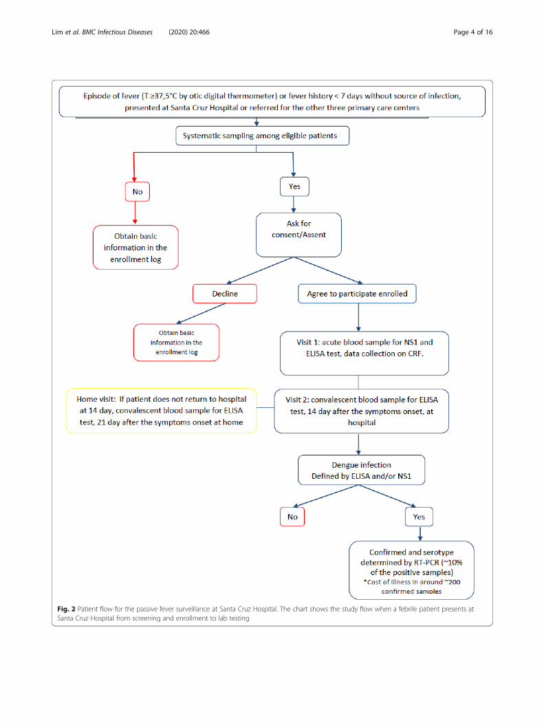
Fig. 2 Patient flow for the passive fever surveillance at Santa Cruz Hospital. The chart shows the study flow when a febrile patient presents atSanta Cruz Hospital from screening and enrollment to lab testing
Lim et al. BMC Infectious Diseases (2020) 20:466 Page 4 of 16
Twelve sequence fragments were obtained for each iso-late, after edition and selection of the best resolved re-gions in the received chromatograms. These fragmentswere assembled, with the ContigExpress Tool within theVector NTI software (Invitrogen, Carlsbad, California,United States), and carefully reviewed to obtain se-quences that contain the complete DENV envelope gene.Total sequence lengths varied by serotype; 1959 bp forDENV-1 isolate, 2653–2347 bp for DENV-3 isolates, and1963–1077 bp for DENV-4 isolates.
Variable construction and statistical analysisFirst, a descriptive summary of characteristics is pre-sented between dengue-positive (combining confirmed-and probable-dengue, following the WHO diagnosticcriteria) and non-dengue patients [11]. Clinical diagnosisat admission, prior to lab-confirmation, was grouped assuspected dengue, undifferentiated fever, and non-dengue. Dichotomous variable was created for yellowfever vaccination history, comparing those self-reportedto have been vaccinated vs. those who did not self-report vaccination or did not remember.Categorical pair-wise comparisons were made between
dengue-positive and non-dengue cases using Chi-square(χ2) and Fisher’s exact tests, with significance at p-value< 0.05. Comparison of continuous variables was per-formed using the student’s t-test and ANOVA. To iden-tify characteristics associated with dengue positivity,independent variables, such as treatment type, and feverduration prior to visit, were investigated in univariableassociations. Associations were expressed in terms ofodds ratios (ORs) with 95% confidence intervals (CIs).All analyses were performed using SAS® version 9.4(SAS Institute, Cary, North Carolina).
Phylogenetic analysisFull-length envelope gene sequence databases were con-structed for each detected DENV serotype with availablesequences in GenBank with known location and sam-pling date (last accessed in November 2018), using theNCBI Mass Sequence Downloader software [15]. Data-sets were downsized by removing identical and highlysimilar (> 99.8%) sequences from the same year andcountry by clustering with the CD-HIT program [16,17]. Resulting datasets (953 sequences for DENV-1, 545for DENV-3 and 862 for DENV-4) were combined withthe sequences in this study and aligned with MAFFTv7.402 software [18]. To identify the corresponding ge-notypes of the isolates, an initial phylogenetic analysiswas performed by maximum likelihood (ML) recon-struction with ultrafast bootstrap approximation(UFBoot) using the IQ-TREE web server [19, 20].To focus on the identified genotypes, sequences from
the same year and country were down sampled when
they were included in monophyletic clades to reduceoverrepresentation. These filtered datasets (117 se-quences for DENV-1, 123 for DENV-3, and 124 forDENV-4) were used to perform a ML analysis, to testthe temporal structure in the data using TempESTv1.5.1 software [21]. With each alignment, a statisticalselection of the best fit models of nucleotide substitutionwas performed with jModelTest software [22, 23]. Priorto molecular clock phylogenetics, the best-fit demo-graphic model (constant-size population, exponentialgrowth population, Bayesian Skyline and Bayesian Sky-Grid priors) and the best fit clock model (strict clockand uncorrelated relaxed clock with log-normal distribu-tion) were selected by estimation of marginal likelihoodvia path-sampling (PS) and stepping-stone sampling (SS)on a 1 million chain sampled for 100 path steps. Phylo-genetic reconstructions were performed using BEASTv1.8.4 software [24] with a 100 million generations Mar-kov Chain Monte Carlo (MCMC). Estimation of substi-tution rates and time of most recent common ancestor(TMRCA) for specific clades were performed usingTracer v1.7 [25]. Maximum clade credibility trees(MCC) were obtained with TreeAnotator v1.8.1 and 10%of the initial MCMC samples were discarded as burn-in.
Ethical considerationsA written informed consent from (ICF) was obtainedfrom each participant. For subjects 7 years old or youn-ger, an informed consent was obtained from at least oneparent or legal guardian. For those aged between 8 and18 years, an assent form was obtained, plus informedconsent from at least one parent or legal guardian. Theprotocol obtained ethical approvals from the EthicsCommittee of the Universidad de Antioquia, Secretariade Salud de Medellin, Metrosalud E.S.E/ Unidad Hospi-talaria Santa Cruz and the Institutional Review Board(IRB) of International Vaccine Institute (IVI).
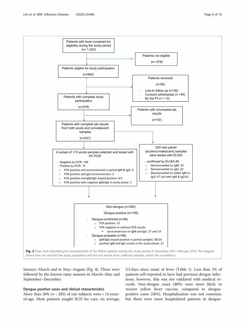
ResultsAmong 1342 febrile patients screened for study partici-pation during the 28-month study period, 664 patientswere eligible and 579 patients agreed to participate inthe study (Fig. 3). After removing 42 of them due to in-complete laboratory and clinical data, the analysis sam-ple included 537 febrile subjects.
General characteristicsAmong 537 subjects, 29% (n = 155) were found to bedengue-positive patients, composed of 56 dengue-confirmed and 99 dengue-probable cases (Table 1). Con-firmed- and probable-dengue were similar in terms ofage, outcome of illness (hospitalization), and symptoms.In terms of monthly distribution of dengue-positivecases, there were peaks of dengue transmission, in
Lim et al. BMC Infectious Diseases (2020) 20:466 Page 5 of 16
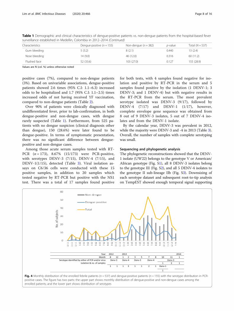
January–March and in May–August (Fig. 4). These werefollowed by the known rainy seasons in March–May andSeptember–December.
Dengue positive cases and clinical characteristicsMore than 50% (n = 285) of our subjects were < 14 years-of-age. Most patients sought SCH for care, on average,
3.5 days since onset of fever (Table 1). Less than 5% ofpatients self-reported to have had previous dengue infec-tions, however, this was not validated with medical re-cords. Non-dengue cases (48%) were more likely toreceive yellow fever vaccine, compared to dengue-positive cases (36%). Hospitalization was not common,but there were more hospitalized patients in dengue-
Fig. 3 Flow chart describing the ascertainment of the febrile patients during the study period of November 2011–February 2014. The diagramshows how we reached the study population and the test results from collected samples, within the surveillance.
Lim et al. BMC Infectious Diseases (2020) 20:466 Page 6 of 16
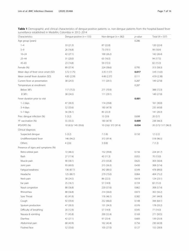
Table 1 Demographic and clinical characteristics of dengue-positive patients vs. non-dengue patients from the hospital-based feversurveillance established in Medellin, Colombia in 2012–2014
Characteristics Dengue-positive (n = 155) Non-dengue (n = 382) p-value Total (N = 537)
Age group (years) 0.286
1–4 33 (21.3) 87 (22.8) 120 (22.4)
5–9 26 (16.8) 73 (19.1) 99 (18.4)
10–24 42 (27.1) 100 (26.2) 142 (26.4)
25–44 31 (20.0) 63 (16.5) 94 (17.5)
45–65 23 (14.8) 59 (15.5) 82 (15.3)
Female (%) 89 (57.4) 224 (58.6) 0.795 313 (58.3)
Mean days of fever since onset (SD) 3.72 (1.75) 3.35 (1.57) 0.017 3.45 (1.63)
Mean overall fever duration (SD) 4.85 (2.39) 4.48 (2.37) 0.111 4.59 (2.38)
Current fever at presentation 38 (24.5) 111 (29.1) 0.287 149 (27.8)
Temperature at enrollment 0.287
Below 38°c 117 (75.5) 271 (70.9) 388 (72.3)
≥ 38°c 38 (24.5) 111 (29.1) 149 (27.8)
Fever duration prior to visit 0.001
1–2 days 47 (30.3) 114 (29.8) 161 (30.0)
3–4 days 52 (33.6) 183 (47.9) 235 (43.8)
5–7 days 56 (36.1) 85 (22.3) 141 (26.3)
Prev. dengue infection (%) 5 (3.2) 15 (3.9) 0.698 20 (3.7)
YF vaccination (%) 55 (35.5) 183 (47.9) 0.009 238 (44.3)
IPD/OPD (%) 10 (6.5)/ 145 (93.6) 10 (2.6)/ 372 (97.4) 0.034 20 (3.7)/ 517 (96.3)
Clinical diagnosis
Suspected dengue 5 (3.2) 7 (1.8) 0.150 12 (2.2)
Undifferentiated fever 146 (94.2) 372 (97.4) 518 (96.5)
Others 4 (2.6) 3 (0.8) 7 (1.3)
Presence of signs and symptoms (%)
Retro-orbital pain 72 (46.5) 152 (39.8) 0.156 224 (41.7)
Rash 27 (17.4) 43 (11.3) 0.055 70 (13.0)
Muscle pain 90 (58.1) 213 (55.8) 0.625 303 (56.4)
Joint pain 93 (60.0) 215 (56.3) 0.430 308 (57.4)
Fatigue/weakness 136 (87.7) 342 (89.5) 0.549 478 (89.0)
Headache 125 (80.7) 279 (73.0) 0.064 404 (75.2)
Neck pain 38 (24.5) 86 (22.5) 0.618 124 (23.1)
Ear pain 25 (16.1) 57 (14.9) 0.724 82 (15.3)
Nasal congestion 88 (56.8) 220 (57.6) 0.862 308 (57.4)
Rhinorrhea 88 (56.8) 214 (56.0) 0.873 302 (56.2)
Sore Throat 65 (41.9) 176 (46.1) 0.382 241 (44.9)
Cough 92 (59.4) 252 (66.0) 0.148 344 (64.1)
Sputum production 47 (30.3) 131 (34.3) 0.376 178 (33.2)
Difficulty of breathing 20 (12.9) 57 (14.9) 0.545 77 (14.3)
Nausea & vomiting 71 (45.8) 200 (52.4) 0.169 271 (50.5)
Diarrhea 42 (27.1) 97 (25.4) 0.683 139 (25.9)
Abdominal pain 68 (43.9) 162 (42.4) 0.756 230 (42.8)
Flushed face 52 (33.6) 103 (27.0) 0.127 155 (28.9)
Lim et al. BMC Infectious Diseases (2020) 20:466 Page 7 of 16
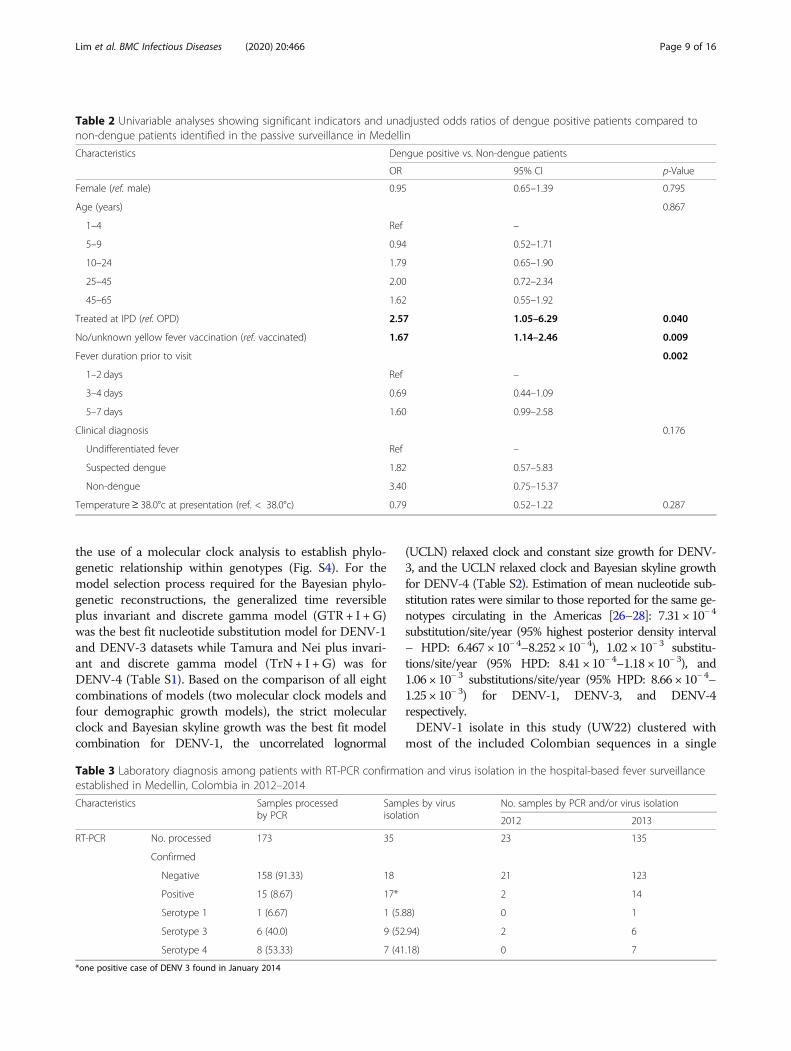
positive cases (7%), compared to non-dengue patients(3%). Based on univariable associations, dengue-positivepatients showed 2.6 times (95% C.I: 1.1–6.3) increasedodds to be hospitalized and 1.7 (95% C.I: 1.1–2.5) timesincreased odds of not having received YF vaccination,compared to non-dengue patients (Table 2).Over 90% of patients were clinically diagnosed with
undifferentiated fever, prior to lab-confirmation, in bothdengue-positive and non-dengue cases, with denguerarely suspected (Table 1). Furthermore, from 525 pa-tients with no dengue suspicion (clinical diagnosis otherthan dengue), 150 (28.6%) were later found to bedengue-positive. In terms of symptomatic presentation,there was no significant difference between dengue-positive and non-dengue cases.Among those acute serum samples tested with RT-
PCR (n = 173), 8.67% (15/173) were PCR-positive,with serotypes DENV-3 (7/15), DENV-4 (7/15), andDENV-1(1/15), detected (Table 3). Viral isolation as-says on C6/36 cells were conducted with these 15positive samples, in addition to 20 samples whichtested negative by RT-PCR but positive with the NS1test. There was a total of 17 samples found positive
for both tests, with 4 samples found negative for iso-lation and positive by RT-PCR in the serum and 5samples found positive by the isolation (1 DENV-1; 3DENV-3; and 1 DENV-4) but with negative results inthe RT-PCR from the serum. The most prevalentserotype isolated was DENV-3 (9/17), followed byDENV-4 (7/17) and DENV-1 (1/17), however,complete envelope gene sequence was obtained from8 out of 9 DENV-3 isolates, 5 out of 7 DENV-4 iso-lates and from the DENV-1 isolate.By the calendar year, DENV-3 was prevalent in 2012,
while the majority were DENV-3 and -4 in 2013 (Table 3).Overall, the number of samples with complete serotypingwas small.
Sequencing and phylogenetic analysisThe phylogenetic reconstructions showed that the DENV-1 isolate (UW22) belongs to the genotype V or American/African genotype (Fig. S1), all 8 DENV-3 isolates belongto the genotype III (Fig. S2), and all 5 DENV-4 isolates tothe genotype II sub-lineage IIb (Fig. S3). Downsizing ofeach serotype dataset and subsequent root-to-tip analysison TempEST showed enough temporal signal supporting
Table 1 Demographic and clinical characteristics of dengue-positive patients vs. non-dengue patients from the hospital-based feversurveillance established in Medellin, Colombia in 2012–2014 (Continued)
Characteristics Dengue-positive (n = 155) Non-dengue (n = 382) p-value Total (N = 537)
Gum bleeding 5 (3.2) 8 (2.1) 0.440 13 (2.4)
Nose bleeding 14 (9.0) 46 (12.0) 0.316 60 (11.2)
Flushed face 52 (33.6) 103 (27.0) 0.127 155 (28.9)
Values are N (col. %) unless otherwise noted
Fig. 4 Monthly distribution of the enrolled febrile patients (n = 537) and dengue-positive patients (n = 155) with the serotype distribution in PCR-positive cases. The figure has two parts: the upper part shows monthly distribution of dengue-positive and non-dengue cases among theenrolled patients; and the lower part shows distribution of serotypes
Lim et al. BMC Infectious Diseases (2020) 20:466 Page 8 of 16
the use of a molecular clock analysis to establish phylo-genetic relationship within genotypes (Fig. S4). For themodel selection process required for the Bayesian phylo-genetic reconstructions, the generalized time reversibleplus invariant and discrete gamma model (GTR + I +G)was the best fit nucleotide substitution model for DENV-1and DENV-3 datasets while Tamura and Nei plus invari-ant and discrete gamma model (TrN + I +G) was forDENV-4 (Table S1). Based on the comparison of all eightcombinations of models (two molecular clock models andfour demographic growth models), the strict molecularclock and Bayesian skyline growth was the best fit modelcombination for DENV-1, the uncorrelated lognormal
(UCLN) relaxed clock and constant size growth for DENV-3, and the UCLN relaxed clock and Bayesian skyline growthfor DENV-4 (Table S2). Estimation of mean nucleotide sub-stitution rates were similar to those reported for the same ge-notypes circulating in the Americas [26–28]: 7.31 × 10− 4
substitution/site/year (95% highest posterior density interval– HPD: 6.467 × 10− 4–8.252 × 10− 4), 1.02 × 10− 3 substitu-tions/site/year (95% HPD: 8.41 × 10− 4–1.18 × 10− 3), and1.06 × 10− 3 substitutions/site/year (95% HPD: 8.66 × 10− 4–1.25 × 10− 3) for DENV-1, DENV-3, and DENV-4respectively.DENV-1 isolate in this study (UW22) clustered with
most of the included Colombian sequences in a single
Table 2 Univariable analyses showing significant indicators and unadjusted odds ratios of dengue positive patients compared tonon-dengue patients identified in the passive surveillance in Medellin
Characteristics Dengue positive vs. Non-dengue patients
OR 95% CI p-Value
Female (ref. male) 0.95 0.65–1.39 0.795
Age (years) 0.867
1–4 Ref –
5–9 0.94 0.52–1.71
10–24 1.79 0.65–1.90
25–45 2.00 0.72–2.34
45–65 1.62 0.55–1.92
Treated at IPD (ref. OPD) 2.57 1.05–6.29 0.040
No/unknown yellow fever vaccination (ref. vaccinated) 1.67 1.14–2.46 0.009
Fever duration prior to visit 0.002
1–2 days Ref –
3–4 days 0.69 0.44–1.09
5–7 days 1.60 0.99–2.58
Clinical diagnosis 0.176
Undifferentiated fever Ref –
Suspected dengue 1.82 0.57–5.83
Non-dengue 3.40 0.75–15.37
Temperature≥ 38.0°c at presentation (ref. < 38.0°c) 0.79 0.52–1.22 0.287
Table 3 Laboratory diagnosis among patients with RT-PCR confirmation and virus isolation in the hospital-based fever surveillanceestablished in Medellin, Colombia in 2012–2014
Characteristics Samples processedby PCR
Samples by virusisolation
No. samples by PCR and/or virus isolation
2012 2013
RT-PCR No. processed 173 35 23 135
Confirmed
Negative 158 (91.33) 18 21 123
Positive 15 (8.67) 17* 2 14
Serotype 1 1 (6.67) 1 (5.88) 0 1
Serotype 3 6 (40.0) 9 (52.94) 2 6
Serotype 4 8 (53.33) 7 (41.18) 0 7
*one positive case of DENV 3 found in January 2014
Lim et al. BMC Infectious Diseases (2020) 20:466 Page 9 of 16
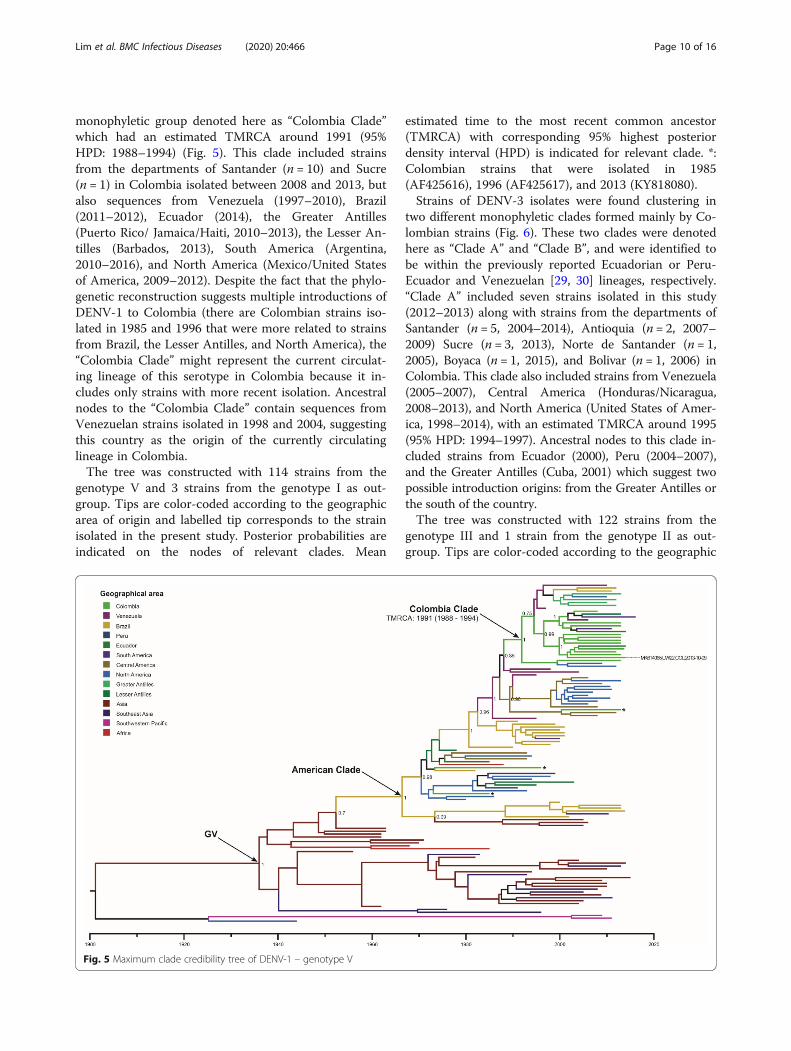
monophyletic group denoted here as “Colombia Clade”which had an estimated TMRCA around 1991 (95%HPD: 1988–1994) (Fig. 5). This clade included strainsfrom the departments of Santander (n = 10) and Sucre(n = 1) in Colombia isolated between 2008 and 2013, butalso sequences from Venezuela (1997–2010), Brazil(2011–2012), Ecuador (2014), the Greater Antilles(Puerto Rico/ Jamaica/Haiti, 2010–2013), the Lesser An-tilles (Barbados, 2013), South America (Argentina,2010–2016), and North America (Mexico/United Statesof America, 2009–2012). Despite the fact that the phylo-genetic reconstruction suggests multiple introductions ofDENV-1 to Colombia (there are Colombian strains iso-lated in 1985 and 1996 that were more related to strainsfrom Brazil, the Lesser Antilles, and North America), the“Colombia Clade” might represent the current circulat-ing lineage of this serotype in Colombia because it in-cludes only strains with more recent isolation. Ancestralnodes to the “Colombia Clade” contain sequences fromVenezuelan strains isolated in 1998 and 2004, suggestingthis country as the origin of the currently circulatinglineage in Colombia.The tree was constructed with 114 strains from the
genotype V and 3 strains from the genotype I as out-group. Tips are color-coded according to the geographicarea of origin and labelled tip corresponds to the strainisolated in the present study. Posterior probabilities areindicated on the nodes of relevant clades. Mean
estimated time to the most recent common ancestor(TMRCA) with corresponding 95% highest posteriordensity interval (HPD) is indicated for relevant clade. *:Colombian strains that were isolated in 1985(AF425616), 1996 (AF425617), and 2013 (KY818080).Strains of DENV-3 isolates were found clustering in
two different monophyletic clades formed mainly by Co-lombian strains (Fig. 6). These two clades were denotedhere as “Clade A” and “Clade B”, and were identified tobe within the previously reported Ecuadorian or Peru-Ecuador and Venezuelan [29, 30] lineages, respectively.“Clade A” included seven strains isolated in this study(2012–2013) along with strains from the departments ofSantander (n = 5, 2004–2014), Antioquia (n = 2, 2007–2009) Sucre (n = 3, 2013), Norte de Santander (n = 1,2005), Boyaca (n = 1, 2015), and Bolivar (n = 1, 2006) inColombia. This clade also included strains from Venezuela(2005–2007), Central America (Honduras/Nicaragua,2008–2013), and North America (United States of Amer-ica, 1998–2014), with an estimated TMRCA around 1995(95% HPD: 1994–1997). Ancestral nodes to this clade in-cluded strains from Ecuador (2000), Peru (2004–2007),and the Greater Antilles (Cuba, 2001) which suggest twopossible introduction origins: from the Greater Antilles orthe south of the country.The tree was constructed with 122 strains from the
genotype III and 1 strain from the genotype II as out-group. Tips are color-coded according to the geographic
Fig. 5 Maximum clade credibility tree of DENV-1 – genotype V
Lim et al. BMC Infectious Diseases (2020) 20:466 Page 10 of 16
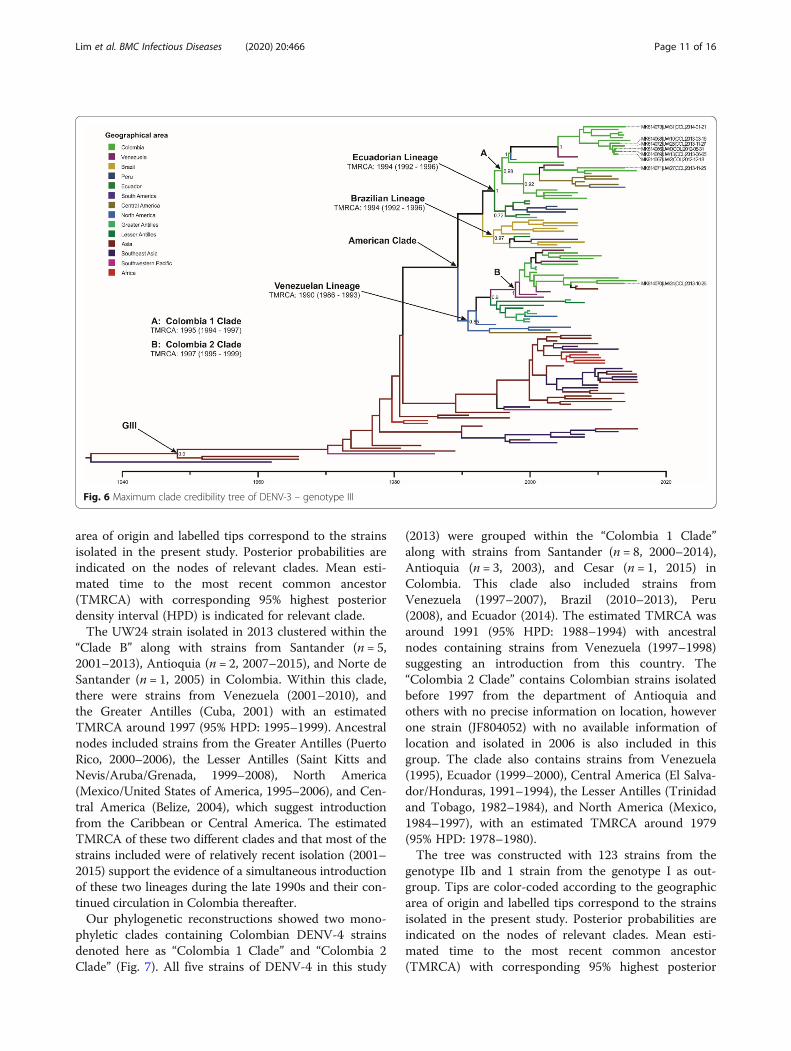
area of origin and labelled tips correspond to the strainsisolated in the present study. Posterior probabilities areindicated on the nodes of relevant clades. Mean esti-mated time to the most recent common ancestor(TMRCA) with corresponding 95% highest posteriordensity interval (HPD) is indicated for relevant clade.The UW24 strain isolated in 2013 clustered within the
“Clade B” along with strains from Santander (n = 5,2001–2013), Antioquia (n = 2, 2007–2015), and Norte deSantander (n = 1, 2005) in Colombia. Within this clade,there were strains from Venezuela (2001–2010), andthe Greater Antilles (Cuba, 2001) with an estimatedTMRCA around 1997 (95% HPD: 1995–1999). Ancestralnodes included strains from the Greater Antilles (PuertoRico, 2000–2006), the Lesser Antilles (Saint Kitts andNevis/Aruba/Grenada, 1999–2008), North America(Mexico/United States of America, 1995–2006), and Cen-tral America (Belize, 2004), which suggest introductionfrom the Caribbean or Central America. The estimatedTMRCA of these two different clades and that most of thestrains included were of relatively recent isolation (2001–2015) support the evidence of a simultaneous introductionof these two lineages during the late 1990s and their con-tinued circulation in Colombia thereafter.Our phylogenetic reconstructions showed two mono-
phyletic clades containing Colombian DENV-4 strainsdenoted here as “Colombia 1 Clade” and “Colombia 2Clade” (Fig. 7). All five strains of DENV-4 in this study
(2013) were grouped within the “Colombia 1 Clade”along with strains from Santander (n = 8, 2000–2014),Antioquia (n = 3, 2003), and Cesar (n = 1, 2015) inColombia. This clade also included strains fromVenezuela (1997–2007), Brazil (2010–2013), Peru(2008), and Ecuador (2014). The estimated TMRCA wasaround 1991 (95% HPD: 1988–1994) with ancestralnodes containing strains from Venezuela (1997–1998)suggesting an introduction from this country. The“Colombia 2 Clade” contains Colombian strains isolatedbefore 1997 from the department of Antioquia andothers with no precise information on location, howeverone strain (JF804052) with no available information oflocation and isolated in 2006 is also included in thisgroup. The clade also contains strains from Venezuela(1995), Ecuador (1999–2000), Central America (El Salva-dor/Honduras, 1991–1994), the Lesser Antilles (Trinidadand Tobago, 1982–1984), and North America (Mexico,1984–1997), with an estimated TMRCA around 1979(95% HPD: 1978–1980).The tree was constructed with 123 strains from the
genotype IIb and 1 strain from the genotype I as out-group. Tips are color-coded according to the geographicarea of origin and labelled tips correspond to the strainsisolated in the present study. Posterior probabilities areindicated on the nodes of relevant clades. Mean esti-mated time to the most recent common ancestor(TMRCA) with corresponding 95% highest posterior
Fig. 6 Maximum clade credibility tree of DENV-3 – genotype III
Lim et al. BMC Infectious Diseases (2020) 20:466 Page 11 of 16
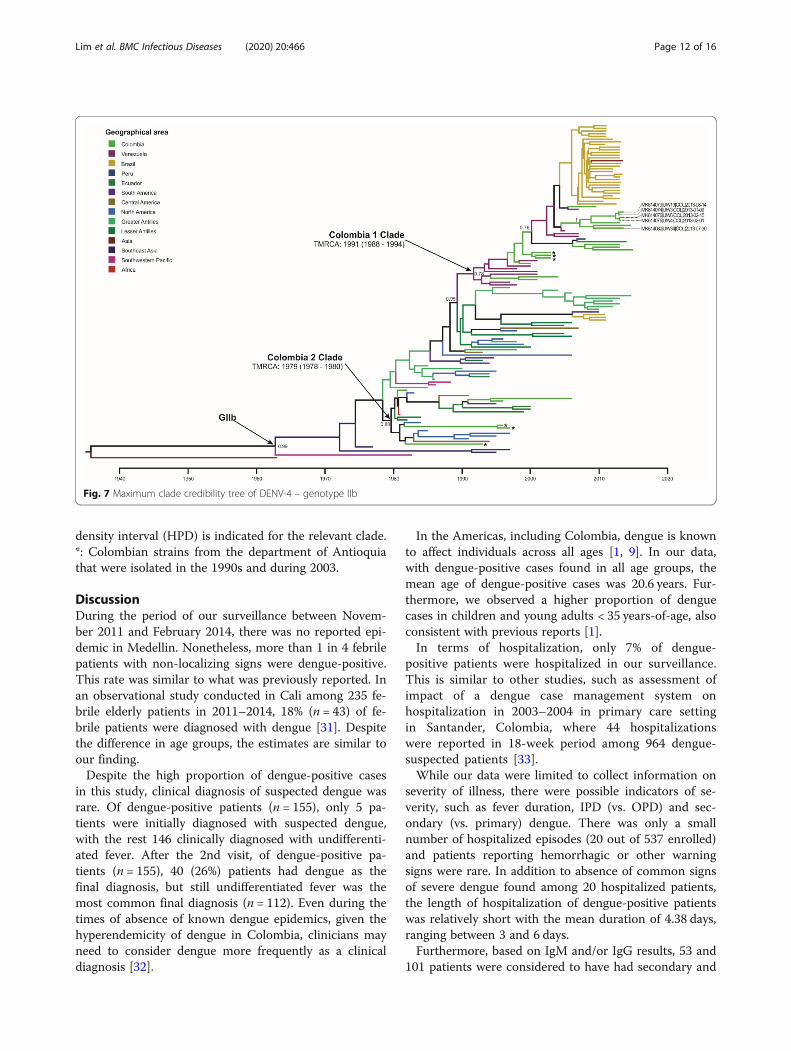
density interval (HPD) is indicated for the relevant clade.*: Colombian strains from the department of Antioquiathat were isolated in the 1990s and during 2003.
DiscussionDuring the period of our surveillance between Novem-ber 2011 and February 2014, there was no reported epi-demic in Medellin. Nonetheless, more than 1 in 4 febrilepatients with non-localizing signs were dengue-positive.This rate was similar to what was previously reported. Inan observational study conducted in Cali among 235 fe-brile elderly patients in 2011–2014, 18% (n = 43) of fe-brile patients were diagnosed with dengue [31]. Despitethe difference in age groups, the estimates are similar toour finding.Despite the high proportion of dengue-positive cases
in this study, clinical diagnosis of suspected dengue wasrare. Of dengue-positive patients (n = 155), only 5 pa-tients were initially diagnosed with suspected dengue,with the rest 146 clinically diagnosed with undifferenti-ated fever. After the 2nd visit, of dengue-positive pa-tients (n = 155), 40 (26%) patients had dengue as thefinal diagnosis, but still undifferentiated fever was themost common final diagnosis (n = 112). Even during thetimes of absence of known dengue epidemics, given thehyperendemicity of dengue in Colombia, clinicians mayneed to consider dengue more frequently as a clinicaldiagnosis [32].
In the Americas, including Colombia, dengue is knownto affect individuals across all ages [1, 9]. In our data,with dengue-positive cases found in all age groups, themean age of dengue-positive cases was 20.6 years. Fur-thermore, we observed a higher proportion of denguecases in children and young adults < 35 years-of-age, alsoconsistent with previous reports [1].In terms of hospitalization, only 7% of dengue-
positive patients were hospitalized in our surveillance.This is similar to other studies, such as assessment ofimpact of a dengue case management system onhospitalization in 2003–2004 in primary care settingin Santander, Colombia, where 44 hospitalizationswere reported in 18-week period among 964 dengue-suspected patients [33].While our data were limited to collect information on
severity of illness, there were possible indicators of se-verity, such as fever duration, IPD (vs. OPD) and sec-ondary (vs. primary) dengue. There was only a smallnumber of hospitalized episodes (20 out of 537 enrolled)and patients reporting hemorrhagic or other warningsigns were rare. In addition to absence of common signsof severe dengue found among 20 hospitalized patients,the length of hospitalization of dengue-positive patientswas relatively short with the mean duration of 4.38 days,ranging between 3 and 6 days.Furthermore, based on IgM and/or IgG results, 53 and
101 patients were considered to have had secondary and
Fig. 7 Maximum clade credibility tree of DENV-4 – genotype IIb
Lim et al. BMC Infectious Diseases (2020) 20:466 Page 12 of 16
primary dengue infection, respectively. While Colombiais considered highly dengue-endemic, there were moreprimary dengue cases than secondary dengue cases inthe studied population. Of 10 dengue-positive casesamong 20 hospitalized patients, 8 of them would be clas-sified as secondary dengue cases. While the majority ofhospitalized dengue patients showed to have secondarydengue than primary dengue infections, interpretationmay be limited due to a small sample size. Our labora-tory data were incomplete in terms of paired IgM and/orIgG results (i.e., only acute results available) for classifi-cation of secondary vs. primary dengue cases. Nonethe-less, our data suggest mildness of dengue disease inMedellin at these times without large epidemics.Taking place prior to the Chikungunya epidemic in
2014 and Zika epidemic in 2015, the molecular epidemi-ology of DENV was captured in the study without inter-ference of other arboviruses and could be used as a basisto observe variation [34, 35]. Also, genetic diversity ofDENV isolates and relatedness to other isolates weredocumented. DENV-1 isolate belonging to the genotypeV, is a well-established variant in the Americas and pre-viously reported to have circulated in Colombia since1977 [36]. This is also consistent with the DENV sero-types and genotypes sequenced in Sanofi Pasteur’s phase3 efficacy (CYD15) trial in Colombia [37]. It was closelyrelated to strains from other parts of Colombia, within asingle clade intermixed with strains from countries, likeVenezuela, Ecuador, and Brazil. There have been reportsof the possible current circulation of two DENV-1 –Genotype V putative lineages in Colombia [36] one ofthem exclusively formed by Colombian isolates, basedon phylogenetic reconstructions using partial segmentsof E gene. Despite the caveat of having only one isolatesequenced for DENV-1, there is no evidence in thisstudy supporting the presence of these two putative line-ages (at least not with the sequences included in thestudy) and this finding is consistent with recent reportsof evolutionary history of DENV-1 in Colombia [28].DENV-3 isolates obtained belonged to the genotype
III, consistent with previous reports [29, 38] and findingsfrom sequencing data in the dengue vaccine phase 3 trialin Colombia [37]. Some studies suggested that most re-cent DENV-3 viruses circulating in South America haveaccumulated enough genetic variation that caused for-mation of clusters totally different from those describedin Central America [39]. Also, there are suggestions ofpossible circulation of an Asian-origin genotype I [38] inLa Guajira (North), Guaviare (Southeast), and Huila(Southwest) in Colombia, but there was no evidence ofcirculation of this genotype in the study. Two differentlineages of genotype III viruses were found to overlap inspace and time during the study which could be an ef-fect of two independent transmission chains since their
official re-introduction in Colombia in 2001 [40] due tocomplete susceptibility of the population to this serotypeafter 24 years with no circulation. Our TMRCA estima-tions suggest that introduction of both lineages toColombia occurred during the late 1990’s (1995 forClade A and 1997 for Clade B), which was earlier thanthe first official report of circulation, indicating ongoinglocal circulation of DENV-3 before its detection andpossibly causing an increase in the number of infectionsduring that period; similar TMRCA estimations havebeen reported previously, supporting the finding of anearlier introduction into the country [41]. This simultan-eous circulation has been reported previously in Medel-lin [42], and more recently in Santander and Valle delCauca, in Colombia [28]. Additionally, it has been docu-mented that the most probable ancestral locations forthe clades we denoted here as “Clade A” and “Clade B”were the Greater Antilles and Central America, respect-ively [28].All DENV-4 isolates belonged to genotype II, to the
IIb linage, which is a variant mainly circulating in theCaribbean. It is consistent with the DENV serotypes andgenotypes sequenced in Sanofi Pasteur’s phase 3 efficacytrial in Colombia [37]. Our isolates showed to groupwith those that were more recently isolated in this re-gion, forming a cluster different from other Colombianstrains isolated before 1997. Recently, circulation of thisgenotype has been reported in at least three other depart-ments of Colombia (Santander, Valle del Cauca, andCesar) and is reported to have caused at least two intro-ductions in the country [28]. Phylogeographic analysisrevealed that the genotype II was circulating along withgenotype I in Brazil during 2010–2011 [43]. However,our study does not provide evidence of the circulation ofthis additional genotype. Nunes and coworkers describedmultiple introductions of genotype II to Brazil in the lastdecade, at least three of them from Colombia andVenezuela, which corresponds with the finding of mul-tiple clusters within the genotypes [43].Commonly, the phylogenetic reconstructions of DENV
have been performed with the complete E gene, as inthis study, creating an imbalance in the amount of avail-able sequences from more ancestral origins for analysis.There are reports supporting that phylogenetic informa-tion obtained from the complete E gene is representativeand similar to what is generally obtained with completegenome sequences of the virus [41, 44–46].Mainly due to the variations on viremia levels, DENV
genetic sequencing generally requires isolation of thevirus on cell monolayers to have enough genetic mater-ial. In our study, isolated viruses were used for sequen-cing and there may be the possibility of mutationsinduced by the cell passaging, however the amount ofpassages was maintained low to reduce this effect.
Lim et al. BMC Infectious Diseases (2020) 20:466 Page 13 of 16
The study has limitations. Due to resource constraints,this study continued for 28 months and was conductedin one area of Medellin. Also, although there was a largenumber of samples collected in the study, only a limitednumber of samples were tested with RT-PCR and virusisolation. Samples to be tested with RT-PCR and virusisolation were selected based on paired ELISA and/orRDT positive results, and there may be selection bias as-sociated, resulting in limited interpretation of our find-ings from the phylogenetic analysis.Furthermore, the rate of detection of dengue positivity
was lower by PCR than serological methods based onIgM/IgG ELISA in our study. Degradation of RNA couldhave contributed to the lower detection rate of PCR.However, PCR was performed in two locations: one inMedellin soon after sample collection and another inUniversity of Wisconsin, and results were comparable.One noteworthy point is the duration of fever prior tohospital visit (enrollment). Given that PCR works betterin the early febrile phase [47], the fact that 70% of ourpatients were enrolled in the study 3 days or later sinceonset of fever could have contributed to the lower rateof case detection by RT-PCR.Another important potential source of bias in our
study is under-ascertainment of the community resi-dents with relevant symptoms. Despite that the SCH isthe main hospital of the comuna, our study may havemissed those eligible patients seeking care at otherhealthcare providers than our facility. Thus, we mayhave missed other mild fever episodes. Therefore, werecognize limited generalizability of our data to repre-sent the general population of the country due to theselimitations.In the surveillance, among these 155 dengue-positive
patients, 99 cases were dengue-probable cases. Given theDENV endemicity in the country, dengue-probablecases, along with confirmed cases, are reported as partof dengue surveillance in the national system inColombia. Furthermore, this surveillance was conductedin the same catchment area population as the repeatedcommunity-based serological surveys measuring sero-prevalence in the same study population [48]. The sero-prevalence study reported 61% of seroprevalance with8.7% of sero-conversion per 1000 person-months [48].Without major epidemics during our study period, theestimated proportion of dengue-positive cases among fe-brile patients as well as the seroprevalence estimate andsero-conversion rate all support high transmission andburden of DENV in the study area.
ConclusionsOur findings confirm that there is considerable dengueburden in Santa Cruz comuna during non-epidemicyears, with the majority with mild illness. Conducted
prior to Chikungunya epidemic in 2014 and Zika epi-demic in 2015, the study findings support genetic diver-sity of DENV isolates and their relatedness to isolatesfrom other nearby countries. Now with emergence ofnew epidemics, our results support the need for contin-ued surveillance and monitoring of dengue and otherarboviruses.
Supplementary informationSupplementary information accompanies this paper at https://doi.org/10.1186/s12879-020-05172-7.
Additional file 1: S1. Checklist: STROBE Checklist. Figure S1. DENV-1Maximum Likelihood tree and root-to-tip regression. A) Linear regressionof root-to-tip divergence and dates of isolation indicating the slope andR-squared value for temporal signal evaluation. Each datapoint is color-coded based on the corresponding genotype within DENV-1. B) Max-imum likelihood tree with 954 full-length E-gene sequences (1485 nt)representing the five genotypes reported for DENV-1. Tips are colored bycorresponding genotype and labelled tip indicate the strain obtain in thisstudy. The tree was rooted with the sequence DENV2-NGC strain as out-group (GenBank: KM204118) and the sequence names are coded as Gen-Bank accession|ISO-3166 Alpha-3 country code|Date of isolation. FigureS2. DENV-3 Maximum Likelihood tree and root-to-tip regression. A) Linearregression of root-to-tip divergence and dates of isolation indicating theslope and R-squared value for temporal signal evaluation. Each datapointis color-coded based on the corresponding genotype within DENV-3. B)Maximum likelihood tree with 553 full-length E-gene sequences (1479 nt)representing the five genotypes reported for DENV-3. Tips are colored bycorresponding genotype and labelled tips indicate the strains obtain inthis study. The tree was rooted with the sequence DENV-1-Hawaii strainas outgroup (GenBank: KM204119) and the sequence names are codedas GenBank accession|ISO-3166 Alpha-3 country code|Date of isolation.Figure S3. DENV-4 Maximum Likelihood tree and root-to-tip regression.A) Linear regression of root-to-tip divergence and dates of isolation indi-cating the slope and R-squared value for temporal signal evaluation. Eachdatapoint is color-coded based on the corresponding genotype withinDENV-4. B) Maximum likelihood tree with 867 full-length E-gene se-quences (1485 nt) representing the four genotypes reported for DENV-4.Tips are colored by corresponding genotype and labelled tips indicatethe strains obtain in this study. The tree was rooted with the sequenceDENV2-NGC strain as outgroup (GenBank: KM204118) and the sequencenames are coded as GenBank accession|ISO-3166 Alpha-3 country code|-Date of isolation. Figure S4. Root-to-tip analysis for identified genotypes.Linear regression of root-to-tip divergence and date of isolation for the E-gene of DENV-1 (GV), DENV-3 (GIII) and DENV-4 (GIIb) to evaluate thetemporal structure of datasets. Each plot shows the R-squared value andslope of the black dashed regression line which indicate the substitutionrate for these viruses. The linear regression supports the use of these datafor molecular clock inferences. Each datapoint is color-coded based onthe geographic area of origin. Table S1. Nucleotide Substitution modelselection. Results for the statistical best fit model selection process withjModelTest for each serotype. Table S2. Molecular clock and demo-graphic growth model selection. Marginal likelihoods calculated withpath-sampling (PS) and stepping-stone sampling (SS) methods for thecombinations of four demographic growth models (constant size, expo-nential, Bayesian Skyline and Bayesian SkyGrid) and two molecular clockmodels (strict clock and uncorrelated relaxed clock with log-normal distri-bution). Bayes factors were calculated against the model combinationwith the lower marginal likelihood estimation which in all three caseswas the constant tree prior and strict clock.
AbbreviationsCI: Confidence Interval; °C: Celsius degrees; CRF: Case Report Form;CYD: Sanofi Pasteur’s phase 3 efficacy trial; DENV: Dengue Virus; DF: DengueFever; DHF: Dengue Hemorrhagic Fever; DSS: Dengue Shock Syndrome;DVI: Dengue Vaccine Initiative; ELISA: Enzyme-Linked Immunosorbent Assay;
Lim et al. BMC Infectious Diseases (2020) 20:466 Page 14 of 16
GTR: Generalized time reversible; HPD: Highest Posterior Density;ICF: Informed Consent Form; IgM/IgG: Immunoglobulin type M and type G;IPD: Inpatient Department; IRB: Institutional Review Board; IVI: InternationalVaccine Institute; ML: Maximum Likelihood; MCC: Maximum Clade Credibilitytrees; MCMC: Markov Chain Monte Carlo; OPD: Outpatient Department;PS: Path-sampling; RDT: Rapid Diagnostic Test; PECET: Programa de Estudio yControl de Enfermedades Tropicales; RT-PCR: Reverse Transcriptase-Polymerase Chain Reaction; SCH: Santa Cruz Hospital; SD: StandardDiagnostics; SS: Stepping-stone Sampling; TMRCA: Time of Most RecentCommon Ancestor; TrN: Tamura and Nei; UCLN: Uncorrelated Lognormal;URI: Upper Respiratory Illness; YF: Yellow fever
AcknowledgementsWe thank the doctors and laboratory staff of Santa Cruz Hospital for theirparticipation in the study. Also, we thank the lab staff at the Department ofPathobiological Sciences of University of Wisconsin, Madison, WI, as well asthe Secretaria de Salud, Medellín, Colombia, and Metrosalud E.S.E / UnidadHospitalaria, comuna Santa Cruz, Medellín, for their support to the project.We thank statisticians and administrative staff at the International VaccineInstitute for their helpful comments during the analysis and preparation ofthis manuscript.
Authors’ contributionsAll persons designated as authors have participated sufficiently in the workto take public responsibility for appropriate portions of the content. And allthe authors contributed in some or all areas of acquisition of funding,conception of the study, collection of data, analysis and interpretation ofdata, drafting the article, article revision, scientific support, and final approvalof the version to be published. The authors meet the criteria for authorshipand qualify for authorship of this manuscript (see below for details). JKL wasresponsible for Conceptualization; Formal analysis; Funding acquisition;Methodology; Supervision; Roles/Writing - original draft; and Writing - review& editing. MC was responsible for Conceptualization; Data curation;Investigation; Methodology; Project administration; Supervision; Roles/Writing- original draft; and Writing - review & editing. EC was responsible for Datacuration; Formal analysis; Investigation; Software; Visualization; Roles/Writing -original draft; and Writing - review & editing. DCV was responsible for Datacuration; Investigation; Project administration; and Resources. AT wasresponsible for Data curation and Investigation. JE was responsible for Datacuration; Investigation; Project administration; Resources; and Supervision.KSL was responsible for Data curation; Investigation; Software; and Validation.IDV was responsible for Conceptualization; Data curation; Investigation;Project administration; Resources; and Supervision. JEO was responsible forConceptualization; Data curation; Funding acquisition; Investigation;Methodology; Project administration; Resources; Supervision; and Writing -review & editing.
FundingThis study was supported by funding from the Bill and Melinda GatesFoundation (grant #: OPP 1016669), as well as from the governments ofSweden, India, and the Republic of Korea.The funders had no role in study design, data collection and analysis,decision to publish, or preparation of the manuscript.
Availability of data and materialsThe dataset supporting the conclusions of this article is included within thearticle and its additional file. All sequences were deposited into the GenBankdatabase under the following accession numbers (which are parts of thesequence names that are shown in the generated phylogenetic trees):MK614065, MK614073, MK614068, MK614072, MK614066, MK614069,MK614067, MK614071, MK614070, MK614079, MK614074, MK614076,MK614075, MK614080.
Ethics approval and consent to participateA written informed consent from (ICF) was obtained from each participant.For subjects 7 years old or younger, a written informed consent wasobtained from at least one parent or legal guardian. For those agedbetween 8 and 18 years, a written assent form was obtained, plus writteninformed consent from at least one parent or legal guardian. The protocolobtained ethical approvals from the Ethics Committee of the Universidad deAntioquia, Secretaria de Salud de Medellin, Metrosalud E.S.E/ Unidad
Hospitalaria Santa Cruz and the Institutional Review Board (IRB) ofInternational Vaccine Institute (IVI).
Consent for publicationNot applicable.
Competing interestsNone. I certify that the authors do not have any relevant financialrelationships or potential conflicts of interest to disclose regarding thematerial discussed in this manuscript.
Author details1Dengue Vaccine Initiative, International Vaccine Institute, SNU Research Park,1 Gwanak-ro, Gwanak-gu, Seoul 08826, Republic of Korea. 2Department ofEpidemiology, Biostatistics and Occupational Health, McGill University, 845Sherbrooke St., W, Montreal, Quebec H3A 0G4, Canada. 3InvestigacionesBiomedicas, Universidad de Sucre, Cra 28 # 5-267, Barrio Puerta Roja,Sincelejo, Sucre, Colombia. 4Programa de Estudio y Control de EnfermedadesTropicales (PECET), Universidad de Antioquia, calle 67 No. 53, 108 Medellín,Antioquia, Colombia. 5Department of Pathobiological Sciences, University ofWisconsin, 500 Lincoln Dr, Madison, WI 53706, USA.
Received: 22 December 2019 Accepted: 17 June 2020
References1. Villar LA, Rojas DP, Besada-Lombana S, Sarti E. Epidemiological trends of
dengue disease in Colombia (2000-2011): a systematic review. PLoS Negl TropDis. 2015;19;9(3):e0003499. https://doi.org/10.1371/journal.pntd.0003499.
2. World Health Organization. Dengue and severe dengue World HealthOrganization; 2009. Updated 2 March 2020. Available from: https://www.who.int/en/news-room/fact-sheets/detail/dengue-and-severe-dengue.
3. Hadinegoro SRS. The revised WHO dengue case classification: does thesystem need to be modified? Paediatr Int Child Health. 2012;32(S1):33–8.
4. World Health Organization: WHO Position Paper on dengue. In. WHO; 2018:457–476.
5. Sridhar S, Luedtke A, Langevin E, Zhu M, Bonaparte M, Machabert T, Savarino S,Zambrano B, Moureau A, Khromava A, et al. Effect of dengue Serostatus ondengue vaccine safety and efficacy. N Engl J Med. 2018;379(4):327–40.
6. Piedrahita LD, Agudelo Salas IY, Marin K, Trujillo AI, Osorio JE, Arboleda-Sanchez SO, Restrepo BN. Risk factors associated with dengue transmissionand spatial distribution of high Seroprevalence in schoolchildren from theurban area of Medellin, Columbia. Canadian Journal of Infectious Diseasesand Medical Microbiology, 2018, 2018.
7. Alvis-Guzmán N, Zakzuk-Sierra J, Vargas-Moranth R, Alcocer-Olaciregui A,Parra-Padilla D. Dengue, Chikunguña y Zika en Colombia 2015–2016.RevMVZ Córdoba. 2017;22(Supl):5994–6003.
8. Pan American Health Organization. Dengue: Datos, mapas y estadísticas:Oficina Regional para las Américas de la Organización Mundial de la Salud.Available from: https://www.paho.org/hq/index.php?option=com_topics&view=readall&cid=3274&Itemid=40734&lang=es.
9. Halstead S. Dengue in the Americas and Southeast Asia: do they differ? RevPanam Salud Publica. 2006;20(6):407–15.
10. Standish K, Kuan G, Avilés W, Balmaseda A, Harris E. High dengue casecapture rate in four years of a cohort study in Nicaragua compared tonational surveillance data. PLoS Negl Trop Dis. 2010;4(3):e633.
11. World Health Organization: Handbook for clinical management of dengue.Geneva, Switzerland: World Health Organization,; 2012.
12. Lanciotti RS, Calisher CH, Gubler DJ, Chang GJ, Vorndam AV. Rapid detectionand typing of dengue viruses from clinical samples by using reversetranscriptase-polymerase chain reaction. J Clin Microbiol. 1992;30(3):545–51.
13. Harris E, Roberts TG, Smith L, Selle J, Kramer LD, Valle S, Sandoval E, BalmasedaA. Typing of dengue viruses in clinical specimens and mosquitoes by single-tube multiplex reverse transcriptase PCR. J Clin Microbiol. 1998;36(9):2634–9.
14. Christenbury J, Aw P, Ong S, Schreiber M, Chow A, Gubler D, Vasudevan S,Ooi E, Hibberd M. A method for full genome sequencing of all fourserotypes of the dengue virus. J Virol Methods. 2010;169(1):202–6.
15. Pina-Martins F, Paulo O: NCBI Mass Sequence Downloader-Large datasetdownloading made easy. In: 5. vol. 80–3, 2016 Jan 1 edn. SoftwareX 2016.
16. Li W, Godzik A: Cd-hit: A fast program for clustering and comparing largesets of protein or nucleotide sequences. In. Bioinformatics [Internet]; 2006.
Lim et al. BMC Infectious Diseases (2020) 20:466 Page 15 of 16
17. Huang Y, Niu B, Gao Y, Fu L, Li W: CD-HIT Suite: A web server for clusteringand comparing biological sequences. In. Bioinformatics [Internet]; 2010.
18. Katoh K, Rozewicki JY, KD MAFFT online service: multiple sequencealignment, interactive sequence choice and visualization. In. Br Bioinform[Internet]; 2017.
19. Hoang D, Chernomor O, Von Haeseler A, Minh B, Vinh L: UFBoot2:improving the ultrafast bootstrap approximation. In. Mol Biol Evol.; 2018.
20. Trifinopoulos J, Nguyen L, von Haeseler A, Minh B: W-IQ-TREE: a fast onlinephylogenetic tool for maximum likelihood analysis. In. Nucleic Acids Res. ;2016.
21. Rambaut A, Lam T, Max CL, Pybus O: Exploring the temporal structure ofheterochronous sequences using TempEst (formerly path-O-gen). Virus Evol2016, 2016.
22. Darriba D, Taboada GL, Doallo R, Posada D. jModelTest 2: more models,new heuristics and parallel computing. Nat Methods. 2012;9(8):772.
23. Guindon S, Gascuel O. A simple, fast, and accurate algorithm to estimatelarge phylogenies by maximum likelihood. Syst Biol. 2003;52(5):696–704.
24. Drummond A, Suchard M, Xie D, Rambaut A: Bayesian phylogenetics withBEAUti and the BEAST 1.7. Mol Biol Evol [Internet] 2012, 29(8):a1969–a1973.
25. Rambaut A, Drummond A, Xie D, Baele G, Suchard M: Posteriorsummarization in Bayesian phylogenetics using Tracer 1.7. Syst Biol 2018.
26. Allicock O, Lemey P, Tatem A, Pybus O, Bennett S, Mueller B, et al:Phylogeography and population dynamics of dengue viruses in theAmericas. Mol Biol Evol 2012.
27. Araújo J, Nogueira R, Schatzmayr H, Zanotto PdA, Bello G: Phylogeographyand evolutionary history of dengue virus type 3. Infect Genet Evol 2009.
28. Jiménez-Silva C, Carreño M, Ortiz-Baez A, Rey L, Villabona-Arenas C,Ocazionez R: Evolutionary history and spatio-temporal dynamics of denguevirus serotypes in an endemic region of Colombia. PLoS One 2018.
29. Ospina MC, Diaz FJ, Osorio JE. Prolonged co-circulation of two distinctdengue virus type 3 lineages in the hyperendemic area of Medellin,Colombia. Am J Trop Med Hyg. 2010;83(3):672–8.
30. Kochel T, Aguilar P, Felices V, Comach G, Cruz C, Alava A, Vargas J, Olson J,Blair P. Molecular epidemiology of dengue virus type 3 in northern SouthAmerica: 2000—2005. Infect Genet Evol. 2008;8(5):682–8.
31. Rosso F, Vanegas S, Rodríguez S, Pacheco R. Prevalencia y curso clínico de lainfección por dengue en adultos mayores con cuadro febril agudo en unhospital de alta complejidad en Cali, Colombia. Biomédica. 2016;36(supl.2).
32. Istúriz R, Gubler D, Brea del Castillo J. Dengue and dengue hemorrhagicfever in Latin America and the Caribbean. Infect Dis Clin N Am. 2000;14(1):121–40 ix.
33. Diaz-Quijano F, Villar-Centeno L, Martínez-Vega R. Reducing hospitalizationwith the use of a dengue management algorithm in Colombia. Rev PanamSalud Publica. 2011;30(3):248–54.
34. Pan American Health Organization. World Health Organization: Zika In:Epidemiological Report. Edited by PAHO/WHO, vol. Colombia. Washington,D.C.; 2017.
35. Moore SM, ten Bosch QA, Siraj AS, Soda KJ, España G, Campo A, Gómez S,Salas D, Raybaud B, Wenger E, et al. Local and regional dynamics ofchikungunya virus transmission in Colombia: the role of mismatched spatialheterogeneity. BMC Med. 2018;16(152).
36. Mendez JA, Usme-Ciro JA, Domingo C, Rey GJ, Sanchez JA, Tenorio A,Gallego-Gomez JC. Phylogenetic history demonstrates two differentlineages of dengue type 1 virus in Colombia. Virol J. 2010;7:226.
37. Rabaa MA, Girerd-Chambaz Y, Hue KDT, Tuan TV, Wills B, Bonaparte M, vander Vliet D, Langevin E, Cortes M, Zambrano B et al: Genetic epidemiologyof dengue viruses in phase III trials of the CYD tetravalent dengue vaccineand implications for efficacy. Elife 2017, 6(pii):e24196.
38. Usme-Ciro JA, Mendez JA, Tenorio A, Rey GJ, Domingo C, Gallego-GomezJC. Simultaneous circulation of genotypes I and III of dengue virus 3 inColombia. Virol J. 2008;5:101.
39. Ramos-Castaneda J, dos Santos FB, Martinez-Vega R, de Araujo JMG, Joint G,Sarti E. Dengue in Latin America: systematic review of molecularepidemiological trends. PLoS Negl Trop Dis. 2017;11(1).
40. Ocazionez R, Cortés F, Villar L, Gómez S: Temporal distribution of denguevirus serotypes in Colombian endemic area and dengue incidence. Re-introduction of dengue-3 associated to mild febrile illness and primaryinfection. Mem Inst Oswaldo Cruz 2006.
41. Jiménez-Silva CL, Carreño MF, Ortiz-Baez AS, Rey LA, Villabona-Arenas CJ,Ocazionez RE. Evolutionary history and spatio-temporal dynamics of denguevirus serotypes in an endemic region of Colombia. PLoS One. 2018;13(8).
42. Ospina MC, Diaz FJ, Osorio JE. Prolonged co-circulation of two distinctDengue virus Type 3 lineages in the hyperendemic area of Medellin,Colombia. Am J Trop Med Hyg. 2010;83(3):672–8. https://doi.org/10.4269/ajtmh.2010.09-0766.
43. Nunes MR, Faria NR, Vasconcelos HB, Medeiros DB, Silva de Lima CP,Carvalho VL, Pinto da Silva EV, Cardoso JF, Sousa EC Jr, Nunes KN, et al.Phylogeography of dengue virus serotype 4, Brazil, 2010-2011. Emerg InfectDis. 2012;18(11):1858–64.
44. Sasmono RT, Kalalo LP, Trismiasih S, Denis D, Yohan B, Hayati RF, HaryantoS. Multiple introductions of dengue virus strains contribute to dengueoutbreaks in East Kalimantan, Indonesia, in 2015–2016. Virol J. 2019;16(1):93.
45. Tuan LV, Thi Tuyet Van N, Hoang Quan N, Tho Duoc P. Phylogeny ofdengue virus type 2 isolated in the central highlands, Vietnam. Rev BiolTrop. 2017;65(2):819–26.
46. Drumond BP, Fagundes LGDS, Rocha RP, Fumagalli MJ, Araki CS, ColomboTE, Nogueira M, Castilho T, da Silveira N, Malaquias L, et al. Phylogeneticanalysis of dengue virus 1 isolated from South Minas Gerais, Brazil. Braz JMicrobiol. 2016;47(1):251–8.
47. Vaughn DW, Green S, Kalayanarooj S, Innis BL, Nimmannitya S, SuntayakornS, Rothman AL, Ennis FA, Nisalak A: Dengue in the early febrile phase:viremia and antibody responses. J Infect Dis 1997, 176 ((August)):322–330.
48. Carabali M, Lim J, Velez D, Trujillo A, Egurrola J, Lee K, Kaufman J, DaSilva L,Velez I, Osorio J. Dengue virus serological prevalence and seroconversionrates in children and adults in Medellin, Colombia: implications for vaccineintroduction. Int J Infect Dis. 2017;58:27–36.
Publisher’s NoteSpringer Nature remains neutral with regard to jurisdictional claims inpublished maps and institutional affiliations.
Lim et al. BMC Infectious Diseases (2020) 20:466 Page 16 of 16