Embed Size (px)
Citation preview
General rights Copyright and moral rights for the publications made accessible in the public portal are retained by the authors and/or other copyright owners and it is a condition of accessing publications that users recognise and abide by the legal requirements associated with these rights.
Users may download and print one copy of any publication from the public portal for the purpose of private study or research.
You may not further distribute the material or use it for any profit-making activity or commercial gain
You may freely distribute the URL identifying the publication in the public portal If you believe that this document breaches copyright please contact us providing details, and we will remove access to the work immediately and investigate your claim.
Downloaded from orbit.dtu.dk on: Mar 22, 2020
Epidemiology and genetic characterization of Peste des petits ruminants virus inBangladesh
Rahman, Mohammed Z.; Haider, Najmul; Gurley, Emily S. ; Ahmed, Sadia ; Osmani, Mozaffar G.;Hossain, Muhammad B. ; Islam, Ariful ; Khan, Shahneaz A. ; Hossain, Mohammad Enayet ; Epstein,Jonathan H.Published in:Veterinary Medicine and Science
Link to article, DOI:10.1002/vms3.98
Publication date:2018
Document VersionPublisher's PDF, also known as Version of record
Link back to DTU Orbit
Citation (APA):Rahman, M. Z., Haider, N., Gurley, E. S., Ahmed, S., Osmani, M. G., Hossain, M. B., ... Rahman, M. (2018).Epidemiology and genetic characterization of Peste des petits ruminants virus in Bangladesh. VeterinaryMedicine and Science, 4(3), 161-171. https://doi.org/10.1002/vms3.98
Epidemiology and genetic characterization of Peste despetits ruminants virus in Bangladesh
Mohammed Z. Rahman*,1 , Najmul Haider*,†,1, Emily S. Gurley*, Sadia Ahmed*,
Mozaffar G. Osmani‡, Muhammad B. Hossain*, Ariful Islam§, Shahneaz A. Khan¶,
Mohammad Enayet Hossain*, Jonathan H. Epstein§,2, Nord Zeidner* and
Mustafizur Rahman**Infectious Diseases Division, icddr,b, Dhaka, Bangladesh, †Division of Diagnostics and Scientific Advice, Technical University of Denmark, Kongens
Lyngby, Denmark, ‡Department of Livestock Services, Ministries of Fisheries and Livestock, Dhaka, Bangladesh, §EcoHealth Alliance, New York, USA and¶Chittagong Veterinary and Animal Sciences University, Khulsi, Chittagong, Bangladesh
Abstract
Peste des petits ruminants (PPR) is an acute, highly contagious disease responsible for high morbidity and mor-tality rates in susceptible sheep and goats. Adequate knowledge of the diversity of circulating strains of PPRvirus will help livestock authorities choose appropriate vaccines. The objective of this study was to describe theepidemiology of PPR and characterize the strains circulating in Bangladesh. Veterinarians enrolled goats show-ing signs consistent with PPR, including diarrhoea, fever and respiratory distress, from three veterinary hospi-tals. Post-treatment follow up was carried out to ascertain health outcomes of the goats. Faecal and throatswab samples were collected from the goats and tested for PPRV RNA using real-time reverse transcriptionpolymerase chain reaction (rRT-PCR). Nucleotide sequence-based phylogenetic analyses of two structuralgenes, the nucleocapsid (N gene), and the haemagglutinin (H gene) were studied to determine the genetic vari-ations of PPRV strains. Of the 539 goats enrolled, 38% (203/539) had detectable RNA for PPRV. We wereable to follow up with 91% (184/203) of the PPRV infected goats; 44 of them died (24%). PPRV was more fre-quently identified in the summer (45%) than in the rainy season (29%) (Odds ratio = 1.9, 95% confidenceinterval: 1.3–3.1). Bangladeshi strains were phylogenetically similar to the lineage IV PPRV strains; showingparticularly strong affiliation with Tibetan and Indian strains. PPR is a common viral infection of the goats inBangladesh, with a high case-fatality rate. This study confirms the circulation of lineage IV PPRV in the coun-try with unique amino acid substitutions in N and H proteins and provides baseline data for vaccine develop-ment and implementation.
Keywords: Peste des petits ruminants virus, PPR, PPRV, case-fatality rate, goats, real time RT-PCR, LineageIV, Bangladesh.
Correspondence: Mohammed Z. Rahman, Infectious Diseases Division, Virology Laboratory, icddr,b, Mohakhali, Dhaka-1212,Bangladesh. E-mail: [email protected]
Introduction
Peste des petits ruminants (PPR) is an acute,
highly contagious animal disease and is responsible
for high morbidity and mortality in sheep and
goats. The disease is characterized by high fever,
depression and loss of appetite, followed by ocular
and nose discharge, erosive mouth lesions, pneu-
monia and severe diarrhoea (Balamurugan et al.
2014). The morbidity rate in a susceptible popula-
tion can reach up to 100% and the mortality rate
can be 23–100% (Chowdhury et al. 2014). The
high rates of morbidity and mortality make PPRV
a significant threat in areas where the local econ-
omy depends on ruminant production. In Bangla-
desh, the rearing of goats is a profitable household
enterprise for rural populations due to the animal’s
prolific breeding potential, survivability and
1Equal contribution.2Correction added on 16 April 2018, after first onlinepublication: the affiliation for Jonathan H. Epstein has beencorrected.
© 2018 The Authors. Veterinary Medicine and Science Published by John Wiley & Sons Ltd.Veterinary Medicine and Science (2018), 4, pp. 161–171This is an open access article under the terms of the Creative Commons Attribution-NonCommercial License, which permits use,distribution and reproduction in any medium, provided the original work is properly cited and is not used for commercialpurposes.
Original Article
DOI: 10.1002/vms3.98
161
consumption of locally collected feed (Islam et al.
2011). The country currently has approximately
25.7 million goats, representing 47% of all rumi-
nants (http://dls.portal.gov.bd).
PPRV belongs to the genus Morbillivirus of the
family Paramyxoviridae and has four genetically
distinct lineages (lineage I, II, III & IV)
(Maganga et al. 2013). Lineages I and II are com-
monly found in Western Africa, lineage III in
Eastern Africa and the Middle East, and lineage
IV is widely distributed in Asia and parts of the
Middle East (El Arbi et al. 2014). Within the
south Asian subcontinent, PPRV, belonging to lin-
eage IV, was first reported in 1987 in the south-
ern part of India (Shaila et al. 1989). In
Bangladesh, PPR was first reported in goats in
1993 and since then it has become endemic in the
country (Islam et al. 2001). Most of the previous
PPR studies conducted in Bangladesh were based
on either serology or clinical signs except a few
recent publications with genetic characterization
(Chowdhury et al. 2014; Rahman et al. 2016). The
overall PPR sero-prevalence was 21% in 2008,
ranging from 6% to 49% in different geographical
locations/districts in Bangladesh (Bhuiyan 2012).
A recent study conducted in 2009–2010 on labora-
tory confirmed outbreaks of PPR in Black Bengal
goats resulted in 75% and 59% flock morbidity
and mortality, respectively, with a case fatality
rate of 74% (Chowdhury et al. 2014). Approxi-
mately 84 000 veterinary clinical cases of PPR
were recorded in 2010 in Bangladesh, but it might
not represent the actual burden of the disease as
they all were not laboratory confirmed and pre-
sumptive diagnosis by veterinarians only has a
moderate predictive value (Chowdhury et al. 2014;
Haider et al. 2016). As the clinical signs of PPR
are similar to other diseases such as foot-and-
mouth disease (FMD), capripox, contagious pustu-
lar dermatitis, bluetongue and contagious caprine
pleuropneumonia (Singh et al. 2009), differential
diagnosis confirmed by appropriate laboratory
tests will increase the diagnostic accuracy of the
veterinarians. A limited number of studies
described laboratory confirmed PPRV in goats
and information about the genotypic diversity of
PPRV in Bangladesh (Chowdhury et al. 2014;
Rahman et al. 2016). These studies identified
Lineage IV PPRV among Bangladeshi goats that
formed a sub-cluster along with recent isolates
from Nepal, Bhutan and China (Rahman et al.
2016).
Investigating the diversity of circulating PPRV
strains is important to assess the impact and
usefulness of the available vaccines in Bangladesh.
Various types of PPR vaccines, such as inacti-
vated, vector based, protein based, recombinant
and live-attenuated, have been developed. Among
them live-attenuated PPR vaccines are considered
to be the best choice for disease prevention in
endemic regions (Balamurugan et al. 2014). The
first attenuated PPR vaccine PPRV/Nigeria/75/1
(goat origin, lineage II) was developed in 1989
and was found to provide substantial protection
against virulent viruses without any adverse effects
(Diallo et al. 1989). In India, three PPR vaccines
(lineage IV), namely PPRV/Sungri/96, PPRV/
Coimbatore/97 (goat origin) and PPRV/Arasur/87
(sheep origin) were extensively tested and found
to be safe and efficacious (Sen et al. 2010). Ban-
gladesh Livestock Research Institute (BLRI)
developed a tissue culture adapted vaccine against
PPRV in 2001 and Department of Livestock Ser-
vices has been using the vaccine to immunize the
goats countrywide since November 2001 (Rahman
et al. 2011). This vaccine required a good cold
chain facility, which was difficult to maintain in
field conditions in Bangladesh and led to BLRI to
develop a thermostable vaccine, named Titu (lin-
eage IV, goat origin) (EFSA-AHAW-Panel, 2015).
However, questions are often raised by the practis-
ing veterinarians and goat farmers about its effec-
tiveness as vaccinated goats are often observed
with the PPR infection.
This study had three objectives: (1) estimate the
prevalence of laboratory-confirmed PPR in goats
presenting with clinical signs compatible with PPRV
diseases from different areas of the country; (2)
describe the clinical and demographic profile of
infected animals and (3) characterize the circulating
PPRV genotypes to understand the possible impact
of currently available vaccines.
© 2018 The Authors. Veterinary Medicine and Science Published by John Wiley & Sons Ltd.Veterinary Medicine and Science (2018), 4, pp. 161–171
M.Z. Rahman et al.162
Materials and methods
Enrolment of goats and collection of specimens
From May 2009 to August 2010, we conducted
surveillance for sick goats at three government vet-
erinary hospitals in three sub-districts of Bangladesh
located in the northwestern (Netrokona), northern
(Dinajpur) and southeastern (Chittagong) areas of
the country. Veterinarians at each of the three hospi-
tals obtained informed consent from the owners/han-
dlers of the goats and enrolled the animals in the
study with any of the clinical signs: diarrhoea, breath-
ing difficulties, and/or measured rectal temperature
greater than or equal to 39.4°C (103 °F). (Encyclope-
dia, 1994; Radostits et al. 2000). After enrolment,
veterinarians interviewed the owners of the animals
to collect demographic data including age, breed, sex
and clinical history. The veterinarians also asked
whether the goats had received PPR vaccination dur-
ing the last year and recorded the addresses of each
owner to follow up on the goats’ clinical outcomes.
Veterinarians collected faecal and nasal swabs from
goats meeting the screening criteria in cryovials con-
taining lysis buffer (RLT buffer, Qiagen, USA).
These samples were then stored in a liquid nitrogen
dry shipper until transferred to the laboratory for
further processing. We visited each household with a
sampled animal between 14 and 60 days after the
clinic visit to ask about the animals’ health outcomes
at 14 days of veterinary hospital visit. We classified
season of patient presentation as winter (November
to February), summer (March to June) and rainy
season (July to October) (Buet 2008).
RNA extraction and polymerase chain reaction
Total RNA was extracted from 100 lL of the swab
sample, which was collected in RLT buffer from the
RNA easy mini kit (Qiagen, Germany) as per manu-
facturer instructions. Real time, one step RT-PCR
was carried out with the BioRad CFX-96 Real-Time
system (BioRad, USA) using the Superscript III/
Platinum Taq One-step qRT-PCR kit (Invitrogen)
for detection of PPRV according to procedures
described elsewhere (Bao et al. 2008). Ten per cent
of PCR positive samples were randomly selected for
genetic characterization by sequencing nucleocapsid
(N) and haemagglutinin (H) genes fragments. These
genes fragments were amplified using the primer sets
Nad1/Pad1 for N gene and hrf3/hre4 for H gene fol-
lowed by direct amplicon sequencing (Couacy-
Hymann et al. 2002), (Balamurugan et al. 2006). The
amplified products were analysed by electrophoresis
through a 1% agarose gel and staining with ethidium
bromide. The PCR products were purified by Exo-
SAP (Affymetrix, CA, USA) treatment for nucleo-
tide sequencing using an automated Genetic
Analyzer ABI 3500 XL (Applied Biosystem, Foster
City, CA) and Big Dye Terminator v3.1 Cycle
Sequencing Kit (Applied Biosystem). The nucleotide
sequences were deposited in GenBank under the
accession numbers KT253989–KT253999 for the N
gene and accession numbers KT254000–KT254005
for the H gene.
Sequence analysis
The electropherogram files of the nucleotide
sequences were examined and edited using Chromas
2.23 (Technelysium). Sequence similarity searches
were performed using the Basic Local Alignment
Search Tool (BLAST) server on the GenBank data-
base. Sequences were aligned using BioEdit 7.0.5
(Hall 1999). Phylogenetic trees were constructed
according to the maximum likelihood method using
the MEGA (Molecular Evolutionary Genetics Anal-
ysis) version 6.0 (Tamura et al. 2011). The bootstrap
probability at each branching point was calculated
with 1000 pseudoreplicate data sets. Evolutionary
distances in the phylogenetic tree were computed
using the Kimura-2 parameter model (Kimura 1980;
Tamura et al. 2011). The prediction of linear B cell
epitopes based on virtual analysis of the amino acid
sequences, derived from the respective H gene
nucleotide sequences, were conducted using web-
based antibody epitope prediction tool (http://tools.
immuneepitope.org/bcell/).
Statistical analysis
We performed descriptive analysis and compared the
frequency of each laboratory confirmed infection
© 2018 The Authors. Veterinary Medicine and Science Published by John Wiley & Sons Ltd.Veterinary Medicine and Science (2018), 4, pp. 161–171
Peste des petits ruminants virus in Bangladesh 163
associated with breed, location of sampling and sea-
son through Chi square or Fishers’ exact test and
report the P value. We calculated the odds ratio
(OR) of laboratory confirmed infections among
goats presenting with compatible clinical signs asso-
ciated with breed, location and season with a 95%
confidence interval (CI).
Results
We enrolled 539 goats from three government veteri-
nary hospitals (Table 1). The mean age of the goats
was 19 months (range: 1–120 months) and 60%
(n = 325) of the goats were female. Only five goats
were reported as being vaccinated against PPR.
Thirty-eight per cent (n = 203) of the goats had
detectable RNA for PPRV. The most common clini-
cal signs in PPRV-positive goats were fever, respira-
tory distress and diarrhoea (Table 1).
Among the 203 goats that had detectable RNA for
PPRV, 91% (n = 184) were followed up. The
address of the remaining animals 9% (n = 19) could
not be identified correctly or the owner could not be
contacted. Among the 184 animals followed up, 24%
(n = 44) goats died. Fifty per cent of the followed up
animals (n = 92) recovered, 11% remained sick dur-
ing the follow up (n = 21), 11% were sold (n = 20)
and 4% were slaughtered (n = 7). The proportion of
PPRV-positive samples varied according to their
location, breed, and season. PPR was more common
in cross-bred (51%; P = 0.009) and Jamunapari
(46%; P = 0.004) breeds than it was in the Black
Bengal (33%) breed. PPR was more common in the
summer (45%, OR = 1.9, CI: 1.3–3.1) than in the
rainy season (Table 2). Among the five goats in our
study previously vaccinated against PPRV, three
were infected with PPRV.
We retrieved 11 PPRV N gene fragments (542 bp)
from a sub set of real time RT-PCR positive samples
(10%, n = 20). The nucleotide sequence similarity of
the N gene fragments revealed that the Bangladeshi
PPRV strains were 99% (�0.5%) identical to each
other and to the Tibetan strains found in China. They
had 96–98% nucleotide conserved identities with
those reported from India. In contrast, less than 90%
identity with African strains (Nigeria, Guinea-Bissau,
Oman, Cote d’Ivoire, Ethiopia and Sudan). Phyloge-
netically, the Bangladeshi PPRV strains clustered with
strains that belong to the PPRV lineage IV (Fig. 1). A
number of amino acid substitutions ranging from sin-
gle to three amino acids were found in the partial N
gene fragments of Bangladeshi strains when com-
pared with others (Fig. 2). Two of those substitutions,
K423Q and E426G, were consistent with the previ-
ously reported Chinese isolates (Chowdhury et al.
2014). Two unique substitutions K439R and I471F
were frequently observed but only in Bangladeshi
strains. The study strains also varied from the previ-
ously reported Bangladeshi strains by a mutation
A ? T amino acid at position 401 (Fig. 2). In accor-
dance to the reference sequence for PPRV
(NC006383), three consecutive amino acid
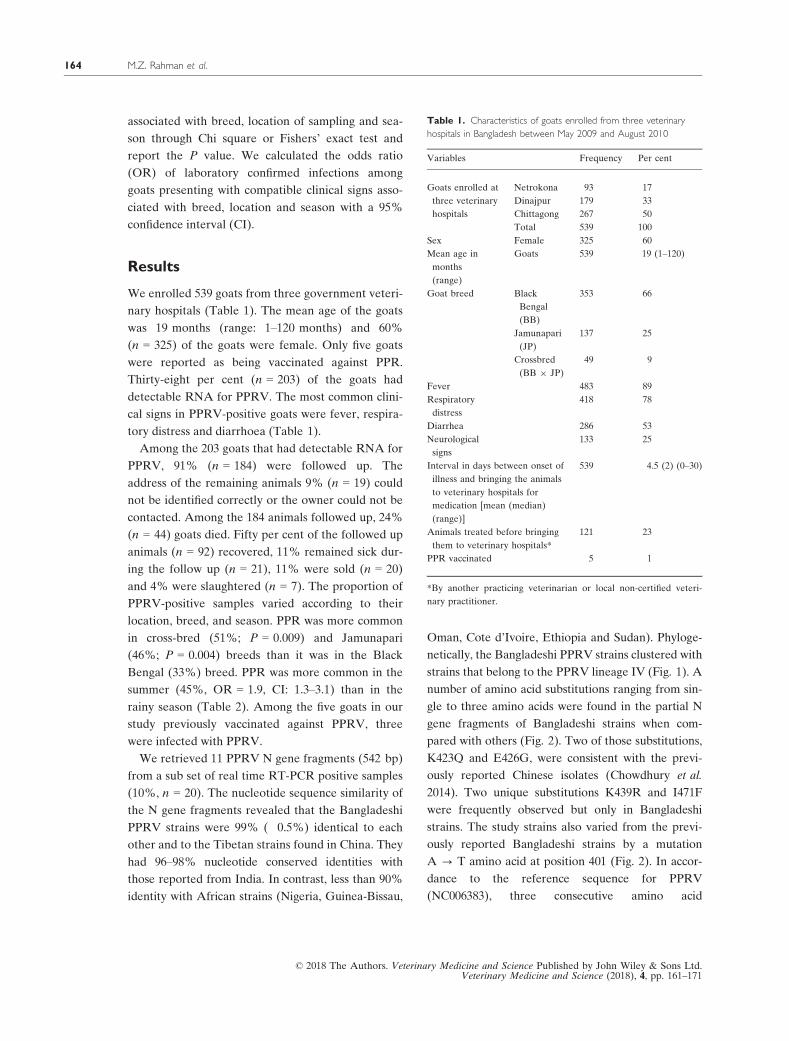
Table 1. Characteristics of goats enrolled from three veterinary
hospitals in Bangladesh between May 2009 and August 2010
Variables Frequency Per cent
Goats enrolled at
three veterinary
hospitals
Netrokona 93 17
Dinajpur 179 33
Chittagong 267 50
Total 539 100
Sex Female 325 60
Mean age in
months
(range)
Goats 539 19 (1–120)
Goat breed Black
Bengal
(BB)
353 66
Jamunapari
(JP)
137 25
Crossbred
(BB 9 JP)
49 9
Fever 483 89
Respiratory
distress
418 78
Diarrhea 286 53
Neurological
signs
133 25
Interval in days between onset of
illness and bringing the animals
to veterinary hospitals for
medication [mean (median)
(range)]
539 4.5 (2) (0–30)
Animals treated before bringing
them to veterinary hospitals*
121 23
PPR vaccinated 5 1
*By another practicing veterinarian or local non-certified veteri-
nary practitioner.
© 2018 The Authors. Veterinary Medicine and Science Published by John Wiley & Sons Ltd.Veterinary Medicine and Science (2018), 4, pp. 161–171
M.Z. Rahman et al.164
substitutions, EIM to DFI, at the positions 471–473
were observed in KT253989, KT253990 and
KT253997 (Fig. 2).
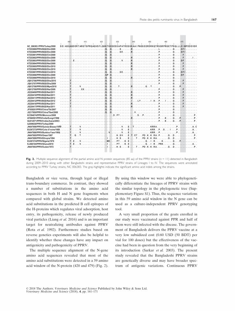
Six H gene nucleotide sequences (523 bp) were
retrieved and phylogenetic analysis of the sequence
data also indicated that the Bangladeshi PPRV
strains were mostly related to the Tibetan strains
found in China, showing 95.9–98.8% nucleotide
sequence identity (Fig. 3). The study strains had
94.7–97.7% nucleotide sequence identity with Indian
strains. Among the Bangladeshi strains, up to 2.9%
divergence was observed in the H gene fragment. A
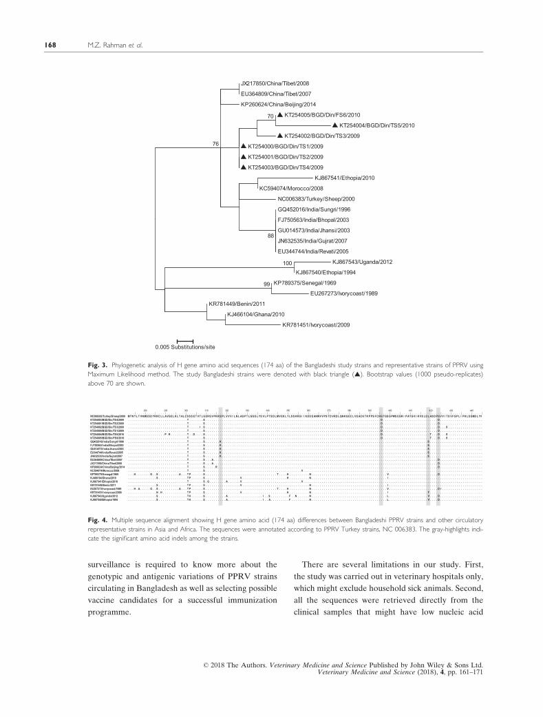
number of amino acid substitutions were found
when compared with the reference PPRV sequence
(NC006383). The study strains had a unique amino
acid substitution, G396D, which was consistently
found among the study strains but not in the Tibe-
tan strains (Fig. 4). The study strains varied from
Indian strains by the substitutions, E ? K at posi-
tion 317, D ? G at positions 396, 424 and L ? S at
position 419 (Fig. 4). In the predicted linear B cell
epitope of SDPGVVI, an amino acid substitution
G424D, was frequently observed among PPRV
strains from Bangladesh and Tibet, China but not
from India (Fig. 4).
Discussion
More than one-third of the goats were positive for
PPRV and one quarter of the enrolled goats died
from the disease. This case fatality rate is consistent
with the previously reported rates from India (30%)
and Pakistan (30%) (Sharma et al. 2007; Khan 2010),
confirming the severity of PPR in goats in this
region. In Bangladesh, not all goats are presented to
veterinary clinics. The goat farmers bring their ani-
mals to veterinary hospitals on foot as they usually
cannot bear the expense of renting vehicle. In addi-
tion, the animals in our study also benefitted from
treatment with antibiotics and anti-histamines
according to the standard practices for PPR clinical
management (Haider et al. 2016; Radostits et al.
2000). Thus, it is possible that the case fatality rate of
PPR among Bangladeshi goats reported elsewhere
and in our study is underestimated.
Our results indicate that the local goats (Black
Bengal) appear less susceptible to PPR than Jamuna-
pari and the cross-bred goats, contrasting with the
previous findings of Mondal et al. (1995) but in
agreement with a recent study (Rony et al. 2017) and
the Food and Agricultural Organization (FAO),
2010) and International Atomic Energy Agency who
reported that the Black Bengal goats had innate
resistance against common diseases and were well
adapted to the local environment.
We found a higher frequency of PPR in goats dur-
ing the summer season, which is in agreement with
previous studies in India, Pakistan, and Bangladesh
(Das et al. 2007; Singh et al. 2009; Khan 2010). Dur-
ing summer, the goats are more frequently allowed
to graze in communal grazing field which might
increase the chance of virus transmission from dis-
eased goat to healthy ones.
The PPRV strains circulating in Bangladeshi goats
were genetically similar although the samples were
collected from different districts, Dinajpur, Netro-
kona and Chittagong bordering Indian districts, Dak-
shin Dinajpur, Meghalaya and Tripura which are
very close to Tibet, China. Genetic analysis reveals
that PPRV strains circulating in this region share a
common phylogenetic lineage IV (Figs. 1 and 3) and
possibly spread either from India and/or China to
Table 2. The odds ratios (OR) of PPR infections among the goats
presenting with compatible clinical signs at three veterinary hospitals
in Bangladesh between May 2009 and August 2010
Name of disease No. of
animals
tested
No. positive
(%)
OR†
(95% CI‡)
Peste des petits
ruminants
(PPR) in goats
539 203 (38) –
By goat breed
Black Bengal 353 115 (33) 1
Jamunapari 137 63 (46) 1.8 (1.2–2.7)*
Crossbred 49 25 (51) 2.1 (1.2–4.1)*
By location
Netrokona 93 12 (8) 1
Dinajpur 179 62 (35) 3.6 (1.7–7.8)*
Chittagong 267 129 (48) 6.3 (3.2–13.3)*
By season
Rainy (July–Oct) 164 48 (29) 1
Winter (Nov–Feb) 171 64 (37) 1.4 (0.9–2.3)
Summer (Mar–Jun) 204 91 (45) 1.9 (1.3–3.1)*
*Significant at P < 0.05. †Odds ratio, ‡Confidence Interval.
© 2018 The Authors. Veterinary Medicine and Science Published by John Wiley & Sons Ltd.Veterinary Medicine and Science (2018), 4, pp. 161–171
Peste des petits ruminants virus in Bangladesh 165

JQ612706/PPRV/BGD/Dha/2010
JQ612707/PPRV/BGD/Dha/2009
JX220412/PPRV/BGD/Net/2012
KT253991/PPRV/BGD/Chi/2009
KT253998/PPRV/BGD/Din/2009
KT253993/PPRV/BGD/Din/2009
KT253989/PPRV/BGD/Din/2009
KT253990/PPRV/BGD/Din/2009
KT253997/PPRV/BGD/Chi/2010
JX220409/PPRV/BGD/Net/2011
JX220410/PPRV/BGD/Net/2011
JX220411/PPRV/BGD/Net/2012
JX220416/PPRV/BGD/Nar/2011
JX220417/PPRV/BGD/Nar/2011
KT253995/PPRV/BGD/Din/2010
KT253996/PPRV/BGD/Din/2010
KT253994/PPRV/BGD/Net/2009
KT253992/PPRV/BGD/Din/2009
KT253999/PPRV/BGD/Din/2010
JQ612708/PPRV/BGD/Mym/2010
JF939201/PPRV/China/Tib/2007
JX217850/PPRV/China/Tibet/2008
NC 006383 /PPRV/Turkey/2000
AJ849636/PPRV/Turkey/2000
KC594074/PPRV/Morocco/2008
AY560591/PPRV/India/Sungri/1996
GU014571/PPRV/India/Jhansi/2003
JN647699/PPRV/Ethopia/1994
JN647693/PPRV/Sudan/1972
JN647695/PPRV/Oman/1987
EU267274/PPRV/Nigeria/1976
KJ466104/PPRV/Ghana/2010
EU267273/PPRV/Cote d’Ivoire/1989
JN647698/PPRV/Guinea-Bissau/1989
JN647696/PPRV/Burkina Faso/1988
9999
96
94
88
97
0.02 Substitutions/site
Lineage IV
Lineage III
Lineage II
Lineage I
Fig. 1. The Phylogenetic analysis of N protein amino acid sequences (85 amino acid residues) of Bangladeshi strains and the representative
strains of each lineage of PPRV using Maximum Likelihood method. The study Bangladeshi strains were denoted with black triangle (▲). Boot-
strap values (1000 pseudo-replicates) above 80 are shown.
© 2018 The Authors. Veterinary Medicine and Science Published by John Wiley & Sons Ltd.Veterinary Medicine and Science (2018), 4, pp. 161–171
M.Z. Rahman et al.166
Bangladesh or vice versa, through legal or illegal
trans-boundary commerce. In contrast, they showed
a number of substitutions in the amino acid
sequences in both H and N gene fragments when
compared with global strains. We detected amino
acid substitutions in the predicted B cell epitopes of
the H-proteins which regulates viral adsorption, host
entry, its pathogenicity, release of newly produced
viral particles (Liang et al. 2016) and is an important
target for neutralizing antibodies against PPRV
(Rota et al. 1992). Furthermore studies based on
reverse genetics experiments will also be helpful to
identify whether these changes have any impact on
antigenicity and pathogenicity of PPRV.
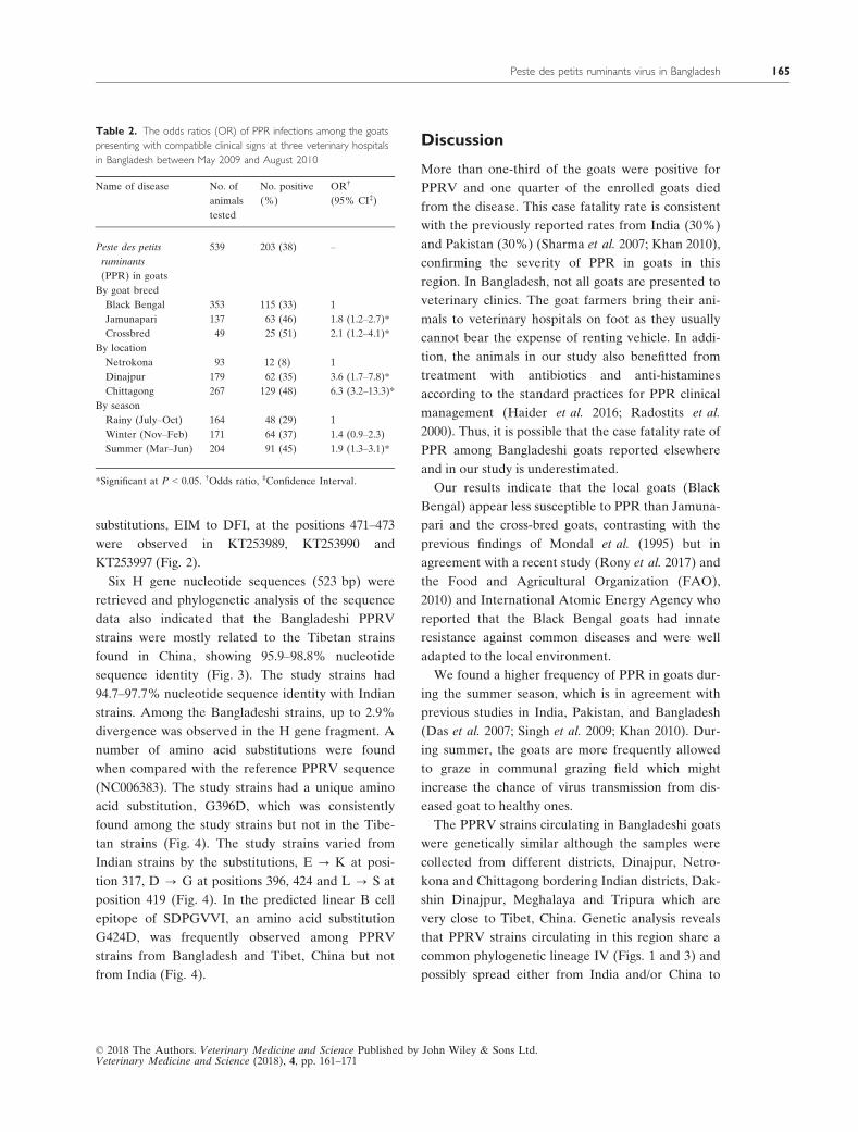
The multiple sequence alignment of the N-gene
amino acid sequences revealed that most of the
amino acid substitutions were detected in a 59 amino
acid window of the N-protein (420 and 479) (Fig. 2).
By using this window we were able to phylogeneti-
cally differentiate the lineages of PPRV strains with
the similar topology in the phylogenetic tree (Sup-
plementary Figure S1). Thus, the sequence variations
in this 59 amino acid window in the N gene can be
used as a culture-independent PPRV genotyping
tool.
A very small proportion of the goats enrolled in
our study were vaccinated against PPR and half of
them were still infected with the disease. The govern-
ment of Bangladesh delivers the PPRV vaccine at a
very low subsidized cost (0.60 USD (50 BDT) per
vial for 100 doses) but the effectiveness of the vac-
cine had been in question from the very beginning of
its introduction (Sarkar et al. 2003). The present
study revealed that the Bangladeshi PPRV strains
are genetically diverse and may have broader spec-
trum of antigenic variations. Continuous PPRV
Fig. 2. Multiple sequence alignment of the partial amino acid N protein sequences (85 aa) of the PPRV strains (n = 11) detected in Bangladesh
during 2009–2010 along with other Bangladeshi strains and representative PPRV strains of Lineages I to IV. The sequences were annotated
according to PPRV Turkey strains, NC 006383. The gray-highlights indicate the significant amino acid indels among the strains.
© 2018 The Authors. Veterinary Medicine and Science Published by John Wiley & Sons Ltd.Veterinary Medicine and Science (2018), 4, pp. 161–171
Peste des petits ruminants virus in Bangladesh 167
surveillance is required to know more about the
genotypic and antigenic variations of PPRV strains
circulating in Bangladesh as well as selecting possible
vaccine candidates for a successful immunization
programme.
There are several limitations in our study. First,
the study was carried out in veterinary hospitals only,
which might exclude household sick animals. Second,
all the sequences were retrieved directly from the
clinical samples that might have low nucleic acid
JX217850/China/Tibet/2008
EU364809/China/Tibet/2007
KP260624/China/Beijing/2014
KT254005/BGD/Din/FS6/2010
KT254004/BGD/Din/TS5/2010
KT254002/BGD/Din/TS3/2009
KT254000/BGD/Din/TS1/2009
KT254001/BGD/Din/TS2/2009
KT254003/BGD/Din/TS4/2009
KJ867541/Ethopia/2010
KC594074/Morocco/2008
NC006383/Turkey/Sheep/2000
GQ452016/India/Sungri/1996
FJ750563/India/Bhopal/2003
GU014573/India/Jhansi/2003
JN632535/India/Gujrat/2007
EU344744/India/Revati/2005
KJ867543/Uganda/2012
KJ867540/Ethopia/1994
KP789375/Senegal/1969
EU267273/Ivorycoast/1989
KR781449/Benin/2011
KJ466104/Ghana/2010
KR781451/Ivorycoast/2009
99
100
70
88
76
0.005 Substitutions/site
Fig. 3. Phylogenetic analysis of H gene amino acid sequences (174 aa) of the Bangladeshi study strains and representative strains of PPRV using
Maximum Likelihood method. The study Bangladeshi strains were denoted with black triangle (▲). Bootstrap values (1000 pseudo-replicates)
above 70 are shown.
Fig. 4. Multiple sequence alignment showing H gene amino acid (174 aa) differences between Bangladeshi PPRV strains and other circulatory
representative strains in Asia and Africa. The sequences were annotated according to PPRV Turkey strains, NC 006383. The gray-highlights indi-
cate the significant amino acid indels among the strains.
© 2018 The Authors. Veterinary Medicine and Science Published by John Wiley & Sons Ltd.Veterinary Medicine and Science (2018), 4, pp. 161–171
M.Z. Rahman et al.168
content of PPRV and remained undetectable, caus-
ing false negatives. Sometimes, PCR produced multi-
ple bands that hindered the quality sequence data.
Third, our genotyping analysis was based on partial
N and H genes sequences. Tissue culture-based
PPRV isolation followed by its whole genome-based
analysis is needed to reveal more about the PPRV
strain diversity and predicted antigenic variations.
Conclusion
PPR is a common infectious disease of goats with a
high case fatality in Bangladesh.
Bangladeshi PPRV strains clustered with strains
that belong to the PPRV lineage IV with unique
amino acid substitutions. Furthermore country-wide
surveillance and monitoring are required to deter-
mine the socioeconomic impact of PPRV strains in
Bangladesh as well as for their pathogenicity, viru-
lence and selection of vaccine type.
Acknowledgements
We acknowledge the Department of Livestock Ser-
vices (DLS) of Bangladesh. We are grateful to Ms.
Gladys Leterme, icddr,b, for her support in editing
this manuscript.
Source of funding
This research was funded by Google and the Rocke-
feller foundation through EcoHealth Alliance. icddr,
b acknowledges with gratitude the commitment of
Google and the Rockefeller foundation to its
research efforts.
Conflicts of interest
Authors have no financial and other conflicts of
interest to declare.
Ethical statement
Our trained veterinary officers obtained informed
written consent from the owners of cattle and goats
enrolled in the respective veterinary hospitals. The
team also took appropriate personal protection dur-
ing the biological specimen collection. All the sam-
pling procedures have been reviewed and approved
by the animal experimentation and ethics committee
of icddr,b.
Contributions
MZR, NH conceived, designed, and supervised the
study; ESG, JHE, NZ protocol supervision and
reviewed the manuscript; MGO, SAK supported
field activities; SA involved in lab testing; MBH
performed epidemiological data analysis; AI
involved in sample and data collection; MZR and
MEH supported lab data analysis and manuscript
preparation; MR reviewed the manuscript.
References
Balamurugan V., Sen A., Saravanan P., Rasool T.J.,
Yadav M.P., Bandyopadhyay S.K. & Singh R.K. (2006)
Development and characterization of a stable vero cell
line constitutively expressing Peste des petits ruminants
virus (PPRV) hemagglutinin protein and its potential
use as antigen in enzyme-linked immunosorbent assay
for serosurveillance of PPRV. Clinical and Vaccine
Immunology 13, 1367–1372.
Balamurugan V., Hemadri D., Gajendragad M.R., Singh
R.K. & Rahman H. (2014) Diagnosis and control of
peste des petits ruminants: a comprehensive review.
Virus Disease 25, 39–56.
Bao J., Li L., Wang Z., Barrett T., Suo L., Zhao W. et al.
(2008) Development of one-step real-time RT-PCR
assay for detection and quantitation of peste des petits
ruminants virus. Journal of Virological Methods 148,
232–236.
Bhuiyan A.R. (2012). Epidemiology and pathology of
peste des petits ruminants (PPR) in Bangladesh and
molecular characterization of the virus. PhD Thesis.
Department of Pathology, Bangladesh Agricultural
University: Mymensingh.
Buet B. 2008. Characterizing long-term change of Bangla-
desh: climate in context of agriculture and irrigation.
Final Report to Ministry of Environment and Forests
and Ministry of Food and Disaster Management, 2008.
(Bangladesh University of Engineering & Technology),
1–7.
Chowdhury E.H., Bhuiyan A.R., Rahman M.M., Siddique
M.S. & Islam M.R. (2014) Natural peste des petits rumi-
nants virus infection in Black Bengal goats: virological,
© 2018 The Authors. Veterinary Medicine and Science Published by John Wiley & Sons Ltd.Veterinary Medicine and Science (2018), 4, pp. 161–171
Peste des petits ruminants virus in Bangladesh 169
pathological and immunohistochemical investigation.
BMC Veterinary Research 10, 263.
Couacy-Hymann E., Roger F., Hurard C., Guillou J.P.,
Libeau G. & Diallo A. (2002) Rapid and sensitive
detection of peste des petits ruminants virus by a poly-
merase chain reaction assay. Journal of Virological
Methods 100, 17–25.
Das K.K., Shil N.K. & Islam M.R. (2007) Sero-epidemio-
logical investigation on Peste des petits ruminants in
black Bengal goats. Bangladesh Journal of Microbiology
24, 143–145.
Diallo A., Taylor W.P., Lefevre P.C. & Provost A. (1989)
Attenuation of a strain of rinderpest virus: potential
homologous live vaccine. Revue d’elevage et de medecine
veterinaire des pays tropicaux 42, 311–319.
EFSA-AHAW-Panel (2015) Scientific opinion on Peste
des petits ruminants. EFSA Journal 13, 94.
El Arbi A.S., El Mamy A.B., Salami H., Isselmou E.,
Kwiatek O., Libeau G. et al. (2014) Peste des petits
ruminants virus, Mauritania. Emerging Infectious Dis-
eases 20, 333–336.
Encyclopedia, A. A. (1994). Body temperature. American
Encyclopedia, B: New York, 357.
Food and Agricultural Organization(FAO), a.I.A.E.A.
(2010). Joint FAO/IAEA Programme: Nuclear Tech-
nique in Food and Agriculture: Livestock: Black Bengal
- A Promising goat genetic resource of Bangladesh Joint
FAO and IAEA Programme Accessed on June 09,
2011.
Haider N., Khan S.U., Islam A., Osmani M.G., Rah-
man M.Z., Epstein J.H. et al. (2016) Efficiency of the
clinical veterinary diagnostic practices and drug
choices for infectious diseases in livestock in Bangla-
desh. Transboundary and Emerging Diseases 64,
1329–1333.
Hall T. (1999). A user-friendly biological sequence align-
ment editor and analysis program for Windows 95/98/
NT. Nucleic Acids Symp Ser, 95–98.
Islam M.R., Shamsuddin M., Rahman M.A., Das P.M. &
Dewan M.L. (2001) An outbreak of peste des petits
ruminants in Black Bengal goats in Mymensingh, 18.
The Bangladesh Veterinarian: Bangladesh.
IslamA., Singha A. & IslamM.A. (2011) Peste des petits
ruminants of goats, outbreak and economic losses. Pakistan
Journal of Scientific and Industrial Research 54, 76–82.
Khan F.M. (2010) Participatory appraisal and scanning
surveillance based contagious diseases risk profile of dis-
trict Rahim ar Khan (Pakistan). The Pakistan Veterinary
Journal 30, 198–202.
Kimura M. (1980) A simple method for estimating evolu-
tionary rates of base substitutions through comparative
studies of nucleotide sequences. Journal of Molecular
Evolution 16, 111–120.
Liang Z., Yuan R., Chen L., Zhu X. & Dou Y. (2016)
Molecular evolution and characterization of hemagglu-
tinin (H) in Peste des Petits ruminants virus. PLoS
ONE 11, e0152587.
Maganga G.D., Verrier D., Zerbinati R.M., Drosten C.,
Drexler J.F. & Leroy E.M. (2013) Molecular typing of
PPRV strains detected during an outbreak in sheep and
goats in south-eastern Gabon in 2011. Virology Journal
10, 82.
Mondal A.K., Chottopadhyay A.P., Sarkar S.D., Saha
G.R. & Bhowmik M.K. (1995) Report on
epizootiological and clinicopathological observations
on peste des petits ruminants (PPR) of goats in
West Bengal. Indian Journal of Animal Health 34,
145–148.
Radostits O.M., Blood D.C., Hinchcliff K.W. (2000).
Veterinary medicine: a textbook of the diseases of cattle,
sheep, pigs, goats and horses, 9th edn, W B Saunders
Company Ltd: London.
Rahman M.M., Bhuiyan A.R., Parvin R., Giasuddin M.,
Haque M.E., Sayem S.M. et al. (2011) Immune
response of goats to thermostable PPR vaccine
in Bangladesh. SAARC Journal of Agriculture 9, 73–
81.
Rahman M.M., Parvin R., Bhuiyan A.R., Giasuddin M.,
Chowdhury S.M.Z.H., Islam M.R. & Chowdhury E.H.
(2016) Genetic characterization of Peste des petits rumi-
nants virus circulating in Bangladesh. British Journal of
Virology 3(4), 115–122.
Rony M.S., Rahman A.K., Alam M.M., Dhand N. & Ward
M.P. (2017) Peste des Petits Ruminants risk factors and
space-time clusters in Mymensingh. Transboundary and
emerging diseases: Bangladesh.
Rota J.S., Hummel K.B., Rota P.A. & Bellini W.J.
(1992) Genetic variability of the glycoprotein genes of
current wild-type measles isolates. Virology 188, 135–
142.
Sarkar J., Sreenivasa B.P., Singh R.P., Dhar P. & Bandy-
opadhyay S.K. (2003) Comparative efficacy of various
chemical stabilizers on the thermostability of a live-atte-
nuated peste des petits ruminants (PPR) vaccine. Vac-
cine 21, 4728–4735.
Sen A., Saravanan P., Balamurugan V., Rajak K.K., Sud-
hakar S.B., Bhanuprakash V. et al. (2010) Vaccines
against peste des petits ruminants virus. Expert Review
of Vaccines 9, 785–796.
Shaila M.S., Purushothaman V., Bhavasar D., Venugopal
K. & Venkatesan R.A. (1989) Peste des petits rumi-
nants of sheep in India. The Veterinary Record 125,
602.
Sharma S., Mahajan V., Kanwar R.S., Filia G., Kumar
H., Singh R. & Bal M.S. (2007) Peste des petits
ruminants (PPR) outbreaks in sheep and goats in
© 2018 The Authors. Veterinary Medicine and Science Published by John Wiley & Sons Ltd.Veterinary Medicine and Science (2018), 4, pp. 161–171
M.Z. Rahman et al.170
Punjab. Indian Journal of Veterinary Pathology 31,
32–35.
Singh R.K., Balamurugan V., Bhanuprakash V., Sen A.,
Saravanan P. & Pal Yadav M. (2009) Possible control
and eradication of peste des petits ruminants from
India: technical aspects. Veterinaria Italiana 45,
449–462.
Tamura K., Peterson D., Peterson N., Stecher G., Nei
M. & Kumar S. (2011) MEGA5: molecular evolu-
tionary genetics analysis using maximum likelihood,
evolutionary distance, and maximum parsimony
methods. Molecular Biology and Evolution 28,
2731–2739.
Supporting information
Additional Supporting Information may be found
online in the supporting information tab for this article:
Figure S1. The Phylogenetic analysis of N protein
amino acid sequences (59 amino acid residues) of
Bangladeshi strains and the representative strains of
each lineage of PPRV by using Maximum Likeli-
hood method. The study Bangladeshi strains were
denoted with black triangle (▲). Bootstrap values
(1000 pseudo-replicates) above 80 are shown.
© 2018 The Authors. Veterinary Medicine and Science Published by John Wiley & Sons Ltd.Veterinary Medicine and Science (2018), 4, pp. 161–171
Peste des petits ruminants virus in Bangladesh 171