Embed Size (px)
Citation preview
Review
Epidemiologic and Molecular Prognostic Review ofGlioblastoma
Jigisha P. Thakkar1,2, Therese A. Dolecek3, Craig Horbinski4, Quinn T. Ostrom5, Donita D. Lightner6,Jill S. Barnholtz-Sloan5, and John L. Villano1,2
AbstractGlioblastoma multiforme (GBM) is the most common and aggressive primary central nervous system
malignancy with a median survival of 15 months. The average incidence rate of GBM is 3.19/100,000
population, and the median age of diagnosis is 64 years. Incidence is higher in men and individuals of white
race and non-Hispanic ethnicity. Many genetic and environmental factors have been studied in GBM, but the
majority are sporadic, and no risk factor accounting for a large proportion of GBMs has been identified.
However, several favorable clinical prognostic factors are identified, including younger age at diagnosis,
cerebellar location, high performance status, and maximal tumor resection. GBMs comprise of primary and
secondary subtypes, which evolve through different genetic pathways, affect patients at different ages, and
have differences in outcomes. We report the current epidemiology of GBM with new data from the Central
Brain Tumor Registry of the United States 2006 to 2010 as well as demonstrate and discuss trends in incidence
and survival. We also provide a concise review on molecular markers in GBM that have helped distinguish
biologically similar subtypes of GBM and have prognostic and predictive value. Cancer Epidemiol Biomarkers
Prev; 23(10); 1985–96. �2014 AACR.
IntroductionGlioblastomamultiforme (GBM) is the most aggressive
diffuse glioma of astrocytic lineage and corresponds tograde 4 based onWHO classification (1). GBM is the mostcommon brain and central nervous system (CNS) malig-nancy, accounting for 45.2% of malignant primary brainand CNS tumors, 54% of all gliomas, and 16% of allprimary brain and CNS tumors (2). GBM remains anincurable disease, with a median survival of 15 months(3, 4). Treatment is complex and initially consists ofmaximal safe surgical resection followed by radiotherapy(RT) with concurrent temozolomide (TMZ) chemothera-py followed by 6 cycles of maintenance TMZ (5).GBMs comprise of primary and secondary subtypes,
which evolve through different genetic pathways, affectpatients at different ages, and have differences in out-comes (6). Primary (de novo) GBMs account for 80% of
GBMs and occur in older patients (mean age, 62 years).Secondary GBMs develop from lower-grade astrocytomaor oligodendrogliomas and occur in younger patients(mean age, 45 years; refs. 6–9). The WHO recently addeda rare subtype of GBM termed "with oligodendrogliomacomponent" (GBM-O), defined as GBM having areas thatresemble anaplastic oligodendroglioma with hallmarkfeatures of GBM, necrosis with or without microvascularproliferation (1).
EpidemiologyIncidence and risk factors
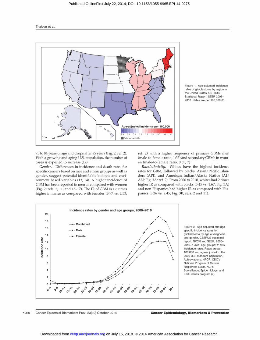
Based on the 2013 Central Brain Tumor Registry of theUnited States (CBTRUS) report, the average annual age-adjusted incidence rate (IR) of GBM is 3.19/100,000 pop-ulation (2). This is the highest IR among brain and CNStumors with malignant behavior followed by diffuseastrocytoma grade 2 (0.56/100,000), and malignant glio-ma not otherwise specified (0.46/100,000; ref. 2). Inci-dence is highest in the northeast and lowest in thesouth-central region of theUnited States (Fig. 1); however,thesedifferences could reflect differences in cancer report-ing by region (2).Many genetic and environmental factorshave been studied in GBMbut no risk factor that accountsfor a large proportion of GBMhas been identified and likemany cancers are sporadic (10).
Age. GBMisprimarilydiagnosedat older ageswith themedian age of diagnosis of 64 years (2, 11). It is uncommonin children accounting only approximately 3% of all brainand CNS tumors reported among 0 to 19 year olds (2). Theincidence continues to rise with increasing age, peaks at
1Department of Medicine, University of Kentucky, Lexington, Kentucky.2Department of Neurology, University of Kentucky, Lexington, Kentucky.3Division of Epidemiology and Biostatistics and Institute for HealthResearch and Policy, School of Public Health, University of Illinois atChicago, Chicago, Illinois. 4Department of Pathology, University of Ken-tucky, Lexington, Kentucky. 5Case Comprehensive Cancer Center, CaseWestern Reserve University School of Medicine, Cleveland, Ohio. 6Depart-ment of Neurology and Pediatrics, University of Kentucky, Lexington,Kentucky.
Corresponding Author: John L. Villano, University of Kentucky, 800 RoseSt., CC447, Lexington, KY 40536-0093. Phone: 859-323-0405; Fax: 859-257-7715; E-mail: [email protected]
doi: 10.1158/1055-9965.EPI-14-0275
�2014 American Association for Cancer Research.
CancerEpidemiology,
Biomarkers& Prevention
www.aacrjournals.org 1985
on July 15, 2018. © 2014 American Association for Cancer Research. cebp.aacrjournals.org Downloaded from
Published OnlineFirst July 22, 2014; DOI: 10.1158/1055-9965.EPI-14-0275
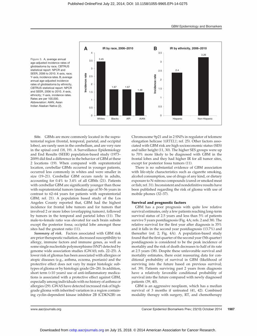
75 to 84 years of age and drops after 85 years (Fig. 2; ref. 2).With a growing and aging U.S. population, the number ofcases is expected to increase (12).
Gender. Differences in incidence and death rates forspecific cancers based on race and ethnic groups aswell asgender, suggest potential identifiable biologic and envi-ronment based variables (13, 14). A higher incidence ofGBMhas been reported inmen as compared withwomen(Fig. 2; refs. 2, 11, and 15–17). The IR of GBM is 1.6 timeshigher in males as compared with females (3.97 vs. 2.53;
ref. 2) with a higher frequency of primary GBMs men(male-to-female ratio, 1:33) and secondary GBMs inwom-en (male-to-female ratio, 0:65; 7).
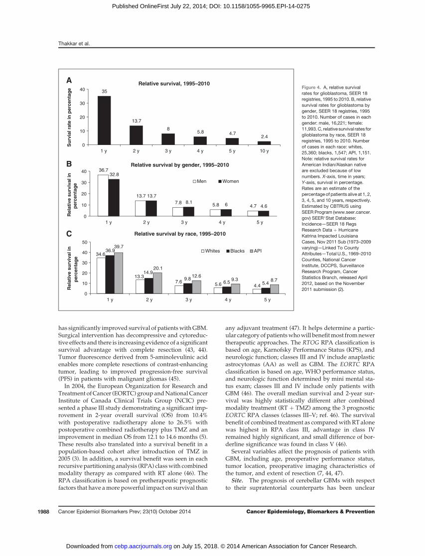
Race/ethnicity. Whites have the highest incidencerates for GBM, followed by blacks, Asian/Pacific Islan-ders (API), and American Indian/Alaska Native (AI/AN; Fig. 3A; ref. 2). From 2006 to 2010, whites had 2 timeshigher IR as compared with blacks (3.45 vs. 1.67; Fig. 3A)and non-Hispanics had higher IR as compared with His-panics (3.26 vs. 2.45; Fig. 3B; refs. 2 and 11).
0
2
4
6
8
10
12
14
16
18
20
Inci
den
ce r
ate
Incidence rates by gender and age groups, 2006–2010
Combined
Male
Female
0–4
5–9
10–1
415
–19
20–2
425
–29
30–3
435
–39
40–4
445
–49
50–5
455
–59
60–6
465
–69
70–7
475
–79
80–8
4
85+
Figure 2. Age-adjusted and age-specific incidence rates forglioblastoma by age at diagnosisand gender, CBTRUS statisticalreport: NPCR and SEER, 2006–2010. X-axis, age groups; Y-axis,incidence rates. Rates are per100,000 and age-adjusted to the2000 U.S. standard population.Abbreviations: NPCR, CDC'sNational Program of CancerRegistries; SEER, NCI'sSurveillance, Epidemiology, andEnd Results program (2).
Age-adjusted incidence per 100,000
2.9 3.0 3.1 3.2 3.3 3.4 3.5 3.6 3.7
Data not available
Figure 1. Age-adjusted incidencerates of glioblastoma by region inthe United States, CBTRUSStatistical Report, SEER 2006–2010. Rates are per 100,000 (2).
Thakkar et al.
Cancer Epidemiol Biomarkers Prev; 23(10) October 2014 Cancer Epidemiology, Biomarkers & Prevention1986
on July 15, 2018. © 2014 American Association for Cancer Research. cebp.aacrjournals.org Downloaded from
Published OnlineFirst July 22, 2014; DOI: 10.1158/1055-9965.EPI-14-0275
Site. GBMs are more commonly located in the supra-tentorial region (frontal, temporal, parietal, and occipitallobes), are rarely seen in the cerebellum, and are very rarein the spinal cord (18, 19). A Surveillance Epidemiologyand End Results (SEER) population-based study (1973–2009) did find a difference in the behavior of GBMat these2 locations (19). When compared with supratentoriallocation, cerebellar GBMs occurred in younger patients,occurred less commonly in whites and were smaller insize (19–21). Cerebellar GBM occurs rarely in adults,accounting for 0.4% to 3.4% of all GBMs (21). Patientswith cerebellar GBM are significantly younger than thosewith supratentorial tumors (median age of 50–56 years incontrast to 62–64 years for patients with supratentorialGBM; ref. 21). A population based study of the LosAngeles County reported that, GBM had the highestincidence for frontal lobe tumors and for tumors thatinvolved 2 or more lobes (overlapping tumors), followedby tumors in the temporal and parietal lobes (11). Themale-to-female ratio was elevated for each brain subsiteexcept the posterior fossa; occipital lobe amongst thesesites had the greatest ratio (11).Summary of risk. Factors associated with GBM risk
are prior therapeutic radiation, decreased susceptibility toallergy, immune factors and immune genes, as well assome single nucleotide polymorphisms (SNP) detected bygenome wide association studies (GWAS; refs. 22–25). Alower risk of gliomas has been associatedwith allergies oratopic diseases (e.g., asthma, eczema, psoriasis) and theprotective effect does not vary by major histologic sub-types of glioma or by histologic grade (26–28). In addition,short term (<10 years) use of anti-inflammatory medica-tions is associated with a protective effect against GBM,especially among individualswith nohistory of asthma orallergies (29). GWAShave detected increased risk of high-grade gliomawith inherited variation in a region contain-ing cyclin-dependent kinase inhibitor 2B (CDKN2B) on
Chromosome 9p21 and in 2 SNPs in regulator of telomereelongation helicase 1(RTEL1; ref. 25). Other factors asso-ciated with GBM risk are high socioeconomic status (SES)and taller height (11, 30). The higher SES groups were upto 70% more likely to be diagnosed with GBM in thefrontal lobes and they had higher IR for all tumor sites,except for posterior fossa tumors (11).
There is no substantial evidence of GBM associationwith life-style characteristics such as cigarette smoking,alcohol consumption, use of drugs of any kind, or dietaryexposure toN-nitroso compounds (cured or smokedmeator fish; ref. 31). Inconsistent andnondefinitive results havebeen published regarding the risk of glioma with use ofmobile phones (32–37).
Survival and prognostic factorsGBM has a poor prognosis with quite low relative
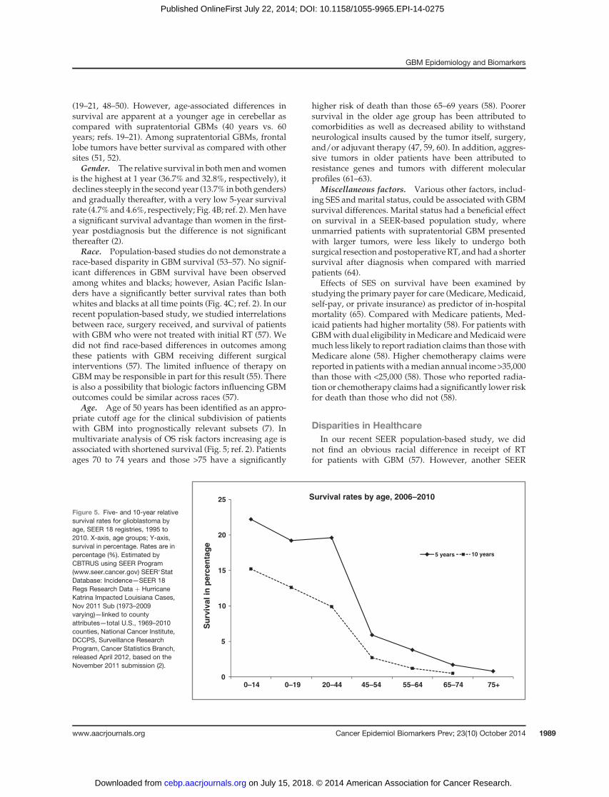
survival estimates, only a fewpatients reaching long-termsurvival status of 2.5 years and less than 5% of patientssurvive 5 years postdiagnosis (Fig. 4A; refs. 2 and 38). Therelative survival for the first year after diagnosis is 35%and it falls in the second year postdiagnosis (13.7%) andthereafter (ref. 2; Fig. 4A). A population-based studyfound that the first quarter of the second year (5th quarter)postdiagnosis is considered to be the peak incidence ofmortality and the risk of death decreases to half of its rateat 2.5 years (38). Despite these unfavorable survival andmortality estimates, there exist reassuring data for con-ditional probability of survival in GBM (likelihood ofsurviving into the future based on previous survival;ref. 39). Patients surviving past 2 years from diagnosishave a relatively favorable conditional probability ofsurvival into the future compared with newly diagnosedpatients (39, 40).
GBM is an aggressive neoplasm, which has a mediansurvival of 3 months if untreated (41, 42). Combinedmodality therapy with surgery, RT, and chemotherapy
3.45
1.67 1.671.48
0
0.5
1
1.5
2
2.5
3
3.5
4
AIANAPIBlacksWhites
Inci
den
ce r
ate
IR by race, 2006–2010 A
2.4
3.26
0
0.5
1
1.5
2
2.5
3
3.5
Non-HispanicHispanic
Inci
den
ce r
ate
IR by ethnicity, 2006–2010 B
Figure 3. A, average annualage-adjusted incidence rates ofglioblastoma by race, CBTRUSstatistical report: NPCR andSEER, 2006 to 2010. X-axis, race;Y-axis, incidence rates. B, averageannual age-adjusted incidencerates of glioblastoma by ethnicity,CBTRUS statistical report: NPCRand SEER, 2006 to 2010. X-axis,ethnicity; Y-axis, incidence rates.Rates are per 100,000.Abbreviation: AIAN, AsianIndian Alaskan Native (2).
GBM Epidemiology and Biomarkers
www.aacrjournals.org Cancer Epidemiol Biomarkers Prev; 23(10) October 2014 1987
on July 15, 2018. © 2014 American Association for Cancer Research. cebp.aacrjournals.org Downloaded from
Published OnlineFirst July 22, 2014; DOI: 10.1158/1055-9965.EPI-14-0275
has significantly improved survival of patientswithGBM.Surgical intervention has decompressive and cytoreduc-tive effects and there is increasing evidence of a significantsurvival advantage with complete resection (43, 44).Tumor fluorescence derived from 5-aminolevulinic acidenables more complete resections of contrast-enhancingtumor, leading to improved progression-free survival(PFS) in patients with malignant gliomas (45).
In 2004, the European Organization for Research andTreatment ofCancer (EORTC) group andNationalCancerInstitute of Canada Clinical Trials Group (NCIC) pre-sented a phase III study demonstrating a significant imp-rovement in 2-year overall survival (OS) from 10.4%with postoperative radiotherapy alone to 26.5% withpostoperative combined radiotherapy plus TMZ and animprovement in median OS from 12.1 to 14.6 months (5).These results also translated into a survival benefit in apopulation-based cohort after introduction of TMZ in2005 (3). In addition, a survival benefit was seen in eachrecursive partitioning analysis (RPA) classwith combinedmodality therapy as compared with RT alone (46). TheRPA classification is based on pretherapeutic prognosticfactors that have amore powerful impact on survival than
any adjuvant treatment (47). It helps determine a partic-ular category ofpatientswhowill benefitmost fromnewertherapeutic approaches. The RTOG RPA classification isbased on age, Karnofsky Performance Status (KPS), andneurologic function; classes III and IV include anaplasticastrocytomas (AA) as well as GBM. The EORTC RPAclassification is based on age, WHO performance status,and neurologic function determined by mini mental sta-tus exam; classes III and IV include only patients withGBM (46). The overall median survival and 2-year sur-vival was highly statistically different after combinedmodality treatment (RT þ TMZ) among the 3 prognosticEORTC RPA classes (classes III–V; ref. 46). The survivalbenefit of combined treatment as comparedwith RT alonewas highest in RPA class III, advantage in class IVremained highly significant, and small difference of bor-derline significance was found in class V (46).
Several variables affect the prognosis of patients withGBM, including age, preoperative performance status,tumor location, preoperative imaging characteristics ofthe tumor, and extent of resection (7, 44, 47).
Site. The prognosis of cerebellar GBMs with respectto their supratentorial counterparts has been unclear
35
13.7
85.8 4.7
2.4
0
10
20
30
40
1 y 2 y 3 y 4 y 5 y 10 y
Su
rvia
l rat
e in
per
cen
tag
e
Relative survival, 1995–2010 A
36.7
13.77.8 5.8 4.7
32.8
13.78.1 6 4.6
0
10
20
30
40
5 y4 y3 y2 y1 y
Rel
ativ
e su
rviv
al in
p
erce
nta
ge
Relative survival by gender, 1995–2010
Men Women
B
34.6
13.37.6 5.6 4.4
36.9
14.99.8
6.5 5.4
39.7
20.1
12.69.3 8.7
0
10
20
30
40
50
5 y4 y3 y2 y1 y
Rel
ativ
e su
rviv
al in
p
erce
nta
ge
Relative survival by race, 1995–2010
Whites Blacks API
C
Figure 4. A, relative survivalrates for glioblastoma, SEER 18registries, 1995 to 2010. B, relativesurvival rates for glioblastoma bygender, SEER 18 registries, 1995to 2010. Number of cases in eachgender: male, 16,221; female:11,993.C, relative survival rates forglioblastoma by race, SEER 18registries, 1995 to 2010. Numberof cases in each race: whites,25,360; blacks, 1,547; API, 1,151.Note: relative survival rates forAmerican Indian/Alaskan nativeare excluded because of lownumbers. X-axis, time in years;Y-axis, survival in percentage.Rates are an estimate of thepercentage of patients alive at 1, 2,3, 4, 5, and 10 years, respectively.Estimated by CBTRUS usingSEER Program (www.seer.cancer.gov) SEER�Stat Database:Incidence—SEER 18 RegsResearch Data þ HurricaneKatrina Impacted LouisianaCases, Nov 2011 Sub (1973–2009varying)—Linked To CountyAttributes—Total U.S., 1969–2010Counties, National CancerInstitute, DCCPS, SurveillanceResearch Program, CancerStatistics Branch, released April2012, based on the November2011 submission (2).
Thakkar et al.
Cancer Epidemiol Biomarkers Prev; 23(10) October 2014 Cancer Epidemiology, Biomarkers & Prevention1988
on July 15, 2018. © 2014 American Association for Cancer Research. cebp.aacrjournals.org Downloaded from
Published OnlineFirst July 22, 2014; DOI: 10.1158/1055-9965.EPI-14-0275
(19–21, 48–50). However, age-associated differences insurvival are apparent at a younger age in cerebellar ascompared with supratentorial GBMs (40 years vs. 60years; refs. 19–21). Among supratentorial GBMs, frontallobe tumors have better survival as compared with othersites (51, 52).Gender. The relative survival in bothmen andwomen
is the highest at 1 year (36.7% and 32.8%, respectively), itdeclines steeply in the secondyear (13.7% inbothgenders)and gradually thereafter, with a very low 5-year survivalrate (4.7%and 4.6%, respectively; Fig. 4B; ref. 2).Menhavea significant survival advantage than women in the first-year postdiagnosis but the difference is not significantthereafter (2).Race. Population-based studies do not demonstrate a
race-based disparity in GBM survival (53–57). No signif-icant differences in GBM survival have been observedamong whites and blacks; however, Asian Pacific Islan-ders have a significantly better survival rates than bothwhites and blacks at all time points (Fig. 4C; ref. 2). In ourrecent population-based study, we studied interrelationsbetween race, surgery received, and survival of patientswith GBM who were not treated with initial RT (57). Wedid not find race-based differences in outcomes amongthese patients with GBM receiving different surgicalinterventions (57). The limited influence of therapy onGBMmay be responsible in part for this result (55). Thereis also a possibility that biologic factors influencing GBMoutcomes could be similar across races (57).Age. Age of 50 years has been identified as an appro-
priate cutoff age for the clinical subdivision of patientswith GBM into prognostically relevant subsets (7). Inmultivariate analysis of OS risk factors increasing age isassociated with shortened survival (Fig. 5; ref. 2). Patientsages 70 to 74 years and those >75 have a significantly
higher risk of death than those 65–69 years (58). Poorersurvival in the older age group has been attributed tocomorbidities as well as decreased ability to withstandneurological insults caused by the tumor itself, surgery,and/or adjuvant therapy (47, 59, 60). In addition, aggres-sive tumors in older patients have been attributed toresistance genes and tumors with different molecularprofiles (61–63).
Miscellaneous factors. Various other factors, includ-ing SES andmarital status, could be associated with GBMsurvival differences. Marital status had a beneficial effecton survival in a SEER-based population study, whereunmarried patients with supratentorial GBM presentedwith larger tumors, were less likely to undergo bothsurgical resection andpostoperative RT, andhad a shortersurvival after diagnosis when compared with marriedpatients (64).
Effects of SES on survival have been examined bystudying the primary payer for care (Medicare, Medicaid,self-pay, or private insurance) as predictor of in-hospitalmortality (65). Compared with Medicare patients, Med-icaid patients had higher mortality (58). For patients withGBMwith dual eligibility inMedicare andMedicaidweremuch less likely to report radiation claims than thosewithMedicare alone (58). Higher chemotherapy claims werereported in patientswith amedian annual income >35,000than those with <25,000 (58). Those who reported radia-tion or chemotherapy claims had a significantly lower riskfor death than those who did not (58).
Disparities in HealthcareIn our recent SEER population-based study, we did
not find an obvious racial difference in receipt of RTfor patients with GBM (57). However, another SEER
0
5
10
15
20
25
0–14 0–19 20–44 45–54 55–64 65–74 75+
Su
rviv
al in
per
cen
tag
e
Survival rates by age, 2006–2010
10 years 5 years
Figure 5. Five- and 10-year relativesurvival rates for glioblastoma byage, SEER 18 registries, 1995 to2010. X-axis, age groups; Y-axis,survival in percentage. Rates are inpercentage (%). Estimated byCBTRUS using SEER Program(www.seer.cancer.gov) SEER�StatDatabase: Incidence—SEER 18Regs Research Data þ HurricaneKatrina Impacted Louisiana Cases,Nov 2011 Sub (1973–2009varying)—linked to countyattributes—total U.S., 1969–2010counties, National Cancer Institute,DCCPS, Surveillance ResearchProgram, Cancer Statistics Branch,released April 2012, based on theNovember 2011 submission (2).
GBM Epidemiology and Biomarkers
www.aacrjournals.org Cancer Epidemiol Biomarkers Prev; 23(10) October 2014 1989
on July 15, 2018. © 2014 American Association for Cancer Research. cebp.aacrjournals.org Downloaded from
Published OnlineFirst July 22, 2014; DOI: 10.1158/1055-9965.EPI-14-0275
population-based study investigating the influence ofregional health system resources on the surgical manage-ment of GBM and receipt of postoperative RT found thatyounger, married patients in health service areas (HSA)with higher median incomes were significantly morelikely to receive both gross total resection and postoper-ative RT (66). For every $10,000 increase in the medianincome of a HSA, a patient’s likelihood of receiving grossresection increased by 7%andpostoperative RT receipt by6.3% (66). Their findings indicated that it may not be thedensity of individual radiation oncologists, but rather theprevalence of radiation oncology centers that influencespostoperative RT receipt, suggesting a dominant role ofhospital-level infrastructure over individual providers foraddressing disparities in GBM management (66). It ispossible that the large variations in treatment of GBMmay be related less to access to neuro-oncology services,but a larger apprehension of physicians to attempt aggres-sive surgery and RT for patients with less favorableprognosis (66).
A significant percentage of patients with GBM (27.3%)do not receive RT in the initial round of therapy andmajority of these are elderly patients (65 years and older;refs. 57 and 67). Similar trends are seen in other SEER-basedpopulation studies for breast and lung cancerwherelower rates of RT with increased age were observed(68, 69). Underuse of RT in a large number of elderlypatients with GBM could be attributed increased risk ofcognitive side-effects associated with RT (67, 70). How-ever, recent data do show benefit of RT in the elderlypopulation (71).
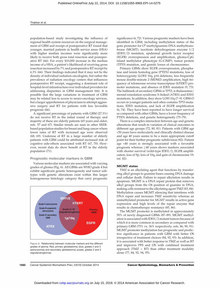
Prognostic molecular markers in GBMVariousmolecular markers are associatedwith varying
grades of glioma (Fig. 6). All GBMs are WHO grade 4 butexhibit significant genetic heterogeneity and tumor sub-types with genetic alterations exist within this largerhomogeneous histologic category that carry prognostic
significance (6, 72). Various prognosticmarkers have beenidentified in GBM, including methylation status of thegene promoter for O6-methylguanine-DNA methyltrans-ferase (MGMT), isocitrate dehydrogenase enzyme 1/2(IDH1/2) mutation, epidermal growth factor receptor(EGFR) overexpression and amplification, glioma-CpGisland methylator phenotype (G-CIMP), tumor protein(TP53) mutation, and genetic losses of chromosomes.
Primary GBMs show EGFR overexpression, phospha-tase and tensin homolog gene (PTEN) mutations, loss ofheterozygosity (LOH) 10q, p16 deletions, less frequentlymouse double-minute 2 (MDM2) amplification, high fre-quency of telomerase reverse transcriptase (hTERT) pro-moter mutations, and absence of IDH1 mutation (9, 73).The hallmark of secondary GBMs is TP53, a thalassemia/mental retardation syndrome X-linked (ATRX) and IDH1mutations. In addition, they showLOH10q (7–9). GBM-Ooccurs in younger patients and often contains TP53muta-tions, IDH1 mutation, and lack of EGFR amplification(6, 74). They have been reported to have longer survivalas compared with other GBMs, have a lower frequency ofPTEN deletions, and genetic heterogeneity (75–79).
There is a complex interaction between age and geneticalterations that result in variation of clinical outcomes indifferent age groups (72, 80, 81). Patients with GBM age<50 years have molecularly and clinically distinct diseaseand age 40 years seems to be a more appropriate cutoffpoint for their further prognostic subdivision (82). Patientage <40 years is strongly associated with a favorableprognosis whereas �40 years shows markers associatedwith shorter survival (wild-type IDH1/2, EGFR amplifi-cation, loss of 9p, loss of 10q, and gain of chromosome 19;ref. 82).
MGMT statusTMZ is an alkylating agent that functions by transfer-
ring alkyl groups to guanine bases causing DNA damageand cellular death. Failure to repair alkylation results inapoptosis. MGMT is a DNA repair protein that removesalkyl groups from the O6 position of guanine in DNA,making cells resistant to the alkylating agent TMZ (83, 84).Methylation causes MGMT silencing that interferes withDNA repair and increases TMZ sensitivity whereas anunmethylated promoter forMGMT results in active geneexpression and high levels of the repair enzyme thatresults in chemotherapy resistance (85, 86).
The MGMT promoter is methylated in approximately50% of newly diagnosed GBMs (87–89). MGMT methyl-ation is associatedwith IDH1/2mutant tumors because ofwhich it is more common in secondary as compared withprimary GBM (75% vs. 36% respectively; refs. 86, 90, 91).MGMT promoter methylation has prognostic and predic-tive significance in patients with GBM with better OSirrespective of treatment choices (84, 92, 93). In addition,it is associated with better response to TMZ as well as RTand improves PFS and OS with combined treatmentapproach (TMZ þ RT) than either treatment modalityalone (77, 84, 92, 94, 95).
IDH1/2
hTERT
ATRX
1p/19q
EGFR
Grade II / III oligodendroglioma
PrimaryGBM
Grade II astrocytoma
Grade III anaplastic astrocytoma
Grade IV secondary GBM
Figure 6. Relationship between molecular markers and the differentgrades of glioma. Red, primary glioblastoma; blue, grades 2 and 3astrocytomas and secondary glioblastoma; purple, grades 2 and 3oligodendrogliomas.
Thakkar et al.
Cancer Epidemiol Biomarkers Prev; 23(10) October 2014 Cancer Epidemiology, Biomarkers & Prevention1990
on July 15, 2018. © 2014 American Association for Cancer Research. cebp.aacrjournals.org Downloaded from
Published OnlineFirst July 22, 2014; DOI: 10.1158/1055-9965.EPI-14-0275
IDH mutationIDH1/2mutations are farmore common in grades 2 and
3 astrocytomas and oligodendrogliomas compared withGBMsand over 90%of themutations involve IDH1 (74, 96–99). The effects of IDH1/2 mutations on gliomagenesis aregreater than inhibition of their wild-type counterparts, andmore likely represent a true gain-of-function geneticchange (100). IDH1-R132H point mutated enzyme (muta-tion in IDH1 at R132) prefers binding to a-ketoglutarateand reduces it to D-2-hydroxyglutarate (100). The levels ofthis oncometabolite are 10- to 100-fold higher in mutantgliomas than their wild-type counterparts (100).IDHmutations tend to occur in younger adults (20- to 60-
year range; refs. 101–104). The relative frequency of IDH1tumors risessharply in the thirddecadeof lifeanddecreasesin the fifth decade (105). IDH1-mutated high-grade gliomasarise by transformation from lower-grade gliomas andhavedistinguishing radiographic, histologic, and transcrip-tional features (frontal location and lesser extent of contrastenhancement and necrosis) that are consistent with a lessaggressive clinical course (105). They are a selective molec-ularmarker of secondaryGBMs anddistinguish them fromprimary GBMs (86, 97, 101). IDH1-mutated high-gradegliomas have a more favorable prognosis than the oneswithout IDH1mutation and the sequence frommore favor-able to poorer outcome is: AA with IDH1 mutation, GBMwith IDH1 mutation, AA without IDH1 mutation, andGBM without IDH1 mutation (97, 106).
G-CIMPAnalysis of the GBM DNA methylation array data gen-
erated by The Cancer Genome Atlas Research Network(TCGA) identifiedG-CIMP,aDNAmethylationphenotypepresent in �10% of GBM (107). This phenotype is stronglyassociated with IDH1mutation and proneural tumor sub-type, and is rare in primary GBM (�5%–8%). There is asignificant OS advantage of patients with G-CIMP, pro-neural tumor subtype and IDH1 mutation (107–109).
EGFREGFR is a transmembrane tyrosine kinase on chromo-
some 7p12 whose downstream signaling pathways mod-ulate a wide range of cellular activities, including growth,migration, and survival (110). In GBMs, EGFR signalingpromotes cell division, tumor invasiveness, and resistanceto RT and chemotherapy (111–113). EGFR activity isenhanced by upregulation of EGFR protein expression,inhibition/deletion of downstream pathway inhibitors,constitutively active EGFR (EGFRvIII), and EGFR ampli-fication (114, 115). EGFR amplification results in over-expression of EGFR (116–118). Alteration of the EGFRgene, results in overexpression of varied mutations,including the most common mutation, EGFRvIII (variantIII), as well as wild-type EGFR (EGFRwt; refs. 119–121).EGFRvIII (variant III) is the most common mutationamong EGFR amplified GBMs and has been described inapproximately 60% to 70% of these tumors (120, 122).EGFRvIII overexpression was found to be a strong pre-
dictor of poor prognosis in presence of EGFR amplifica-tion (123).
About 40% of all GBMs have EGFR amplification, and itis more common in primary as compared with secondaryGBMs (7–9, 124, 125). There is experimental evidence thatEGFR amplification may result in a less favorable prog-nosis; however, clinical studies are inconclusive (72, 117,118). Some have shown the degree of EGFR amplificationto impact survival with higher levels associated withlonger median survival whereas others have found it tobe differentially prognostic with age (115, 126). EGFRoverexpression was associated with worse prognosis inyounger patients and better prognosis in older patients(41, 72, 127). Data also suggest the existence of a complexrelationship of survival in GBM with the patient’s age,p53, and EGFR amplification. Poorer survival was notedin younger patients whose tumors overexpressed EFGRbut had normal p53 immunohistochemistry (72). In addi-tion, lower levels of amplification correlated with worseresponse to TMZ-containing adjuvant therapeutic regi-mens compared with GBMs with high amplification ornone at all (126).
TP53 mutationMutationof theTP53genehasbeen found in 60% to 70%
of secondary GBMs, 25% to 30% of primary GBMs, andoccurs more frequently in younger patients (9). Studies ofTP53 mutations as a prognostic marker have not beendefinitive (72, 128, 129).
ATRX mutationMutations in ATRX have been identified in multiple
tumor types and seem to cause alternative lengthening oftelomeres (ALT), a presumed precursor to genomic insta-bility (130, 131). ATRX alterations are present mainly intumors of an astrocytic lineage and are specific to astro-cytic tumors carrying IDH1/2 and TP53 mutations (132).They are more common in secondary as compared withprimary GBMs (132, 133).
ATRX is frequently mutated in grade 2 and 3 astrocy-tomas (71%), oligoastrocytomas (68%), and secondaryGBMs (57%), but is infrequent in primary (4%) and pedi-atric GBMs (20%) aswell as pure oligodendroglial tumors(14%; ref. 133). ATRXmutations are associatedwith ALTsphenotype among GBMs (133, 134). They cluster withIDH1 and TP53 mutations in secondary GBMs (133).
In a prospective cohort of patients with astrocytictumors, those harboring ATRX loss had a significantlybetter prognosis than the ones that expressed ATRX andhad IDHmutation (135). Jiao and colleagues described theprognostic molecular classification of gliomas and basedon 3 gene signatures (133). The I-A signature was definedby alterations in ATRX and IDH; with ALT and TP53mutations. These tumorswere grade 2 and 3 astrocytomasand secondary GBMs, were often diagnosed in the fourthdecade of life, and had a median survival of 51 months(133). I-CF signature was defined by IDH mutations andby alterations in either capicua transcriptional repressor
GBM Epidemiology and Biomarkers
www.aacrjournals.org Cancer Epidemiol Biomarkers Prev; 23(10) October 2014 1991
on July 15, 2018. © 2014 American Association for Cancer Research. cebp.aacrjournals.org Downloaded from
Published OnlineFirst July 22, 2014; DOI: 10.1158/1055-9965.EPI-14-0275
(CIC), far upstream element (FUSE) binding protein 1(FUBP1) and/or 1p/19q, rarely displayed ALT. Thesetumors typically had an oligodendroglial component andwere often diagnosed in the fifth decade of life with amedian survival of 96 months (133). I-X signature wasdefined by lack of IDHorATRXmutations. I-X tumors area genetically heterogeneous group, associated with poorpatient survival 13months, advanced patient age, and aresimilar to primary GBMs (133, 134).
TERTTERT is involved in telomere maintenance, which is
essential for actively growing cells. TERTmutation is oneof the most frequent genetic alterations in primary adultGBMs and is significantly higher in these tumors ascompared with secondary adult or any pediatric GBMs(73, 136). In GBMs, TERT mutations are significantlycorrelated with EGFR amplification but have an inversecorrelation with IDH and TP53 mutations (73, 136).Although TERT mutations have yet to be directly com-pared with ATRXmutations, it is highly probable that the2 aremutually exclusive. GBMswith TERTmutation havea shorter survival than those without TERT mutations(136). However, when adjusted for GBM subtype (prima-ry and secondary), they do not have a significant impacton survival (136).
Genetic losses of chromosomesLosses on chromosome 10. Some of the most frequent
genetic alterations in GBMs are genetic losses on chromo-some 10 (80%–90%), occurring either as loss of the entirechromosome or as loss of only the long or short arms (9).Phosphatase and tensin (PTEN), located at 10q23.3, wasthe first tumor suppressor gene identifiedon chromosome10 and is mutated in 20% to 40% of GBMs and almostexclusively in primary GBM (7, 137). Prognostic role of10q deletion in GBM are controversial, with some studiessuggesting 10q loss as an indicator of poor outcomewhereas others did not report a significant role as prog-nostic factor (127, 129, 138, 139).
1p/19q status. 1p/19q deletions (loss of the short armof chromosome 1 and the long arm of chromosome 19)predict response to chemotherapy and better prognosis inanaplastic oligodendrogliomas (140). However, it has noutility in histologically unequivocal GBMs (141).
ConclusionAlthough many studies have investigated the basis of
incidence differences by gender, age, race, and risk factorsforGBM,manyof these studies had inconclusive findings.
Although ionizing RT increases risk and allergiesdecrease risk, these factors do not account for a largeproportion ofGBMs.Hence, further studies arewarrantedto untangle the complexities of GBM etiology.
Advances have beenmade in development of prognos-tic tools and identifying molecular markers that helppredict prognosis and response to therapy. Progress ininvestigating the molecular biology has led to identifica-tion of GBM subsets that are biologically similar, aremoresusceptible to standard therapy, and have a better prog-nosis. Efforts to establish the role of IDH1 and MGMT inpredicting therapeutic response is ongoing. Understand-ing the role of IDH1/2 mutations in promoting glioma-genesis, its effect on prognosis, and targeting IDH1/2mutations in novel therapies holds promise to makeadvances in preventive and treatment strategies (86).
In summary, GBM represents a molecularly heteroge-neous diseasewith numerous subclassifications. The fieldhas invested significant resources on this characterizationand is now poised to advance therapies specific to thegenetic abnormalities of each subtype. The success ofmTOR pathway inhibition for subependymal giant-cellastrocytomas and the possibility of identifying a subtypeof GBM sensitive to upfront treatment with bevacizumabare examples, but we need much more (142, 143). Thecomplex molecular changes associated with GBM willlikely make personalized therapy challenging andalthough clinical advances in GBM are rare, we are in anew era in cancer biology. Whether an immune-basedtherapy or treating multiple targets will provide thebreakthroughs is yet unknown, butwe expectmeaningfulclinical advances to occur—and soon.
Disclosure of Potential Conflicts of InterestNo potential conflicts of interest were disclosed.
Grant SupportJ. Thakkar, T. Dolecek, and J. Villano were supported by the National
Cancer Institute (R03CA156561). Q. Ostrom and J. Barnholtz-Sloan weresupported in part by the Case Comprehensive Cancer Center SupportGrant (NIH/NCI P30 CA043703), and provided in part by the CBTRUS,which received support from the National Brain Tumor Society, thePediatric Brain Tumor Foundation, Novocure Inc., private donations, andfrom the Cooperative Agreement 5U58DP003831 from the Centers forDisease Control and Prevention. C. Horbinski was supported by theNational Cancer Institute (K08CA155764) and a 2P20 RR020171 COBREpilot grant (National Institute of General Medical Sciences). D. Lightnerdid not receive funding.
The costs of publication of this article were defrayed in part by the pay-ment of page charges. This article must therefore be hereby marked adver-tisement in accordancewith 18 U.S.C. Section 1734 solely to indicate this fact.
Received March 13, 2014; revised June 16, 2014; accepted July 16, 2014;published OnlineFirst July 22, 2014.
References1. Louis DN, Ohgaki H,Wiestler OD, CaveneeWK, Burger PC, Jouvet A,
et al. The 2007 WHO classification of tumours of the central nervoussystem. Acta Neuropathol 2007;114:97–109.
2. Ostrom QT, Gittleman H, Farah P, Ondracek A, Chen Y, Wolinsky Y,et al. CBTRUS statistical report: primary brain and central nervous
system tumors diagnosed in the United States in 2006–2010. NeuroOncol 2013;15 Suppl 2:ii1–ii56.
3. KoshyM,VillanoJL,DolecekTA,HowardA,MahmoodU,ChmuraSJ,et al. Improved survival time trends for glioblastoma using the SEER17 population-based registries. J Neurooncol 2012;107:207–12.
Thakkar et al.
Cancer Epidemiol Biomarkers Prev; 23(10) October 2014 Cancer Epidemiology, Biomarkers & Prevention1992
on July 15, 2018. © 2014 American Association for Cancer Research. cebp.aacrjournals.org Downloaded from
Published OnlineFirst July 22, 2014; DOI: 10.1158/1055-9965.EPI-14-0275
4. Tran B, Rosenthal MA. Survival comparison between glioblastomamultiforme and other incurable cancers. J Clin Neurosci 2010;17:417–21.
5. Stupp R, MasonWP, van den Bent MJ, Weller M, Fisher B, TaphoornMJ, et al. Radiotherapy plus concomitant and adjuvant temozolo-mide for glioblastoma. N Engl J Med 2005;352:987–96.
6. Kleihues P, Ohgaki H. Phenotype vs genotype in the evolution ofastrocytic brain tumors. Toxicol Pathol 2000;28:164–70.
7. Ohgaki H, Dessen P, Jourde B, Horstmann S, Nishikawa T, Di PatrePL, et al. Genetic pathways to glioblastoma: a population-basedstudy. Cancer Res 2004;64:6892–9.
8. Ohgaki H, Kleihues P. Genetic alterations and signaling pathways inthe evolution of gliomas. Cancer Sci 2009;100:2235–41.
9. Ohgaki H, Kleihues P. Genetic pathways to primary and secondaryglioblastoma. Am J Pathol 2007;170:1445–53.
10. Wrensch M, Minn Y, Chew T, Bondy M, Berger MS. Epidemiology ofprimary brain tumors: current concepts and review of the literature.Neuro Oncol 2002;4:278–99.
11. Chakrabarti I, Cockburn M, Cozen W, Wang YP, Preston-Martin S. Apopulation-based description of glioblastoma multiforme in LosAngeles County, 1974–1999. Cancer 2005;104:2798–806.
12. Yabroff KR, Harlan L, Zeruto C, Abrams J, Mann B. Patterns of careand survival for patients with glioblastoma multiforme diagnosedduring 2006. Neuro Oncol 2012;14:351–9.
13. American Cancer Society. Cancer Facts & Figures 2010. Atlanta:American Cancer Society. 2010.
14. Sloane D. Cancer epidemiology in the United States: racial, social,and economic factors. Methods Mol Biol 2009;471:65–83.
15. McKinley BP, Michalek AM, Fenstermaker RA, Plunkett RJ. Theimpact of age and sex on the incidence of glial tumors in New Yorkstate from 1976 to 1995. J Neurosurg 2000;93:932–9.
16. Fleury A, Menegoz F, Grosclaude P, Daures J-P, Henry-Amar M,Raverdy N, et al. Descriptive epidemiology of cerebral gliomas inFrance. Cancer 1997;79:1195–202.
17. Walker AE, RobinsM,Weinfeld FD. Epidemiology of brain tumors: thenational survey of intracranial neoplasms. Neurology 1985;35:219–26.
18. EngelhardHH, Villano JL, Porter KR,Stewart AK, BaruaM,Barker FG,et al. Clinical presentation, histology, and treatment in 430 patientswith primary tumors of the spinal cord, spinal meninges, or caudaequina. J Neurosurg Spine 2010;13:67–77.
19. Adams H, Chaichana KL, Avendano J, Liu B, Raza SM, Quinones-Hinojosa A. Adult cerebellar glioblastoma: understanding survivaland prognostic factors using a population-based database from1973 to 2009. World Neurosurg 2013;80:e237–43.
20. Jeswani S, Nuno M, Folkerts V, Mukherjee D, Black KL, Patil CG.Comparison of survival between cerebellar and supratentorial glio-blastoma patients: surveillance, epidemiology, and end results(SEER) analysis. Neurosurgery 2013;73:240–6; discussion 6; quiz 6.
21. BabuR, SharmaR, Karikari IO, Owens TR, FriedmanAH, AdamsonC.Outcome and prognostic factors in adult cerebellar glioblastoma. JClin Neurosci 2013;20:1117–21.
22. Hodges LC, Smith JL, Garrett A, Tate S. Prevalence of glioblastomamultiforme in subjects with prior therapeutic radiation. J NeurosciNurs 1992;24:79–83.
23. Schwartzbaum JA, Ahlbom A, Lonn S, Malmer B, Wigertz A, AuvinenA, et al. An international case-control study of interleukin-4Ralpha,interleukin-13, and cyclooxygenase-2 polymorphisms and glioblas-toma risk. Cancer Epidemiol Biomarkers Prev 2007;16:2448–54.
24. Schwartzbaum JA, Xiao Y, Liu Y, Tsavachidis S, Berger MS, BondyML, et al. Inherited variation in immune genes and pathways andglioblastoma risk. Carcinogenesis 2010;31:1770–7.
25. WrenschM, Jenkins RB, Chang JS, Yeh RF, Xiao Y, Decker PA, et al.Variants in theCDKN2B andRTEL1 regions are associatedwith high-grade glioma susceptibility. Nat Genet 2009;41:905–8.
26. Brenner AV, Linet MS, Fine HA, Shapiro WR, Selker RG, Black PM,et al. History of allergies and autoimmune diseases and risk of braintumors in adults. Int J Cancer 2002;99:252–9.
27. Schlehofer B, Blettner M, Preston-Martin S, Niehoff D, Wahrendorf J,Arslan A, et al. Role of medical history in brain tumour development.
Results from the international adult brain tumour study. Int J Cancer1999;82:155–60.
28. Lachance DH, Yang P, Johnson DR, Decker PA, Kollmeyer TM,McCoy LS, et al. Associations of high-grade glioma with glioma riskalleles and histories of allergy and smoking. Am J Epidemiol 2011;174:574–81.
29. Scheurer ME, Amirian ES, Davlin SL, Rice T, Wrensch M, Bondy ML.Effects of antihistamine and anti-inflammatorymedication use on riskof specific glioma histologies. Int J Cancer 2011;129:2290–6.
30. Kitahara CM, Wang SS, Melin BS, Wang Z, Braganza M, Inskip PD,et al. Association between adult height, genetic susceptibility and riskof glioma. Int J Epidemiol 2012;41:1075–85.
31. Hochberg F, Toniolo P, Cole P, Salcman M. Nonoccupational riskindicators of glioblastoma in adults. J Neurooncol 1990;8:55–60.
32. Frei P, Poulsen AH, JohansenC, Olsen JH, Steding-JessenM, SchuzJ. Use of mobile phones and risk of brain tumours: update of Danishcohort study. BMJ 2011;343:d6387.
33. Lagorio S, RoosliM.Mobile phone use and risk of intracranial tumors:a consistency analysis. Bioelectromagnetics 2014;35:79–90.
34. Hardell L, Carlberg M, Soderqvist F, Mild KH. Case-control study ofthe association between malignant brain tumours diagnosedbetween 2007 and 2009 and mobile and cordless phone use. Int JOncol 2013;43:1833–45.
35. Group IS.Brain tumour risk in relation tomobile telephoneuse: resultsof the INTERPHONE international case-control study. Int J Epidemiol2010;39:675–94.
36. Benson VS, Pirie K, Schuz J, Reeves GK, Beral V, Green J, et al.Mobile phone use and risk of brain neoplasms and other cancers:prospective study. Int J Epidemiol 2013;42:792–802.
37. Deltour I, Auvinen A, FeychtingM, Johansen C, Klaeboe L, Sankila R,et al. Mobile phone use and incidence of glioma in the Nordiccountries 1979–2008: consistency check. Epidemiology 2012;23:301–7.
38. Smoll NR, Schaller K, Gautschi OP. Long-term survival of patientswith glioblastoma multiforme (GBM). J Clin Neurosci 2013;20:670–5.
39. Johnson DR, Ma DJ, Buckner JC, Hammack JE. Conditional prob-ability of long-term survival in glioblastoma: a population-basedanalysis. Cancer 2012;118:5608–13.
40. Porter KR,McCarthyBJ, BerbaumML,Davis FG.Conditional survivalof all primary brain tumor patients by age, behavior, and histology.Neuroepidemiology 2011;36:230–9.
41. Malmstrom A, Gronberg BH, Marosi C, Stupp R, Frappaz D, SchultzH, et al. Temozolomide versus standard 6-week radiotherapy versushypofractionated radiotherapy in patients older than 60 years withglioblastoma: the Nordic randomised, phase 3 trial. Lancet Oncol2012;13:916–26.
42. Percy AK, Elveback LR, Okazaki H, Kurland LT. Neoplasms of thecentral nervous system. Epidemiologic considerations. Neurology1972;22:40–8.
43. StummerW,ReulenHJ,Meinel T, PichlmeierU,SchumacherW, TonnJC, et al. Extent of resection and survival in glioblastoma multiforme:identification of and adjustment for bias. Neurosurgery 2008;62:564–76; discussion -76.
44. Lacroix M, Abi-Said D, Fourney DR, Gokaslan ZL, Shi W, DeMonte F,et al. A multivariate analysis of 416 patients with glioblastoma multi-forme: prognosis, extent of resection, and survival. J Neurosurg2001;95:190–8.
45. Stummer W, Pichlmeier U, Meinel T, Wiestler OD, Zanella F, ReulenHJ. Fluorescence-guided surgery with 5-aminolevulinic acid forresection of malignant glioma: a randomised controlled multicentrephase III trial. Lancet Oncol 2006;7:392–401.
46. Mirimanoff RO, Gorlia T, Mason W, Van den Bent MJ, Kortmann RD,Fisher B, et al. Radiotherapy and temozolomide for newly diagnosedglioblastoma: recursive partitioning analysis of the EORTC 26981/22981-NCIC CE3 phase III randomized trial. J Clin Oncol 2006;24:2563–9.
47. Lamborn KR, Chang SM, Prados MD. Prognostic factors for survivalof patients with glioblastoma: recursive partitioning analysis. NeuroOncol 2004;6:227–35.
GBM Epidemiology and Biomarkers
www.aacrjournals.org Cancer Epidemiol Biomarkers Prev; 23(10) October 2014 1993
on July 15, 2018. © 2014 American Association for Cancer Research. cebp.aacrjournals.org Downloaded from
Published OnlineFirst July 22, 2014; DOI: 10.1158/1055-9965.EPI-14-0275
48. Weber DC, Miller RC, Villa S, Hanssens P, Baumert BG, Castadot P,et al. Outcome and prognostic factors in cerebellar glioblastomamultiforme in adults: a retrospective study from the Rare CancerNetwork. Int J Radiat Oncol Biol Phys 2006;66:179–86.
49. Djalilian HR, Hall WA. Malignant gliomas of the cerebellum: ananalytic review. J Neurooncol 1998;36:247–57.
50. Levine SA, McKeever PE, Greenberg HS. Primary cerebellar glio-blastoma multiforme. J Neurooncol 1987;5:231–6.
51. Simpson JR, Horton J, Scott C, Curran WJ, Rubin P, Fischbach J,et al. Influence of location and extent of surgical resection on survivalof patients with glioblastomamultiforme: results of three consecutiveRadiation Therapy Oncology Group (RTOG) clinical trials. Int J RadiatOncol Biol Phys 1993;26:239–44.
52. JeremicB,Grujicic D, Antunovic V, Djuric L, StojanovicM, ShibamotoY. Influence of extent of surgery and tumor location on treatmentoutcome of patients with glioblastoma multiforme treated with com-bined modality approach. J Neurooncol 1994;21:177–85.
53. Barnholtz-Sloan JS, Maldonado JL, Williams VL, Curry WT, RodkeyEA, Barker FG 2nd, et al. Racial/ethnic differences in survival amongelderly patients with a primary glioblastoma. J Neurooncol 2007;85:171–80.
54. McLendonRE, Robinson JS Jr., Chambers DB,GruffermanS, BurgerPC. The glioblastoma multiforme in Georgia, 1977–1981. Cancer1985;56:894–7.
55. Simpson JR, Scott CB, Rotman M, Curran WJ, Constine LS 3rd,Fischbach AJ, et al. Race and prognosis of brain tumor patientsentering multicenter clinical trials. A report from the Radiation Ther-apy Oncology Group. Am J Clin Oncol 1996;19:114–20.
56. Robertson JT, Gunter BC, Somes GW. Racial differences in theincidence of gliomas: a retrospective study from Memphis, Tennes-see. Br J Neurosurg 2002;16:562–6.
57. Thakkar JP, Dolecek TA, Popa AM, Villano JL. Racial disparities insurvival of glioblastoma patients not treated with radiation. AmericanSociety of Clinical Oncology; 2013 ASCO Annual Meeting; Chicago,IL. J Clin Oncol 31 (suppl; abstr 2069).
58. SherwoodPR,DahmanBA,DonovanHS,MintzA,GivenCW,BradleyCJ. Treatment disparities following the diagnosis of an astrocytoma.J Neurooncol 2011;101:67–74.
59. Krex D, Klink B, Hartmann C, Von Deimling A, Pietsch T, Simon M,et al. Long-term survival with glioblastoma multiforme. Brain2007;130:2596–606.
60. Laws ER, Parney IF, HuangW, Anderson F, Morris AM, Asher A, et al.Survival following surgery and prognostic factors for recently diag-nosed malignant glioma: data from the Glioma Outcomes Project. JNeurosurg 2003;99:467–73.
61. Alonso M, Hamelin R, Kim M, Porwancher K, Sung T, Parhar P, et al.Microsatellite instability occurs in distinct subtypes of pediatricbut not adult central nervous system tumors. Cancer Res 2001;61:2124–8.
62. Rickert CH, Strater R, Kaatsch P, Wassmann H, Jurgens H, Dock-horn-Dworniczak B, et al. Pediatric high-grade astrocytomas showchromosomal imbalances distinct from adult cases. Am J Pathol2001;158:1525–32.
63. Sung T, Miller DC, Hayes RL, Alonso M, Yee H, Newcomb EW.Preferential inactivation of the p53 tumor suppressor pathway andlack of EGFR amplification distinguish de novo high grade pediatricastrocytomas from de novo adult astrocytomas. Brain Pathol 2000;10:249–59.
64. Chang SM, Barker FG 2nd. Marital status, treatment, and survival inpatients with glioblastoma multiforme: a population based study.Cancer 2005;104:1975–84.
65. Curry WT Jr., Barker FG 2nd. Racial, ethnic and socioeconomicdisparities in the treatment of brain tumors. J Neurooncol 2009;93:25–39.
66. Aneja S, Khullar D, Yu JB. The influence of regional health systemcharacteristics on the surgical management and receipt of postoperative radiation therapy for glioblastoma multiforme. J Neuroon-col 2013;112:393–401.
67. Barnholtz-Sloan JS, Williams VL, Maldonado JL, Shahani D, Stock-well HG, ChamberlainM, et al. Patterns of care and outcomes among
elderly individuals with primary malignant astrocytoma. J Neurosurg2008;108:642–8.
68. Schonberg MA, Marcantonio ER, Li D, Silliman RA, Ngo L, McCarthyEP. Breast cancer among the oldest old: tumor characteristics,treatment choices, and survival. J Clin Oncol 2010;28:2038–45.
69. Owonikoko TK, Ragin CC, Belani CP, Oton AB, GoodingWE, Taioli E,et al. Lung cancer in elderly patients: an analysis of the surveillance,epidemiology, and end results database. J Clin Oncol 2007;25:5570–7.
70. Weller M, Platten M, Roth P, Wick W. Geriatric neuro-oncology: frommythology to biology. Curr Opin Neurol 2011;24:599–604.
71. Gzell C,Wheeler H, Guo L, KastelanM, BackM. Elderly patients aged65–75 years with glioblastoma multiforme may benefit from longcourse radiation therapywith temozolomide. J Neurooncol 2014May15. [Epub ahead of print]
72. Simmons ML, Lamborn KR, Takahashi M, Chen P, Israel MA, BergerMS, et al. Analysis of complex relationships between age, p53,epidermal growth factor receptor, and survival in glioblastomapatients. Cancer Res 2001;61:1122–8.
73. Killela PJ, Reitman ZJ, Jiao Y, Bettegowda C, Agrawal N, Diaz LA Jr.,et al. TERT promoter mutations occur frequently in gliomas and asubset of tumors derived from cells with low rates of self-renewal.Proc Natl Acad Sci U S A 2013;110:6021–6.
74. Parsons DW, Jones S, Zhang X, Lin JC, Leary RJ, Angenendt P, et al.An integrated genomic analysis of human glioblastoma multiforme.Science 2008;321:1807–12.
75. Appin CL, Gao J, Chisolm C, Torian M, Alexis D, Vincentelli C, et al.Glioblastoma with oligodendroglioma component (GBM-O): molec-ular genetic and clinical characteristics. Brain Pathol 2013;23:454–61.
76. Ha SY, Kang SY, Do IG, Suh YL. Glioblastoma with oligodendroglialcomponent represents a subgroup of glioblastoma with high prev-alence of IDH1 mutation and association with younger age. J Neu-rooncol 2013;112:439–48.
77. Hegi ME, Janzer RC, Lambiv WL, Gorlia T, Kouwenhoven MC,HartmannC, et al. Presenceof anoligodendroglioma-like componentin newly diagnosed glioblastoma identifies a pathogenetically het-erogeneous subgroup and lacks prognostic value: central pathologyreview of the EORTC_26981/NCIC_CE.3 trial. Acta Neuropathol2012;123:841–52.
78. Wang Y, Li S, Chen L, You G, Bao Z, Yan W, et al. Glioblastoma withan oligodendroglioma component: distinct clinical behavior, geneticalterations, and outcome. Neuro Oncol 2012;14:518–25.
79. Franco-Hernandez C, Martinez-Glez V, de Campos JM, Isla A,Vaquero J, Gutierrez M, et al. Allelic status of 1p and 19q in oligo-dendrogliomas and glioblastomas: multiplex ligation-dependentprobe amplification versus loss of heterozygosity. Cancer GenetCytogenet 2009;190:93–6.
80. Ciammella P, Podgornii A, Galeandro M, D'Abbiero N, Pisanello A,Botti A, et al. Hypofractionated stereotactic radiation therapy forrecurrent glioblastoma: single institutional experience. Radiat Oncol2013;8:222.
81. Kleinberg L. Polifeprosan 20, 3.85% carmustine slow-release waferin malignant glioma: evidence for role in era of standard adjuvanttemozolomide. Core Evid 2012;7:115–30.
82. Korshunov A, Sycheva R, Golanov A. The prognostic relevance ofmolecular alterations in glioblastomas for patients age <50 years.Cancer 2005;104:825–32.
83. Tabatabai G, Hegi M, Stupp R, Weller M. Clinical implications ofmolecular neuropathology and biomarkers formalignant glioma. CurrNeurol Neurosci Rep 2012;12:302–7.
84. Hegi ME, Diserens AC, Gorlia T, Hamou MF, de Tribolet N, Weller M,et al. MGMT gene silencing and benefit from temozolomide inglioblastoma. N Engl J Med 2005;352:997–1003.
85. Nakagawachi T, Soejima H, Urano T, Zhao W, Higashimoto K, SatohY, et al. Silencing effect of CpG island hypermethylation and histonemodifications onO6-methylguanine-DNAmethyltransferase (MGMT)gene expression in human cancer. Oncogene 2003;22:8835–44.
86. Horbinski C. What do we know about IDH1/2 mutations so far, andhow do we use it? Acta Neuropathol 2013;125:621–36.
Thakkar et al.
Cancer Epidemiol Biomarkers Prev; 23(10) October 2014 Cancer Epidemiology, Biomarkers & Prevention1994
on July 15, 2018. © 2014 American Association for Cancer Research. cebp.aacrjournals.org Downloaded from
Published OnlineFirst July 22, 2014; DOI: 10.1158/1055-9965.EPI-14-0275
87. Mellai M, Monzeglio O, Piazzi A, Caldera V, Annovazzi L, Cassoni P,et al. MGMT promoter hypermethylation and its associations withgenetic alterations in a series of 350 brain tumors. J Neurooncol2012;107:617–31.
88. Ohka F, Natsume A, Motomura K, Kishida Y, Kondo Y, Abe T, et al.The global DNA methylation surrogate LINE-1 methylation is corre-lated with MGMT promoter methylation and is a better prognosticfactor for glioma. PLoS ONE 2011;6:e23332.
89. Preusser M, Charles Janzer R, Felsberg J, Reifenberger G, HamouMF, Diserens AC, et al. Anti-O6-methylguanine-methyltransferase(MGMT) immunohistochemistry in glioblastomamultiforme: observervariability and lack of association with patient survival impede its useas clinical biomarker. Brain Pathol 2008;18:520–32.
90. Nakamura M, Watanabe T, Yonekawa Y, Kleihues P, Ohgaki H.Promotermethylation of the DNA repair geneMGMT in astrocytomasis frequently associated with G:C –> A:Tmutations of the TP53 tumorsuppressor gene. Carcinogenesis 2001;22:1715–9.
91. EoliM,Menghi F, BruzzoneMG,DeSimoneT, Valletta L, PolloB, et al.Methylation of O6-methylguanine DNAmethyltransferase and loss ofheterozygosity on 19q and/or 17p are overlapping features of sec-ondary glioblastomaswith prolonged survival. Clin Cancer Res 2007;13:2606–13.
92. Olson RA, Brastianos PK, Palma DA. Prognostic and predictive valueof epigenetic silencing of MGMT in patients with high grade gliomas:a systematic review and meta-analysis. J Neurooncol 2011;105:325–35.
93. Hegi ME, Diserens AC, Godard S, Dietrich PY, Regli L, Ostermann S,et al. Clinical trial substantiates the predictive value of O-6-methyl-guanine-DNA methyltransferase promoter methylation in glioblasto-ma patients treated with temozolomide. Clin Cancer Res 2004;10:1871–4.
94. Rivera AL, Pelloski CE, Gilbert MR, Colman H, De La Cruz C, SulmanEP, et al. MGMT promoter methylation is predictive of response toradiotherapy and prognostic in the absence of adjuvant alkylatingchemotherapy for glioblastoma. Neuro Oncol 2010;12:116–21.
95. Stupp R, Hegi ME, Mason WP, van den Bent MJ, Taphoorn MJ,Janzer RC, et al. Effects of radiotherapy with concomitant andadjuvant temozolomide versus radiotherapy alone on survival inglioblastoma in a randomised phase III study: 5-year analysis of theEORTC-NCIC trial. Lancet Oncol 2009;10:459–66.
96. Bleeker FE, Lamba S, Leenstra S, Troost D, Hulsebos T, VandertopWP, et al. IDH1 mutations at residue p.R132 (IDH1(R132)) occurfrequently in high-grade gliomas but not in other solid tumors. HumMutat 2009;30:7–11.
97. Yan H, Parsons DW, Jin G, McLendon R, Rasheed BA, YuanW, et al.IDH1 and IDH2mutations in gliomas. NEngl JMed2009;360:765–73.
98. Horbinski C, Kofler J, Kelly LM, Murdoch GH, Nikiforova MN. Diag-nostic use of IDH1/2 mutation analysis in routine clinical testing offormalin-fixed, paraffin-embeddedglioma tissues. JNeuropathol ExpNeurol 2009;68:1319–25.
99. Hartmann C, Meyer J, Balss J, Capper D, Mueller W, Christians A,et al. Type and frequency of IDH1 and IDH2 mutations are related toastrocytic and oligodendroglial differentiation and age: a study of1,010 diffuse gliomas. Acta Neuropathol 2009;118:469–74.
100. Dang L, White DW, Gross S, Bennett BD, Bittinger MA, Driggers EM,et al. Cancer-associated IDH1 mutations produce 2-hydroxygluta-rate. Nature 2009;462:739–44.
101. Nobusawa S, Watanabe T, Kleihues P, Ohgaki H. IDH1 mutations asmolecular signature and predictive factor of secondary glioblasto-mas. Clin Cancer Res 2009;15:6002–7.
102. Watanabe T, Nobusawa S, Kleihues P, Ohgaki H. IDH1mutations areearly events in the development of astrocytomas and oligodendro-gliomas. Am J Pathol 2009;174:1149–53.
103. Pollack IF, Hamilton RL, Sobol RW, NikiforovaMN, Lyons-WeilerMA,LaFramboise WA, et al. IDH1 mutations are common in malignantgliomas arising in adolescents: a report from theChildren's OncologyGroup. Childs Nerv Syst 2011;27:87–94.
104. SongTao Q, Lei Y, Si G, YanQing D, HuiXia H, XueLin Z, et al. IDHmutations predict longer survival and response to temozolomide insecondary glioblastoma. Cancer Sci 2012;103:269–73.
105. Lai A, Kharbanda S, Pope WB, Tran A, Solis OE, Peale F, et al.Evidence for sequenced molecular evolution of IDH1 mutant glio-blastoma from a distinct cell of origin. J Clin Oncol 2011;29:4482–90.
106. Hartmann C, Hentschel B, Wick W, Capper D, Felsberg J, Simon M,et al. Patients with IDH1 wild type anaplastic astrocytomas exhibitworse prognosis than IDH1-mutated glioblastomas, and IDH1 muta-tion status accounts for the unfavorable prognostic effect of higherage: implications for classification of gliomas. Acta Neuropathol2010;120:707–18.
107. Noushmehr H, Weisenberger DJ, Diefes K, Phillips HS, Pujara K,BermanBP, et al. Identification of aCpG islandmethylator phenotypethat defines a distinct subgroup of glioma. Cancer Cell 2010;17:510–22.
108. Brennan CW, Verhaak RG, McKenna A, Campos B, Noushmehr H,Salama SR, et al. The somatic genomic landscape of glioblastoma.Cell 2013;155:462–77.
109. Verhaak RG, Hoadley KA, Purdom E, Wang V, Qi Y, Wilkerson MD,et al. Integrated genomic analysis identifies clinically relevant sub-types of glioblastoma characterized by abnormalities in PDGFRA,IDH1, EGFR, and NF1. Cancer Cell 2010;17:98–110.
110. Nagane M, Coufal F, Lin H, Bogler O, Cavenee WK, Huang HJ. Acommonmutant epidermal growth factor receptor confers enhancedtumorigenicity on human glioblastoma cells by increasing prolifera-tion and reducing apoptosis. Cancer Res 1996;56:5079–86.
111. Chakravarti A, Chakladar A, DelaneyMA, LathamDE, Loeffler JS. Theepidermal growth factor receptor pathway mediates resistance tosequential administration of radiation and chemotherapy in primaryhuman glioblastoma cells in a RAS-dependent manner. Cancer Res2002;62:4307–15.
112. Mazzoleni S, Politi LS, Pala M, Cominelli M, Franzin A, Sergi Sergi L,et al. Epidermal growth factor receptor expression identifies func-tionally and molecularly distinct tumor-initiating cells in human glio-blastoma multiforme and is required for gliomagenesis. Cancer Res2010;70:7500–13.
113. Hatanpaa KJ, Burma S, Zhao D, Habib AA. Epidermal growth factorreceptor in glioma: signal transduction, neuropathology, imaging,and radioresistance. Neoplasia 2010;12:675–84.
114. Aldape KD, Ballman K, Furth A, Buckner JC, Giannini C, Burger PC,et al. Immunohistochemical detection of EGFRvIII in high malignancygrade astrocytomas and evaluation of prognostic significance. JNeuropathol Exp Neurol 2004;63:700–7.
115. Smith JS, Tachibana I, Passe SM, Huntley BK, Borell TJ, Iturria N,et al. PTEN mutation, EGFR amplification, and outcome in patientswith anaplastic astrocytoma and glioblastoma multiforme. J NatlCancer Inst 2001;93:1246–56.
116. von Deimling A, Louis DN, von Ammon K, Petersen I, Hoell T, ChungRY, et al. Association of epidermal growth factor receptor geneamplification with loss of chromosome 10 in human glioblastomamultiforme. J Neurosurg 1992;77:295–301.
117. Waha A, Baumann A, Wolf HK, Fimmers R, Neumann J, KindermannD, et al. Lack of prognostic relevance of alterations in the epidermalgrowth factor receptor-transforming growth factor-alpha pathway inhuman astrocytic gliomas. J Neurosurg 1996;85:634–41.
118. Galanis E, Buckner J, Kimmel D, Jenkins R, Alderete B, O'Fallon J,et al. Gene amplification as a prognostic factor in primary andsecondary high-grade malignant gliomas. Int J Oncol 1998;13:717–24.
119. WongAJ, Bigner SH, Bigner DD,Kinzler KW,Hamilton SR, VogelsteinB. Increased expression of the epidermal growth factor receptor genein malignant gliomas is invariably associated with gene amplification.Proc Natl Acad Sci U S A 1987;84:6899–903.
120. Frederick L, Wang XY, Eley G, James CD. Diversity and frequency ofepidermal growth factor receptor mutations in human glioblastomas.Cancer Res 2000;60:1383–7.
121. Ekstrand AJ, Sugawa N, James CD, Collins VP. Amplified and rear-ranged epidermal growth factor receptor genes in human glioblas-tomas reveal deletions of sequences encodingportions of theN- and/or C-terminal tails. Proc Natl Acad Sci U S A 1992;89:4309–13.
122. SchwechheimerK,HuangS,CaveneeWK.EGFRgeneamplification–rearrangement in human glioblastomas. Int J Cancer 1995;62:145–8.
www.aacrjournals.org Cancer Epidemiol Biomarkers Prev; 23(10) October 2014 1995
GBM Epidemiology and Biomarkers
on July 15, 2018. © 2014 American Association for Cancer Research. cebp.aacrjournals.org Downloaded from
Published OnlineFirst July 22, 2014; DOI: 10.1158/1055-9965.EPI-14-0275
123. Shinojima N, Tada K, Shiraishi S, Kamiryo T, Kochi M, Nakamura H,et al. Prognostic value of epidermal growth factor receptor in patientswith glioblastoma multiforme. Cancer Res 2003;63:6962–70.
124. Ekstrand AJ, James CD, Cavenee WK, Seliger B, Pettersson RF,Collins VP. Genes for epidermal growth factor receptor, transforminggrowth factor alpha, and epidermal growth factor and their expres-sion in human gliomas in vivo. Cancer Res 1991;51:2164–72.
125. Benito R, Gil-Benso R, Quilis V, Perez M, Gregori-Romero M, RoldanP, et al. Primary glioblastomas with and without EGFR amplification:relationship to genetic alterations and clinicopathological features.Neuropathology 2010;30:392–400.
126. Hobbs J, Nikiforova MN, Fardo DW, Bortoluzzi S, Cieply K, HamiltonRL, et al. Paradoxical relationship between the degree of EGFRamplification and outcome in glioblastomas. Am J Surg Pathol 2012;36:1186–93.
127. Batchelor TT, Betensky RA, Esposito JM, Pham LD, Dorfman MV,Piscatelli N, et al. Age-dependent prognostic effects of geneticalterations in glioblastoma. Clin Cancer Res 2004;10:228–33.
128. Leenstra S, Oskam NT, Bijleveld EH, Bosch DA, Troost D, HulsebosTJ. Genetic sub-types of human malignant astrocytoma correlatewith survival. Int J Cancer 1998;79:159–65.
129. Hill C, Hunter SB, Brat DJ. Genetic markers in glioblastoma: prog-nostic significance and future therapeutic implications. Adv AnatPathol 2003;10:212–7.
130. Schwartzentruber J, KorshunovA, Liu XY, JonesDT, Pfaff E, JacobK,et al. Driver mutations in histone H3.3 and chromatin remodellinggenes in paediatric glioblastoma. Nature 2012;482:226–31.
131. Lovejoy CA, Li W, Reisenweber S, Thongthip S, Bruno J, de Lange T,et al. Loss of ATRX, genome instability, and an altered DNA damageresponse are hallmarks of the alternative lengthening of telomerespathway. PLoS Genet 2012;8:e1002772.
132. Liu XY, Gerges N, Korshunov A, Sabha N, Khuong-Quang DA,Fontebasso AM, et al. Frequent ATRX mutations and loss of expres-sion in adult diffuse astrocytic tumors carrying IDH1/IDH2 and TP53mutations. Acta Neuropathol 2012;124:615–25.
133. Jiao Y, Killela PJ, Reitman ZJ, RasheedAB,HeaphyCM, deWilde RF,et al. Frequent ATRX, CIC, FUBP1 and IDH1 mutations refine theclassification of malignant gliomas. Oncotarget 2012;3:709–22.
134. Heaphy CM, de Wilde RF, Jiao Y, Klein AP, Edil BH, Shi C, et al.Altered telomeres in tumorswith ATRX andDAXXmutations. Science2011;333:425.
135. Wiestler B, Capper D, Holland-Letz T, Korshunov A, von Deimling A,Pfister SM, et al. ATRX loss refines the classification of anaplasticgliomas and identifies a subgroup of IDH mutant astrocytic tumorswith better prognosis. Acta Neuropathol 2013;126:443–51.
136. Nonoguchi N, Ohta T, Oh JE, Kim YH, Kleihues P, Ohgaki H. TERTpromoter mutations in primary and secondary glioblastomas. ActaNeuropathol 2013;126:931–7.
137. Knobbe CB, Merlo A, Reifenberger G. Pten signaling in gliomas.Neuro Oncol 2002;4:196–211.
138. Houillier C, Lejeune J, Benouaich-Amiel A, Laigle-DonadeyF,CriniereE, Mokhtari K, et al. Prognostic impact of molecular markers in aseries of 220 primary glioblastomas. Cancer 2006;106:2218–23.
139. Felsberg J, Rapp M, Loeser S, Fimmers R, Stummer W, Goeppert M,et al. Prognostic significance of molecular markers and extent ofresection in primary glioblastoma patients. Clin Cancer Res 2009;15:6683–93.
140. Cairncross JG, Ueki K, Zlatescu MC, Lisle DK, Finkelstein DM,HammondRR, et al. Specific genetic predictors of chemotherapeuticresponse and survival in patients with anaplastic oligodendroglio-mas. J Natl Cancer Inst 1998;90:1473–9.
141. Clark KH, Villano JL, Nikiforova MN, Hamilton RL, Horbinski C. 1p/19q testing has no significance in the workup of glioblastomas.Neuropathol Appl Neurobiol 2013;39:706–17.
142. KruegerDA,CareMM,HollandK,AgricolaK, TudorC,MangeshkarP,et al. Everolimus for subependymal giant-cell astrocytomas in tuber-ous sclerosis. N Engl J Med 2010;363:1801–11.
143. Aldape KD. Impact of Molecular Signatures on Clinical Outcome.American Society of Clinical Oncology (ASCO) 50th Annual MeetingMay 30-June 3, 2014; Chicago, IL.
Cancer Epidemiol Biomarkers Prev; 23(10) October 2014 Cancer Epidemiology, Biomarkers & Prevention1996
Thakkar et al.
on July 15, 2018. © 2014 American Association for Cancer Research. cebp.aacrjournals.org Downloaded from
Published OnlineFirst July 22, 2014; DOI: 10.1158/1055-9965.EPI-14-0275
2014;23:1985-1996. Published OnlineFirst July 22, 2014.Cancer Epidemiol Biomarkers Prev Jigisha P. Thakkar, Therese A. Dolecek, Craig Horbinski, et al. Epidemiologic and Molecular Prognostic Review of Glioblastoma
Updated version
10.1158/1055-9965.EPI-14-0275doi:
Access the most recent version of this article at:
Cited articles
http://cebp.aacrjournals.org/content/23/10/1985.full#ref-list-1
This article cites 139 articles, 25 of which you can access for free at:
Citing articles
http://cebp.aacrjournals.org/content/23/10/1985.full#related-urls
This article has been cited by 6 HighWire-hosted articles. Access the articles at:
E-mail alerts related to this article or journal.Sign up to receive free email-alerts
Subscriptions
Reprints and
To order reprints of this article or to subscribe to the journal, contact the AACR Publications Department
Permissions
Rightslink site. Click on "Request Permissions" which will take you to the Copyright Clearance Center's (CCC)
.http://cebp.aacrjournals.org/content/23/10/1985To request permission to re-use all or part of this article, use this link
on July 15, 2018. © 2014 American Association for Cancer Research. cebp.aacrjournals.org Downloaded from
Published OnlineFirst July 22, 2014; DOI: 10.1158/1055-9965.EPI-14-0275