Embed Size (px)
Citation preview
Research article
2210 TheJournalofClinicalInvestigation http://www.jci.org Volume 121 Number 6 June 2011
Epicutaneous challenge of orally immunized mice redirects antigen-specific
gut-homing T cells to the skinMichiko K. Oyoshi,1,2 Abdallah Elkhal,1,2 Jordan E. Scott,1,2 Marc-Andre Wurbel,2,3
Jason L. Hornick,4 James J. Campbell,5 and Raif S. Geha1,2
1Division of Immunology, Children’s Hospital, Boston, Massachusetts, USA. 2Department of Pediatrics, Harvard Medical School, Boston, Massachusetts, USA. 3Division of Gastroenterology/Nutrition, Children’s Hospital, Boston, Massachusetts, USA.
4Department of Pathology and 5Department of Dermatology, Brigham and Women’s Hospital, Boston, Massachusetts, USA.
Patientswithatopicdermatitis(AD)oftensufferfromfoodallergyanddevelopflaresuponskincontactwithfoodallergens.However,itisunclearwhetherTcellssensitizedtoallergensinthegutpromotethisskininflam-mation.Toaddressthisquestion,weorallyimmunizedWTmiceandmicelackingtheskin-homingchemokinereceptorCcr4(Ccr4–/–mice)withOVAandthenchallengedthemepicutaneouslywithantigen.AllergicskininflammationdevelopedintheWTmicebutnotinthemutantsandwascharacterizedbyepidermalthicken-ing,dermalinfiltrationbyeosinophilsandCD4+Tcells,andupregulationofTh2cytokines.Tcellspurifiedfrommesentericlymphnodes(MLNs)oforallyimmunizedWTmicetransferredallergicskininflammationtonaiverecipientscutaneouslychallengedwithantigen,butthiseffectwaslostinTcellspurifiedfromCcr4–/–mice.Inaddition,theabilityofadoptivelytransferredOVA-activatedTcellstohometotheskinfollowingcutaneousOVAchallengewasablatedinmicethatlackedlymphnodes.Theseresultsindicatethatcutaneousexposuretofoodantigenscanreprogramgut-homingeffectorTcellsinLNstoexpressskin-homingreceptors,elicitingskinlesionsuponfoodallergencontactinorallysensitizedADpatients.
IntroductionMore than 40% of children with atopic dermatitis (AD) have food allergy (1). Ingestion of food allergens can exacerbate AD and is accompanied by expansion of circulating T cells that express cuta-neous lymphocyte antigen (2–4). Cutaneous contact with food allergen can also trigger skin flares in patients with AD (5). These observations suggest that T cells sensitized following antigen expo-sure via the gut might act to elicit allergic skin inflammation.
Naive T cells migrate to lymphoid organs such as spleen, LNs, and Peyer patches, but are normally excluded from nonlymphoid tissues. Following activation by antigen, T cells change their expression pattern of adhesion receptors and acquire the ability to migrate into tissues. This occurs through a multistep process that involves complex interactions between lymphocytes and endothelial cells (6). Chemokines displayed on the endothelial cell surface regulate lymphocyte traffic by activating integrins, which provide a signal for leukocytes to firmly adhere to endo-thelium, then extravasate into tissue (6). Migration of T cells into the skin involves interaction between E and P selectin on endothelial cells and their ligands (E-lig and P-lig) on T cells (6). Skin-homing T cells express CC chemokine receptor 4 (CCR4) and respond to its ligands thymus and activation regulated che-mokine (CCL17) and macrophage-derived chemokine (CCL22) by firm adhesion to the cell-adhesion molecule ICAM-1 and rapid arrest under physiological flow (7). Increased expression of TARC and MDC in the skin is thought to contribute to the influx of T cells to AD skin lesions (8).
T cell trafficking to the small intestinal lamina propria and mucosal epithelium involves the chemokine receptor CCR9, which binds the intestinal epithelium-derived chemokine CCL25, and α4β7 integrin, which binds to MAdCAM-1 on endo-thelium (6, 9, 10). Skin-homing T cells, which express CCR4, are α4β7
– (7). Conversely, gut-homing α4β7+ memory T cells
rarely express CCR4 or respond to CCR4 ligands. Induction of tissue-homing receptors is dependent on DCs. Activation of CD8+ TCR transgenic T cells in the presence of DCs from Peyer patches induces higher proportions and numbers of α4β7
hi CCR9-expressing intestinal homing cells than activation in the presence of DCs from peripheral LNs (PLNs) (11). Conversely, DCs from skin-draining PLNs induce higher levels of P and E selectin ligands on TCR transgenic CD8+ T cells than DCs from mesenteric lymph nodes (MLNs) (12). There is also evidence that stromal cells play a critical role in the induction of tissue-spe-cific homing receptors (13).
The physiologic relevance of the plasticity in expression of tissue-specific homing receptors by T cells has not yet been established. Here we provide evidence for reprogramming of gut-sensitized CD4+α4β7
+E-lig–CCR4– T cells into skin-homing T cells that can mediate allergic skin inflammation in a model of food allergen–induced AD.
ResultsEpicutaneous challenge of mice orally immunized with OVA elicits allergic skin inflammation. BALB/c mice orally immunized with OVA plus cholera toxin (CT) developed OVA-specific IgG1, IgG2a, and IgE antibodies (Supplemental Figure 1, A and B; supplemental materi-al available online with this article; doi:10.1172/JCI43586DS1), as previously shown (14, 15). Their splenocytes proliferated (Supple-mental Figure 1C) and secreted significant amounts of IL-4, IL-13, and IFN-γ following in vitro stimulation with OVA (Supplemental
Authorshipnote: Michiko K. Oyoshi and Abdallah Elkhal contributed equally to this work.
Conflictofinterest: The authors have declared that no conflict of interest exists.
Citationforthisarticle: J Clin Invest. 2011;121(6):2210–2220. doi:10.1172/JCI43586.
research article
TheJournalofClinicalInvestigation http://www.jci.org Volume 121 Number 6 June 2011 2211
Figure 1D), and their small intestine had increased infiltration with eosinophils and increased Il4, Il13, and Ifng mRNA expres-sion, but no morphologic changes (Supplemental Figure 2, A–C). In the absence of CT, there was a weak IgG antibody response to OVA feeding, but no detectable IgE antibody, no splenocyte pro-liferation or cytokine secretion in response to OVA stimulation, and no detectable intestinal inflammation (Supplemental Figure 2). The presence of a weak IgG antibody to oral feeding with OVA alone is consistent with the detection of low levels of IgG antibody to commonly ingested food antigens in normal subjects (16).
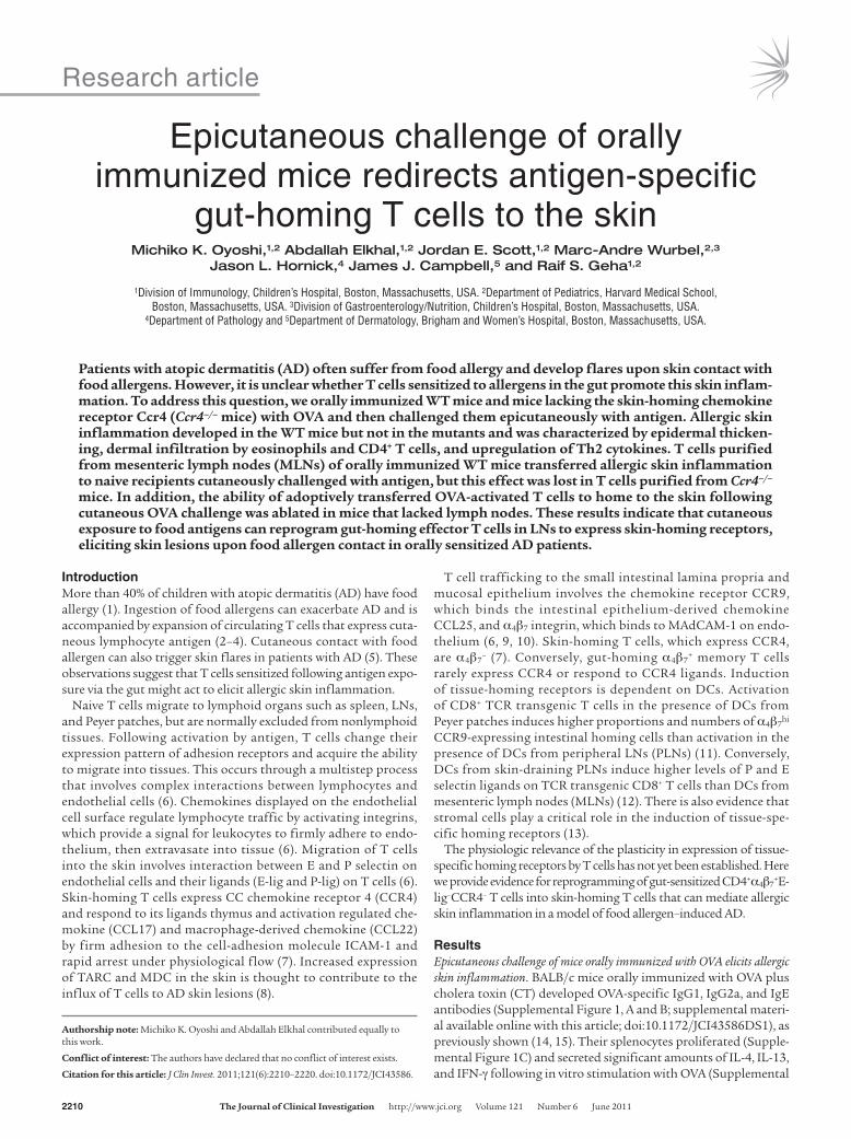
Epicutaneous (EC) challenge with OVA of mice orally immunized with OVA plus CT caused a significant increase in epidermal thick-ness (Figure 1, A and B), dermal infiltration by eosinophils and
CD4+ cells (Figure 1, A and C), and expression of mRNA encoding the Th2 cytokines Il4 and Il13, but not the Th1 cytokine Ifng, com-pared with mice subjected to EC challenge with saline (Figure 1D). EC challenge with OVA of BALB/c mice orally sham immunized with saline caused no significant skin changes compared with EC challenge with saline (Figure 1). These results indicate that orally immunized mice develop allergic skin inflammation in response to cutaneous challenge with antigen.
Allergic skin inflammation following cutaneous antigen challenge in orally immunized mice is dependent on CCR4. CCR4 is the major skin-homing receptor (7). Oral immunization with OVA induced robust serum levels of OVA-specific IgG1, IgG2a, and IgE antibodies in Ccr4–/– mice and genetically matched WT C57BL/6 controls (Supplemental
Figure 1Dermal infiltrates and cytokine expression in EC-challenged skin of orally immunized BALB/c mice. (A) Representative photomicrographs of H&E sections from EC-challenged skin of orally sensitized mice with Sal/CT or OVA/CT. Scale bars: 10 μm. Arrows point to eosinophils. (B) Epidermal thickness. (C) Numbers per HPF of eosinophils and CD4+ cells infiltrating the dermis. (D) Relative cytokine mRNA expression. The mean for saline-challenged skin of saline immunized mice was arbitrarily set at 1. Values represent individual mice, and bars represent mean (n = 6–7 mice per group). Two-tailed Student’s t test was used to determine statistical differences between each set of 2 groups. Similar results were obtained in 2 other independent experiments with 4 mice per group.
research article
2212 TheJournalofClinicalInvestigation http://www.jci.org Volume 121 Number 6 June 2011
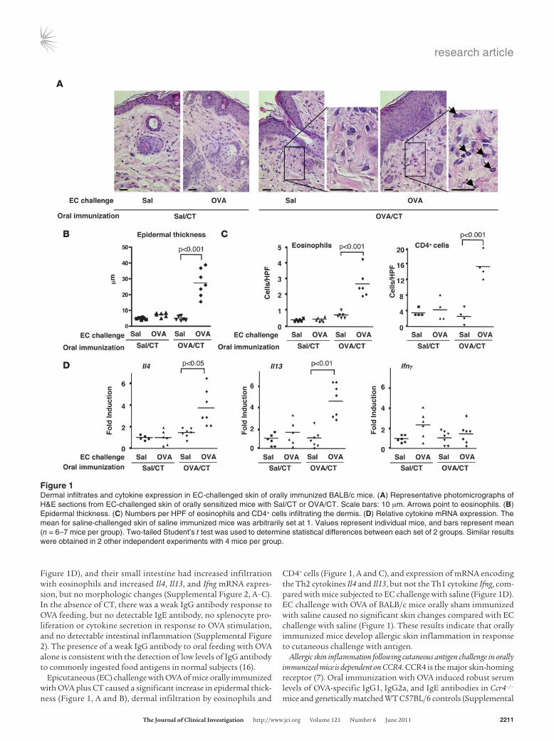
Figure 3), indicating that CCR4 is not required for the systemic immune response to oral immunization. Antigen challenge of back skin of orally immunized WT C57BL/6 mice with OVA caused a sig-nificant increase in epidermal thickness and dermal infiltration by eosinophils and CD4+ cells, and in Il4, Il13, and Ifng mRNA expres-sion, compared with challenge with saline (Figure 2). In contrast, Ccr4–/– mice orally immunized with OVA developed no detectable allergic skin inflammation following cutaneous antigen challenge.
Thus, elicitation of allergic skin inflammation following EC chal-lenge of orally sensitized mice is dependent on CCR4.
CD4+α4β7+E-lig–CCR4– T cells from MLNs of orally immunized mice
transfer allergic skin inflammation. To determine whether CD4+α4β7+
T cells can mediate allergic skin inflammation, MLN cells from orally immunized BALB/c mice were cultured with OVA for 4 days; then CD4+α4β7
+ T cells were purified by FACS sorting and 3 × 10⁶ were adoptively transferred by intravenous injection into naive WT
Figure 2Dermal infiltrates and cytokine expression in EC-challenged skin of orally immunized C57BL/6 WT and Ccr4–/– mice. (A) Epidermal thickness. (B) Numbers per HPF of eosinophils and CD4+ cells infiltrating the dermis. (C) Relative cytokine mRNA expression. The mean for saline-challenged skin of saline-immunized mice was arbitrarily set at 1. Values represent individual mice, and bars represent means (n = 5–6 mice per group). Two-tailed Student’s t test was used to determine statistical differences between each set of 2 groups. Similar results were obtained in another independent experiment with 4 mice per group.
research article
TheJournalofClinicalInvestigation http://www.jci.org Volume 121 Number 6 June 2011 2213
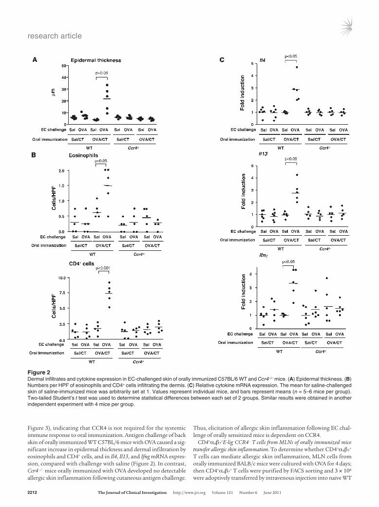
BALB/c recipients. Recipients were EC challenged on the back with OVA on days 0 and 3 of the transfer, and skin was examined 7 days later. Adoptively transferred cells were more than 95% CD4+ (data not shown) and more than 95% α4β7
+ and contained less than 0.1% E-lig+ or CCR4+ cells (Figure 3A). RT-PCR analysis revealed no
detectable expression of Ccr4 mRNA after 25 cycles of amplifica-tion of 100 ng cDNA, whereas Ccr4 mRNA was readily detected in splenocytes (Figure 3B). Under these conditions, the RT-PCR assay would have detected fewer than 10 CCR4+ cells in 3 × 10⁶ Ccr4–/– T cells (Supplemental Figure 4).
Figure 3Adoptive transfer of allergic skin inflammation by CD4+α4β7
+ T cells from MLNs of orally immunized BALB/c mice with OVA. (A) FACS analysis of α4β7, E-lig, and CCR4 expression by OVA-stimulated purified CD4+α4β7
+ MLN T cells used in adoptive transfer (representative of 3 experi-ments). Numbers indicate the percentage of cells in each quadrant in purified CD4+α4β7
+ cells. (B) RT-PCR analysis of integrin β7 (Itgb7) and Ccr4 expression in adoptively transferred CD4+α4β7
+ MLN T cells. Spleen cDNA was used as a positive control. β2 microglobulin (β2m) was used as a housekeeping gene. RNA from purified MLN cells used as a control for contamination by genomic DNA gave no detectable signal. Similar data were obtained in 2 other independent experiments. (C) Epidermal thickness. (D) Numbers per HPF of eosinophils and CD4+ cells infiltrating the dermis in OVA-challenged skin of recipients of CD4+α4β7
+ T cells from MLNs of donors orally immunized with saline or OVA. (E) Relative cytokine mRNA expression in OVA-challenged skin. The mean for OVA-challenged skin of recipients of CD4+α4β7
+ T cells from MLNs of donors orally immunized with saline was arbitrarily set at 1. Values represent individual mice, and mean bars are shown (n = 4–7 mice per group). Two-tailed Student’s t test was used to determine statistical differences between each set of 2 groups. Similar results were obtained in 2 other independent experiments with 3 mice per group.
research article
2214 TheJournalofClinicalInvestigation http://www.jci.org Volume 121 Number 6 June 2011
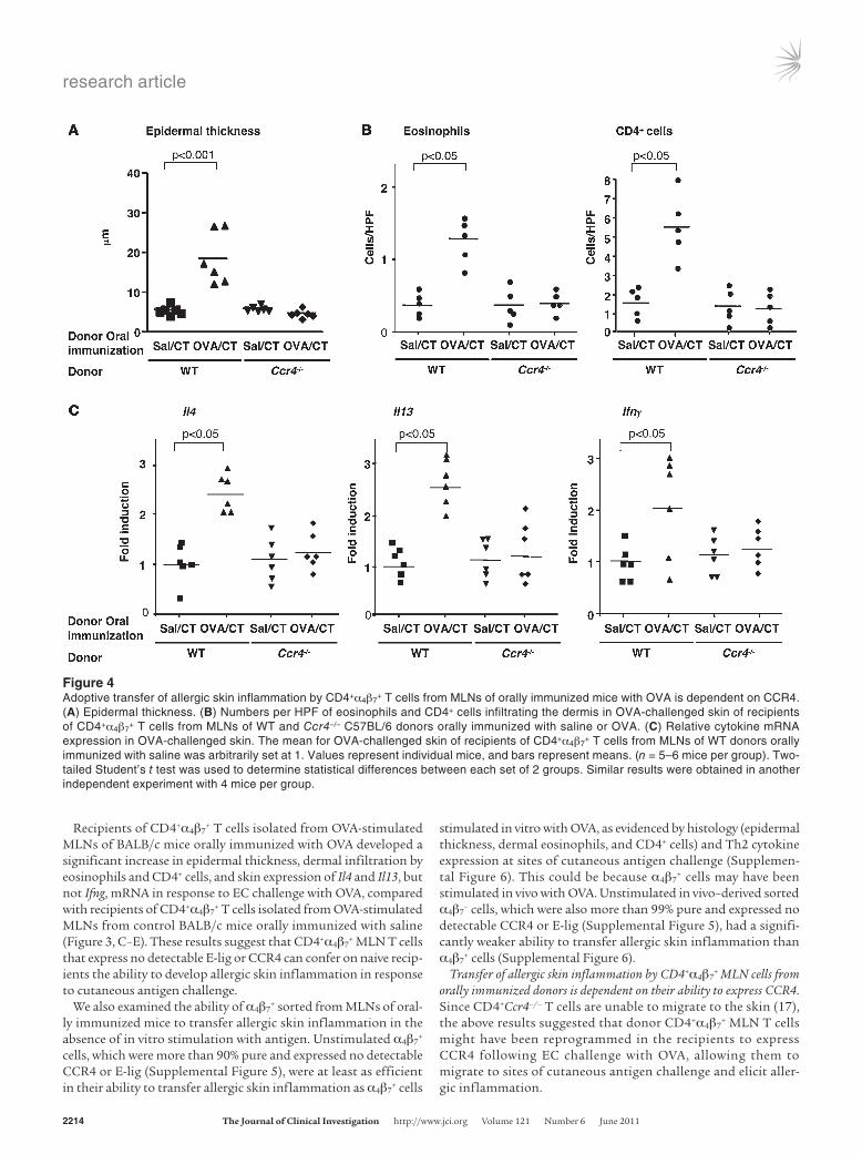
Recipients of CD4+α4β7+ T cells isolated from OVA-stimulated
MLNs of BALB/c mice orally immunized with OVA developed a significant increase in epidermal thickness, dermal infiltration by eosinophils and CD4+ cells, and skin expression of Il4 and Il13, but not Ifng, mRNA in response to EC challenge with OVA, compared with recipients of CD4+α4β7
+ T cells isolated from OVA-stimulated MLNs from control BALB/c mice orally immunized with saline (Figure 3, C–E). These results suggest that CD4+α4β7
+ MLN T cells that express no detectable E-lig or CCR4 can confer on naive recip-ients the ability to develop allergic skin inflammation in response to cutaneous antigen challenge.
We also examined the ability of α4β7+ sorted from MLNs of oral-
ly immunized mice to transfer allergic skin inflammation in the absence of in vitro stimulation with antigen. Unstimulated α4β7
+ cells, which were more than 90% pure and expressed no detectable CCR4 or E-lig (Supplemental Figure 5), were at least as efficient in their ability to transfer allergic skin inflammation as α4β7
+ cells
stimulated in vitro with OVA, as evidenced by histology (epidermal thickness, dermal eosinophils, and CD4+ cells) and Th2 cytokine expression at sites of cutaneous antigen challenge (Supplemen-tal Figure 6). This could be because α4β7
+ cells may have been stimulated in vivo with OVA. Unstimulated in vivo–derived sorted α4β7
– cells, which were also more than 99% pure and expressed no detectable CCR4 or E-lig (Supplemental Figure 5), had a signifi-cantly weaker ability to transfer allergic skin inflammation than α4β7
+ cells (Supplemental Figure 6).Transfer of allergic skin inflammation by CD4+α4β7
+ MLN cells from orally immunized donors is dependent on their ability to express CCR4. Since CD4+Ccr4–/– T cells are unable to migrate to the skin (17), the above results suggested that donor CD4+α4β7
+ MLN T cells might have been reprogrammed in the recipients to express CCR4 following EC challenge with OVA, allowing them to migrate to sites of cutaneous antigen challenge and elicit aller-gic inflammation.
Figure 4Adoptive transfer of allergic skin inflammation by CD4+α4β7
+ T cells from MLNs of orally immunized mice with OVA is dependent on CCR4. (A) Epidermal thickness. (B) Numbers per HPF of eosinophils and CD4+ cells infiltrating the dermis in OVA-challenged skin of recipients of CD4+α4β7
+ T cells from MLNs of WT and Ccr4–/– C57BL/6 donors orally immunized with saline or OVA. (C) Relative cytokine mRNA expression in OVA-challenged skin. The mean for OVA-challenged skin of recipients of CD4+α4β7
+ T cells from MLNs of WT donors orally immunized with saline was arbitrarily set at 1. Values represent individual mice, and bars represent means. (n = 5–6 mice per group). Two-tailed Student’s t test was used to determine statistical differences between each set of 2 groups. Similar results were obtained in another independent experiment with 4 mice per group.
research article
TheJournalofClinicalInvestigation http://www.jci.org Volume 121 Number 6 June 2011 2215
To determine whether the ability to express CCR4 is required for the transfer of allergic skin inflammation by CD4+α4β7
+ cells, CD4+α4β7
+ T cells were purified from OVA-stimulated MLNs of orally immunized Ccr4–/– mice and C57BL/6 WT controls and administered to genetically matched WT recipients. The trans-ferred cells were more than 95% CD4+ (data not shown), more than 90% α4β7
+, less than 0.1% E-lig+, less than 0.2% CCR4+, and showed no detectable Ccr4 mRNA by RT-PCR (Supplemental Fig-ure 7). OVA-challenged back skin from recipients of CD4+α4β7
+ T cells isolated from OVA-stimulated MLNs of WT donors orally immunized with OVA exhibited significant increase in epidermal thickness, dermal infiltration by eosinophils and CD4+ cells, and significant upregulation of Il4, Il13, and Ifng mRNA expression compared with OVA-challenged skin from recipients of CD4+α4β7
+ cells from WT donors orally immunized with saline (Figure 4). In
contrast, OVA-challenged back skin of recipients of CD4+α4β7+ T
cells isolated from OVA-stimulated MLNs of Ccr4–/– mice orally immunized with OVA failed to develop allergic inflammation. This result indicates that the ability to express CCR4 is essential for the transfer of allergic skin inflammation by antigen-stimu-lated CD4+α4β7
+ cells from orally immunized mice.CD4+α4β7
+ effector T cells migrate to antigen-challenged skin sites. To directly determine whether CD4+α4β7
+ cells can migrate to cutane-ous sites of antigen exposure, CD4+α4β7
+ cells were purified from MLNs of DO11.10 mice that had been fed with OVA plus CT weekly for 3 weeks, and transferred to naive BALB/c recipients. Recipient ears were challenged with OVA plus CT or saline plus CT control on days 0 and 3 after transfer and analyzed on day 7 for CD4+ cells that expressed the donor-derived TCR clonotype, KJ1.26. FACS analysis revealed that more than 85% of the transferred cells were
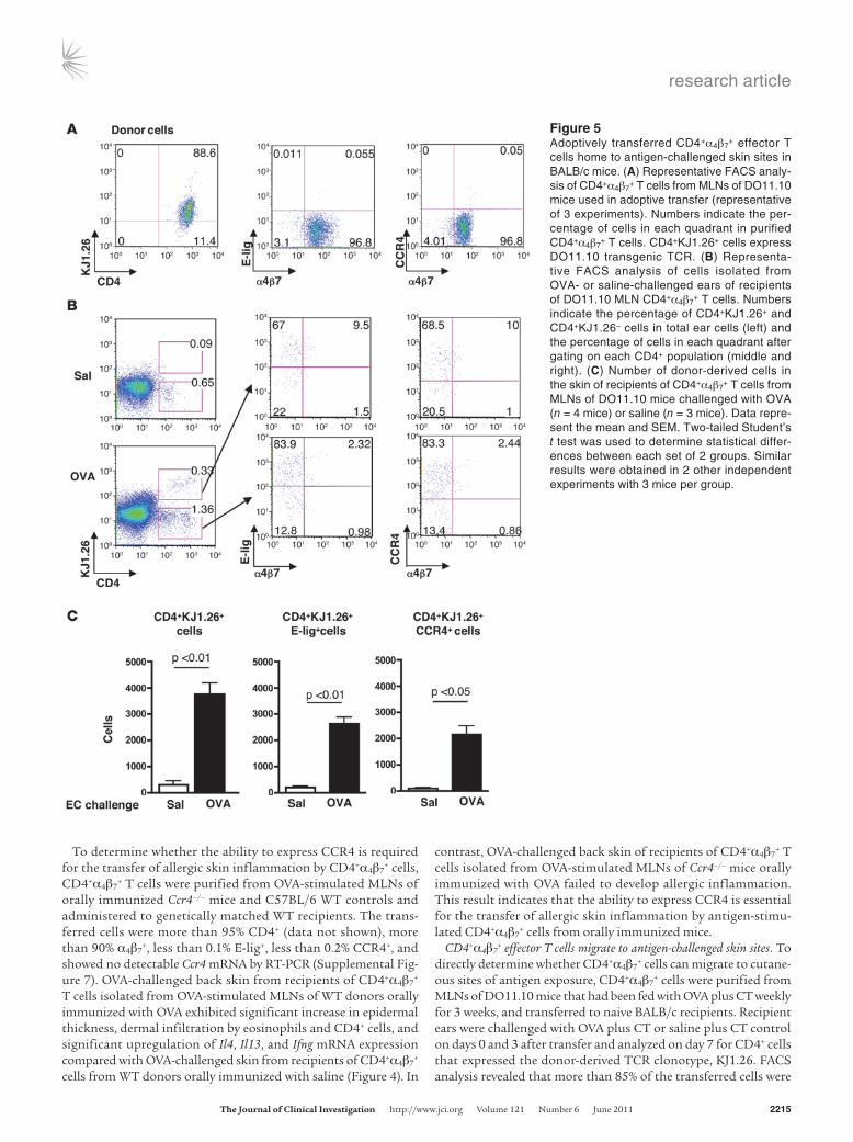
Figure 5Adoptively transferred CD4+α4β7
+ effector T cells home to antigen-challenged skin sites in BALB/c mice. (A) Representative FACS analy-sis of CD4+α4β7
+ T cells from MLNs of DO11.10 mice used in adoptive transfer (representative of 3 experiments). Numbers indicate the per-centage of cells in each quadrant in purified CD4+α4β7
+ T cells. CD4+KJ1.26+ cells express DO11.10 transgenic TCR. (B) Representa-tive FACS analysis of cells isolated from OVA- or saline-challenged ears of recipients of DO11.10 MLN CD4+α4β7
+ T cells. Numbers indicate the percentage of CD4+KJ1.26+ and CD4+KJ1.26– cells in total ear cells (left) and the percentage of cells in each quadrant after gating on each CD4+ population (middle and right). (C) Number of donor-derived cells in the skin of recipients of CD4+α4β7
+ T cells from MLNs of DO11.10 mice challenged with OVA (n = 4 mice) or saline (n = 3 mice). Data repre-sent the mean and SEM. Two-tailed Student’s t test was used to determine statistical differ-ences between each set of 2 groups. Similar results were obtained in 2 other independent experiments with 3 mice per group.
research article
2216 TheJournalofClinicalInvestigation http://www.jci.org Volume 121 Number 6 June 2011
research article
TheJournalofClinicalInvestigation http://www.jci.org Volume 121 Number 6 June 2011 2217
KJ1.26+, more than 95% α4β7+, less than 0.1% E-lig+, and less than
0.1% CCR4+ (Figure 5A). CD4+KJ1.26+ cells were virtually absent from saline-challenged ears, but were readily detectable in OVA-challenged ears (Figure 5B). The majority (69.33% ± 2.49%, n = 4) of CD4+KJ1.26+ cells in OVA-challenged ears expressed E-lig without α4β7, a small fraction (9.8% ± 0.17%) expressed E-lig together with α4β7, and very few (2.1% ± 0.35%) expressed α4β7 but not E-lig. The expression pattern of CCR4 on donor-derived CD4+KJ1.26+ cells in OVA-challenged skin was similar to that of E-lig: 72.45% ± 2.28% CCR4+α4β7
–, 10.40% ± 0.23% CCR4+α4β7+, and 1.51% ± 0.30%
CCR4–α4β7+ (n = 4). There was marked and significant accumula-
tion of CD4+KJ1.26+, CD4+KJ1.26+E-lig+, and CD4+KJ1.26+CCR4+ cells in OVA-challenged ears compared with saline-challenged ears (Figure 5C). No CD4+KJ1.26+ cells were detected in ears of naive mice that received no donor cells (data not shown). OVA challenge resulted in an approximately 2-fold increase in recipi-ent-derived CD4+KJ1.26– cells compared with saline challenge (1.24% ± 0.13% vs. 0.61% ± 0.05%, P < 0.05). E-lig and CCR4 were expressed on the majority of recipient-derived cells in OVA-chal-lenged ears (81.83% ± 1.18% and 80.76% ± 1.47% respectively, n = 4); very few of these cells (4.09% ± 0.46%) expressed α4β7. There was comparable expression of these homing receptors on recipient-derived cells in saline-challenged ears (data not shown). Analysis of cervical and axillary draining LNs (DLNs) revealed a signifi-cant increase in CD4+KJ1.26+ cells in DLNs of OVA-challenged ears compared with DLNs of saline-challenged ears (Supple-mental Figure 8A). The percentages of E-lig+ and CCR4+ cells among donor-derived CD4+KJ1.26+ cells in DLNs of OVA-chal-lenged ears (8.13% ± 0.16% and 11.74% ± 0.34% respectively, n = 4) were comparable to their percentages among recipient-derived CD4+KJ1.26– cells in the same ears (9.42% ± 0.42% and 11.8% ± 0.50% respectively) (Supplemental Figure 8A). There was drasti-cally more accumulation of CD4+KJ1.26+, CD4+KJ1.26+E-lig+, and CD4+KJ1.26+CCR4+ cells in DLNs of OVA-challenged ears
compared with DLNs of saline-challenged ears (Supplemental Figure 8B), indicating antigen-driven accumulation of the adop-tively transferred transgenic cells in skin DLNs and acquisition of skin-homing receptors by a fraction of these cells. Very few donor-derived cells in DLNs of OVA-challenged ears retained expression of α4β7 (3.17% ± 0.03%), indicating that they have downregu-lated α4β7 expression. Very few recipient-derived cells in these DLNs expressed α4β7 (1.59% ± 0.06%). Analysis of CD4+KJ1.26+ donor cells present in DLNs of saline-challenged skin revealed that they lost α4β7 expression and that only a small percentage expressed E-lig and CCR4 (0.52% ± 0.30% and 1.55% ± 0.66% respectively) (Supplemental Figure 8A). This could reflect the fact that these cells express, in addition to the OVA-specific transgenic αβTCR, TCRs consisting of the transgenic TCR-β chain paired with endogenous TCR-α chain, which could recognize antigens and/or superantigens introduced through saline-challenged skin. Alternatively, these cells may have responded to skin-homing signals present in DLNs without any antigen stimulation. These results suggest that expression of skin-homing receptors on donor T cells found in skin DLNs is primarily dependent on cutaneous exposure to antigen.
In contrast to the markedly higher (~12-fold) accumulation of donor T cells in skin EC challenged with OVA versus saline (Figure 5C), there were comparable numbers of donor T cells in the gut and blood of recipient EC challenged with OVA versus saline (Supplemental Figure 9A). Also in contrast to the drastically higher (~50-fold) accumulation of donor T cells in DLNs of skin EC challenged with OVA versus saline (Supplemental Figure 8B), there was only a modest (~2-fold) increase in the number of donor T cells in MLNs and spleens in recipient EC challenged with OVA versus saline (Supplemental Figure 9B).
LNs are essential for in vivo redirection of CD4+α4β7+ T cells to the
skin. Circulating T cells encounter cutaneously introduced anti-gen in DLNs. We tested the hypothesis that reprogramming of CD4+α4β7
+ cells into E-lig+CCR4+ skin-homing T cells in vivo occurs in LNs. CD4+α4β7
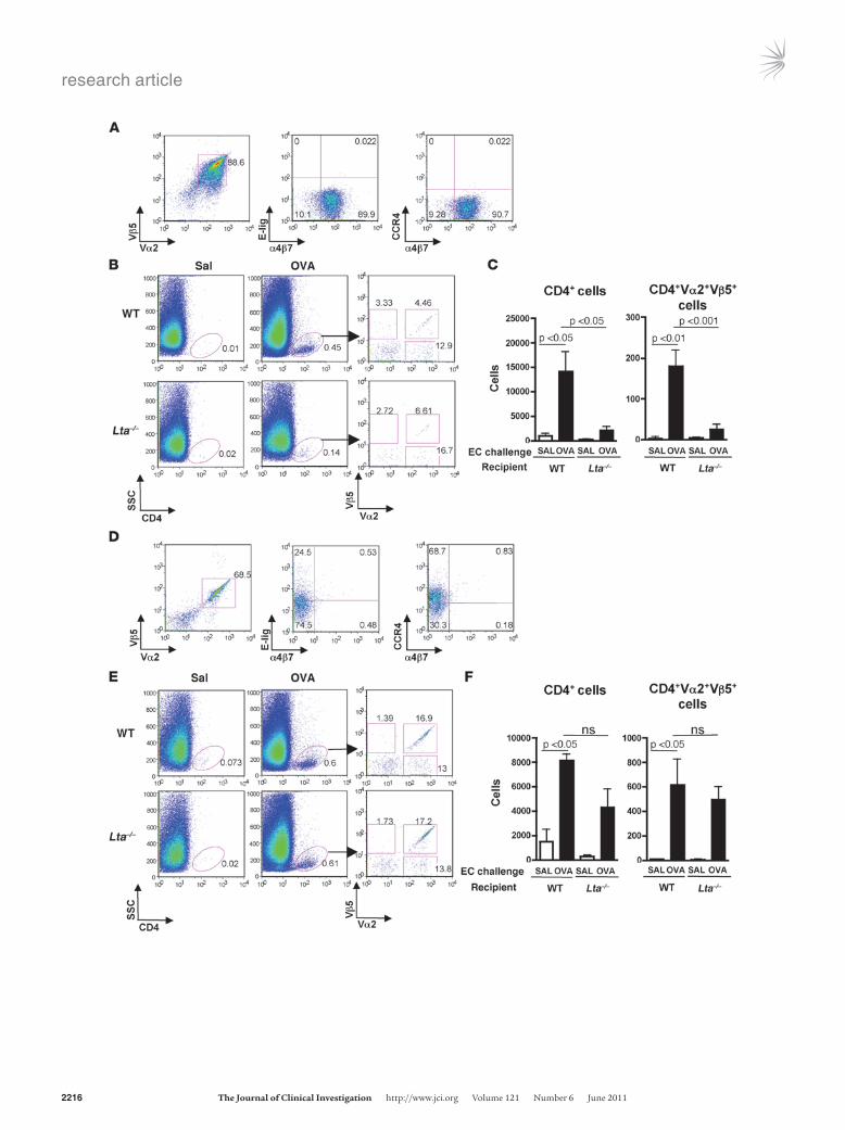
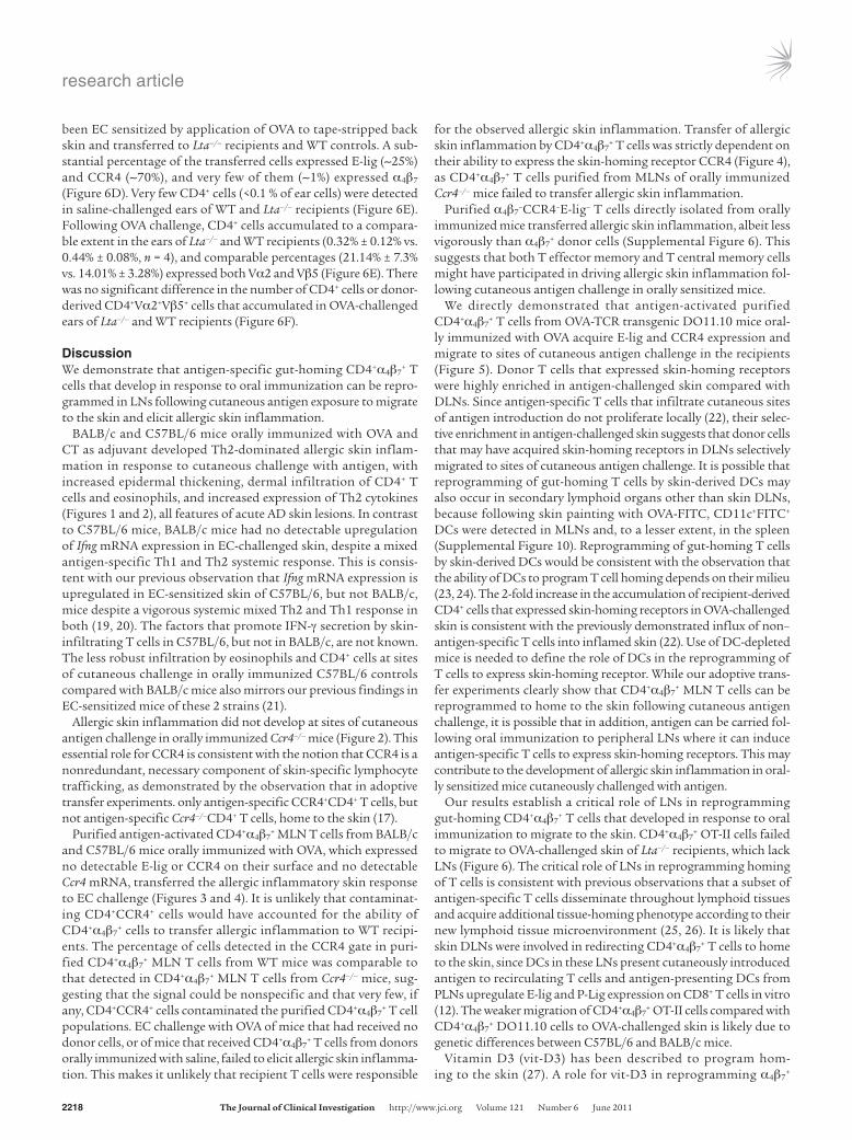
+ T cells isolated from MLNs of OVA-fed TCR-OVA transgenic OT-II mice were adoptively transferred into Lta–/– mice, which lack LNs (18), and C57BL/6 WT controls. Recipient ears were challenged with antigen and examined for the presence of donor-derived CD4+ cells that express both Vα2 and Vβ5 TCR chains used by the OT-II transgenic TCR. FACS analy-sis revealed that the transferred cells were more than 95% CD4+ (data not shown), approximately 90% Vα2+Vβ5+, approximately 90% α4β7
+, less than 0.05% E-lig+, and less than 0.05% CCR4+ (Fig-ure 6A). Very few CD4+ cells (<0.1% of ear cells) were detected in saline-challenged ears of Lta–/– and WT recipients (Figure 6B). The percentage of CD4+ cells in OVA-challenged ears was significantly less in Lta–/– recipients than in WT recipients (0.15% ± 0.05% ver-sus 0.46% ± 0.12% n = 7, P < 0.05), but comparable percentages of them (6.65% ± 1.99% vs. 9.14% ± 1.84%) expressed both Vα2 and Vβ5 (Figure 6B). The numbers of CD4+ cells and donor-derived CD4+Vα2+Vβ5+ cells that accumulated in OVA-challenged ears were significantly reduced in Lta–/– recipients compared with in WT recipients (Figure 6C).
The failure of CD4+α4β7+ effector T cells to home to antigen-chal-
lenged skin of Lta–/– recipients was not due to a general problem in T cell trafficking to the skin. This was demonstrated by examining the trafficking of activated CD4+ effector T cells derived from skin DLNs of WT OT-II mice to the skin of Lta–/– mice. CD4+ T cells were purified from axillary and inguinal LN cells of OT-II mice that had
Figure 6Adoptively transferred CD4+α4β7
+ effector T cells fail to home to antigen-challenged skin sites of Lta–/– mice. (A) FACS analysis of CD4+α4β7
+ T cells from MLNs of OT-II mice used in adoptive transfer (representative of 3 experiments). (B) Representative FACS analysis of cells isolated from saline-challenged ears (n = 6 mice) and OVA-challenged ears (n = 7 mice) of recipients of MLN cells. Ears from each mouse were pooled. (C) Number of CD4+ cells and donor-derived CD4+Vα2+Vβ5+ cells in the challenged ears of recipients of MLN cells. n = 6 and 7 mice, respectively, in saline-challenged versus OVA-chal-lenged groups. (D) Representative FACS analysis of CD4+ T cells from PLNs of OT-II mice used in adoptive transfer (representative of 3 experiments). (E) Representative FACS analysis of cells isolated from saline-challenged ears and OVA-challenged ears of recipients of PLN cells. Ears from each mouse were pooled. (F) Number of CD4+ cells and of donor-derived CD4+Vα2+Vβ5+ cells in the challenged ears of recipients of PLN cells. n = 3 and 4 mice, respectively, in saline-chal-lenged versus OVA-challenged groups. Numbers in A and D indicate the percentage of Vα2+Vβ5+ OT-II transgenic TCR-expressing cells (left) and the percentage of cells in each quadrant (middle and right) in purified CD4+α4β7
+ T cells. Numbers in B and E indicate the percent-age of CD4+ cells in total ear cells (left and middle) and the percentage of Vα2+, Vβ5+, and Vα2+Vβ5+ cells after gating on CD4+ cells (right). Data in C and F represent the mean and SEM. One-way ANOVA was used to determine statistical differences between groups. For each of the MLN and PLN cell transfer experiments, similar results were obtained in another independent experiment with 3 mice per group.
research article
2218 TheJournalofClinicalInvestigation http://www.jci.org Volume 121 Number 6 June 2011
been EC sensitized by application of OVA to tape-stripped back skin and transferred to Lta–/– recipients and WT controls. A sub-stantial percentage of the transferred cells expressed E-lig (~25%) and CCR4 (~70%), and very few of them (~1%) expressed α4β7 (Figure 6D). Very few CD4+ cells (<0.1 % of ear cells) were detected in saline-challenged ears of WT and Lta–/– recipients (Figure 6E). Following OVA challenge, CD4+ cells accumulated to a compara-ble extent in the ears of Lta–/– and WT recipients (0.32% ± 0.12% vs. 0.44% ± 0.08%, n = 4), and comparable percentages (21.14% ± 7.3% vs. 14.01% ± 3.28%) expressed both Vα2 and Vβ5 (Figure 6E). There was no significant difference in the number of CD4+ cells or donor-derived CD4+Vα2+Vβ5+ cells that accumulated in OVA-challenged ears of Lta–/– and WT recipients (Figure 6F).
DiscussionWe demonstrate that antigen-specific gut-homing CD4+α4β7
+ T cells that develop in response to oral immunization can be repro-grammed in LNs following cutaneous antigen exposure to migrate to the skin and elicit allergic skin inflammation.
BALB/c and C57BL/6 mice orally immunized with OVA and CT as adjuvant developed Th2-dominated allergic skin inflam-mation in response to cutaneous challenge with antigen, with increased epidermal thickening, dermal infiltration of CD4+ T cells and eosinophils, and increased expression of Th2 cytokines (Figures 1 and 2), all features of acute AD skin lesions. In contrast to C57BL/6 mice, BALB/c mice had no detectable upregulation of Ifng mRNA expression in EC-challenged skin, despite a mixed antigen-specific Th1 and Th2 systemic response. This is consis-tent with our previous observation that Ifng mRNA expression is upregulated in EC-sensitized skin of C57BL/6, but not BALB/c, mice despite a vigorous systemic mixed Th2 and Th1 response in both (19, 20). The factors that promote IFN-γ secretion by skin-infiltrating T cells in C57BL/6, but not in BALB/c, are not known. The less robust infiltration by eosinophils and CD4+ cells at sites of cutaneous challenge in orally immunized C57BL/6 controls compared with BALB/c mice also mirrors our previous findings in EC-sensitized mice of these 2 strains (21).
Allergic skin inflammation did not develop at sites of cutaneous antigen challenge in orally immunized Ccr4–/– mice (Figure 2). This essential role for CCR4 is consistent with the notion that CCR4 is a nonredundant, necessary component of skin-specific lymphocyte trafficking, as demonstrated by the observation that in adoptive transfer experiments. only antigen-specific CCR4+CD4+ T cells, but not antigen-specific Ccr4–/–CD4+ T cells, home to the skin (17).
Purified antigen-activated CD4+α4β7+ MLN T cells from BALB/c
and C57BL/6 mice orally immunized with OVA, which expressed no detectable E-lig or CCR4 on their surface and no detectable Ccr4 mRNA, transferred the allergic inflammatory skin response to EC challenge (Figures 3 and 4). It is unlikely that contaminat-ing CD4+CCR4+ cells would have accounted for the ability of CD4+α4β7
+ cells to transfer allergic inflammation to WT recipi-ents. The percentage of cells detected in the CCR4 gate in puri-fied CD4+α4β7
+ MLN T cells from WT mice was comparable to that detected in CD4+α4β7
+ MLN T cells from Ccr4–/– mice, sug-gesting that the signal could be nonspecific and that very few, if any, CD4+CCR4+ cells contaminated the purified CD4+α4β7
+ T cell populations. EC challenge with OVA of mice that had received no donor cells, or of mice that received CD4+α4β7
+ T cells from donors orally immunized with saline, failed to elicit allergic skin inflamma-tion. This makes it unlikely that recipient T cells were responsible
for the observed allergic skin inflammation. Transfer of allergic skin inflammation by CD4+α4β7
+ T cells was strictly dependent on their ability to express the skin-homing receptor CCR4 (Figure 4), as CD4+α4β7
+ T cells purified from MLNs of orally immunized Ccr4–/– mice failed to transfer allergic skin inflammation.
Purified α4β7–CCR4–E-lig– T cells directly isolated from orally
immunized mice transferred allergic skin inflammation, albeit less vigorously than α4β7
+ donor cells (Supplemental Figure 6). This suggests that both T effector memory and T central memory cells might have participated in driving allergic skin inflammation fol-lowing cutaneous antigen challenge in orally sensitized mice.
We directly demonstrated that antigen-activated purified CD4+α4β7
+ T cells from OVA-TCR transgenic DO11.10 mice oral-ly immunized with OVA acquire E-lig and CCR4 expression and migrate to sites of cutaneous antigen challenge in the recipients (Figure 5). Donor T cells that expressed skin-homing receptors were highly enriched in antigen-challenged skin compared with DLNs. Since antigen-specific T cells that infiltrate cutaneous sites of antigen introduction do not proliferate locally (22), their selec-tive enrichment in antigen-challenged skin suggests that donor cells that may have acquired skin-homing receptors in DLNs selectively migrated to sites of cutaneous antigen challenge. It is possible that reprogramming of gut-homing T cells by skin-derived DCs may also occur in secondary lymphoid organs other than skin DLNs, because following skin painting with OVA-FITC, CD11c+FITC+ DCs were detected in MLNs and, to a lesser extent, in the spleen (Supplemental Figure 10). Reprogramming of gut-homing T cells by skin-derived DCs would be consistent with the observation that the ability of DCs to program T cell homing depends on their milieu (23, 24). The 2-fold increase in the accumulation of recipient-derived CD4+ cells that expressed skin-homing receptors in OVA-challenged skin is consistent with the previously demonstrated influx of non–antigen-specific T cells into inflamed skin (22). Use of DC-depleted mice is needed to define the role of DCs in the reprogramming of T cells to express skin-homing receptor. While our adoptive trans-fer experiments clearly show that CD4+α4β7
+ MLN T cells can be reprogrammed to home to the skin following cutaneous antigen challenge, it is possible that in addition, antigen can be carried fol-lowing oral immunization to peripheral LNs where it can induce antigen-specific T cells to express skin-homing receptors. This may contribute to the development of allergic skin inflammation in oral-ly sensitized mice cutaneously challenged with antigen.
Our results establish a critical role of LNs in reprogramming gut-homing CD4+α4β7
+ T cells that developed in response to oral immunization to migrate to the skin. CD4+α4β7
+ OT-II cells failed to migrate to OVA-challenged skin of Lta–/– recipients, which lack LNs (Figure 6). The critical role of LNs in reprogramming homing of T cells is consistent with previous observations that a subset of antigen-specific T cells disseminate throughout lymphoid tissues and acquire additional tissue-homing phenotype according to their new lymphoid tissue microenvironment (25, 26). It is likely that skin DLNs were involved in redirecting CD4+α4β7
+ T cells to home to the skin, since DCs in these LNs present cutaneously introduced antigen to recirculating T cells and antigen-presenting DCs from PLNs upregulate E-lig and P-Lig expression on CD8+ T cells in vitro (12). The weaker migration of CD4+α4β7
+ OT-II cells compared with CD4+α4β7
+ DO11.10 cells to OVA-challenged skin is likely due to genetic differences between C57BL/6 and BALB/c mice.
Vitamin D3 (vit-D3) has been described to program hom-ing to the skin (27). A role for vit-D3 in reprogramming α4β7
+
research article
TheJournalofClinicalInvestigation http://www.jci.org Volume 121 Number 6 June 2011 2219
cells to home to the skin was supported by the observation that E-lig+ donor T cells isolated from DLNs of OVA-sensitized skin upregulated mRNA expression of the vit-D3–metabolizing enzymes 25-hydroxylase Cyp27a1 and 1-hydroxylase Cyp27b1 com-pared with E-lig– cells isolated from the same DLNs. There was no detectable signal for Cyp2r1 in either E-lig+ or E-lig– cells (Supple-mental Figure 11A). In addition, CD11c+ DCs isolated from skin DLNs of unsensitized mice expressed significantly higher levels of all 3 enzymes compared with CD11c+ DCs isolated from MLNs from the same mice (Supplemental Figure 11B). This is consis-tent with a role for DC-derived vit-D3 in reprogramming T cells to express skin-homing receptors. Furthermore, tape stripping significantly upregulated Cyp27a1 and Cyp2r1 mRNA expression in CD11c+ DCs isolated from the skin 6 hours after mechanical injury, compared with CD11c+ DCs isolated from unmanipulated skin (Supplemental Figure 11C). This suggests that upregulation of vit-D3–metabolizing enzymes following mechanical injury, such as inflicted by scratching in AD patients, may contribute to the ability of DCs that carry cutaneously introduced antigens to program T cells to express skin-homing receptors.
The resetting of the migratory program of gut-homing antigen-specific T cells following cutaneous challenge with antigen dem-onstrated in this study may explain the flare-up of skin lesions fol-lowing cutaneous contact with food allergens in orally sensitized patients with AD.
MethodsMice. WT mice (Jackson Laboratory) and Ccr4–/– mice provided by A.E. Proudfoot (Merck Serono Geneva Research Center, Geneva, Switzerland) (28, 29) were kept in a specific pathogen–free environment. All animal studies were approved by the Animal Care and Use Committee at Chil-dren’s Hospital, Boston.
Oral sensitization and EC challenge. 4- to 6-week-old mice were fed intragas-trically once a week for 7 weeks (total, 8 times) 5 mg OVA and 10 μg CT in 200 μl saline or with CT alone in saline. Shaved-back skin of anesthetized mice was tape stripped 6 times, then challenged with 100 μg OVA in 100 μl normal saline, or 100 μl normal saline as previously described (19). Three days later, the procedure was repeated, and the mice were studied at day 7.
Serum antibody and splenocyte proliferation and cytokine production. These assays were performed as previously described (19).
Histological analysis, quantitative RT-PCR for cytokines, and chemokines in skin. All assays were performed as previously described (30). Cells were counted in 10–20 high-power fields (HPFs) at ×400 magnification by a blinded observer. Total mRNA was extracted from back skin that included epider-mis and dermis and thus contained keratinocytes, fibroblasts, epidermal/
dermal DCs, mast cells, eosinophils, and CD4+ cells. For some assays, the number of samples analyzed was less than that of the mice due to technical limitations or accidental loss.
Adoptive T cell transfer. Single-cell suspensions from MLNs were cultured in complete DMEM with 50 μg/ml OVA for 4 days. Cells were then collect-ed, purified with CD4 MicroBeads (Miltenyi Biotec), incubated with PE-conjugated anti-α4β7 mAb (anti-LPAM-1; BD Biosciences) for 30 minutes on ice, and washed once; then α4β7
+ cells were sorted by FACS and isotype control mAb was used to set up the gate. Sorted cells were administered intravenously at 3 × 10⁶ cells/mouse to naive 8-week-old WT recipients, which were EC challenged with 100 μg OVA on days 0 and 3 of the adoptive transfer. Challenged skin sites were examined 7 days later.
Antibodies and flow cytometry. Antibodies for cell-surface staining were purchased from BD Biosciences. E-lig staining was performed as previ-ously described (29). CCL22-Fc fusion protein was provided by D.J. Camp-bell (Benaroya Research Institute, Seattle, Washington, USA) and used for surface CCR4 staining as previously described (31). Flow cytometry was performed on FACSCanto (BD), and data were analyzed using FlowJo soft-ware (Tree Star Inc.).
Semiquantitative RT-PCR. Total RNA prepared using RNAqueous Extraction Kit (Ambion Inc.) was treated with DNase (Ambion Inc.), and cDNA was syn-thesized using an I-Script cDNA synthesis kit (Bio-Rad Laboratories). Prim-ers were as follows: integrin β7 (Itgβ7), 5′-CCTACGACTCTGGGCTCTTG-3′ and 5′-ACAGGTCAGCCTCAGAGCAT-3′; Ccr4 and for β2 microglobulin (β2m), as previously described (19, 32). PCR cycling conditions were 94°C for 1 minute, 56°C for 1 minute, and 68°C for 1 minute for 25 cycles.
Statistics. Statistical significance was determined using GraphPad Prism, version 4.0a. Statistical differences were determined by 2-tailed Student’s t test (between 2 groups) and a 1-way ANOVA (among multiple groups). P < 0.05 was considered to indicate statistical significance.
AcknowledgmentsWe thank Daniel J. Campbell for his gift of CCL22-IgG; Hans C. Oettgen and Michel J. Massaad for reading the manuscript; and Christopher Lewis for technical assistance. This work was sup-ported by NIH/NIAID grants AR-047417, N01-AI-40030 (to R.S. Geha), and AI-046784 (to J.J. Campbell).
Received for publication May 3, 2010, and accepted in revised form March 2, 2011.
Address correspondence to: Raif Geha, Division of Immunology, Children’s Hospital, Boston, Massachusetts 02115, USA. Phone: 617.919.2482; Fax: 617.730.0528; E-mail: [email protected].
1. Sicherer SH, Sampson HA. Food hypersensitivity and atopic dermatitis: pathophysiology, epidemi-ology, diagnosis, and management. J Allergy Clin Immunol. 1999;104(3 pt 2):S114–S122.
2. Sampson HA. Role of immediate food hypersen-sitivity in the pathogenesis of atopic dermatitis. J Allergy Clin Immunol. 1983;71(5):473–480.
3. Abernathy-Carver KJ, Sampson HA, Picker LJ, Leung DY. Milk-induced eczema is associated with the expansion of T cells expressing cutaneous lym-phocyte antigen. J Clin Invest. 1995;95(2):913–918.
4. Niggemann B. Role of oral food challenges in the diagnostic work-up of food allergy in atop-ic eczema dermatitis syndrome. Allergy. 2004; 59(suppl 78):32–34.
5. Darsow U, et al. The prevalence of positive reactions in the atopy patch test with aeroallergens and food allergens in subjects with atopic eczema: a European
multicenter study. Allergy. 2004;59(12):1318–1325. 6. Butcher EC, Williams M, Youngman K, Rott L,
Briskin M. Lymphocyte trafficking and regional immunity. Adv Immunol. 1999;72:209–253.
7. Campbell JJ, et al. The chemokine receptor CCR4 in vascular recognition by cutaneous but not intestinal memory T cells. Nature. 1999; 400(6746):776–780.
8. Homey B, Steinhoff M, Ruzicka T, Leung DY. Cytokines and chemokines orchestrate atopic skin inflammation. J Allergy Clin Immunol. 2006; 118(1):178–189.
9. Hamann A, Andrew DP, Jablonski-Westrich D, Hol-zmann B, Butcher EC. Role of alpha 4-integrins in lymphocyte homing to mucosal tissues in vivo. J Immunol. 1994;152(7):3282–3293.
10. Svensson M, et al. CCL25 mediates the localization of recently activated CD8alphabeta(+) lympho-
cytes to the small-intestinal mucosa. J Clin Invest. 2002;110(8):1113–1121.
11. Mora JR, et al. Selective imprinting of gut-hom-ing T cells by Peyer’s patch dendritic cells. Nature. 2003;424(6944):88–93.
12. Mora JR, Cheng G, Picarella D, Briskin M, Buchan-an N, von Andrian UH. Reciprocal and dynamic control of CD8 T cell homing by dendritic cells from skin- and gut-associated lymphoid tissues. J Exp Med. 2005;201(2):303–316.
13. Hammerschmidt SI, et al. Stromal mesenteric lymph node cells are essential for the generation of gut-homing T cells in vivo. J Exp Med. 2008; 205(11):2483–2490.
14. Perrier C, Thierry AC, Mercenier A, Corthesy B. Allergen-specific antibody and cytokine respons-es, mast cell reactivity and intestinal permeabil-ity upon oral challenge of sensitized and tolerized
research article
2220 TheJournalofClinicalInvestigation http://www.jci.org Volume 121 Number 6 June 2011
mice. Clin Exp Allergy. 2010;40(1):153–162. 15. Ganeshan K, Neilsen CV, Hadsaitong A, Schleimer
RP, Luo X, Bryce PJ. Impairing oral tolerance promotes allergy and anaphylaxis: a new murine food allergy model. J Allergy Clin Immunol. 2009; 123(1):231–238.
16. Tay SS, Clark AT, Deighton J, King Y, Ewan PW. Patterns of immunoglobulin G responses to egg and peanut allergens are distinct: ovalbumin-spe-cific immunoglobulin responses are ubiquitous, but peanut-specific immunoglobulin responses are up-regulated in peanut allergy. Clin Exp Allergy. 2007;37(10):1512–1518.
17. Campbell JJ, O’Connell DJ, Wurbel MA. Cutting Edge: Chemokine receptor CCR4 is necessary for antigen-driven cutaneous accumulation of CD4 T cells under physiological conditions. J Immunol. 2007;178(6):3358–3362.
18. Banks TA, et al. Lymphotoxin-alpha-deficient mice. Effects on secondary lymphoid organ development and humoral immune responsiveness. J Immunol. 1995;155(4):1685–1693.
19. Spergel JM, Mizoguchi E, Brewer JP, Martin TR, Bhan AK, Geha RS. Epicutaneous sensitization with protein antigen induces localized allergic dermatitis and hyperresponsiveness to methacho-line after single exposure to aerosolized antigen in
mice. J Clin Invest. 1998;101(8):1614–1622. 20. Spergel JM, Mizoguchi E, Oettgen H, Bhan AK, Geha
RS. Roles of TH1 and TH2 cytokines in a murine model of allergic dermatitis. J Clin Invest. 1999; 103(8):1103–1111.
21. Alenius H, et al. Mast cells regulate IFN-gamma expression in the skin and circulating IgE levels in allergen-induced skin inflammation. J Allergy Clin Immunol. 2002;109(1):106–113.
22. Reinhardt RL, Bullard DC, Weaver CT, Jenkins MK. Preferential accumulation of antigen-specific effec-tor CD4 T cells at an antigen injection site involves CD62E-dependent migration but not local prolif-eration. J Exp Med. 2003;197(6):751–762.
23. Dudda JC, et al. Dendritic cells govern induction and reprogramming of polarized tissue-selective homing receptor patterns of T cells: important roles for soluble factors and tissue microenviron-ments. Eur J Immunol. 2005;35(4):1056–1065.
24. Woodland DL, Kohlmeier JE. Migration, mainte-nance and recall of memory T cells in peripheral tissues. Nat Rev Immunol. 2009;9(3):153–161.
25. Liu L, Fuhlbrigge RC, Karibian K, Tian T, Kupper TS. Dynamic programming of CD8+ T cell traffick-ing after live viral immunization. Immunity. 2006; 25(3):511–520.
26. Liu L, Zhong Q, Tian T, Dubin K, Athale SK, Kup-
per TS. Epidermal injury and infection during pox-virus immunization is crucial for the generation of highly protective T cell-mediated immunity. Nat Med. 2010;16(2):224–227.
27. Sigmundsdottir H, et al. DCs metabolize sunlight-induced vitamin D3 to ‘program’ T cell attraction to the epidermal chemokine CCL27. Nat Immunol. 2007;8(3):285–293.
28. Chvatchko Y, et al. A key role for CC chemokine receptor 4 in lipopolysaccharide-induced endotoxic shock. J Exp Med. 2000;191(10):1755–1764.
29. Baekkevold ES, et al. A role for CCR4 in develop-ment of mature circulating cutaneous T helper memory cell populations. J Exp Med. 2005; 201(7):1045–1051.
30. He R, Oyoshi MK, Jin H, Geha RS. Epicutaneous antigen exposure induces a Th17 response that drives airway inflammation after inhalation challenge. Proc Natl Acad Sci U S A. 2007;104(40):15817–15822.
31. Sather BD, et al. Altering the distribution of Foxp3(+) regulatory T cells results in tissue-specific inflamma-tory disease. J Exp Med. 2007;204(6):1335–1347.
32. Lloyd CM, et al. CC chemokine receptor (CCR)3/eotaxin is followed by CCR4/monocyte-derived chemokine in mediating pulmonary T helper lymphocyte type 2 recruitment after serial antigen challenge in vivo. J Exp Med. 2000;191(2):265–274.