Embed Size (px)
Citation preview
Free Radical Biology & Medicine xxx (2011) xxx–xxx
FRB-10805; No. of pages: 10; 4C:
Contents lists available at SciVerse ScienceDirect
Free Radical Biology & Medicine
j ourna l homepage: www.e lsev ie r .com/ locate / f reeradb iomed
Original Contribution
Epicatechin lowers blood pressure, restores endothelial function, and decreasesoxidative stress and endothelin-1 and NADPH oxidase activity inDOCA-salt hypertension
Manuel Gómez-Guzmán a, Rosario Jiménez a, Manuel Sánchez a, María José Zarzuelo a, Pilar Galindo a,Ana María Quintela a, Rocío López-Sepúlveda a, Miguel Romero a, Juan Tamargo b, Félix Vargas c,Francisco Pérez-Vizcaíno b,d, Juan Duarte a,⁎a Department of Pharmacology, School of Pharmacy, School of Medicine, University of Granada, 18071 Granada, Spainb Instituto de Investigación Sanitaria del Hospital Clínico San Carlos, Department of Pharmacology, School of Medicine, University Complutense of Madrid, 28040 Madrid, Spainc Department of Physiology, School of Medicine, University of Granada, 18071 Granada, Spaind Ciber Enfermedades Respiratorias, Madrid, Spain
Abbreviations: Ang II, angiotensin II; DAPI, 4,6-diamiddrate; DHE, dihydroethidium; DOCA, deoxycorticosteriodonium; eNOS, endothelial nitric oxide synthase;glutamylcysteine synthetase; HMOX1, heme oxygenasedin F2α; Keap-1, Kelch-like erythroid cell-derived protein-methyl ester; MDA, malondialdehyde; NADPH, nicotinamphate; NQO1, NADPH:quinone oxidoreductase 1; Nrelated factor-2/antioxidant-response element; O2
•−, soxygen species; RT-PCR, reverse transcriptase–polymerablood pressure.⁎ Corresponding author. Fax: +34 958 248964.
E-mail address: [email protected] (J. Duarte).
0891-5849/$ – see front matter © 2011 Elsevier Inc. Alldoi:10.1016/j.freeradbiomed.2011.09.015
Please cite this article as: Gómez-Guzmán,stress and endothelin-1 and NADPH oxidase
a b s t r a c t
a r t i c l e i n f oArticle history:Received 17 February 2011Revised 13 September 2011Accepted 14 September 2011Available online xxxx
Keywords:EpicatechinDOCA-salt hypertensionEndothelial dysfunctionNADPH oxidaseEndothelin-1Nrf2 pathwayFree radicals
Flavanol-rich diets have been reported to exert beneficial effects in preventing cardiovascular diseases, suchas hypertension. We studied the effects of chronic treatment with epicatechin on blood pressure, endothelialfunction, and oxidative status in deoxycorticosterone acetate (DOCA)-salt-induced hypertension. Rats weretreated for 5 weeks with (−)-epicatechin at 2 or 10 mg kg−1 day−1. The high dose of epicatechin preventedboth the increase in systolic blood pressure and the proteinuria induced by DOCA-salt. Plasma endothelin-1and malondialdehyde levels and urinary iso-prostaglandin F2α excretion were increased in animals of theDOCA-salt group and reduced by the epicatechin 10 mg kg−1 treatment. Aortic superoxide levels were en-hanced in the DOCA-salt group and abolished by both doses of epicatechin. However, only epicatechin at10 mg kg−1 reduced the rise in aortic nicotinamide adenine dinucleotide phosphate (NADPH) oxidase activ-ity and p47phox and p22phox gene overexpression found in DOCA-salt animals. Epicatechin increased the tran-scription of nuclear factor-E2-related factor-2 (Nrf2) and Nrf2 target genes in aortas from control rats.Epicatechin also improved the impaired endothelium-dependent relaxation response to acetylcholine andincreased the phosphorylation of both Akt and eNOS in aortic rings. In conclusion, epicatechin preventshypertension, proteinuria, and vascular dysfunction. Epicatechin also induced a reduction in ET-1 release,systemic and vascular oxidative stress, and inhibition of NADPH oxidase activity.
ino-2-phenylindole dichlorohy-one acetate; DPI, diphenyleneET-1, endothelin-1; GCLC, γ--1; iso-PGF2α, 8-iso-prostaglan-1; L-NAME, NG-nitro-L-arginineide adenine dinucleotide phos-rf2/ARE, nuclear factor-E2-uperoxide anion; ROS, reactivese chain reaction; SBP, systolic
rights reserved.
M.; et al., Epicatechin lowers blood pressure, reactivity..., Free Radic. Biol. Med. (2011), doi:10.
© 2011 Elsevier Inc. All rights reserved.
Flavanols, such epicatechin and catechin, and their oligomers, theprocyanidins, represent a major class of flavonoids commonly presentin higher plants. They are found at high concentrations in certain foodplants, such as Vitis vinifera (wine grape), Camellia sinensis (tea), andTheobroma cacao (cocoa). Several epidemiological investigations anddietary interventions in humans using flavanol-containing foods
indicate an inverse relationship between flavanol intake and therisk of cardiovascular disease [1,2]. A very wide range of biologicalactions of flavanol-rich foods supports these potential cardiovascularprotective effects including the improvement of vasodilation andendothelial function [3–9], blood pressure [10–13], and insulin resis-tance and glucose tolerance [14]; the attenuation of platelet reactivity[15]; and the improvement of immune responses and antioxidantdefense system [16,17]. In contrast, some studies have shown thatblack tea does not modify blood pressure, platelet aggregation, or sev-eral markers of oxidative stress or inflammation in coronary arterydisease patients [4,18]. However, little is known about the molecularmechanisms of flavanol-mediated bioactivities in both humans andanimals. The reasons for these shortcomings are, at least in part,based on the fact that food matrices contain a multitude of phyto-chemical constituents, of which an unknown number may exertphysiological effects. However, the effect of high-flavanol cocoa wasmimicked by oral intake of pure epicatechin isolated from cocoa,and the maximum effect on endothelial function coincided with the
stores endothelial function, and decreases oxidative1016/j.freeradbiomed.2011.09.015
2 M. Gómez-Guzmán et al. / Free Radical Biology & Medicine xxx (2011) xxx–xxx
peak of the plasma level of epicatechin metabolites [8]. Therefore, epi-catechin seems to be a major bioactive constituent of cocoa and otherflavanol-rich foods and beverages. Epicatechin controls vascular tone,inducing endothelium-independent and endothelium-dependent nitricoxide (NO)-mediated vasodilation in resistance arteries [19,20] andimproving endothelial dysfunction found in pathological conditionssuch as diabetes and hypertension [21]. Epicatechin induced endotheli-al NO synthase (eNOS) activation through several mechanisms: (i)eNOS phosphorylation under normal calcium conditions through theparticipation of the phosphatidylinositol 3-kinase pathway [22], (ii)activationmediated via the Ca2+/calmodulin-dependent kinase II path-way [22], and (iii) eNOS phosphorylation under calcium-depletedconditions [23].
Hypertension is a well-established risk factor for the developmentand acceleration of atherosclerosis. Oxidative stress and the inactivationof NO by vascular superoxide anion (O2
•−) play critical roles in the path-ogenesis of vascular disease, including hypertension [24]. Arterial O2
•− iselevated in angiotensin II (Ang II)-induced hypertension, attributableto a large extent to nicotinamide adenine dinucleotide phosphate(NADPH) oxidase activation by Ang II [25,26]. However, an excess ofvascular O2
•− production has also been found in deoxycorticosteroneacetate (DOCA) salt hypertension [27–29], a model with a markedlydepressed plasma renin activity because of sodium retention [30].Patients with low renin (i.e., salt-sensitive hypertension) representapproximately 30% of the essential hypertensives and show a poor ther-apeutic response to angiotensin-converting enzyme inhibitors andangiotensin receptor blockers. Endothelin-1 (ET-1) has been shown tocontribute to the pathogenesis of salt-sensitive hypertension in animalsand humans, secondary to a low-renin state [31,32]. ET-1 is the mostpotent vasoconstrictor produced in the blood vessel wall and also aug-ments vascular O2
•− production, at least in part, via the ETA/NADPHoxidase pathway [33], leading to endothelial dysfunction and hyperten-sion. Because flavanols [34,35] decreased ET-1 synthesis in culturedbovine and human aortic endothelial cells by suppressing transcriptionof the ET-1 gene, we hypothesized that in vivo epicatechin would affectthe development of DOCA-salt hypertension and its vascular features byinterferingwith ET-1 production. Therefore, the aim of this studywas toexamine whether chronic intake of epicatechin prevents DOCA-salt-induced hypertension and endothelial dysfunction and, if so, to deter-mine the underlying mechanism, focusing on the involvement of ET-1and oxidative stress.
Materials and methods
DOCA-salt hypertensive rats and in vivo pharmacological intervention
The experimental protocol followed the European Union guide-lines for animal care and protection. Twelve-week-old male Wistarrats (150–180 g) were obtained from the Laboratory Animal Serviceof the University of Granada (Spain). All rats were kept at five percage at a constant temperature (24±1 °C), with a 12-h dark/lightcycle and on standard rat chow. An adaptation period of 2 weeks forvehicle administration and systolic blood pressure (SBP) measure-ments was allowed before the initiation of experiments.
Rats were randomly divided into five groups: control (1 ml of 1%methylcellulose once daily, n=10), (−)-epicatechin (EPI10; 10 mgkg−1 po by gavage, mixed in 1 ml of 1% methylcellulose, once daily,n=10), DOCA-salt (1 ml of 1% methylcellulose once daily, n=9),DOCA-salt–EPI2 (2 mg kg−1 po by gavage, mixed in 1 ml of 1% meth-ylcellulose, once daily, n=10), and DOCA-salt–EPI10 (10 mg kg−1 poby gavage, mixed in 1 ml of 1% methylcellulose, once daily, n=9).DOCA-salt hypertension was created as previously described [34].Briefly, DOCA-salt animals were anesthetized with 2.5 ml/kg equiten-sin (500 ml contains 43% w/v chloral hydrate in 81 ml ethanol,4.86 mg Nembutal, 198 ml propylene glycol, 10.63 g MgSO4, distilledwater) (ip) and uninephrectomized. On the following day, they
Please cite this article as: Gómez-Guzmán, M.; et al., Epicatechin lowersstress and endothelin-1 and NADPH oxidase activity..., Free Radic. Biol. Me
were administered DOCA subcutaneously at a dose of 12.5 mg/ratper week for 5 weeks. The rest of two groups received a sham opera-tion. During the experimental period, DOCA-salt-treated rats wereallowed free access to water containing 1% NaCl. Epicatechin treat-ment was stopped 2 days before the end of the experiments, tostudy its long-term effects without the involvement of acute adminis-tration effects. All rats of each group were then housed in metaboliccages with free access to food and their respective drinking fluids,to measure urine output for 24 h. After 5 weeks of treatment, therats were anesthetized with 2.5 ml/kg equitensin (ip) and bloodwas collected from the abdominal aorta.
Blood pressure measurements
SBP and heart rate were determined once a week, in the morning,18–20 h after administration of the drugs in conscious, prewarmed,restrained rats by tail-cuff plethysmography (digital pressure meterLE 5000; Letica S.A., Barcelona, Spain). At least seven determinationswere made in every session and the mean of the lowest three valueswithin 5 mm Hg was taken as the SBP level.
Cardiac and renal weight indices
At the end of the experimental period, animals were anesthetizedwith 2.5 ml/kg equitensin (ip) and blood was collected from theabdominal aorta. The animals were sacrificed and kidneys and heartsexcised, cleaned, and weighed. The atria and the right ventricle werethen removed and the remaining left ventriclewasweighed. The cardiac,left-ventricular, and renal weight indiceswere calculated by dividing theheart, left-ventricle, and kidney weight by the body weight.
Plasma and urinary determinations
Plasma was obtained by blood centrifugation at 2000 g for 15 min,aliquotted, and frozen until analysis. Plasma malondialdehyde (MDA)content was evaluated as described by Esterbauer and Cheeseman [36].One hundred microliters of plasma was reacted with a chromogenicreagent, 1-methyl-2-phenylindole (10.3 mM) in acetonitrile and 37%aqueous HCl (10.4 M). After incubation of the reaction mixture for40 min in a 45 °C water bath, the absorbance was measured at 586 nmin a GBC 920 spectrophotometer. Plasma ET-1 levels were determinedusing a commercially available enzyme-linked immunosorbent assaykit (R&D Systems, Minneapolis, MN, USA) according to the instructionsof the manufacturer.
For total 8-iso-prostaglandin (iso-PG) F2α determination, 50 μl ofurine was used for assay. The total iso-PGF2α concentration was mea-sured using a competitive enzyme immunoassay kit (CaymanChemical),and the results were expressed as nanograms excreted during 24 h.
Proteinuria was determined according to Bradford, using bovineserum albumin as standard, and the results were expressed as milli-grams of protein excreted per 100 g of rat during 24 h.
Vascular O2•− levels
Vascular O2•− was assayed with oxidative dihydroethidium (DHE)
fluorescence as previously described [37,38]. Unfixed thoracic aorticrings were cryopreserved (phosphate buffer solution 0.1 M, plus 30%sucrose for 1–2 h), placed in optimum cutting temperature compoundmedium (Tissue-Tek; Sakura Finetechnical, Tokyo, Japan), and frozen(−80 °C), and 10-μm cross sections were obtained in a cryostat(Microm International Model HM500 OM). Sections were incubated ina humidified chamber for 30 min in Hepes-buffered solution (in mM:NaCl 130, KCl 5, MgCl2 1.2, glucose 10, and Hepes 10, pH 7.3 withNaOH) at 37 °C, further incubated for 30 min in Hepes solution contain-ing DHE (10−5 M) in the dark, counterstained with the nuclear stain4,6-diamidino-2-phenylindole dichlorohydrate (DAPI; 3×10⁻⁷ M),
blood pressure, restores endothelial function, and decreases oxidatived. (2011), doi:10.1016/j.freeradbiomed.2011.09.015
3M. Gómez-Guzmán et al. / Free Radical Biology & Medicine xxx (2011) xxx–xxx
and examined on a fluorescence microscope (Leica DM IRB; Wetzlar,Germany). Four sections of each preparation were photographed, andethidium and DAPI fluorescence was quantified using ImageJ (version1.32j; NIH, http://rsb.info.nih/ij/). O2
•− content was estimated from theratio of ethidium/DAPI fluorescence. In preliminary experiments, DHEfluorescence was almost abolished by the O2
•− scavenger tiron, indicat-ing the specificity of this reaction.
Vascular NADPH oxidase activity
The lucigenin-enhanced chemiluminescence assay was used todetermine NADPH oxidase activity in intact aortic rings, as previouslydescribed [29]. Aortic rings from all experimental groups were incubat-ed for 30 min at 37 °C in Hepes-containing physiological salt solution(pH 7.4) of the following composition (in mM): NaCl 119, Hepes 20,KCl 4.6, MgSO4 1, Na2HPO4 0.15, KH2PO4 0.4, NaHCO3 1, CaCl2 1.2, andglucose 5.5. Aortic production of O2
•− was stimulated by addition ofNADPH (100 μM). Rings were then placed in tubes containing physio-logical salt solution, with orwithout NADPH, and lucigeninwas injectedautomatically at a final concentration of 5 μM to avoid known artifactswhen used a higher concentrations. NADPH oxidase activity was deter-mined by measuring luminescence over 200 s in a scintillation counter(Lumat LB 9507; Berthold, Germany) at 5-s intervals andwas calculatedby subtracting the basal values from those in the presence of NADPH.Vessels were then dried, and dry weight was determined. NADPH oxi-dase activity is expressed as relative luminescence units/min/mg dryaortic tissue.
Western blot analysis
Aortic homogenates were run on a sodium dodecyl sulfate–polyacrylamide electrophoresis gel. Proteins were transferred to poly-vinylidene difluoride membranes and incubated with primary mono-clonal mouse anti-eNOS antibody (Transduction Laboratories, SanDiego, CA, USA), anti-phospho-eNOS (Ser-1177) (Cell Signaling Tech-nology, Danvers, MA, USA), rabbit anti-phospho-Akt (Ser-473), rabbitanti-Akt (Cell Signaling Technology), polyclonal goat anti-p22phox
(Santa Cruz Biotechnology, Santa Cruz, C, USA), or polyclonal rabbitanti-p47phox (Upstate Cell Signaling) overnight, all at 1:1000 dilution,and with the corresponding secondary peroxidase-conjugated anti-body. Antibody binding was detected by an ECL system (AmershamPharmacia Biotech, Amersham, UK) and densitometric analysis wasperformed using Scion Image release beta 4.02 software (http://www.scioncorp.com). Samples were reprobed for expression of smoothmus-cle α-actin. Films were scanned and densitometric analysis was per-formed as previously described.
Nrf-2 nuclear protein expression was measured in the nuclearextract of aortic homogenates. Aortae were lysed with buffer A(10 mMHepes–KOH (pH 7.9), 10 mM KCl, 0.1 mM EDTA, 0.5% NonidetP-40, 1 mM dithiothreitol, 0.5 mM phenylmethylsulfonyl fluoride) onice for 20 min and then centrifuged at 14,000 g for 15 min at 4 °C. Thesupernatants were saved as the cytoplasmic fraction. The nuclear pel-lets were washed three times with buffer A and resuspended in bufferB (20 mM Hepes, 0.5 M KCl, 1 mM EDTA, 1 mM dithiothreitol, 1 mMphenylmethylsulfonyl fluoride, pH 7.9) for 30 min at 4 °C on a rotatingwheel and then centrifuged at 14,000 g for 15 min at 4 °C. The nuclear
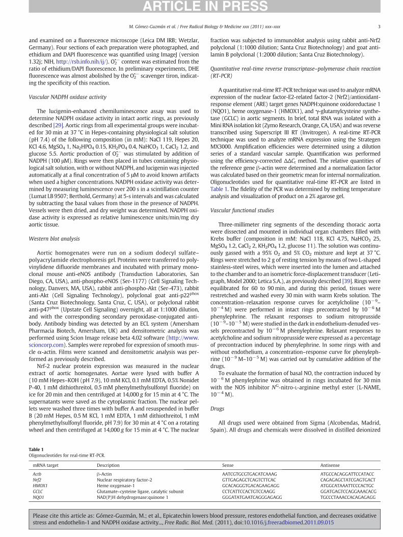
Table 1Oligonucleotides for real-time RT-PCR.
mRNA target Description
Actb β-ActinNrf2 Nuclear respiratory factor-2HMOX1 Heme oxygenase-1GCLC Glutamate–cysteine ligase, catalytic subunitNQO1 NAD(P)H dehydrogenase:quinone 1
Please cite this article as: Gómez-Guzmán, M.; et al., Epicatechin lowersstress and endothelin-1 and NADPH oxidase activity..., Free Radic. Biol. Me
fraction was subjected to immunoblot analysis using rabbit anti-Nrf2polyclonal (1:1000 dilution; Santa Cruz Biotechnology) and goat anti-lamin B polyclonal (1:2000 dilution; Santa Cruz Biotechnology).
Quantitative real-time reverse transcriptase–polymerase chain reaction(RT-PCR)
Aquantitative real-timeRT-PCR techniquewas used to analyzemRNAexpression of the nuclear factor-E2-related factor-2 (Nrf2)/antioxidant-response element (ARE) target genes NADPH:quinone oxidoreductase 1(NQO1), heme oxygenase-1 (HMOX1), and γ-glutamylcysteine synthe-tase (GCLC) in aortic segments. In brief, total RNA was isolated with aMini RNA isolation kit (ZymoResearch, Orange, CA, USA) andwas reversetranscribed using Superscript III RT (Invitrogen). A real-time RT-PCRtechnique was used to analyze mRNA expression using the StrategenMX3000. Amplification efficiencies were determined using a dilutionseries of a standard vascular sample. Quantification was performedusing the efficiency-corrected ΔΔCt method. The relative quantities ofthe reference gene β-actin were determined and a normalization factorwas calculated based on their geometricmean for internal normalization.Oligonucleotides used for quantitative real-time RT-PCR are listed inTable 1. The fidelity of the PCR was determined by melting temperatureanalysis and visualization of product on a 2% agarose gel.
Vascular functional studies
Three-millimeter ring segments of the descending thoracic aortawere dissected and mounted in individual organ chambers filled withKrebs buffer (composition in mM: NaCl 118, KCl 4.75, NaHCO3 25,MgSO4 1.2, CaCl2 2, KH2PO4 1.2, glucose 11). The solution was continu-ously gassed with a 95% O2 and 5% CO2 mixture and kept at 37 °C.Rings were stretched to 2 g of resting tension bymeans of two L-shapedstainless-steel wires, which were inserted into the lumen and attachedto the chamber and to an isometric force-displacement transducer (Leti-graph,Model 2000; Letica S.A.), as previously described [39]. Rings wereequilibrated for 60 to 90 min, and during this period, tissues wererestretched and washed every 30 min with warm Krebs solution. Theconcentration–relaxation response curves for acetylcholine (10−9–
10−4 M) were performed in intact rings precontracted by 10−6 Mphenylephrine. The relaxant responses to sodium nitroprusside(10−9–10−5 M)were studied in the dark in endothelium-denuded ves-sels precontracted by 10−6 M phenylephrine. Relaxant responses toacetylcholine and sodium nitroprusside were expressed as a percentageof precontraction induced by phenylephrine. In some rings with andwithout endothelium, a concentration–response curve for phenyleph-rine (10−9 M–10−5 M) was carried out by cumulative addition of thedrugs.
To evaluate the formation of basal NO, the contraction induced by10−6 M phenylephrine was obtained in rings incubated for 30 minwith the NOS inhibitor NG-nitro-L-arginine methyl ester (L-NAME,10−4 M).
Drugs
All drugs used were obtained from Sigma (Alcobendas, Madrid,Spain). All drugs and chemicals were dissolved in distilled deionized
Sense Antisense
AATCGTGCGTGACATCAAAG ATGCCACAGGATTCCATACCGTTGAGAGCTCAGTCTTCAC CAGAGAGCTATCGAGTGACTGCACAGGGTGACAGAAGAGG ATGGCATAAATTCCCACTGCCCTCATTCCACTGTCCAAGG GGATGAGTCCAGGAAACACGGGGATATGAATCAGGGAGAGG TGCCCTAAACCACAGAGAGG
blood pressure, restores endothelial function, and decreases oxidatived. (2011), doi:10.1016/j.freeradbiomed.2011.09.015
4 M. Gómez-Guzmán et al. / Free Radical Biology & Medicine xxx (2011) xxx–xxx
water, except for DOCA, which was mixed with ethanol/sesame oil(1/5, v/v).
Statistical analysis
Results are expressed as means±standard error of the mean(SEM) of measurements. For statistical analysis, we used one-wayanalysis of variance followed by Bonferroni's multiple comparisonstest. For all comparisons, differences were considered significant ata value of Pb0.05.
Results
Blood pressure, cardiac and renal hypertrophy, and urinary protein excretion
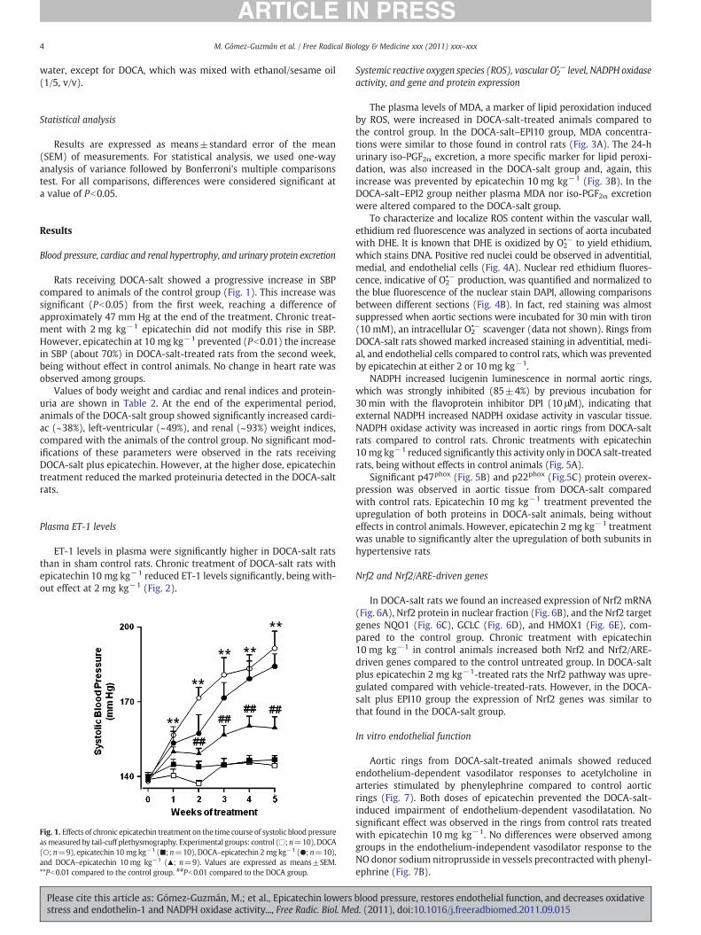
Rats receiving DOCA-salt showed a progressive increase in SBPcompared to animals of the control group (Fig. 1). This increase wassignificant (Pb0.05) from the first week, reaching a difference ofapproximately 47 mm Hg at the end of the treatment. Chronic treat-ment with 2 mg kg−1 epicatechin did not modify this rise in SBP.However, epicatechin at 10 mg kg−1 prevented (Pb0.01) the increasein SBP (about 70%) in DOCA-salt-treated rats from the second week,being without effect in control animals. No change in heart rate wasobserved among groups.
Values of body weight and cardiac and renal indices and protein-uria are shown in Table 2. At the end of the experimental period,animals of the DOCA-salt group showed significantly increased cardi-ac (~38%), left-ventricular (~49%), and renal (~93%) weight indices,compared with the animals of the control group. No significant mod-ifications of these parameters were observed in the rats receivingDOCA-salt plus epicatechin. However, at the higher dose, epicatechintreatment reduced the marked proteinuria detected in the DOCA-saltrats.
Plasma ET-1 levels
ET-1 levels in plasma were significantly higher in DOCA-salt ratsthan in sham control rats. Chronic treatment of DOCA-salt rats withepicatechin 10 mg kg−1 reduced ET-1 levels significantly, being with-out effect at 2 mg kg−1 (Fig. 2).
Fig. 1. Effects of chronic epicatechin treatment on the time course of systolic blood pressureasmeasured by tail-cuff plethysmography. Experimental groups: control (□; n=10), DOCA(○; n=9), epicatechin 10 mg kg−1 (■; n=10), DOCA–epicatechin 2 mg kg−1 (●; n=10),and DOCA–epicatechin 10 mg kg−1 (▲; n=9). Values are expressed as means±SEM.**Pb0.01 compared to the control group. ##Pb0.01 compared to the DOCA group.
Please cite this article as: Gómez-Guzmán, M.; et al., Epicatechin lowersstress and endothelin-1 and NADPH oxidase activity..., Free Radic. Biol. Me
Systemic reactive oxygen species (ROS), vascular O2•− level, NADPH oxidase
activity, and gene and protein expression
The plasma levels of MDA, a marker of lipid peroxidation inducedby ROS, were increased in DOCA-salt-treated animals compared tothe control group. In the DOCA-salt–EPI10 group, MDA concentra-tions were similar to those found in control rats (Fig. 3A). The 24-hurinary iso-PGF2α excretion, a more specific marker for lipid peroxi-dation, was also increased in the DOCA-salt group and, again, thisincrease was prevented by epicatechin 10 mg kg−1 (Fig. 3B). In theDOCA-salt–EPI2 group neither plasma MDA nor iso-PGF2α excretionwere altered compared to the DOCA-salt group.
To characterize and localize ROS content within the vascular wall,ethidium red fluorescence was analyzed in sections of aorta incubatedwith DHE. It is known that DHE is oxidized by O2
•− to yield ethidium,which stains DNA. Positive red nuclei could be observed in adventitial,medial, and endothelial cells (Fig. 4A). Nuclear red ethidium fluores-cence, indicative of O2
•− production, was quantified and normalized tothe blue fluorescence of the nuclear stain DAPI, allowing comparisonsbetween different sections (Fig. 4B). In fact, red staining was almostsuppressed when aortic sections were incubated for 30 min with tiron(10 mM), an intracellular O2
•− scavenger (data not shown). Rings fromDOCA-salt rats showed marked increased staining in adventitial, medi-al, and endothelial cells compared to control rats, which was preventedby epicatechin at either 2 or 10 mg kg−1.
NADPH increased lucigenin luminescence in normal aortic rings,which was strongly inhibited (85±4%) by previous incubation for30 min with the flavoprotein inhibitor DPI (10 μM), indicating thatexternal NADPH increased NADPH oxidase activity in vascular tissue.NADPH oxidase activity was increased in aortic rings from DOCA-saltrats compared to control rats. Chronic treatments with epicatechin10 mg kg−1 reduced significantly this activity only inDOCA salt-treatedrats, being without effects in control animals (Fig. 5A).
Significant p47phox (Fig. 5B) and p22phox (Fig.5C) protein overex-pression was observed in aortic tissue from DOCA-salt comparedwith control rats. Epicatechin 10 mg kg−1 treatment prevented theupregulation of both proteins in DOCA-salt animals, being withouteffects in control animals. However, epicatechin 2 mg kg−1 treatmentwas unable to significantly alter the upregulation of both subunits inhypertensive rats
Nrf2 and Nrf2/ARE-driven genes
In DOCA-salt rats we found an increased expression of Nrf2 mRNA(Fig. 6A), Nrf2 protein in nuclear fraction (Fig. 6B), and the Nrf2 targetgenes NQO1 (Fig. 6C), GCLC (Fig. 6D), and HMOX1 (Fig. 6E), com-pared to the control group. Chronic treatment with epicatechin10 mg kg−1 in control animals increased both Nrf2 and Nrf2/ARE-driven genes compared to the control untreated group. In DOCA-saltplus epicatechin 2 mg kg−1-treated rats the Nrf2 pathway was upre-gulated compared with vehicle-treated-rats. However, in the DOCA-salt plus EPI10 group the expression of Nrf2 genes was similar tothat found in the DOCA-salt group.
In vitro endothelial function
Aortic rings from DOCA-salt-treated animals showed reducedendothelium-dependent vasodilator responses to acetylcholine inarteries stimulated by phenylephrine compared to control aorticrings (Fig. 7). Both doses of epicatechin prevented the DOCA-salt-induced impairment of endothelium-dependent vasodilatation. Nosignificant effect was observed in the rings from control rats treatedwith epicatechin 10 mg kg−1. No differences were observed amonggroups in the endothelium-independent vasodilator response to theNO donor sodium nitroprusside in vessels precontracted with phenyl-ephrine (Fig. 7B).
blood pressure, restores endothelial function, and decreases oxidatived. (2011), doi:10.1016/j.freeradbiomed.2011.09.015
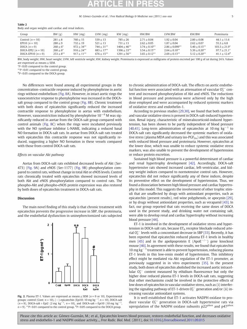
Table 2Body and organ weights and cardiac and renal indices.
Group BW (g) HW (mg) LVW (mg) KW (mg) HW/BW LVW/BW KW/BW Proteinuria
Control (n=10) 281±8 760±15 539±13 785±26 2.71±0.04 1.92±0.04 2.80±0.08 44.1±11.6EPI10 (n=10) 280±9 732±19 521±13 731±13 2.62±0.05 1.87±0.04 2.62±0.07 55.3±6.8DOCA (n=9) 260±6* 972±34** 744±31** 1404±46** 3.74±0.10** 2.86±0.099** 5.40±0.15** 103.3±21.9*DOCA-EPI2 (n=10) 260±6* 916±24** 683±17** 1396±33** 3.54±0.15** 2.64±0.10** 5.39±0.20** 97.7±21.1*DOCA-EPI10 (n=9) 253±8** 917±11** 676±15** 1291±56** 3.65±0.12** 2.69±0.13** 5.12±0.20** 41.1±12.4#
BW, body weight; HW, heart weight; LVW, left ventricle weight; KW, kidney weight. Proteinuria is expressed as milligrams of protein excreted per 100 g of rat during 24 h. Valuesare expressed as means±SEM.*Pb0.05 compared to the control group.**Pb0.01 compared to the control group.#Pb0.05 compared to the DOCA group.
5M. Gómez-Guzmán et al. / Free Radical Biology & Medicine xxx (2011) xxx–xxx
No differences were found among all experimental groups in theconcentration–contractile response induced by phenylephrine in aorticrings without endothelium (Fig. 8A). However, in intact aortic rings thevasoconstrictor response to phenylephrine was increased in the DOCA-salt group compared to the control group (Fig. 8B). Chronic treatmentwith both doses of epicatechin significantly reduced the increasedcontractile response to phenylephrine in aortas with endothelium.However, vasoconstriction induced by phenylephrine 10−6 M was sig-nificantly reduced in aortae from the DOCA-salt group compared withcontrol animals (Fig. 8C) when the rings were incubated previouslywith the NO synthase inhibitor L-NAME, indicating a reduced basalNO formation in DOCA-salt rats. In aortae from DOCA-salt rats treatedwith epicatechin this contractile response was not significantly re-duced, suggesting a higher NO formation in these vessels comparedwith those from control DOCA-salt rats.
Effects on vascular Akt pathway
Aortas from DOCA-salt rats exhibited decreased levels of Akt (Ser-473) (Fig. 9A) and eNOS (Ser-1177) (Fig. 9B) phosphorylation com-pared to control rats,without change in total Akt or eNOS levels. Controlrats chronically treated with epicatechin showed increased levels ofboth Akt and eNOS phosphorylation compared to control rats. Thephospho-Akt and phospho-eNOS protein expression was also restoredby both doses of epicatechin treatment in DOCA-salt rats.
Discussion
The main novel finding of this study is that chronic treatment withepicatechin prevents the progressive increase in SBP, the proteinuria,and the endothelial dysfunction in uninephrectomized rats subjected
Fig. 2. Plasma ET-1. Values are expressed as means±SEM (n=9 or 10). Experimentalgroups: control (Cont; n=10), (−)-epicatechin (Epi10; 10 mg kg−1, n=10), DOCA-salt(n=9), DOCA-salt+Epi2 (2 mg kg−1, n=10), and DOCA-salt+Epi10 (10 mg kg−1,n=9). **Pb0.01 compared to the control group. #Pb0.05 compared to the DOCA group.
Please cite this article as: Gómez-Guzmán, M.; et al., Epicatechin lowersstress and endothelin-1 and NADPH oxidase activity..., Free Radic. Biol. Me
to chronic administration of DOCA-salt. The effects on aortic endothe-lial function were associated with an attenuation of vascular O2
•− con-tent and increased phosphorylation of Akt and eNOS. The reductionsin blood pressure and proteinuria were achieved only by the highdose employed and were accompanied by reduced systemic markersof oxidative stress and endothelin-1.
According to previous studies [29,30], we found that both systemicand vascular oxidative stress is present in DOCA-salt-induced hyperten-sion. Renal injury, characteristic of mineralocorticoid-induced hyper-tension, has been shown to be partly independent of blood pressure[40,41]. Long-term administration of epicatechin at 10 mg kg−1 toDOCA-salt rats significantly decreased the systemic markers of oxida-tive stress (plasmaMDA and urinary iso-PGF2α) and this was associatedwith reduced blood pressure and proteinuria. However, epicatechin atthe lower dose, which was unable to reduce systemic oxidative stressmarkers, was also unable to prevent the development of hypertensionor reduce protein excretion.
Sustained high blood pressure is a powerful determinant of cardiacand renal hypertrophy development [42]. Accordingly, DOCA-salthypertensive rats showed increased cardiac, left-ventricular, and kid-ney weight indices compared to normotensive control rats. However,epicatechin did not reduce significantly any of these indices, despiteits preventive effect on the development of hypertension. Thus, wefound a dissociation between high blood pressure and cardiac hypertro-phy in this model. This suggests the involvement of other trophic stim-uli that are unaffected by drugs with antioxidant properties, such asepicatechin (present results), red wine polyphenols, or apocynin [29],or by drugs without antioxidant properties, such as verapamil [43]. Infact, our group reported that rats receiving the same doses of DOCAused in the present study, and drinking water not containing salt,were able to develop renal and cardiac hypertrophy without increasingblood pressure [44].
ET-1 is involved in the development of oxidative stress and hyper-tension in DOCA-salt rats, because ETA receptor blockade reduced arte-rial O2
•− levels with a concomitant decrease in SBP [33]. Recently, it hasbeen reported that epicatechin reduced plasma ET-1 levels in healthymen [45] and in the apolipoprotein E (ApoE−/−) gene knockoutmouse [46]. In agreement with these results, we found that epicatechin10 mgkg−1 treatment is able to prevent hypertension, reducing plasmaET-1 levels in this low-renin model of hypertension. This inhibitoryeffect might be mediated via Akt regulation of the ET-1 promoter, aspreviously suggested in in vitro experiments [35]. In the presentstudy, both doses of epicatechin abolished the increased aortic intracel-lular O2
•− content measured by ethidium fluorescence but only thehigher dose reduced plasma ET-1 levels in DOCA-salt rats, suggestingthat other mechanisms could be involved in the protective effects oflowdoses of epicatechin in vascular oxidative stress, such as (i) interfer-ing the signaling pathway of ET-1-driven O2
•− generation and/or (ii) in-creasing vascular antioxidant systems.
It is well established that ET-1 activates NADPH oxidase to pro-duce vascular O2
•− generation in DOCA-salt hypertensive rats viaupregulation of the NADPH oxidase subunits p22phox and p47phox
blood pressure, restores endothelial function, and decreases oxidatived. (2011), doi:10.1016/j.freeradbiomed.2011.09.015
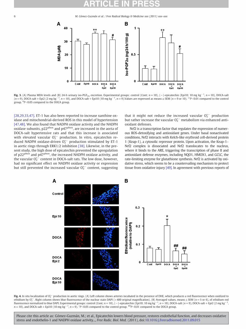
Fig. 3. (A) Plasma MDA levels and (B) 24-h urinary iso-PGF2α excretion. Experimental groups: control (Cont; n=10), (−)-epicatechin (Epi10; 10 mg kg−1, n=10), DOCA-salt(n=9), DOCA-salt+Epi2 (2 mg kg−1, n=10), and DOCA-salt+Epi10 (10 mg kg−1, n=9).Values are expressed as means±SEM (n=9 or 10). **Pb0.01 compared to the controlgroup. #Pb0.05 compared to the DOCA group.
6 M. Gómez-Guzmán et al. / Free Radical Biology & Medicine xxx (2011) xxx–xxx
[28,29,33,47]. ET-1 has also been reported to increase xanthine ox-idase and mitochondrial-derived ROS in this model of hypertension[47,48]. We also found that NADPH oxidase activity and the NADPHoxidase subunits, p22phox and p47phox, are increased in the aorta ofDOCA-salt hypertensive rats and that this increase is associatedwith elevated vascular O2
•− production. In vitro, epicatechin re-duced NADPH oxidase-driven O2
•− production stimulated by ET-1in aortic rings through ERK1/2 inhibition [38]. Likewise, in the pre-sent study, the high dose of epicatechin prevented the upregulationof p22phox and p47phox, the increased NADPH oxidase activity, andthe vascular O2
•− content in DOCA-salt rats. The low dose, however,had no significant effect on NADPH oxidase activity or expressionbut still prevented the increased vascular O2
•− content, suggesting
Fig. 4. In situ localization of O2•− production in aortic rings. (A) Left column shows arteries i
ethidium by O2•−. Right column shows blue fluorescence of the nuclear stain DAPI (×400 or
fluorescence normalized to blue DAPI. Experimental groups: control (Cont; n=10), (−)-epin=10), and DOCA-salt+Epi10 (10 mg kg−1, n=9). *Pb0.05 compared to the control grou
Please cite this article as: Gómez-Guzmán, M.; et al., Epicatechin lowersstress and endothelin-1 and NADPH oxidase activity..., Free Radic. Biol. Me
that it might not reduce the increased vascular O2•− production
but rather increase the vascular O2•− metabolism via enhanced anti-
oxidant defenses.Nrf2 is a transcription factor that regulates the expression of numer-
ous ROS-detoxifying and antioxidant genes. Under basal nonactivatedconditions, Nrf2 interacts with Kelch-like erythroid cell-derived protein1 (Keap-1), a cytosolic repressor protein. Upon activation, the Keap-1–Nrf2 complex is dissociated and Nrf2 translocates to the nucleus,where it binds to the ARE, triggering the transcription of phase II andantioxidant defense enzymes, including NQO1, HMOX1, and GCLC, therate-limiting enzyme for glutathione synthesis. Nrf2 is activated by oxi-dative stress, which seems to be a countervailing mechanism to protecttissue from oxidative injury [49]. In agreement with previous reports of
ncubated in the presence of DHE, which produces a red fluorescence when oxidized toiginal magnification). (B) Averaged values, means±SEM (n=5 or 6), of ethidium redcatechin (Epi10; 10 mg kg−1, n=10), DOCA-salt (n=9), DOCA-salt+Epi2 (2 mg kg−1,p. ##Pb0.01 compared to the DOCA group.
blood pressure, restores endothelial function, and decreases oxidatived. (2011), doi:10.1016/j.freeradbiomed.2011.09.015
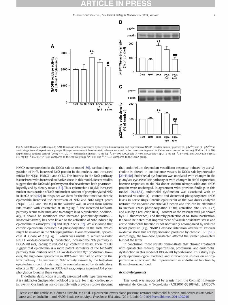
Fig. 5. NADPH oxidase pathway. (A) NADPH oxidase activity measured by lucigenin luminescence and expression of NADPH oxidase subunit proteins (B) p47phox and (C) p22phox inaortic rings from all experimental groups. Histograms represent densitometric values normalized to the corresponding α-actin. Values are expressed as means±SEM (n=9 or 10).Experimental groups: control (Cont; n=10), (−)-epicatechin (Epi10; 10 mg kg−1, n=10), DOCA-salt (n=9), DOCA-salt+Epi2 (2 mg kg−1, n=10), and DOCA-salt+Epi10(10 mg kg−1, n=9). **Pb0.01 compared to the control group. #Pb0.05 and ##Pb0.01 compared to the DOCA group.
7M. Gómez-Guzmán et al. / Free Radical Biology & Medicine xxx (2011) xxx–xxx
HMOX overexpression in the DOCA-salt rat model [50], we found upre-gulation of Nrf2, increased Nrf2 protein in the nucleus, and increasedmRNA for NQO1, HMOX1, and GCLC. This increase in the Nrf2 pathwayis consistentwith increased oxidative stress in thismodel. Recent studiessuggest that theNrf2/ARE pathway can also be activated both pharmaco-logically and by dietary means [51]. Thus, epicatechin (10 μM) increasednuclear translocation of Nrf2 and nuclear content of phosphorylatedNrf2in HepG2 cells [52]. In this paper we show for the first time that chronicepicatechin increased the expression of Nrf2 and Nrf2 target genes(NQO1, GCLC, and HMOX) in the vascular wall. In aorta from controlrats treated with epicatechin at 10 mg kg−1, the increased Nrf2/AREpathway seems to be unrelated to changes in ROS production. Addition-ally, it should be mentioned that increased phosphatidylinositol-3-kinase/Akt activity has been linked to the activation of Nrf2 induced byepicatechin in astrocytes [53] and HepG2 cells [52]. We also found thatchronic epicatechin increased Akt phosphorylation in the aorta, whichmight be involved in the Nrf2 upregulation. In our experiments, epicate-chin at a dose of 2 mg kg−1, which was unable to reduce vascularNADPH oxidase-derived O2
•− production, increased the Nrf2 pathway inDOCA-salt rats, leading to reduced O2
•− content in vessel. These resultssuggest that epicatechin is a more potent stimulator of the Nrf2/AREpathway than inhibitor of NADPH oxidase-driven O2
•− production. How-ever, the high-dose epicatechin in DOCA-salt rats had no effect on theNrf2 pathway. The increase in Nrf2 activity evoked by the high-doseepicatechin in control rats might be counterbalanced by its inhibitoryeffects on O2
•− production in DOCA-salt rats, despite increased Akt phos-phorylation found in these vessels.
Endothelial dysfunction is usually associated with hypertension andit is a risk factor (independent of blood pressure) formajor cardiovascu-lar events. Our findings are compatible with previous studies showing
Please cite this article as: Gómez-Guzmán, M.; et al., Epicatechin lowersstress and endothelin-1 and NADPH oxidase activity..., Free Radic. Biol. Me
that endothelium-dependent vasodilator response induced by acetyl-choline is altered in conductance vessels in DOCA-salt hypertension[29,43,50]. Endothelial dysfunction was unrelated with changes in theguanylate cyclase/cGMP pathway or with changes in eNOS expression,because responses to the NO donor sodium nitroprusside and eNOSprotein were unchanged. In agreement with previous findings in thismodel [29,43,54], endothelial dysfunction was associated with anincreased vascular O2
•− content and decreased phosphorylated eNOSlevels in aortic rings. Chronic epicatechin at the two doses analyzedrestored the impaired endothelial function and this can be attributedto increase eNOS phosphorylation at the activation site (Ser-1177)and also by a reduction in O2
•− content at the vascular wall (as shownby DHE fluorescence), and thereby protection of NO from inactivation.It should be noted that improvement of vascular oxidative stress andaortic endothelial function is not necessarily accompanied by reducedblood pressure (e.g., NADPH oxidase inhibition attenuates vascularoxidative stress but not hypertension produced by chronic ET-1 [55]).Accordingly, the low-dose epicatechin affected the former parametersbut not the latter.
In conclusion, these results demonstrate that chronic treatmentwith epicatechin reduces hypertension, proteinuria, and endothelialdysfunction in this model of DOCA-salt hypertension. This study sup-ports epidemiological evidence and intervention studies on antihy-pertensive effects and the improvement in endothelial function byflavanol-enriched food.
Acknowledgments
This work was supported by grants from the Comisión Intermi-nisterial de Ciencia y Tecnología (AGL2007-66108/ALI, SAF2007-
blood pressure, restores endothelial function, and decreases oxidatived. (2011), doi:10.1016/j.freeradbiomed.2011.09.015
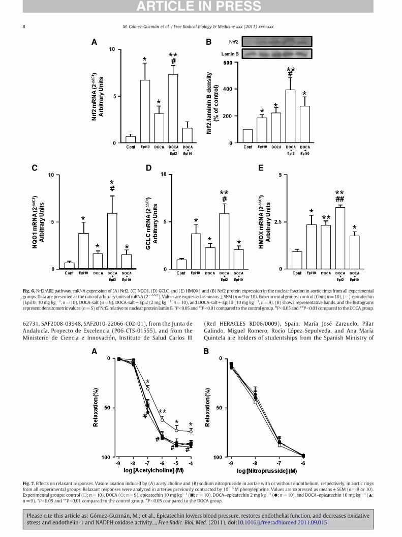
Fig. 6. Nrf2/ARE pathway. mRNA expression of (A) Nrf2, (C) NQO1, (D) GCLC, and (E) HMOX1 and (B) Nrf2 protein expression in the nuclear fraction in aortic rings from all experimentalgroups. Data are presented as the ratio of arbitrary units ofmRNA (2−ΔΔCt). Values are expressed asmeans±SEM(n=9or 10). Experimental groups: control (Cont;n=10), (−)-epicatechin(Epi10; 10 mg kg−1, n=10), DOCA-salt (n=9), DOCA-salt+Epi2 (2 mg kg−1, n=10), and DOCA-salt+Epi10 (10 mg kg−1, n=9). (B) shows representative bands, and the histogramsrepresent densitometric values (n=5) of Nrf2 relative to nuclear protein lamin B. *Pb0.05 and **Pb0.01 compared to the control group. #Pb0.05 and ##Pb0.01 compared to theDOCAgroup.
8 M. Gómez-Guzmán et al. / Free Radical Biology & Medicine xxx (2011) xxx–xxx
62731, SAF2008-03948, SAF2010-22066-C02-01), from the Junta deAndalucía, Proyecto de Excelencia (P06-CTS-01555), and from theMinisterio de Ciencia e Innovación, Instituto de Salud Carlos III
Fig. 7. Effects on relaxant responses. Vasorelaxation induced by (A) acetylcholine and (B) sofrom all experimental groups. Relaxant responses were analyzed in arteries previously contExperimental groups: control (□; n=10), DOCA (○; n=9), epicatechin 10 mg kg−1 (■; n=n=9). *Pb0.05 and **Pb0.01 compared to the control group. #Pb0.05 compared to the DO
Please cite this article as: Gómez-Guzmán, M.; et al., Epicatechin lowersstress and endothelin-1 and NADPH oxidase activity..., Free Radic. Biol. Me
(Red HERACLES RD06/0009), Spain. María José Zarzuelo, PilarGalindo, Miguel Romero, Rocío López-Sepulveda, and Ana MaríaQuintela are holders of studentships from the Spanish Ministry of
dium nitroprusside in aortae with or without endothelium, respectively, in aortic ringsracted by 10−6 M phenylephrine. Values are expressed as means±SEM (n=9 or 10).10), DOCA–epicatechin 2 mg kg−1 (●; n=10), and DOCA–epicatechin 10 mg kg−1 (▲;CA group.
blood pressure, restores endothelial function, and decreases oxidatived. (2011), doi:10.1016/j.freeradbiomed.2011.09.015
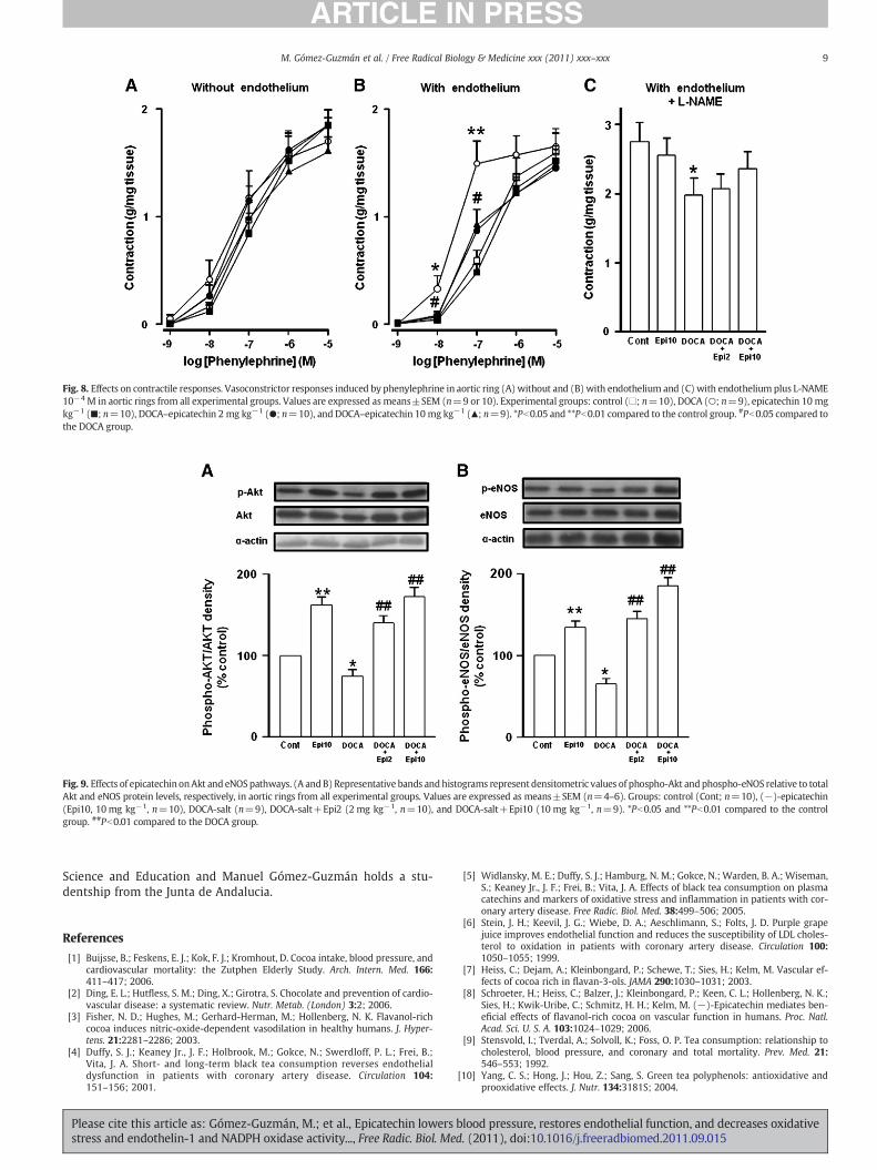
Fig. 8. Effects on contractile responses. Vasoconstrictor responses induced by phenylephrine in aortic ring (A)without and (B) with endothelium and (C) with endothelium plus L-NAME10−4 M in aortic rings from all experimental groups. Values are expressed as means±SEM (n=9 or 10). Experimental groups: control (□; n=10), DOCA (○; n=9), epicatechin 10 mgkg−1 (■; n=10), DOCA–epicatechin 2 mg kg−1 (●; n=10), and DOCA–epicatechin 10 mg kg−1 (▲; n=9). *Pb0.05 and **Pb0.01 compared to the control group. #Pb0.05 compared tothe DOCA group.
Fig. 9. Effects of epicatechin onAkt and eNOSpathways. (A andB) Representative bands andhistograms represent densitometric values of phospho-Akt andphospho-eNOS relative to totalAkt and eNOS protein levels, respectively, in aortic rings from all experimental groups. Values are expressed as means±SEM (n=4–6). Groups: control (Cont; n=10), (−)-epicatechin(Epi10, 10 mg kg−1, n=10), DOCA-salt (n=9), DOCA-salt+Epi2 (2 mg kg−1, n=10), and DOCA-salt+Epi10 (10 mg kg−1, n=9). *Pb0.05 and **Pb0.01 compared to the controlgroup. ##Pb0.01 compared to the DOCA group.
9M. Gómez-Guzmán et al. / Free Radical Biology & Medicine xxx (2011) xxx–xxx
Science and Education and Manuel Gómez-Guzmán holds a stu-dentship from the Junta de Andalucia.
References
[1] Buijsse, B.; Feskens, E. J.; Kok, F. J.; Kromhout, D. Cocoa intake, blood pressure, andcardiovascular mortality: the Zutphen Elderly Study. Arch. Intern. Med. 166:411–417; 2006.
[2] Ding, E. L.; Hutfless, S. M.; Ding, X.; Girotra, S. Chocolate and prevention of cardio-vascular disease: a systematic review. Nutr. Metab. (London) 3:2; 2006.
[3] Fisher, N. D.; Hughes, M.; Gerhard-Herman, M.; Hollenberg, N. K. Flavanol-richcocoa induces nitric-oxide-dependent vasodilation in healthy humans. J. Hyper-tens. 21:2281–2286; 2003.
[4] Duffy, S. J.; Keaney Jr., J. F.; Holbrook, M.; Gokce, N.; Swerdloff, P. L.; Frei, B.;Vita, J. A. Short- and long-term black tea consumption reverses endothelialdysfunction in patients with coronary artery disease. Circulation 104:151–156; 2001.
Please cite this article as: Gómez-Guzmán, M.; et al., Epicatechin lowersstress and endothelin-1 and NADPH oxidase activity..., Free Radic. Biol. Me
[5] Widlansky, M. E.; Duffy, S. J.; Hamburg, N. M.; Gokce, N.; Warden, B. A.; Wiseman,S.; Keaney Jr., J. F.; Frei, B.; Vita, J. A. Effects of black tea consumption on plasmacatechins and markers of oxidative stress and inflammation in patients with cor-onary artery disease. Free Radic. Biol. Med. 38:499–506; 2005.
[6] Stein, J. H.; Keevil, J. G.; Wiebe, D. A.; Aeschlimann, S.; Folts, J. D. Purple grapejuice improves endothelial function and reduces the susceptibility of LDL choles-terol to oxidation in patients with coronary artery disease. Circulation 100:1050–1055; 1999.
[7] Heiss, C.; Dejam, A.; Kleinbongard, P.; Schewe, T.; Sies, H.; Kelm, M. Vascular ef-fects of cocoa rich in flavan-3-ols. JAMA 290:1030–1031; 2003.
[8] Schroeter, H.; Heiss, C.; Balzer, J.; Kleinbongard, P.; Keen, C. L.; Hollenberg, N. K.;Sies, H.; Kwik-Uribe, C.; Schmitz, H. H.; Kelm, M. (−)-Epicatechin mediates ben-eficial effects of flavanol-rich cocoa on vascular function in humans. Proc. Natl.Acad. Sci. U. S. A. 103:1024–1029; 2006.
[9] Stensvold, I.; Tverdal, A.; Solvoll, K.; Foss, O. P. Tea consumption: relationship tocholesterol, blood pressure, and coronary and total mortality. Prev. Med. 21:546–553; 1992.
[10] Yang, C. S.; Hong, J.; Hou, Z.; Sang, S. Green tea polyphenols: antioxidative andprooxidative effects. J. Nutr. 134:3181S; 2004.
blood pressure, restores endothelial function, and decreases oxidatived. (2011), doi:10.1016/j.freeradbiomed.2011.09.015
10 M. Gómez-Guzmán et al. / Free Radical Biology & Medicine xxx (2011) xxx–xxx
[11] Hodgson, J. M.; Devine, A.; Puddey, I. B.; Chan, S. Y.; Beilin, L. J.; Prince, R. L. Teaintake is inversely related to blood pressure in older women. J. Nutr. 133:2883–2886; 2003.
[12] Taubert, D.; Berkels, R.; Roesen, R.; Klaus, W. Chocolate and blood pressure in el-derly individuals with isolated systolic hypertension. JAMA 290:1029–1030;2003.
[13] Taubert, D.; Roesen, R.; Lehmann, C.; Jung, N.; Schömig, E. Effects of low habitualcocoa intake on blood pressure and bioactive nitric oxide: a randomized con-trolled trial. JAMA 298:49–60; 2007.
[14] Grassi, D.; Lippi, C.; Necozione, S.; Desideri, G.; Ferri, C. Short-term adminis-tration of dark chocolate is followed by a significant increase in insulin sensi-tivity and a decrease in blood pressure in healthy persons. Am. J. Clin. Nutr. 81:611–614; 2005.
[15] Holt, R. R.; Lazarus, S. A.; Sullards, M. C.; Zhu, Q. Y.; Schramm, D. D.; Hammer-stone, J. F.; Fraga, C. G.; Schmitz, H. H.; Keen, C. L. Procyanidin dimer B2 [epicate-chin-(4beta-8)-epicatechin] in human plasma after the consumption of aflavanol-rich cocoa. Am. J. Clin. Nutr. 76:798–804; 2002.
[16] Sies, H.; Schewe, T.; Heiss, C.; Kelm, M. Cocoa polyphenols and inflammatory me-diators. Am. J. Clin. Nutr. 81:304S–312S; 2005.
[17] Duarte, J.; Perez-Vizcaino, F.; Utrilla, P.; Jimenez, J.; Tamargo, J.; Zarzuelo, A. Vaso-dilatory effects of flavonoids in rat aortic smooth muscle: structure–activity rela-tionships. Gen. Pharmacol. 24:857–862; 1993.
[18] Duffy, S. J.; Vita, J. A.; Holbrook, M.; Swerdloff, P. L.; Keaney Jr., J. F. Effect of acuteand chronic tea consumption on platelet aggregation in patients with coronaryartery disease. Arterioscler. Thromb. Vasc. Biol. 21:1084–1089; 2001.
[19] Huang, Y.; Zhang, A.; Lau, C. W.; Chen, Z. Y. Vasorelaxant effects of purifiedgreen tea epicatechin derivatives in rat mesenteric artery. Life Sci. 63:275–283; 1998.
[20] Huang, Y.; Chan, N. W.; Lau, C. W.; Yao, X. Q.; Chan, F. L.; Chen, Z. Y. Involvementof endothelium/nitric oxide in vasorelaxation induced by purified green tea (−)epicatechin. Biochim. Biophys. Acta 1427:322–328; 1999.
[21] Ihm, S. H.; Lee, J. O.; Kim, S. J.; Seung, K. B.; Schini-Kerth, V. B.; Chang, K.; Oak, M. H.Catechin prevents endothelial dysfunction in the prediabetic stage of OLETF ratsby reducing vascular NADPH oxidase activity and expression. Atherosclerosis206:47–53; 2009.
[22] Ramirez-Sanchez, I.; Maya, L.; Ceballos, G.; Villarreal, F. (−)-Epicatechin activa-tion of endothelial cell endothelial nitric oxide synthase, nitric oxide, and relatedsignaling pathways. Hypertension 55:1398–1405; 2010.
[23] Ramirez-Sanchez, I.; Maya, L.; Ceballos, G.; Villarreal, F. J. (−)-Epicatechin in-duces calcium and translocation independent eNOS activation in arterial endo-thelial cells. Am. J. Physiol. Cell Physiol. 300:C880–C887; 2011.
[24] Griendling, K. K.; Alexander, R. W. Oxidative stress and cardiovascular disease.Circulation 96:3264–3265; 1997.
[25] Rajagopalan, S.; Kurz, S.; Munzel, T.; Tarpey, M.; Freeman, B. A.; Griendling,K. K.; Harrison, D. G. Angiotensin II-mediated hypertension in the rat increasesvascular superoxide production via membrane NADH/NADPH oxidase activa-tion: contribution to alterations of vasomotor tone. J. Clin. Invest. 97:1916–1923; 1996.
[26] Pagano, P. J.; Clark, J. K.; Cifuentes-Pagano,M. E.; Clark, S.M.; Callis, G.M.; Quinn,M. T.Localization of a constitutively active, phagocyte-like NADPH oxidase in rabbit aorticadventitia: enhancement by angiotensin II. Proc. Natl. Acad. Sci. U. S. A. 94:14483–14488; 1997.
[27] Wu, R.; Millette, E.; Wu, L.; de Champlain, J. Enhanced superoxide anion forma-tion in vascular tissues from spontaneously hypertensive and desoxycorticoster-one acetate-salt hypertensive rats. J. Hypertens. 19:741–748; 2001.
[28] Beswick, R. A.; Dorrance, A. M.; Leite, R.; Webb, R. C. NADH/NADPH oxidase andenhanced superoxide production in the mineralocorticoid hypertensive rat. Hy-pertension 38:1107–1111; 2001.
[29] Jiménez, R.; López-Sepúlveda, R.; Kadmiri, M.; Romero, M.; Vera, R.; Sánchez,M.; Vargas, F.; O'Valle, F.; Zarzuelo, A.; Dueñas, M.; Santos-Buelga, C.;Duarte, J. Polyphenols restore endothelial function in DOCA-salt hyperten-sion: role of endothelin-1 and NADPH oxidase. Free Radic. Biol. Med. 43:462–473; 2007.
[30] Gavras, H.; Brunner, H. R.; Laragh, J. H.; Vaughan Jr., E. D.; Koss, M.; Cote, L. J.;Gavras, I. Malignant hypertension resulting from deoxycorticosterone acetateand salt excess: role of renin and sodium in vascular changes. Circ. Res. 36:300–309; 1975.
[31] Letizia, C.; Cerci, S.; De Toma, G.; D'Ambrosio, C.; De Ciocchis, A.; Coassin, S.;Scavo, D. High plasma endothelin-1 levels in hypertensive patients with low-renin essential hypertension. J. Hum. Hypertens. 11:447–451; 1997.
[32] Elijovich, F.; Laffer, C. L.; Amador, E.; Gavras, H.; Bresnahan, M. R.; Schiffrin, E. L.Regulation of plasma endothelin by salt in salt-sensitive hypertension. Circulation103:263–268; 2001.
[33] Li, L.; Fink, G. D.; Watts, S. W.; Northcott, C. A.; Galligan, J. J.; Pagano, P. J.; Chen,A. F. Endothelin-1 increases vascular superoxide via endothelin(A)–NADPH oxi-dase pathway in low-renin hypertension. Circulation 107:1053–1058; 2003.
[34] Khan, N. Q.; Lees, D. M.; Douthwaite, J. A.; Carrier, M. J.; Corder, R. Comparison ofred wine extract and polyphenol constituents on endothelin-1 synthesis by cul-tured endothelial cells. Clin. Sci. (London) 103:72S–75S; 2002.
[35] Reiter, C. E.; Kim, J. A.; Quon, M. J. Green tea polyphenol epigallocatechin gallatereduces endothelin-1 expression and secretion in vascular endothelial cells:
Please cite this article as: Gómez-Guzmán, M.; et al., Epicatechin lowersstress and endothelin-1 and NADPH oxidase activity..., Free Radic. Biol. Me
roles for AMP-activated protein kinase, Akt, and FOXO1. Endocrinology 151:103–114; 2010.
[36] Esterbauer, H.; Cheeseman, K. H. Determination of aldehydic lipid peroxidationproducts: malonaldehyde and 4-hydroxynonenal. Methods Enzymol. 186:407–421; 1990.
[37] Romero, M.; Jimenez, R.; Sanchez, M.; López-Sepúlveda, R.; Zarzuelo, A.; Tamargo,J.; Pérez-Vizcaíno, F.; Duarte, J. Vascular superoxide production by endothelin-1requires Src non-receptor protein tyrosine kinase and MAPK activation. Athero-sclerosis 212:78–85; 2010.
[38] Lopez-Sepulveda, R.; Gomez-Guzman, M.; Zarzuelo, M. J.; Romero, M.; Sanchez,M.; Quintela, A. M.; Galindo, P.; O'Valle, F.; Tamargo, J.; Perez-Vizcaino, F.; Duarte,J.; Jiménez, R. Red wine polyphenols prevent endothelial dysfunction induced byendothelin-1 in rat aorta: role of NADPH oxidase. Clin. Sci. (London) 120:321–333;2011.
[39] Lopez-Sepulveda, R.; Jimenez, R.; Romero, M.; Zarzuelo, M. J.; Sanchez, M.;Gomez-Guzman, M.; Vargas, F.; O'Valle, F.; Zarzuelo, A.; Perez-Vizcaino, F.;Duarte, J. Wine polyphenols improve endothelial function in large vesselsof female spontaneously hypertensive rats. Hypertension 51:1088–1095;2008.
[40] de Gracia, M. C.; Osuna, A.; O'Valle, F.; del Moral, R. G.; Wangensteen, R.; delRio, C. G.; Vargas, F. Deoxycorticosterone suppresses the effects of losartanin nitric oxide-deficient hypertensive rats. J. Am. Soc. Nephrol. 11:1995–2000;2000.
[41] Jin, L.; Beswick, R. A.; Yamamoto, T.; Palmer, T.; Taylor, T. A.; Pollock, J. S.; Pollock,D. M.; Brands, M. W.; Webb, R. C. Increased reactive oxygen species contributes tokidney injury in mineralocorticoid hypertensive rats. J. Physiol. Pharmacol. 57:343–357; 2006.
[42] Frohlich, E. D.; Apstein, C.; Chobanian, A. V.; Devereux, R. B.; Dustan, H. P.;Dzau, V.; Fauad-Tarazi, F.; Horan, M. J.; Marcus, M.; Massie, B.; Pfefer, M.; Re,R.; Roccella, E.; Savage, D.; Shub, C. The heart in hypertension. N. Engl. J.Med. 327:998–1008; 1992.
[43] Galisteo, M.; Garcia-Saura, M. F.; Jimenez, R.; Villar, I. C.; Wangensteen, R.;Zarzuelo, A.; Vargas, F.; Duarte, J. Effects of quercetin treatment on vascularfunction in deoxycorticosterone acetate-salt hypertensive rats: comparativestudy with verapamil. Planta Med. 70:334–341; 2004.
[44] Wangensteen, R.; O'Valle, F.; Del Moral, R.; Vargas, F.; Osuna, A. Chronic alpha1-adrenergic blockade improves hypertension and renal injury in L-NAME and low-renin L-NAME-DOCA hypertensive rats. Med. Sci. Monit. 8:BR378–BR384; 2002.
[45] Loke, W. M.; Hodgson, J. M.; Proudfoot, J. M.; McKinley, A. J.; Puddey, I. B.; Croft,K. D. Pure dietary flavonoids quercetin and (−)-epicatechin augment nitricoxide products and reduce endothelin-1 acutely in healthy men. Am. J. Clin.Nutr. 88:1018–1025; 2008.
[46] Loke, W. M.; Proudfoot, J. M.; Hodgson, J. M.; McKinley, A. J.; Hime, N.; Magat, M.;Stocker, R.; Croft, K. D. Specific dietary polyphenols attenuate atherosclerosis inapolipoprotein E-knockout mice by alleviating inflammation and endothelial dys-function. Arterioscler. Thromb. Vasc. Biol. 30:749–757; 2010.
[47] Callera, G. E.; Tostes, R. C.; Yogi, A.; Montezano, A. C.; Touyz, R. M. Endothelin-1-induced oxidative stress in DOCA-salt hypertension involves NADPH-oxidase-independent mechanisms. Clin. Sci. (London) 110:243–253; 2006.
[48] Viel, E. C.; Benkirane, K.; Javeshghani, D.; Touyz, R. M.; Schiffrin, E. L. Xanthine ox-idase and mitochondria contribute to vascular superoxide anion generation inDOCA-salt hypertensive rats. Am. J. Physiol. Heart Circ. Physiol. 295:H281–H288;2008.
[49] Singh, S.; Vrishni, S.; Singh, B. K.; Rahman, I.; Kakkar, P. Nrf2–ARE stress responsemechanism: a control point in oxidative stress-mediated dysfunctions and chron-ic inflammatory diseases. Free Radic. Res. 44:1267–1288; 2010.
[50] Nath, K. A.; d'Uscio, L. V.; Juncos, J. P.; Croatt, A. J.; Manriquez, M. C.; Pittock,S. T.; Katusic, Z. S. An analysis of the DOCA-salt model of hypertension inHO-1−/− mice and the Gunn rat. Am. J. Physiol. Heart Circ. Physiol. 293:H333–H342; 2007.
[51] Pearson, K. J.; Lewis, K. N.; Price, N. L.; Chang, J.W.; Perez, E.; Cascajo,M. V.; Tamashiro,K. L.; Poosala, S.; Csiszar, A.; Ungvari, Z.; Kensler, T. W.; Yamamoto, M.; Egan, J. M.;Longo, D. L.; Ingram, D. K.; Navas, P.; de Cabo, R. Nrf2 mediates cancer protectionbut not prolongevity induced by caloric restriction. Proc. Natl. Acad. Sci. U. S. A. 105:2325–2330; 2008.
[52] Granado-Serrano, A. B.; Martín, M. A.; Haegeman, G.; Goya, L.; Bravo, L.; Ramos, S.Epicatechin induces NF-kappaB, activator protein-1 (AP-1) and nuclear transcrip-tion factor erythroid 2p45-related factor-2 (Nrf2) via phosphatidylinositol-3-kinase/protein kinase B (PI3K/AKT) and extracellular regulated kinase (ERK) signal-ling in HepG2 cells. Br. J. Nutr. 103:168–179; 2010.
[53] Bahia, P. K.; Rattray, M.; Williams, R. J. Dietary flavonoid (−)epicatechin stimu-lates phosphatidylinositol 3-kinase-dependent anti-oxidant response elementactivity and up-regulates glutathione in cortical astrocytes. J. Neurochem. 106:2194–2204; 2008.
[54] Lima, V. V.; Giachini, F. R.; Choi, H.; Carneiro, F. S.; Carneiro, Z. N.; Fortes, Z. B.;Carvalho, M. H.; Webb, R. C.; Tostes, R. C. Impaired vasodilator activity indeoxycorticosterone acetate-salt hypertension is associated with increasedprotein O-GlcN acylation. Hypertension 53:166–174; 2009.
[55] Elmarakby, A. A.; Loomis, E. D.; Pollock, J. S.; Pollock, D. M. NADPH oxidase inhi-bition attenuates oxidative stress but not hypertension produced by chronic ET-1. Hypertension 45:283–287; 2005.
blood pressure, restores endothelial function, and decreases oxidatived. (2011), doi:10.1016/j.freeradbiomed.2011.09.015