Embed Size (px)
Citation preview
TitleEpac2-deficiency leads to more severe retinal swelling, glialreactivity and oxidative stress in transient middle cerebral arteryocclusion induced ischemic retinopathy
Author(s) LIU, J; Yeung, PKK; Cheng, L; Lo, ACY; Chung, SM; Chung, SK
Citation Science China Life Sciences, 2015, v.58 n. 6, p. 521-530
Issued Date 2015
URL http://hdl.handle.net/10722/218641
Rights Creative Commons: Attribution 3.0 Hong Kong License
SCIENCE CHINA Life Sciences
© The Author(s) 2015. This article is published with open access at link.springer.com life.scichina.com link.springer.com
*Corresponding author (email: [email protected])
SPECIAL TOPIC: Diabetic retinopathy special edition June 2015 Vol.58 No.6: 521–530
• RESEARCH PAPER • doi: 10.1007/s11427-015-4860-1
Epac2-deficiency leads to more severe retinal swelling, glial reactivity and oxidative stress in transient middle cerebral artery
occlusion induced ischemic retinopathy
LIU Jin1, YEUNG Patrick Ka Kit1, CHENG Lu1, LO Amy Cheuk Yin2,3, CHUNG Stephen Sum Man4 & CHUNG Sookja Kim1,3,5*
1Department of Anatomy, Li Ka Shing Faculty of Medicine, The University of Hong Kong, Hong Kong, China; 2Department of Ophthalmology, Li Ka Shing Faculty of Medicine, The University of Hong Kong, Hong Kong, China;
3Research Centre of Heart, Brain, Hormone and Healthy Aging, Li Ka Shing Faculty of Medicine, The University of Hong Kong, Hong Kong, China;
4Division of Science and Technology, United International College, Zhuhai 519085, China 5State Key Laboratory of Pharmaceutical Biotechnology, The University of Hong Kong, Hong Kong, China
Received December 12, 2014; accepted March 13, 2015; published online May 18, 2015
Ischemia occurs in diabetic retinopathy with neuronal loss, edema, glial cell reactivity and oxidative stress. Epacs, consisting of Epac1 and Epac2, are cAMP mediators playing important roles in maintenance of endothelial barrier and neuronal functions. To investigate the roles of Epacs in the pathogenesis of ischemic retinopathy, transient middle cerebral artery occlusion (tMCAO) was performed on Epac1-deficient (Epac1) mice, Epac2-deficient (Epac2) mice, and their wild type counter-parts (Epac1+/+ and Epac2+/+). Two-hour occlusion and 22-hour reperfusion were conducted to induce ischemia/reperfusion injury to the retina. After tMCAO, the contralateral retinae displayed similar morphology between different genotypes. Neu-ronal loss, retinal edema and increase in immunoreactivity for aquaporin 4 (AQP4), glial fibrillary acidic protein (GFAP), peroxiredoxin 6 (Prx6) were observed in ipsilateral retinae. Epac2 ipsilateral retinae showed more neuronal loss in retinal ganglion cell layer, increased retinal thickness and stronger immunostaining of AQP4, GFAP, and Prx6 than those of Epac2+/+. However, Epac1 ipsilateral retinae displayed similar pathology as those in Epac1+/+ mice. Our observations suggest that Epac2-deficiency led to more severe ischemic retinopathy after retinal ischemia/reperfusion injury.
Epac, retina, ischemia, retinopathy
Citation: Liu J, Yeung PKK, Cheng L, Lo ACY, Chung SSM, Chung SK. Epac2-deficiency leads to more severe retinal swelling, glial reactivity and oxidative stress in transient middle cerebral artery occlusion induced ischemic retinopathy. Sci China Life Sci, 2015, 58: 521–530, doi: 10.1007/s11427-015-4860-1
Ischemia occurs in several retinal diseases, including dia-betic retinopathy, glaucoma, and central retinal artery oc-clusion [1]. Dysfunction of retinal capillary is the major cause of retinal ischemia. Deprivation of blood supply in-duces a series of pathophysiological changes in the retina. Neuronal cell death in the retinal ganglion cell layer (GCL) is observed as pyknotic nuclei in the GCL [25]. Glial acti-
vation is evident by up-regulation of glial fibrillary acidic protein (GFAP) in response to deleterious release of gluta-mate from over excited neurons after ischemia [6,7]. Retinal edema also occurs, because of the increased water transpor-tation and accumulation as a result of increased extracellular fluid volume, increased aquaporin 4 (AQP4) immunoreac-tivity and swelling of retinal glial cells [8,9]. Oxidative stress is present under ischemic condition, which further exacerbates the progression of retinopathy [10,11].

522 Liu J, et al. Sci China Life Sci June (2015) Vol.58 No.6
Exchange proteins directly activated by cAMP (Epacs), consisting of Epac1 and Epac2, are cAMP mediators inde-pendent of protein kinase A (PKA). As a mediator of cAMP, an important second messenger for intracellular signal transduction, Epacs take part in many biological functions (reviewed in [12]). Epac1 is involved in the maintenance of cell-cell junctions [13]. The activation of Epac1 reduces the permeability of endothelial cells in vitro [14]. Epac2 partic-ipates in the modulation of neuronal activities, including neurotransmitter release and synaptic plasticity [1517]. Both Epac1 and Epac2 are expressed in the brain [18], and retina [19], which is part of central nervous system (CNS) and can be viewed using non-invasive methods.
Therefore, we determine the molecular significance of Epac in retinal neurons and in the pathogenesis of ischemic retinopathy using the Epac1-deficient (Epac1) and Epac2-deficient (Epac2) mice under transient ischemic retinopathy condition. Here, we report that, after transient middle cerebral artery occlusion (tMCAO), more severe ischemic retinopathy was observed in the ipsilateral retinae of Epac2 mice when compared with that of Epac2+/+ mice, whereas there was no obvious difference in retinae of Epac1and Epac1+/+ mice.
1 Materials and methods
1.1 Animals
All animals used in this study were housed in a tempera-ture-controlled room with diurnal light conditions and al-lowed access to food and water freely in the Laboratory Animal Unit of The University of Hong Kong.
Epac1 mice were generated previously [20], and they were backcrossed to C57BL/6N mice for more than 10 generations. Age-matched Epac1+/+ littermates served as the control group. Epac2 mice were backcrossed to C57BL/6N mice for more than 10 generations (a kind gift from Prof. S. Seino of Kobe University); thus they were considered congenic with C57BL/6N mice. C57BL/6N mice were therefore used as the control group (Epac2+/+) for Epac2 mice as in previous publication [21]. Three to six animals were included in each group.
1.2 Genotyping of Epac1- and Epac2-deficient mice
Genomic DNA was extracted from tail biopsy of each mouse. Polymerase chain reaction (PCR) was used to iden-tify the genotype of each mouse. The sequences of primers used are as follows: Epac1 (forward) 5′-GTTCTGCTCTTT- GAACCACACAGCAA-3′, Epac1 (reverse1) 5′-AGAACT- CAATCACAGCCTGTCCCTACC-3′, and Epac1 (reverse2) 5′-ATCAGCAGCCTCTGTTCCAC-3′, for Epac1 genotyp-ing; Epac2 (forward1) 5′-TGTGATAAACATTCTCGATT- 3′, Epac2 (forward2) 5′-GCATACATTATACGAAGT-
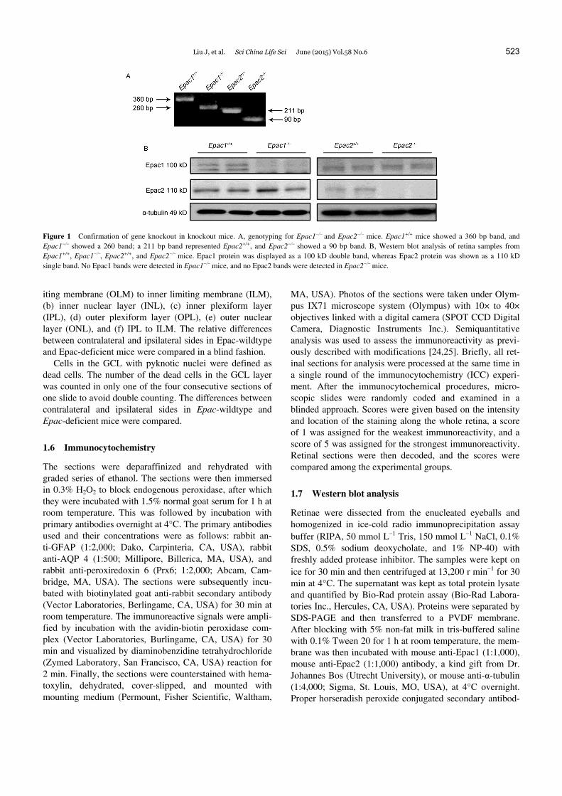
TATC-3′, and Epac2 (reverse) 5′-CTGATCACATTAGC- AAGCTC-3′ for Epac2 genotyping. The genotyping was performed in a 50 μL PCR reaction system, and the anneal-ing temperatures were 58°C and 51°C for Epac1 and Epac2, respectively. The PCR products were loaded to a 1.5% aga-rose gel with ethidium bromide for electrophoresis. The bands for PCR products were visualized under ultraviolet light. For Epac1 genotyping, bands with two sizes could be seen: the upper 360 bp band indicated Epac1+/+, and the lower 260 bp band was for Epac1; for Epac2 genotyping, the upper 211 bp band represented Epac2+/+, whereas the lower 90 bp band was for Epac2 (Figure 1A).
1.3 Transient middle cerebral artery occlusion (tMCAO)
tMCAO was performed as previously described [11,22,23]. Briefly, after the mice (812 weeks old) were anesthetized (2% halothane in 70% N2O/30% O2 for induction and 1% halothane in 70% N2O/30% O2 for maintenance), a nylon filament was inserted into the common carotid artery, di-rected to the right internal carotid artery, and finally reached the bifurcation between the middle and anterior cerebral arteries. This blocked the middle cerebral artery and its branch, the ophthalmic artery, leading to an interruption in the blood supply to the eye. Laser Doppler flowmetry (Perimed, Jarfalla, Sweden) was used to measure the rela-tive cerebral blood flow (rCBF) of middle cerebral artery territory so as to ensure the successful occlusion and reper-fusion of the middle cerebral artery. The filament was left for 2 h and was pulled out afterwards for a 22-h reperfusion after which the mice were sacrificed for sample collection. The ischemia was introduced to right eye (ipsilateral side) while the left eye served as a contralateral control side.
1.4 Tissue processing
The eyeball was enucleated 24 h after induction of ischemia. The cornea and lens were removed; the eye cup was fixed in 4% paraformaldehyde at room temperature for 2 h. After dehydration with graded series of ethanol, the eye cup was embedded in paraffin wax. Seven micron-thick sections were cut for morphological analysis.
1.5 Morphometric analysis
The thickness of the retina layers 300 μm adjacent to the optic nerve was measured in paraffin embedded retinal sec-tions stained with hematoxylin and eosin (H&E). The mi-crophotographs were taken under 20× objective of an Olympus IX71 microscope system (Olympus, Japan) linked with a digital camera (SPOT CCD Digital Camera, Diag-nostic Instruments Inc., Sterling Heights, MI, USA). The retinal layers being assessed were as follows: (a) outer lim-
Liu J, et al. Sci China Life Sci June (2015) Vol.58 No.6 523
Figure 1 Confirmation of gene knockout in knockout mice. A, genotyping for Epac1and Epac2 mice. Epac1+/+ mice showed a 360 bp band, and Epac1 showed a 260 band; a 211 bp band represented Epac2+/+, and Epac2 showed a 90 bp band. B, Western blot analysis of retina samples from Epac1+/+, Epac1, Epac2+/+, and Epac2mice. Epac1 protein was displayed as a 100 kD double band, whereas Epac2 protein was shown as a 110 kD single band. No Epac1 bands were detected in Epac1 mice, and no Epac2 bands were detected in Epac2 mice.
iting membrane (OLM) to inner limiting membrane (ILM), (b) inner nuclear layer (INL), (c) inner plexiform layer (IPL), (d) outer plexiform layer (OPL), (e) outer nuclear layer (ONL), and (f) IPL to ILM. The relative differences between contralateral and ipsilateral sides in Epac-wildtype and Epac-deficient mice were compared in a blind fashion.
Cells in the GCL with pyknotic nuclei were defined as dead cells. The number of the dead cells in the GCL layer was counted in only one of the four consecutive sections of one slide to avoid double counting. The differences between contralateral and ipsilateral sides in Epac-wildtype and Epac-deficient mice were compared.
1.6 Immunocytochemistry
The sections were deparaffinized and rehydrated with graded series of ethanol. The sections were then immersed in 0.3% H2O2 to block endogenous peroxidase, after which they were incubated with 1.5% normal goat serum for 1 h at room temperature. This was followed by incubation with primary antibodies overnight at 4°C. The primary antibodies used and their concentrations were as follows: rabbit an-ti-GFAP (1:2,000; Dako, Carpinteria, CA, USA), rabbit anti-AQP 4 (1:500; Millipore, Billerica, MA, USA), and rabbit anti-peroxiredoxin 6 (Prx6; 1:2,000; Abcam, Cam-bridge, MA, USA). The sections were subsequently incu-bated with biotinylated goat anti-rabbit secondary antibody (Vector Laboratories, Berlingame, CA, USA) for 30 min at room temperature. The immunoreactive signals were ampli-fied by incubation with the avidin-biotin peroxidase com-plex (Vector Laboratories, Burlingame, CA, USA) for 30 min and visualized by diaminobenzidine tetrahydrochloride (Zymed Laboratory, San Francisco, CA, USA) reaction for 2 min. Finally, the sections were counterstained with hema-toxylin, dehydrated, cover-slipped, and mounted with mounting medium (Permount, Fisher Scientific, Waltham,
MA, USA). Photos of the sections were taken under Olym-pus IX71 microscope system (Olympus) with 10× to 40× objectives linked with a digital camera (SPOT CCD Digital Camera, Diagnostic Instruments Inc.). Semiquantitative analysis was used to assess the immunoreactivity as previ-ously described with modifications [24,25]. Briefly, all ret-inal sections for analysis were processed at the same time in a single round of the immunocytochemistry (ICC) experi-ment. After the immunocytochemical procedures, micro-scopic slides were randomly coded and examined in a blinded approach. Scores were given based on the intensity and location of the staining along the whole retina, a score of 1 was assigned for the weakest immunoreactivity, and a score of 5 was assigned for the strongest immunoreactivity. Retinal sections were then decoded, and the scores were compared among the experimental groups.
1.7 Western blot analysis
Retinae were dissected from the enucleated eyeballs and homogenized in ice-cold radio immunoprecipitation assay buffer (RIPA, 50 mmol L1 Tris, 150 mmol L1 NaCl, 0.1% SDS, 0.5% sodium deoxycholate, and 1% NP-40) with freshly added protease inhibitor. The samples were kept on ice for 30 min and then centrifuged at 13,200 r min1 for 30 min at 4°C. The supernatant was kept as total protein lysate and quantified by Bio-Rad protein assay (Bio-Rad Labora-tories Inc., Hercules, CA, USA). Proteins were separated by SDS-PAGE and then transferred to a PVDF membrane. After blocking with 5% non-fat milk in tris-buffered saline with 0.1% Tween 20 for 1 h at room temperature, the mem-brane was then incubated with mouse anti-Epac1 (1:1,000), mouse anti-Epac2 (1:1,000) antibody, a kind gift from Dr. Johannes Bos (Utrecht University), or mouse anti-α-tubulin (1:4,000; Sigma, St. Louis, MO, USA), at 4°C overnight. Proper horseradish peroxide conjugated secondary antibod-
524 Liu J, et al. Sci China Life Sci June (2015) Vol.58 No.6
ies were used to probe the primary antibodies in the follow-ing day, and the protein bands were visualized using En-hanced Chemiluminescence Reagent (GE Healthcare, Buckinghamshire, UK).
1.8 Statistical analysis
All the data were expressed as mean±SEM. Statistical anal-yses were performed using two-way analysis of variations (ANOVA) followed by Bonferroni post-test (GraphPad Prism software, San Diego, CA, USA). Difference with P<0.05 was considered as statistically significant.
2 Results
2.1 Confirmation of Epac deficiency
Western blot analysis showed that Epac1 bands could be seen in Epac1+/+, Epac2+/+ and Epac2 retinal samples, but not in Epac1 retinal samples; Epac2 bands could be seen in Epac1+/+, Epac1, and Epac2+/+ samples, but no bands were seen in Epac2 samples. The absence of Epac1 pro-tein in Epac1 mice retinae and the absence of Epac2 pro-tein in Epac2 mice retinae demonstrated that the Epac gene was successfully knocked out in the retinae of Epac1 and Epac2 mice, respectively (Figure 1B).
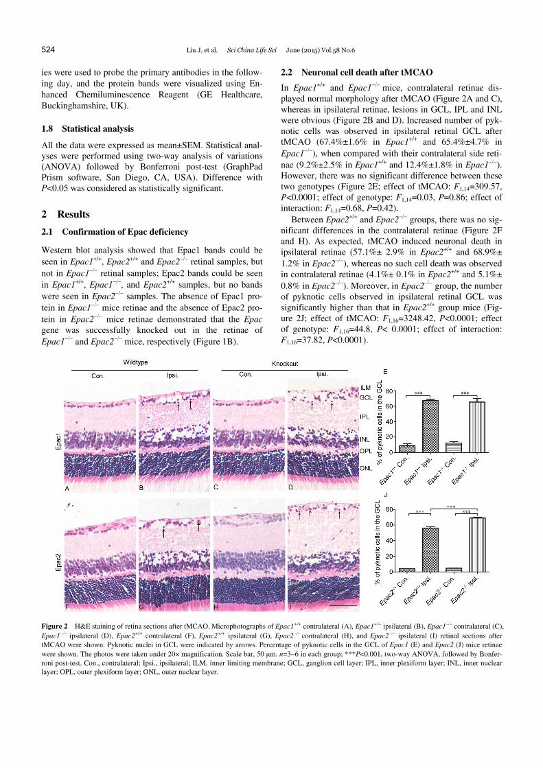
2.2 Neuronal cell death after tMCAO
In Epac1+/+ and Epac1mice, contralateral retinae dis- played normal morphology after tMCAO (Figure 2A and C), whereas in ipsilateral retinae, lesions in GCL, IPL and INL were obvious (Figure 2B and D). Increased number of pyk-notic cells was observed in ipsilateral retinal GCL after tMCAO (67.4%±1.6% in Epac1+/+ and 65.4%±4.7% in Epac1), when compared with their contralateral side reti-nae (9.2%±2.5% in Epac1+/+ and 12.4%±1.8% in Epac1). However, there was no significant difference between these two genotypes (Figure 2E; effect of tMCAO: F1,14=309.57, P<0.0001; effect of genotype: F1,14=0.03, P=0.86; effect of interaction: F1,14=0.68, P=0.42).
Between Epac2+/+ and Epac2 groups, there was no sig-nificant differences in the contralateral retinae (Figure 2F and H). As expected, tMCAO induced neuronal death in ipsilateral retinae (57.1%± 2.9% in Epac2+/+ and 68.9%± 1.2% in Epac2), whereas no such cell death was observed in contralateral retinae (4.1%± 0.1% in Epac2+/+ and 5.1%± 0.8% in Epac2). Moreover, in Epac2 group, the number of pyknotic cells observed in ipsilateral retinal GCL was significantly higher than that in Epac2+/+ group mice (Fig-ure 2J; effect of tMCAO: F1,16=3248.42, P<0.0001; effect of genotype: F1,16=44.8, P< 0.0001; effect of interaction: F1,16=37.82, P<0.0001).
Figure 2 H&E staining of retina sections after tMCAO. Microphotographs of Epac1+/+ contralateral (A), Epac1+/+ ipsilateral (B), Epac1 contralateral (C), Epac1 ipsilateral (D), Epac2+/+ contralateral (F), Epac2+/+ ipsilateral (G), Epac2contralateral (H), and Epac2 ipsilateral (I) retinal sections after tMCAO were shown. Pyknotic nuclei in GCL were indicated by arrows. Percentage of pyknotic cells in the GCL of Epac1 (E) and Epac2 (J) mice retinae were shown. The photos were taken under 20× magnification. Scale bar, 50 μm. n=36 in each group; ***P<0.001, two-way ANOVA, followed by Bonfer-roni post-test. Con., contralateral; Ipsi., ipsilateral; ILM, inner limiting membrane; GCL, ganglion cell layer; IPL, inner plexiform layer; INL, inner nuclear layer; OPL, outer plexiform layer; ONL, outer nuclear layer.
Liu J, et al. Sci China Life Sci June (2015) Vol.58 No.6 525
2.3 Retinal swelling after tMCAO
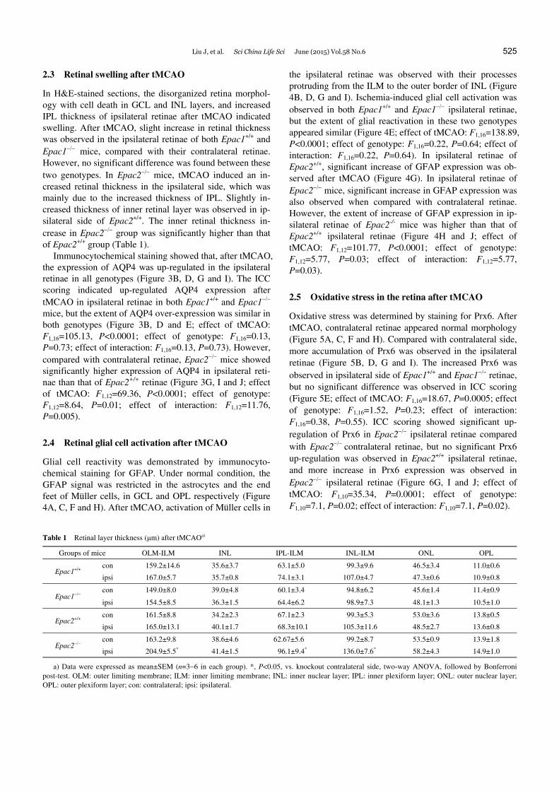
In H&E-stained sections, the disorganized retina morphol-ogy with cell death in GCL and INL layers, and increased IPL thickness of ipsilateral retinae after tMCAO indicated swelling. After tMCAO, slight increase in retinal thickness was observed in the ipsilateral retinae of both Epac1+/+ and Epac1 mice, compared with their contralateral retinae. However, no significant difference was found between these two genotypes. In Epac2 mice, tMCAO induced an in-creased retinal thickness in the ipsilateral side, which was mainly due to the increased thickness of IPL. Slightly in-creased thickness of inner retinal layer was observed in ip-silateral side of Epac2+/+. The inner retinal thickness in-crease in Epac2 group was significantly higher than that of Epac2+/+ group (Table 1).
Immunocytochemical staining showed that, after tMCAO, the expression of AQP4 was up-regulated in the ipsilateral retinae in all genotypes (Figure 3B, D, G and I). The ICC scoring indicated up-regulated AQP4 expression after tMCAO in ipsilateral retinae in both Epac1+/+ and Epac1 mice, but the extent of AQP4 over-expression was similar in both genotypes (Figure 3B, D and E; effect of tMCAO: F1,16=105.13, P<0.0001; effect of genotype: F1,16=0.13, P=0.73; effect of interaction: F1,16=0.13, P=0.73). However, compared with contralateral retinae, Epac2 mice showed significantly higher expression of AQP4 in ipsilateral reti-nae than that of Epac2+/+ retinae (Figure 3G, I and J; effect of tMCAO: F1,12=69.36, P<0.0001; effect of genotype: F1,12=8.64, P=0.01; effect of interaction: F1,12=11.76, P=0.005).
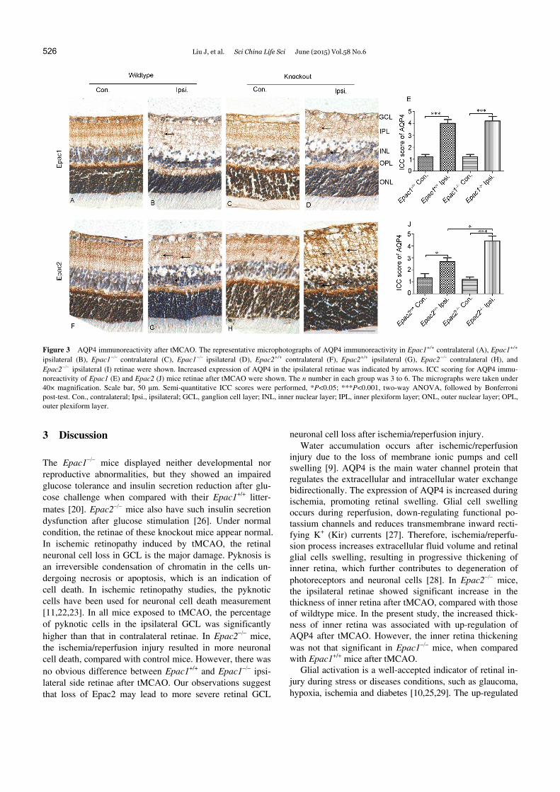
2.4 Retinal glial cell activation after tMCAO
Glial cell reactivity was demonstrated by immunocyto-chemical staining for GFAP. Under normal condition, the GFAP signal was restricted in the astrocytes and the end feet of Müller cells, in GCL and OPL respectively (Figure 4A, C, F and H). After tMCAO, activation of Müller cells in
the ipsilateral retinae was observed with their processes protruding from the ILM to the outer border of INL (Figure 4B, D, G and I). Ischemia-induced glial cell activation was observed in both Epac1+/+ and Epac1 ipsilateral retinae, but the extent of glial reactivation in these two genotypes appeared similar (Figure 4E; effect of tMCAO: F1,16=138.89, P<0.0001; effect of genotype: F1,16=0.22, P=0.64; effect of interaction: F1,16=0.22, P=0.64). In ipsilateral retinae of Epac2+/+, significant increase of GFAP expression was ob-served after tMCAO (Figure 4G). In ipsilateral retinae of Epac2 mice, significant increase in GFAP expression was also observed when compared with contralateral retinae. However, the extent of increase of GFAP expression in ip-silateral retinae of Epac2-/- mice was higher than that of Epac2+/+ ipsilateral retinae (Figure 4H and J; effect of tMCAO: F1,12=101.77, P<0.0001; effect of genotype: F1,12=5.77, P=0.03; effect of interaction: F1,12=5.77, P=0.03).
2.5 Oxidative stress in the retina after tMCAO
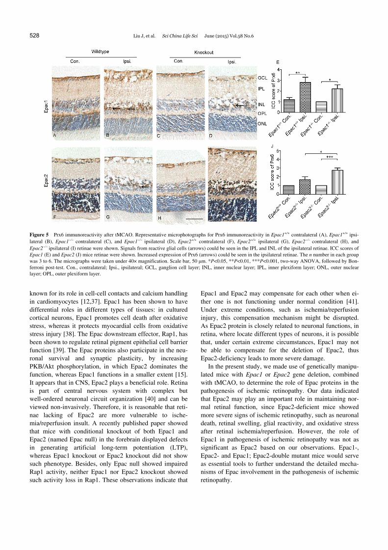
Oxidative stress was determined by staining for Prx6. After tMCAO, contralateral retinae appeared normal morphology (Figure 5A, C, F and H). Compared with contralateral side, more accumulation of Prx6 was observed in the ipsilateral retinae (Figure 5B, D, G and I). The increased Prx6 was observed in ipsilateral side of Epac1+/+ and Epac1 retinae, but no significant difference was observed in ICC scoring (Figure 5E; effect of tMCAO: F1,16=18.67, P=0.0005; effect of genotype: F1,16=1.52, P=0.23; effect of interaction: F1,16=0.38, P=0.55). ICC scoring showed significant up- regulation of Prx6 in Epac2 ipsilateral retinae compared with Epac2 contralateral retinae, but no significant Prx6 up-regulation was observed in Epac2+/+ ipsilateral retinae, and more increase in Prx6 expression was observed in Epac2 ipsilateral retinae (Figure 6G, I and J; effect of tMCAO: F1,10=35.34, P=0.0001; effect of genotype: F1,10=7.1, P=0.02; effect of interaction: F1,10=7.1, P=0.02).
Table 1 Retinal layer thickness (μm) after tMCAOa)
Groups of mice OLM-ILM INL IPL-ILM INL-ILM ONL OPL
Epac1+/+ con 159.2±14.6 35.6±3.7 63.1±5.0 99.3±9.6 46.5±3.4 11.0±0.6
ipsi 167.0±5.7 35.7±0.8 74.1±3.1 107.0±4.7 47.3±0.6 10.9±0.8
Epac1 con 149.0±8.0 39.0±4.8 60.1±3.4 94.8±6.2 45.6±1.4 11.4±0.9
ipsi 154.5±8.5 36.3±1.5 64.4±6.2 98.9±7.3 48.1±1.3 10.5±1.0
Epac2+/+ con 161.5±8.8 34.2±2.3 67.1±2.3 99.3±5.3 53.0±3.6 13.8±0.5
ipsi 165.0±13.1 40.1±1.7 68.3±10.1 105.3±11.6 48.5±2.7 13.6±0.8
Epac2 con 163.2±9.8 38.6±4.6 62.67±5.6 99.2±8.7 53.5±0.9 13.9±1.8
ipsi 204.9±5.5* 41.4±1.5 96.1±9.4* 136.0±7.6* 58.2±4.3 14.9±1.0
a) Data were expressed as mean±SEM (n=36 in each group). *, P<0.05, vs. knockout contralateral side, two-way ANOVA, followed by Bonferroni post-test. OLM: outer limiting membrane; ILM: inner limiting membrane; INL: inner nuclear layer; IPL: inner plexiform layer; ONL: outer nuclear layer; OPL: outer plexiform layer; con: contralateral; ipsi: ipsilateral.
526 Liu J, et al. Sci China Life Sci June (2015) Vol.58 No.6
Figure 3 AQP4 immunoreactivity after tMCAO. The representative microphotographs of AQP4 immunoreactivity in Epac1+/+ contralateral (A), Epac1+/+ ipsilateral (B), Epac1 contralateral (C), Epac1 ipsilateral (D), Epac2+/+ contralateral (F), Epac2+/+ ipsilateral (G), Epac2 contralateral (H), and Epac2ipsilateral (I) retinae were shown. Increased expression of AQP4 in the ipsilateral retinae was indicated by arrows. ICC scoring for AQP4 immu-noreactivity of Epac1 (E) and Epac2 (J) mice retinae after tMCAO were shown. The n number in each group was 3 to 6. The micrographs were taken under 40× magnification. Scale bar, 50 μm. Semi-quantitative ICC scores were performed, *P<0.05; ***P<0.001, two-way ANOVA, followed by Bonferroni post-test. Con., contralateral; Ipsi., ipsilateral; GCL, ganglion cell layer; INL, inner nuclear layer; IPL, inner plexiform layer; ONL, outer nuclear layer; OPL, outer plexiform layer.
3 Discussion
The Epac1 mice displayed neither developmental nor reproductive abnormalities, but they showed an impaired glucose tolerance and insulin secretion reduction after glu-cose challenge when compared with their Epac1+/+ litter-mates [20]. Epac2 mice also have such insulin secretion dysfunction after glucose stimulation [26]. Under normal condition, the retinae of these knockout mice appear normal. In ischemic retinopathy induced by tMCAO, the retinal neuronal cell loss in GCL is the major damage. Pyknosis is an irreversible condensation of chromatin in the cells un-dergoing necrosis or apoptosis, which is an indication of cell death. In ischemic retinopathy studies, the pyknotic cells have been used for neuronal cell death measurement [11,22,23]. In all mice exposed to tMCAO, the percentage of pyknotic cells in the ipsilateral GCL was significantly higher than that in contralateral retinae. In Epac2 mice, the ischemia/reperfusion injury resulted in more neuronal cell death, compared with control mice. However, there was no obvious difference between Epac1+/+ and Epac1 ipsi-lateral side retinae after tMCAO. Our observations suggest that loss of Epac2 may lead to more severe retinal GCL
neuronal cell loss after ischemia/reperfusion injury. Water accumulation occurs after ischemic/reperfusion
injury due to the loss of membrane ionic pumps and cell swelling [9]. AQP4 is the main water channel protein that regulates the extracellular and intracellular water exchange bidirectionally. The expression of AQP4 is increased during ischemia, promoting retinal swelling. Glial cell swelling occurs during reperfusion, down-regulating functional po-tassium channels and reduces transmembrane inward recti-fying K+ (Kir) currents [27]. Therefore, ischemia/reperfu- sion process increases extracellular fluid volume and retinal glial cells swelling, resulting in progressive thickening of inner retina, which further contributes to degeneration of photoreceptors and neuronal cells [28]. In Epac2 mice, the ipsilateral retinae showed significant increase in the thickness of inner retina after tMCAO, compared with those of wildtype mice. In the present study, the increased thick-ness of inner retina was associated with up-regulation of AQP4 after tMCAO. However, the inner retina thickening was not that significant in Epac1 mice, when compared with Epac1+/+ mice after tMCAO.
Glial activation is a well-accepted indicator of retinal in-jury during stress or diseases conditions, such as glaucoma, hypoxia, ischemia and diabetes [10,25,29]. The up-regulated
Liu J, et al. Sci China Life Sci June (2015) Vol.58 No.6 527
Figure 4 GFAP immunoreactivity after tMCAO. Micrographs for Epac1+/+ contralateral (A), Epac1+/+ ipsilateral (B), Epac1contralateral (C), and Epac1 ipsilateral retinae (D), Epac2+/+ contralateral (F), Epac2+/+ ipsilateral (G), Epac2 contralateral (H), and Epac2 ipsilateral (I) retinae were shown. GFAP positive astrocytes (arrowheads) were restricted in GCL. Processes of activated Müller cells (arrows) could be seen in the ipsilateral retinae (B, D, G and I). The n number in each group was 3 to 6. The micrographs were taken under 40× magnification. Scale bar, 50 μm. Semi-quantitative ICC scores for Epac1 (E) and Epac2 (J) retinae were shown, *P<0.05, ***P<0.001, two-way ANOVA followed by Bonferroni post-test. Con., contralateral; Ipsi., ipsilat-eral; GCL, ganglion cell layer; INL, inner nuclear layer; IPL, inner plexiform layer; ONL, outer nuclear layer; OPL, outer plexiform layer.
immunoactivity of GFAP, a marker of astrocytes and im-portant component of Müller cells, is a well-known sign for glial activation. Under normal conditions, the astrocytes and Müller cells reside in GCL and INL respectively, and the GFAP staining signals are restricted in these two retinal layers. After ischemia/reperfusion insult, the expression of GFAP increased, shown as hypertrophy of astrocytes, and Müller cell processes protruding from GCL to outer ONL. In a 15 months old type 2 diabetes mice model, in which retinal ischemia also occurred, the retinal GFAP expression was higher than that in their non-diabetic littermates [10]. In other studies using tMCAO to induce retinal ische-mia/reperfusion injury, GFAP expression is higher in the ipsilateral side retinae compared to their contralateral con-trol retinae [11,22,25]. Currently, it is clear that the more intense GFAP staining is an indirect effect due to the more severe ischemic retinopathy induced. Our observations on intense GFAP staining in ipsilateral retinae were consistent with previous data. More GFAP staining was observed in Epac2 ipsilateral retinae when compared with Epac2+/+ ipsilateral retinae-the Epac2 retinae suffered more severe retinopathy after tMCAO. In contrast, the difference in GFAP expression in the ipsilateral retinae was not observed in the ipsilateral retinae between Epac1+/+ and Epac1 mice.
Ischemia/reperfusion injury is often accompanied with oxidative stress. When ischemia/reperfusion process occurs, the free radicals and proteases are formed which further disrupt brain-cell membranes, causing irreversible damage. Therefore in this study, oxidative stress level was deter-mined to reflect the extent of retinopathy. Prx6 is a member of antioxidant enzyme that belongs to peroxiredoxin family. It functions as a protective factor in ischemia/reperfusion injury [3032]. Increased expression of Prx6 has been ob-served in the brain after tMCAO [33]. In our case, increased Prx6 expression in the ipsilateral retinae was observed in parallel with other observations. The extent of the increase of Prx6 was higher in Epac2 mice, but not in other three genotypes.
Epac1 and Epac2 are two isoforms of Epac proteins, with differential expression patterns, which have been reported since the discovery of Epac proteins [18]. Both Epac1 and Epac2 are expressed in the retina [19]. It has been shown that Epacs have a profound role in cognition, loss of Epacs leads to impairment in memory retrieval in a fear condi-tioning paradigm [34], and it is also noted that in depressed patients, the Epac2 expression but not Epac1 is altered in the cortex [35]. Besides, Epac2 participates in synaptic plasticity [17,36]. Such experimental evidence indicates that Epac2 is more involved in CNS. Epac1 protein has been
528 Liu J, et al. Sci China Life Sci June (2015) Vol.58 No.6
Figure 5 Prx6 immunoreactivity after tMCAO. Representative microphotographs for Prx6 immunoreactivity in Epac1+/+ contralateral (A), Epac1+/+ ipsi-lateral (B), Epac1 contralateral (C), and Epac1 ipsilateral (D), Epac2+/+ contralateral (F), Epac2+/+ ipsilateral (G), Epac2 contralateral (H), and Epac2ipsilateral (I) retinae were shown. Signals from reactive glial cells (arrows) could be seen in the IPL and INL of the ipsilateral retinae. ICC scores of Epac1 (E) and Epac2 (J) mice retinae were shown. Increased expression of Prx6 (arrows) could be seen in the ipsilateral retinae. The n number in each group was 3 to 6. The micrographs were taken under 40× magnification. Scale bar, 50 μm. *P<0.05, **P<0.01, ***P<0.001, two-way ANOVA, followed by Bon-ferroni post-test. Con., contralateral; Ipsi., ipsilateral; GCL, ganglion cell layer; INL, inner nuclear layer; IPL, inner plexiform layer; ONL, outer nuclear layer; OPL, outer plexiform layer.
known for its role in cell-cell contacts and calcium handling in cardiomyocytes [12,37]. Epac1 has been shown to have differential roles in different types of tissues: in cultured cortical neurons, Epac1 promotes cell death after oxidative stress, whereas it protects myocardial cells from oxidative stress injury [38]. The Epac downstream effector, Rap1, has been shown to regulate retinal pigment epithelial cell barrier function [39]. The Epac proteins also participate in the neu-ronal survival and synaptic plasticity, by increasing PKB/Akt phosphorylation, in which Epac2 dominates the function, whereas Epac1 functions in a smaller extent [15]. It appears that in CNS, Epac2 plays a beneficial role. Retina is part of central nervous system with complex but well-ordered neuronal circuit organization [40] and can be viewed non-invasively. Therefore, it is reasonable that reti-nae lacking of Epac2 are more vulnerable to ische-mia/reperfusion insult. A recently published paper showed that mice with conditional knockout of both Epac1 and Epac2 (named Epac null) in the forebrain displayed defects in generating artificial long-term potentiation (LTP), whereas Epac1 knockout or Epac2 knockout did not show such phenotype. Besides, only Epac null showed impaired Rap1 activity, neither Epac1 nor Epac2 knockout showed such activity loss in Rap1. These observations indicate that
Epac1 and Epac2 may compensate for each other when ei-ther one is not functioning under normal condition [41]. Under extreme conditions, such as ischemia/reperfusion injury, this compensation mechanism might be disrupted. As Epac2 protein is closely related to neuronal functions, in retina, where locate different types of neurons, it is possible that, under certain extreme circumstances, Epac1 may not be able to compensate for the deletion of Epac2, thus Epac2-deficiency leads to more severe damage.
In the present study, we made use of genetically manipu-lated mice with Epac1 or Epac2 gene deletion, combined with tMCAO, to determine the role of Epac proteins in the pathogenesis of ischemic retinopathy. Our data indicated that Epac2 may play an important role in maintaining nor-mal retinal function, since Epac2-deficient mice showed more severe signs of ischemic retinopathy, such as neuronal death, retinal swelling, glial reactivity, and oxidative stress after retinal ischemia/reperfusion. However, the role of Epac1 in pathogenesis of ischemic retinopathy was not as significant as Epac2 based on our observations. Epac1-, Epac2- and Epac1; Epac2-double mutant mice would serve as essential tools to further understand the detailed mecha-nisms of Epac involvement in the pathogenesis of ischemic retinopathy.
Liu J, et al. Sci China Life Sci June (2015) Vol.58 No.6 529
The authors declare that they have no conflict of interest. All animal ex-periments in the study were conducted according to the requirements of the Cap. 340 Animals (Control of Experiments) Ordinance and Regulation, and relevant legislation and Codes of Practice in Hong Kong. All the ex-periments performed were strictly following the experimental guidelines approved by the Committee on the Use of Live Animals for Teaching and Researching (CULATR No. 2239-10).
This work was supported by the Research Grants Council of Hong Kong (RGC) HKU 764008M to Sookja Kim Chung. We thank Professor S. Seino from Kobe University for providing Epac2 mice, and Professor J.L. Bos from Utrecht University for providing Epac antibodies.
1 Osborne NN, Casson RJ, Wood JPM, Chidlow G, Graham M, Mele-na J. Retinal ischemia: mechanisms of damage and potential thera-peutic strategies. Prog Retin Eye Res, 2004, 23: 91–147
2 Buchi ER. Cell death in the rat retina after a pressure-induced is-chaemia-reperfusion insult: an electron microscopic study. I. Gangli-on cell layer and inner nuclear layer. Exp Eye Res, 1992, 55: 605–613
3 Lafuente MP, Villegas-Pérez MP, Sellés-Navarro I, Mayor- Torroglosa S, de Imperial JM, Vidal-Sanz M. Retinal ganglion cell death after acute retinal ischemia is an ongoing process whose sever-ity and duration depends on the duration of the insult. Neuroscience, 2002, 109: 157–168
4 Nucci C, Tartaglione R, Rombolà L, Morrone LA, Fazzi E, Bagetta G. Neurochemical evidence to implicate elevated glutamate in the mechanisms of high intraocular pressure (IOP)-induced retinal gan-glion cell death in rat. Neurotoxicology, 2005, 26: 935–941
5 Yamamoto H, Schmidt-Kastner R, Hamasaki DI, Yamamoto H, Parel JM. Complex neurodegeneration in retina following moderate ische-mia induced by bilateral common carotid artery occlusion in Wistar rats. Exp Eye Res, 2006, 82: 767–779
6 Bringmann A, Francke M, Pannicke T, Biedermann B, Faude F, Enzmann V, Wiedemann P, Reichelt W, Reichenbach A. Human Muller glial cells: altered potassium channel activity in proliferative vitreoretinopathy. Invest Ophthalmol Vis Sci, 1999, 40: 3316–3323
7 Kim IB, Kim KY, Joo CK, Lee MY, Oh SJ, Chung JW, Chun MH. Reaction of Muller cells after increased intraocular pressure in the rat retina. Exp Brain Res, 1998, 121: 419–424
8 Da T, Verkman AS. Aquaporin-4 gene disruption in mice protects against impaired retinal function and cell death after ischemia. Invest Ophthalmol Vis Sci, 2004, 45: 4477–4483
9 Yanoff M, Fine BS, Brucker AJ, Eagle Jr. Ralph C. Pathology of human cystoid macular edema. Surv Ophthalmol, 1984, 28(Suppl): 505–511
10 Cheung AK, Fung MK, Lo AC, Lam TT, So KF, Chung SS, Chung SK. Aldose reductase deficiency prevents diabetes-induced blood- retinal barrier breakdown, apoptosis, and glial reactivation in the ret-ina of db/db mice. Diabetes, 2005, 54: 3119–3125
11 Cheung AK, Lo AC, So KF, Chung SS, Chung SK. Gene deletion and pharmacological inhibition of aldose reductase protect against retinal ischemic injury. Exp Eye Res, 2007, 85: 608–616
12 Gloerich M, Bos JL. Epac: defining a new mechanism for cAMP ac-tion. Annu Rev Pharmacol Toxicol, 2010, 50: 355–375
13 Bos JL. Linking Rap to cell adhesion. Curr Opin Cell Biol, 2005, 17: 123–128
14 Rampersad SN, Ovens JD, Huston E, Umana MB, Wilson LS, Neth-erton SJ, Lynch MJ, Baillie GS, Houslay MD, Maurice DH. Cyclic AMP phosphodiesterase 4D (PDE4D) Tethers EPAC1 in a vascular endothelial cadherin (VE-Cad)-based signaling complex and controls cAMP-mediated vascular permeability. J Biol Chem, 2010, 285: 33614–33622
15 Nijholt IM, Dolga AM, Ostroveanu A, Luiten PG, Schmidt M, Eisel UL. Neuronal AKAP150 coordinates PKA and Epac-mediated PKB/Akt phosphorylation. Cell Signal, 2008, 20: 1715–1724
16 Penzes P, Woolfrey KM, Srivastava DP. Epac2-mediated dendritic spine remodeling: implications for disease. Mol Cell Neurosci, 2011, 46: 368–380
17 Woolfrey KM, Srivastava DP, Photowala H, Yamashita M, Barbolina MV, Cahill ME, Xie Z, Jones KA, Quilliam LA, Prakriya M, Penzes P. Epac2 induces synapse remodeling and depression and its dis-ease-associated forms alter spines. Nat Neurosci, 2009, 12: 1275–1284
18 Kawasaki H, Springett GM, Mochizuki N, Toki S, Nakaya M, Matsuda M, Housman DE, Graybiel AM. A family of cAMP-binding proteins that directly activate Rap1. Science, 1998, 282: 2275–2279
19 Whitaker CM, Cooper NGF. Differential distribution of exchange proteins directly activated by cyclic AMP within the adult rat retina. Neuroscience, 2010, 165: 955–967
20 Kai AK, Lam AK, Chen Y, Tai AC, Zhang X, Lai AK, Yeung PK, Tam S, Wang J, Lam KS, Vanhoutte PM, Bos JL, Chung SS, Xu A, Chung SK. Exchange protein activated by cAMP 1 (Epac1)-deficient mice develop beta-cell dysfunction and metabolic syndrome. FASEB J, 2013, 27: 4122–4135
21 Zhang CL, Katoh M, Shibasaki T, Minami K, Sunaga Y, Takahashi H, Yokoi N, Iwasaki M, Miki T, Seino S. The cAMP sensor Epac2 is a direct target of antidiabetic sulfonylurea drugs. Science, 2009, 325: 607–610
22 Cheung SS, Leung JW, Lam AK, Lam KS, Chung SS, Lo AC, Chung SK. Selective over-expression of endothelin-1 in endothelial cells exacerbates inner retinal edema and neuronal death in ischemic retina. PLoS One, 2011, 6: e26184
23 Li SY, Fu ZJ, Ma H, Jang WC, So KF, Wong D, Lo AC. Effect of lutein on retinal neurons and oxidative stress in a model of acute ret-inal ischemia/reperfusion. Invest Ophthalmol Vis Sci, 2009, 50: 836–843
24 Fu ZJ, Li SY, Kociok N, Wong D, Chung SK, Lo AC. Aldose reduc-tase deficiency reduced vascular changes in neonatal mouse retina in oxygen-induced retinopathy. Invest Ophthalmol Vis Sci, 2012, 53: 5698–5712
25 Li SY, Yang D, Yeung CM, Yu WY, Chang RC, So KF, Wong D, Lo AC. Lycium barbarum polysaccharides reduce neuronal damage, blood-retinal barrier disruption and oxidative stress in retinal ische-mia/reperfusion injury. PLoS One, 2011, 6: e16380
26 Shibasaki T, Takahashi H, Miki T, Sunaga Y, Matsumura K, Yama-naka M, Zhang C, Tamamoto A, Satoh T, Miyazaki J, Seino S. Es-sential role of Epac2/Rap1 signaling in regulation of insulin granule dynamics by cAMP. Proc Natl Acad Sci USA, 2007, 104: 19333–19338
27 Pannicke T, Uckermann O, Iandiev I, Biedermann B, Wiedemann P, Perlman I, Reichenbach A, Bringmann A. Altered membrane physi-ology in Muller glial cells after transient ischemia of the rat retina. Glia, 2005, 50: 1–11
28 Tso MO. Pathology of cystoid macular edema. Ophthalmology, 1982, 89: 902–915
29 Lo AC, Chen AY, Hung VK, Yaw LP, Fung MK, Ho MC, Tsang MC, Chung SS, Chung SK. Endothelin-1 overexpression leads to further water accumulation and brain edema after middle cerebral artery oc-clusion via aquaporin 4 expression in astrocytic end-feet. J Cereb Blood Flow Metab, 2005, 25: 998–1011
30 Avellini C, Baccarani U, Trevisan G, Cesaratto L, Vascotto C, D’Aurizio F, Pandolfi M, Adani GL, Tell G. Redox proteomics and immunohistology to study molecular events during ischemia- reperfusion in human liver. Transplant Proc, 2007, 39: 1755–1760
31 Eismann T, Huber N, Shin T, Kuboki S, Galloway E, Wyder M, Ed-wards MJ, Greis KD, Shertzer HG, Fisher AB, Lentsch AB. Perox-iredoxin-6 protects against mitochondrial dysfunction and liver injury during ischemia-reperfusion in mice. Am J Physiol Gastrointest Liver Physiol, 2009, 296: G266–274
32 Nagy N, Malik G, Fisher AB, Das DK. Targeted disruption of perox-iredoxin 6 gene renders the heart vulnerable to ischemia-reperfusion injury. Am J Physiol Heart Circ Physiol, 2006, 291: H2636–2640
33 Zhang X, Yeung PK, McAlonan GM, Chung SS, Chung SK. Trans-genic mice over-expressing endothelial endothelin-1 show cognitive
530 Liu J, et al. Sci China Life Sci June (2015) Vol.58 No.6
deficit with blood-brain barrier breakdown after transient ischemia with long-term reperfusion. Neurobiol Learn Mem, 2013, 101: 46–54
34 Ouyang M, Zhang L, Zhu JJ, Schwede F, Thomas SA. Epac signaling is required for hippocampus-dependent memory retrieval. Proc Natl Acad Sci USA, 2008, 105: 11993–11997
35 Dwivedi Y, Mondal AC, Rizavi HS, Faludi G, Palkovits M, Sarosi A, Conley RR, Pandey GN. Differential and brain region-specific regu-lation of Rap-1 and Epac in depressed suicide victims. Arch Gen Psychiatry, 2006, 63: 639–648
36 Gekel I, Neher E. Application of an Epac activator enhances neuro-transmitter release at excitatory central synapses. J Neurosci, 2008, 28: 7991–8002
37 Roscioni SS, Elzinga CR, Schmidt M. Epac: effectors and biological functions. Naunyn Schmiedebergs Arch Pharmacol, 2008, 377: 345–357
38 Suzuki S, Yokoyama U, Abe T, Kiyonari H, Yamashita N, Kato Y, Kurotani R, Sato M, Okumura S, Ishikawa Y. Differential roles of Epac in regulating cell death in neuronal and myocardial cells. J Biol Chem, 2010, 285: 24248–24259
39 Wittchen ES, Hartnett ME. The small GTPase Rap1 is a novel regu-lator of RPE cell barrier function. Invest Ophthalmol Vis Sci, 2011, 52: 7455–7463
40 Kolb H. The architecture of functional neural circuits in the verte-brate retina. The Proctor Lecture. Invest Ophthalmol Vis Sci, 1994, 35: 2385–2404
41 Yang Y, Shu X, Liu D, Shang Y, Wu Y, Pei L, Xu X, Tian Q, Zhang J, Qian K, Wang YX, Petralia RS, Tu W, Zhu LQ, Wang JZ, Lu Y. EPAC null mutation impairs learning and social interactions via ab-errant regulation of miR-124 and Zif268 translation. Neuron, 2012, 73: 774–788
Open Access This article is distributed under the terms of the Creative Commons Attribution License which permits any use, distribution, and reproduction
in any medium, provided the original author(s) and source are credited.