Embed Size (px)
Citation preview
Eosinophils are a feature of upper and lower airway pathology
in non-atopic asthma, irrespective of the presence of rhinitis
M. GAGA, P. LAMBROU, N. PAPAGEORGIOU, N. G. KOULOURIS, E. KOSMAS,S. FRAGAKIS, C. SOFIOS, A. RASIDAKIS and J. JORDANOGLOU
Department of Respiratory Medicine, Athens University, School of Medicine and ENT Department of Sotiria Hospital,
Athens, Greece
Summary
Background Asthma and rhinitis often co-exist and there are data to suggest that they may
be two ends of the same disease spectrum. Immunohistochemical studies have shown that
eosinophilia in the airways is a feature of rhinitic patients without asthma.
Objective The aim of our study was to examine whether cellular in®ltration exists in the
nasal mucosa of asthmatics even in the absence of symptoms and signs of rhinitis.
Methods Nasal mucosa biopsies were taken from 27 non-atopic subjects and comprised
nine asthmatic rhinitic patients (AR), eight asthmatic non-rhinitic patients (ANR) and 10
healthy control subjects (N). Bronchial mucosa biopsies were also taken simultaneously
from some of the patients (n� 10) to determine whether there was an association between
cellular in®ltration in the nose and the lungs. The alkaline phosphatase-anti-alkaline
phosphatase (APAAP) method was used on 6 mm thick cryostat sections using monoclonal
antibodies against T cells (CD4, CD8), eosinophils (EG2) and mast cells (mast cell
tryptase). Slides were counted blind and results expressed as cells per ®eld.
Results The results showed that eosinophil counts were higher in both asthma groups
compared with control nasal biopsies (median values AR 8.3, ANR 9.2, N 2.1 cells per ®eld,
P < 0.01). Furthermore, there was a signi®cant correlation between eosinophil cell counts
in the nose and the airways (r� 0.851 P < 0.001). No differences in eosinophil numbers
were detected between the two groups of asthmatics. Also, no differences were noted for
any other cell type (i.e. CD4, CD8, tryptase) among the three study groups.
Conclusions These results show that eosinophil in®ltration was present in the nasal
mucosa of asthmatic patients even in the absence of rhinitis, and add further support to
the hypothesis that asthma and rhinitis are clinical expressions of the same disease entity.
Keywords: asthma, rhinitis, eosinophils, non-atopic
Clinical and Experimental Allergy, Vol. 30, pp. 663±669. Submitted 31 December 1998;
revised 10 June 1999; accepted 30 September 1999.
Introduction
Asthma and rhinitis are common diseases that frequently
co-exist [1±3] and share many clinical features. Both show
intermittent symptoms or ¯uctuations in severity, are char-
acterized by in¯ammation and obstruction, and a speci®c
allergen elicits early and late phase reactions [4±6]. This
association is not surprising as the upper and lower airways
are lined by the same pseudostrati®ed columnar epithelium
and the mucosal surface is contiguous throughout the
respiratory tract. Therefore, both regions of the airway
share a common susceptibility to various agents such as
allergens, infectious agents, occupational sensitizers and
drugs. Although several studies have shown that rhinitis
and asthma are characterized by a similar in¯ammatory
process [7±12], few have examined the association between
the two diseases and most of the data are from atopic
patients. Previous investigators showed that eosinophilia,
Clinical and Experimental Allergy, 2000, Volume 30, pages 663±669
663q 2000 Blackwell Science Ltd
Correspondence: M. Gaga, Department of Respiratory Medicine, Medical
School, Athens University, Sotiria Hospital, 152, Mesogion Avenue,
Athens 115 27, Greece.
a hallmark of asthmatic in¯ammation, is present in the
bronchi of rhinitic patients without asthma [13,14]. Further-
more, it has been shown that patients with rhinitis
and no history of asthma develop bronchoconstriction and
demonstrate in¯ammatory changes in their bronchi when
challenged with inhaled allergens [15±20], or when exposed
to high allergen load such as during the pollen season. These
data indicate that there is a pathogenetic link between
rhinitis and asthma, and that the condition of the upper
airways in¯uences the lower airways. However, it is not
clear whether the state of the lower airways in¯uences the
upper airway.
The aim of this study was to examine whether nasal
mucosal in¯ammation, with particular reference to eosino-
phils, exists in asthma, irrespective of symptoms and clini-
cal signs of rhinitis. Non-atopic patients and non-atopic
normal control subjects were examined so that atopy was
not a confounding factor in the study. The hypothesis
was that the two conditions are part of the same disease
spectrum. To test this hypothesis further, it was also
examined whether a correlation exists between cell counts
in the nose and the airways in asthma.
Materials and methods
Subjects
A total of 27 subjects participated in the study: nine patients
with asthma and rhinitis, eight patients with asthma without
rhinitis, and 10 healthy, non-atopic control subjects.
Asthma was de®ned as clear clinical history with current
symptoms, based on diary card symptoms scores plus 15%
reversibility in FEV1 after two puffs of b2 agonist, and/or
positive methacholine challenge. Methacholine challenge
was considered positive when a 20% drop in FEV1 was
induced at a cummulative dose of #1 mg. The challenge
was performed with the equipment and the protocol
described in the European Community Respiratory Health
Survey (ECRHS) [21].
A diagnosis of rhinitis was based on clinical history
and symptoms, direct examination of the nasal mucosa
and Youlten inspiratory peak ¯ow readings. All patients
answered a questionnaire on nasal symptoms, including
itch, sneeze, discharge and blocking, and were excluded if
they had recurrent symptoms of rhinitis. Patients with nasal
polyps or signi®cant deviation of the septum were also
excluded. It should be mentioned that out of the 66 asth-
matic patients screened, only 10 had no symptoms or signs
of rhinitis (of which only two were atopic).
All subjects were non-atopic. Atopy was examined by
skin prick tests to a panel of extracts of common aero-
allergens, including house dust mite, grass, parietatia
and olive tree pollen, cat dander, dog hair, cladosporium,
alternaria and aspergillus (Allergopharma, Germany),
and/or raised total and speci®c IgE levels (Pharmacia,
Sweden).
All patients were non-smokers, were receiving no oral,
inhaled or topical treatment other than inhaled b2 agonists
prn, and had no evidence of respiratory infection in the
previous 4 weeks. The study was approved by the Ethics
Committee of Sotiria Hospital and was performed with
the informed consent of the patients. Clinical data for the
patients are shown in Table 1.
Study design
Asthmatic patients and healthy control subjects ®lled in a
questionnaire on symptoms and duration of asthma and
rhinitis, and had baseline and post-dilatation spirometry
and inspiratory nasal peak ¯ow measurement [22]. Within
a week from lung function tests, nasal biopsies were taken
between 09.00 and 23.00 h. A small subgroup of asthmatic
patients (®ve asthmatic±rhinitic and ®ve asthmatic non-
rhinitic) underwent simultaneous nasal and bronchial
biopsy. In this case, bronchoscopy was performed between
08.00 and 10.00 h, and nasal biopsy was taken imme-
diately after bronchoscopy from the contralateral nostril
of the one used at bronchoscopy.
Nasal biopsy
Local anaesthesia was achieved by applying a small cotton
wool plug soaked in 3% cocaine and 0.025% epinephrine
immediately below the inferior turbinate for 10 min. A
2.5 mm biopsy was taken using Gerritsma forceps [23].
Any local bleeding was controlled by cautery using a
silver nitrate stick. Patients were observed for 1 h and
discharged.
664 M. Gaga et al.
q 2000 Blackwell Science Ltd, Clinical and Experimental Allergy, 30, 663±669
Table 1. Patient clinical characteristics
Normal
Asthma±rhinitis Asthma±no rhinitis control
n 9 8 10
Gender 2M:7F 3M:5F 6M:4F
Age 42 47 33
(22±61) (24±64) (20±66)
FEV1 % pred 81.2 80 94.3
(58±98) (72±89) (86±102)
Nasal PIFR 140 215 230
(70±170) (170±275) (180±345)
Values given as mean (range). M � male, F � female.
Fibreoptic bronchoscopy and biopsies
Bronchoscopy was performed using an Olympus model
BFP20 bronchoscope with a 2.2 mm channel (Olympus
Corporation, Tokyo, Japan). All subjects underwent bron-
choscopy following pre-medication with 2.5 mg salbutamol
by nebulizer, and 0.6 mg of atropine and 5±10 mg mida-
zolam administered intravenously. Local anaesthesia of
the vocal cords, trachea and bronchial tree was induced
with 2% lidocaine. After inspection of the airways, biopsies
were taken from the right middle, lower and upper lobe
bronchi using Olympus forceps (FP-15C).
Immunohistology
Nasal biopsies were immediately placed in phosphate-
buffered saline and within 15 min, transferred to a drop of
OCT embedding medium (BDH) on a small piece of card.
The biopsy was snap-frozen in isopentane pre-cooled in
liquid nitrogen and stored at ÿ808C for a maximum of 1
month. Cryostat sections 6-mm thick were cut, air-dried
for 1 h, and then ®xed by immersion in a mixture of
acetone and methanol (60:40) for 7 min. After a further
1 h drying period, slides were stored at ÿ208C until staining.
Immunostaining was performed using a modi®ed alkaline
phosphatase anti-alkaline phosphatase (APAAP) method
described in detail in previous publications [24,25]. Mono-
clonal antibodies against CD4� T cells (Leu-3a, Becton-
Dickinson, San Jose, CA USA) CD8� T cells (CD8,
Clone DK25, Dako A/S, Denmark) and eosinophils (EG2,
Pharmacia Diagnostics, Sweden).
Counting method
Slides were counted blind in coded random order.
An Olympus BH-2 microscope (Olympus Optical Co.
Ltd, Tokyo, Japan) was used with an eyepiece graticule
(0.202 mm2) at ´ 200. At least two sections and a median
of six ®elds were counted for each monoclonal antibody.
The graticule was orientated along the epithelial basement
membrane, and sub-epithelial counts were expressed as
mean counts per high-power ®eld. The mean intraobserver
coef®cient of variation for repeat counts was 8%.
Statistical analysis
Data were analysed using a statistical package (Sigma stat
2.0, Jandel Scienti®c, San Rafael, CA, USA). Three groups
of patients were used in the analysis: (a) normal control
subjects, (b) asthmatic non-rhinitic patients and (c) asth-
matic rhinitic patients. For each group of patients, data are
expressed as median and IQ range. When comparisons were
made between groups, signi®cant between-group variability
was ®rst established using the Kruskal±Wallis ANOVA on
ranks test. The Mann±Whitney U-test was then used for
between-group comparison. Correlation coef®cients
between nasal and bronchial cell counts were obtained by
Spearman's rank method. Correlation was examined for
each asthma group separately and for all patients together.
P-values less than 0.05 were considered signi®cant.
Results
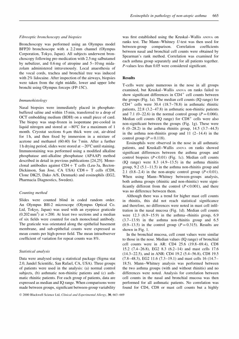
T-cells were quite numerous in the nose in all groups
examined, but Kruskal±Wallis ANOVA on ranks failed to
show signi®cant differences in CD4� cell counts between
the groups (Fig. 1a). The median cell counts (IQ range) for
CD4� cells were 30.4 (18.7±78.8) in asthmatic rhinitic
patients, 22.8 (3.2±47.8) in asthmatic non-rhinitic patients
and 7.1 (0±22.6) in the normal control group (P� 0.066).
Median cell counts (IQ range) for CD8� cells were also
non-signi®cant between the groups (Fig. 1g). These were
6 (0±28.2) in the asthma rhinitis group, 14.5 (3.7±44.5)
in the asthma non-rhinitis group and 11 (2±14.4) in the
control group (P� 0.118).
Eosinophils were observed in the nose in all asthmatic
patients, and Kruskall±Wallis ANOVA on ranks showed
signi®cant differences between the asthma groups and
control biopsies (P < 0.01) (Fig. 1c). Median cell counts
(IQ range) were 8.3 (4.9±13.5) in the asthma rhinitis
group, 9.2 (5.1±11.5) in the asthma non-rhinitis group and
2.1 (0.8±2.4) in the non-atopic control group (P < 0.01).
When using Mann±Whitney between-groups analysis,
both asthma groups (rhinitic and non-rhinitic) were signi-
®cantly different from the control (P< 0.001), and there
was no difference between them.
Although there was a trend for higher mast cell counts
in rhinitis, this did not reach statistical signi®cance
and therefore, no differences were noted in mast cell in®l-
tration in the nasal mucosa (Fig. 1d). Median cell counts
were 12.3 (6.9±15.9) in the asthma±rhinitis group, 6.9
(3.7±13.9) in the asthma non-rhinitis group and 6.5
(0.9±13.5) in the control group (P� 0.315). Results are
shown in Fig. 1.
In the bronchial mucosa, cell count values were similar
to those in the nose. Median values (IQ range) of bronchial
cell counts were in AR: CD4 25.6 (19.8±69.4), CD8
15.2 (7.4±26.8), EG2 8.3 (6.2±14) and mast cells 17.6
(14.3±22.5), and in ANR: CD4 19.2 (5.4±56.8), CD8 19.5
(7.8±48.3), EG2 11.6 (7.3±19.1) and mast cells 16 (14.7±
18.5). Mann±Whitney analysis was performed between
the two asthma groups (with and without rhinitis) and no
differences were noted. Analysis for correlation between
cell counts in the nasal and bronchial mucosa was then
performed for all asthmatic patients. No correlation was
found for CD4, CD8 or mast cell counts but a highly
Eosinophils in pathology of non-atopic asthma 665
q 2000 Blackwell Science Ltd, Clinical and Experimental Allergy, 30, 663±669
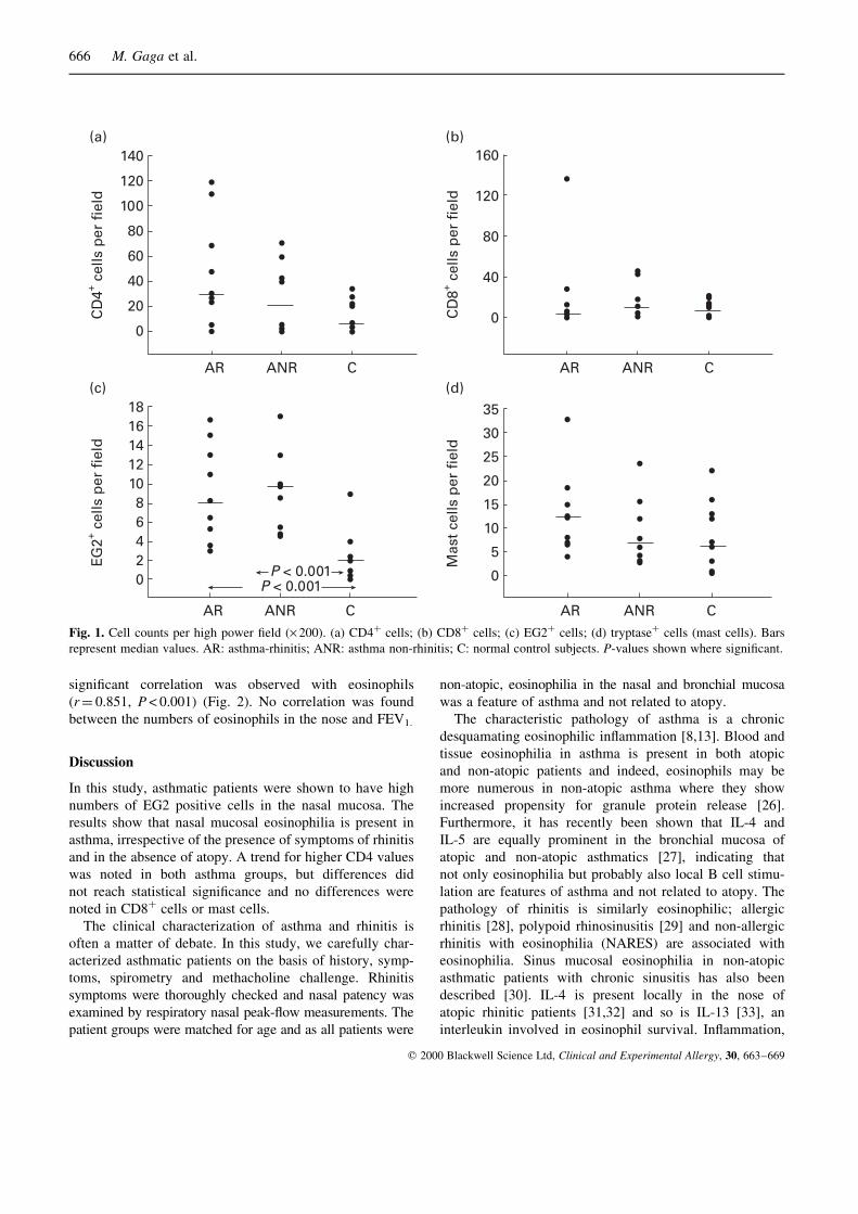
signi®cant correlation was observed with eosinophils
(r� 0.851, P< 0.001) (Fig. 2). No correlation was found
between the numbers of eosinophils in the nose and FEV1.
Discussion
In this study, asthmatic patients were shown to have high
numbers of EG2 positive cells in the nasal mucosa. The
results show that nasal mucosal eosinophilia is present in
asthma, irrespective of the presence of symptoms of rhinitis
and in the absence of atopy. A trend for higher CD4 values
was noted in both asthma groups, but differences did
not reach statistical signi®cance and no differences were
noted in CD8� cells or mast cells.
The clinical characterization of asthma and rhinitis is
often a matter of debate. In this study, we carefully char-
acterized asthmatic patients on the basis of history, symp-
toms, spirometry and methacholine challenge. Rhinitis
symptoms were thoroughly checked and nasal patency was
examined by respiratory nasal peak-¯ow measurements. The
patient groups were matched for age and as all patients were
non-atopic, eosinophilia in the nasal and bronchial mucosa
was a feature of asthma and not related to atopy.
The characteristic pathology of asthma is a chronic
desquamating eosinophilic in¯ammation [8,13]. Blood and
tissue eosinophilia in asthma is present in both atopic
and non-atopic patients and indeed, eosinophils may be
more numerous in non-atopic asthma where they show
increased propensity for granule protein release [26].
Furthermore, it has recently been shown that IL-4 and
IL-5 are equally prominent in the bronchial mucosa of
atopic and non-atopic asthmatics [27], indicating that
not only eosinophilia but probably also local B cell stimu-
lation are features of asthma and not related to atopy. The
pathology of rhinitis is similarly eosinophilic; allergic
rhinitis [28], polypoid rhinosinusitis [29] and non-allergic
rhinitis with eosinophilia (NARES) are associated with
eosinophilia. Sinus mucosal eosinophilia in non-atopic
asthmatic patients with chronic sinusitis has also been
described [30]. IL-4 is present locally in the nose of
atopic rhinitic patients [31,32] and so is IL-13 [33], an
interleukin involved in eosinophil survival. In¯ammation,
666 M. Gaga et al.
q 2000 Blackwell Science Ltd, Clinical and Experimental Allergy, 30, 663±669
AR ANR C
CD
4+ cel
ls p
er f
ield
140
120
100
80
60
40
20
0
(a)
AR ANR C
Mas
t ce
lls p
er f
ield
35
30
25
20
15
10
5
0
(d)AR ANR C
CD
8+ cel
ls p
er f
ield
160
120
80
40
0
(b)
AR ANR C
EG2+ c
ells
per
fie
ld
181614121086420
(c)
P < 0.001P < 0.001
Fig. 1. Cell counts per high power ®eld (´ 200). (a) CD4� cells; (b) CD8� cells; (c) EG2� cells; (d) tryptase� cells (mast cells). Bars
represent median values. AR: asthma-rhinitis; ANR: asthma non-rhinitis; C: normal control subjects. P-values shown where signi®cant.

therefore, in rhinitis and in asthma is quite similar. On the
other hand, it seems that there is a link between rhinitis
and asthma, and this link is the subject of some excellent
reviews [34,35]. Previous studies reported eosinophilia
and loss of mast cell granular content in the bronchi of
rhinitic patients without asthma [13,14], and elevated
histamine levels in the bronchoalveolar lavage of allergic
rhinitic patients with levels intermediate between atopic
asthmatics and normal controls [36]. Furthermore, bronchial
challenge with methacholine or allergen often results in
bronchoconstriction in rhinitis patients without asthma [37],
and treatment of rhinitis with topical steroids reduces
bronchial hyperactivity [38,39]. A possible explanation for
these phenomena might be the fact that when the nose is
by-passed due to obstruction, as is often the case in rhinitis,
air is inhaled through the mouth and reaches the bronchi
colder, less humid and un®ltered. These changes in inhaled
air may lead to hyperactivity and in¯ammation. Therefore,
rhinitis may be linked to asthma through increased load
of sensitizing agents. This should not be the case in the
reverse situation.
Our study shows eosinophilia in the nose of asthmatic
patients in the absence of clinical rhinitis. In addition, we
found a signi®cant correlation between eosinophil counts
in the nose and the lung in asthma, as indeed between large
and small airways in asthma [13]. These results show that
the airway epithelium is affected all along its length in
asthma, and further support the hypothesis that asthma
and rhinitis are indeed parts of the same spectrum. Eosino-
phils are effector cells causing bronchial tissue damage
and leading to remodelling in asthma. In rhinitis, there is
also eosinophil granule protein release, and ECP and EPO
are found in nasal biopsies and lavage ¯uid [40]. LTC4 is
also formed by eosinophils [41] and probably contributes
to blocking and rhinorrea. However, the presence of eosino-
phils in the nose is not always related to clinical disease,
which is similar to ®ndings reported in the bronchi [14].
The reason for this is unclear, but it may be that other
mechanisms, such as T cell or neurogenic in¯ammation,
are absent or that the threshold of patients differs. Further-
more, eosinophils do not seem to damage the nasal mucosa
in rhinitis [42] although they are associated with epi-
thelial damage in nasal polyps [43]. In a recent paper,
increased basement membrane thickness was shown in
the nasal epithelium of asthmatic-rhinitic patients with
perennial rhinitis [44] but without epithelium shedding.
In our study, measurement of basement membrane thick-
ness would not have been accurate as we used frozen
tissue but again, no epithelium shedding was noted, so
although eosinophils are present in both asthma and
rhinitis, it seems there may be differences in the nasal
and bronchial tissue reaction to them. Although common
mechanisms are implicated in the pathogenesis of both
rhinitis and asthma, the end organs differ. The nose is the
®rst contact of the respiratory system with the environment
and acts as a barrier ®ltering inhaled air. It also has a very
quick clearance rate so that noxious substances do not
remain in contact with the mucosa for a long time. Perhaps,
then, the nasal mucosa may withstand more assault without
damage.
CD4� T cells are believed to be important in the patho-
genesis of chronic asthma, but studies in mild asthma
did not show signi®cantly increased numbers of CD4�
cells compared with controls, similar to our results. Mast
cells play an important role in allergen-induced asthma
and rhinitis, and it has been shown that seasonal pollen
exposure results in epithelial migration of mast cells in the
nose [9,27,45]. However, in both atopic asthma and rhini-
tis, mast cell numbers in the submucosa of the upper and
lower airways are not different from controls [46] and this
is in accordance with our data.
The most important ®nding in this study was the presence
of eosinophils in the nasal mucosa of asthmatic patients
without symptoms of rhinitis, an eosinophilia that correlates
well with eosinophilia in the airways of the same patients.
These ®ndings further support the hypothesis that asthma
and rhinitis are clinical expressions of the same disease entity.
Guidelines exist for the classi®cation and management
in both rhinitis and asthma [47,48]. However, the terms of
classi®cation differ (e.g. intrinsic asthma, idiopathic rhini-
tis) and so does the approach to management. Perhaps
those guidelines should be reconsidered and the upper and
Eosinophils in pathology of non-atopic asthma 667
q 2000 Blackwell Science Ltd, Clinical and Experimental Allergy, 30, 663±669
Fig. 2. Correlation between EG2� cells in the bronchial and the
nasal mucosa.

lower respiratory tract treated as one system using the same
approach to classi®cation and management.
References
1 European Community Respiratory Health Survey. Variations
in the prevalence of respiratory symptoms, self-reported asthma
attacks, and use of asthma medication in the European
Community Respiratory Health Survey (ECRHS). Eur Respir
J 1996; 9:687±95.
2 Sibbald B, Rink E. Epidemiology of seasonal and perennial
rhinitis: clinical presentation and medical history. Thorax
1991; 46:895±901.
3 Broder I, Higgins MW, Matthews KP, Keller JP. Epidemiology
of asthma and allergic rhinitis in a total community, Tecumseh,
Michigan. III. Second survey of the community. J Allergy Clin
Immunol 1974; 53:127±38.
4 Iliopoulos O, Proud D, Adkinson Franklin N, Jr et al. Relation-
ship between the early, late, and rechallenge reaction to nasal
challenge with antigen: Observations on the role of in¯a-
mmatory mediators and cells. J Allergy Clin Immunol 1990;
86:851±61.
5 Rak S, Jacobson MR, Sudderick RM et al. In¯uence of
prolonged treatment with topical corticosteroid (¯uticasone
propionate) on early and late phase nasal responses and cellular
in®ltration in the nasal mucosa after allergen challenge. Clin
Exp Allergy 1994; 24:930±9.
6 Naclerio RM, Proud D, Togias AG et al. In¯ammatory
mediators in late antigen-induced rhinitis. N Eng J Med
1985; 313:65±70.
7 Bousquet J, Vignola AM, Campbell AM, Michel F-B. Patho-
physiology of allergic rhinitis. Int Arch Allergy Immunol 1996;
110:207±19.
8 Bousquet J, Chanez P, Lacoste JY et al. Eosinophilic in¯am-
mation in asthma. N Eng J Med 1990; 323:1033±9.
9 Bentley AM, Jacobson MR, Cumberworth V et al. Immuno-
histology of the nasal mucosa in seasonal allergic rhinitis:
increases in activated eosinophils and epithelial mast cells.
J Allergy Clin Immunol 1992; 89:877±83.
10 Togias A, Naclerio RM, Proud D et al. Studies on the allergic
and nonallergic nasal in¯ammation. J Allergy Clin Immunol
1988; 81:782±90.
11 Bentley AM, Menz G, Storz C et al. Identi®cation of T
lymphocytes, macrophages, and activated eosinophils in the
bronchial mucosa in intrinsic asthma. Relationship to symp-
toms and bronchial responsiveness. Am Rev Respir Dis 1992;
146:500±6.
12 Naclerio RM, Baroody F. Understanding the in¯ammatory
processes in upper allergic airway disease and asthma. J
Allergy Clin Immunol 1998; 101:S345±51.
13 Azzawi M, Bradley B, Jeffery PK et al. Identi®cation of
activated T lymphocytes and eosinophils in bronchial biopsies
in stable atopic asthma. Am Rev Respir Dis 1990; 142:
1407±13.
14 Djukanovic R, Lai CKW, Wilson JW et al. Bronchial
mucosal manifestations of atopy: a comparison of markers
of in¯ammation between atopic asthmatics, atopic
non-asthmatics and healthy controls. Eur Respir J 1992; 5:
538±44.
15 Muller AB, Cheryl AL, Smith RM, Suelzer MT, Richerson
HB. Comparisons of speci®c and non-speci®c broncho-
provocation in subjects with asthma, rhinitis, and healthy
subjects. J Allergy Clin Immunol 1993; 91:758±72.
16 Bonavia M, Crimi E, Quaglia A, Brusasco V. Comparison
of early and late responses between patients with allergic
rhinitis and mild asthma. Eur Respir J 1996; 9:905±9.
17 Townley RG, Ryo UK, Kolotzin MB, Kang B. Bronchial
sensitivity to methacholine in current and former asthmatic
and allergic rhinitis patients and control subjects. J Allergy
Clin Immunol 1975; 56:429±42.
18 Fish JE, Ankin MG, Kelly JF, Peterman VI. Comparison of
responses to pollen extract in subjects with allergic asthma
and non-asthmatic subjects with allergic rhinitis. J Allergy Clin
Immunol 1980; 65:154±61.
19 Bruce CA, Rosenthal RR, Lichtenstein LM, Norman PS.
Quantitative inhalation bronchial challenge in ragweed hay
fever patients: a comparison with ragweed-allergic asthmatics.
J Allergy Clin Immunol 1975; 56:331±7.
20 Sotomayor H, Badier M, Vervoloet D, Orcheck J. Seasonal
increase of carbachol airway responsiveness in patients
allergic to grass pollen. Reversal by corticosteroids. Am Rev
Respir Dis 1984; 130:56±8.
21 Burney PGJ, Luczynska Chinn S, Jarvis D. The European
Community Respiratory Health Survey. Eur Respir J 1994;
7:954±60.
22 Gleeson MJ, Youlten LJF, Shelton DM, Siodlak MZ,
Eiser NM, Wengraf CL. Assessment of nasal airway patency:
a comparison of four methods. Clin Otolaryngol 1986; 11:
99±107.
23 Fokkens WJ, Vroom TM, Gerritsma V, Rijntjes E. A biopsy
method to obtain high quality specimens of nasal mucosa.
Rhinology 1988; 26:293±5.
24 Mason DY, Sammons R. Alkaline phosphatase and peroxidase
for double immunising labelling of cellular contacts. J Clin
Pathol 1978; 31:454±60.
25 Frew AJ, Kay AB. The relationship between in®ltrating
CD4 positive lymphocytes, activated eosinophils and the
magnitude of the allergen-induced late phase cutaneous
reaction in man. J Immunol 1988; 141:4158±64.
26 Carlson M, HaÊkansson L, Peterson C, StaÊlenheim G, Venge P.
Secretion of granule proteins from eosinophils and neutro-
phils is increased in asthma. J Allergy Clin Immunol 1991;
87:27±33.
27 Humbert M, Durham SR, Ying S et al. IL-4 and IL-5 mRNA
and protein in bronchial biopsies from patients with atopic
and nonatopic asthma: evidence against `intrinsic' asthma
being a distinct immunopathologic entity. Am J Respir Crit
Care Med 1996; 154:1497±504.
28 Varney VA, Jacobson MR, Sudderick RM et al. Immuno-
histology of the nasal mucosa following allergen-induced
rhinitis. Am Rev Respir Dis 1992; 146:170±6.
29 Jankowski R, BeÁneÁ MC, Moneret-Vautrin AD et al. Immuno-
histological characteristics of nasal polyps. A comparison with
668 M. Gaga et al.
q 2000 Blackwell Science Ltd, Clinical and Experimental Allergy, 30, 663±669

healthy mucosa and chronic sinusitis. Rhinol 1989; Suppl.
8:51±8.
30 Harlin SL, Ansel DG, Lane SR, Myers J, Kephart GM,
Gleich GJ. A clinical and pathological study of chronic
sinusitis: the role of the eosinophil. J Allergy Clin Immunol
1988; 81:867±75.
31 Ying S, Durham S, Jacobson MR et al. T lymphocytes and
mast cells express messenger for the interleukin-4 in the nasal
mucosa in allergen-induced rhinitis. Immunol 1994; 82:200±6.
32 Durham SR, Ying S, Varney VA et al. Cytokine messenger
RNA expression for IL-3, IL-4, IL-5 and GM-CSF in the nasal
mucosa after local allergen provocation: relationship to tissue
eosinophilia. J Immunol 1992; 148:2390±4.
33 Ghaffar O, Laberge S, Jacobson MR et al. IL-13 mRNA and
immunoreactivity in allergen-induced rhinitis: comparison
with IL-4 expression and modulation by topical glucocorticoid
therapy. Am J Respir Cell Mol Biol 1997; 17:17±24.
34 Rowe-Jones JM. The link between the nose and the lung,
perennial rhinitis and asthma ± is it the same disease? Allergy
1997; 52 (Suppl.36):20±8.
35 Howarth PH, LundbaÈck B, Durham SR et al. Rhinitis and
asthma: links and optimal treatment. Clin Exp Allergy 1997; 28
(Suppl. 2):1±40.
36 Casale TB, Wood D, Richerson HB et al. Elevated broncho-
alveolar lavage ¯uid histame levels in allergic asthmatics are
associated with methacholine bronchial hyperresponsiveness.
J Clin Invest 1987; 79:1197±203.
37 Cockcroft DW, Ruf®n RE, Dolovich J, Hargreave FE.
Allergen-induced increase in non-allergic bronchial reactivity.
Clin Allergy 1977; 7:503±13.
38 Watson WTA, Becker AB, Simons FER. Treatment of allergic
rhinitis with intranasal corticosteroids in patients with mild
asthma: Effect on lower airway responsiveness. J Allergy Clin
Immunol 1993; 91:97±101.
39 Pelucchi A, Chiapparino A, Mastropasqua B, Marazzini L,
Hernadez A, Foresi A. Effect of intranasal azelastine and
beclomethasone dipropionate on nasal symptoms, nasal cytol-
ogy and bronchial responsiveness to methacholine in allergic
rhinitis in response to grass pollens. J Allergy Clin Immunol
1995; 95:512±23.
40 Erjefalt JS, Greiff L, Morgan A et al. Allergen-induced
eosinophil cytolysis is a primary mechanism for granule
protein release in human upper airways. Am Respir Crit Care
Med 1999; 160:304±12.
41 Wilson SJ, Lau L, Howarth PH. In¯ammatory mediators in
naturally occuring rhinitis. Clin Exp Allergy 1998; 28:220±7.
42 Lozewicz S, Davies RJ. In¯ammatory changes in the nasal
and bronchial mucous membrane in allergic rhinitis and
asthma. In: Mygind N, Pipkorn U, Dahl R, eds. Rhinitis
and Asthma. Copenhagen: Munksgaard, 1990:47±64.
43 Wladislavosky-Waserman P, Kern EB, Holley KE,
Eisenbrey AB, Gleich GJ. Epithelial damage in nasal polyps.
Clin Allergy 1984; 14:241±7.
44 Chanez P, Vignola AM, Vic P et al. Comparison between
nasal and bronchial in¯ammation in asthma and control sub-
jects. Am J Respir Crit Care Med 1999; 159:588±95.
45 Enerback L, Pipkorn U, Granerus G. Intraepithelial migration
of nasal mucosal mast cells in hay fever. Int Arch Allergy Appl
Immunol 1986; 80:44.
46 Bradding P, Feather IH, Wilson S et al. Immunolocalization
of cytokines in the nasal mucosa of normal and perennial
rhinitic subjects. J Immunol 1993; 151:3853±65.
47 International Rhinitis Management Working Group. Inter-
national Consensus Report on the Diagnosis and Management
of Rhinitis. Allergy 1994; 49(S):1±34.
48 Second Expert Panel on the Management of Asthma. Guide-
lines for the Diagnosis and Management of Asthma 1997:
ii±viii, 1±50. National Institutes of Health. National Heart,
Lung, and Blood Institute (NHLBI). NIH Publication No
97±4051A, May 1997.
Eosinophils in pathology of non-atopic asthma 669
q 2000 Blackwell Science Ltd, Clinical and Experimental Allergy, 30, 663±669


![Diagnosis and Management of Rhinitis: Complete Guidelines ... · different forms of rhinitis (allergic, non-allergic, occupational rhinitis, hormonal rhinitis [pregnancy and hypothyroidism],](https://img.pdfslide.us/doc/110x75/5d61f07588c993197b8b51b8/diagnosis-and-management-of-rhinitis-complete-guidelines-different-forms.jpg)


![ANTI-Ige: An Overview such as atopic dermatitis, asthma, allergic rhinitis and eosinophilic . esophagitis [6]. The response to drug treatment in asthma is a complex trait and . is](https://img.pdfslide.us/doc/110x75/5f10c73c7e708231d44ac5b3/anti-ige-an-overview-such-as-atopic-dermatitis-asthma-allergic-rhinitis-and-eosinophilic.jpg)