Embed Size (px)
Citation preview

Immunology, 1969, 17, 777.
Eosinophil Granule Lysis in vitro Induced bySoluble Antigen-Antibody Complexes
G. T. ARCHER, MARGARET NELSON AND JILL JOHNSTON
New South Wales Red Cross Blood Transfusion Service, Sydney, Australia
(Received 31st March 1969)
Summary. A simple test system is described, for the demonstration of antigen-antibody reactions capable of causing eosinophil granule lysis in vitro. The antigenpreparations used were extracts of the nematode Amplicaecum robertsi and bodyfluid of Ascaris suum. Antisera were obtained from rats infested with Amplicaecum.Eosinophils were obtained from the peritoneal cavity of normal rats. Centrifuga-tion of the cells to form a cell button was an essential step in the procedure.Lysis of eosinophils occurred with antiserum obtained from the animals betweenthe 12th and 32nd days of infestation with Amplicaecum, and was accompaniedby vacuole formation in macrophages and mast cell disruption. The reaction wasmost pronounced during the 3rd week. Serum from adrenalectomized infestedanimals caused the most marked changes in eosinophils. Serum from cortisone-treated infested animals failed to cause eosinophil changes.
Attempts at purification ofthe antigen in Ascaris body fluid resulted in two fractionswith marked activity in the test system. The same two fractions were found to formprecipitin lines on agarose gel diffusion against rat antiserum.
It is postulated that antigen-antibody complexes soluble in low concentrationwere responsible for the changes observed in the eosinophils, macrophages and mastcells. One or more labile factors in the serum were found to be necessary for eosino-phil granule lysis. The evidence, though incomplete, would favour the suggestionthat both labile antibody and complement were necessary.
INTRODUCTIONPhagocytosis by eosinophils of insoluble antigen-antibody complexes is known to be
followed by lysis of those eosinophil granules near the phagocytic vacuole (Archer, 1969).Eosinophil granule lysis has been observed in preparations containing fresh serum,antigen and antibody, and in red cell antigen-antibody systems studied, granule lysisoccurred at the same time as red cell lysis (Archer and Hirsch, 1963). It has, therefore,been suggested that complement factors may be responsible for eosinophil granule lysis.This paper reports the finding of rapid degranulation of eosinophils in the presence ofsoluble antigen-antibody complexes. Labile serum factors were found to be necessary forgranule lysis, and complement was fixed during the reaction.
MATERIALS AND METHODSAntigensThe antigens were obtained from the nematode parasites Amplicaecum robertsi (Sprent,
1963) and Ascaris suum. Adult worms ofAmplicaecum were obtained from the stomach of the
777

778 G. T. Archer, Margaret Nelson and Jill Johnstoncarpet python and homogenized with a small volume of physiological saline in a teflontissue grinder. Insoluble material was deposited by centrifugation. The clear supernatanthad a protein concentration of approximately 5 mg/ml. Ascaris body fluid was obtainedfrom adult worms by cutting off the posterior tip of the worms and expressing the fluid.Insoluble material was removed by centrifugation and the clear supernatant was dialysedagainst 0-005 M phosphate buffer at pH 8-0. Four millilitres of the dialysate was appliedto a DEAE-cellulose column 16 cm high x 1-6 cm wide, equilibrated with the phosphatebuffer. Gradient elution was performed using 100 ml of each of the following phosphatebuffer solutions: 0f01 M at pH 8-0; 0 05 M at pH 8-0; 0-1 M at pH 8-0; 0-15 M at pH 6-0;0-15 M at pH 6-0 in 2 M sodium chloride; 0-15 M at pH 6-0 in 4 M sodium chloride. Twohundred drop fractions (approximately 6 ml) were collected. The major protein fractionwas concentrated in a rotary evaporator after dialysis against distilled water. The residuewas dissolved in saline and applied to a Sephadex G-200 column, 32 x 1 -6 cm, equilibratedwith saline. Fifty-drop fractions (1.5 ml) were collected.
Agarose block electrophoresis of nematode proteinAgarose 1 per cent in veronal buffer at pH 8-6 and ionic strength 0 04 was used in blocks
measuring 27 x 10x 1 cm. Veronal buffer at pH 8 fi6 and ionic strength 0 08 was used in theelectrode chambers. The protein sample was injected together with a warm solution of2 per cent agarose into a slit approximately 1 cm wide cut towards the cathode end of theblock. Electrophoresis was performed at 70 for 18 hours at 50 V and 50 mA. Strips 1 cmwide were cut across the block. Each strip was homogenized in approximately 25 mlphysiological saline and filtered. The filtrates were placed in Visking casing and dialysedagainst 10 per cent Carbowax in water for 20 hours. The sacs were then dialysed againstsaline for approximately 3 hours, the contents made up to 10 ml with saline, and centri-fuged. The supernatant solutions were analysed for protein and for activity in promotingeosinophil degranulation in vitro.
Protein was estimated by the Lowry modification of the Folin-Ciocalten method, usingcrystalline lysozyme as a protein standard (Lowry, Rosebrough, Farr and Randall, 1951).
AntiseraYoung adult male rats of the Long Evans strain were each injected with 2000 larvae of
Amplicaecum robertsi by the intramuscular route (Archer and McGovern, 1968). Bloodsamples were collected from the jugular vein by venepuncture, the overlying hair beingfirst removed with a depilatory cream. Between 10 and 20 days before infestation onegroup of six rats was submitted to bilateral adrenalectomy; another group of six (shamoperated controls) had a laparatomy performed and an incision made into the retroperi-toneal tissue surrounding each -adrenal gland. After infestation with Amplicaecum, a thirdgroup of six rats was given 1 mg hydrocortisone acetate ('Cortef', kindly made available byUpjohn Pty Ltd, Australia), injected into the right hind limb, and the dose was repeatedevery 2nd day for 20 days.
Antigen-antibody precipitateThe method of gel diffusion described by Ouchterlony (1962) was used to demonstrate
precipitating antibody to worm antigens. Micro plates were made up with 1 per centngarose in saline. Immunoelectrophoresis was performed, using 1 per cent agarose in

Eosinophil Granule Lysis In Vitro 779
veronal buffer at pH 8-6, ionic strength 0-04. A layer 1-mm thick was poured on to micro-scope slides. Electrophoresis was performed for 90 minutes at 100 V and 20 mA.
Peritoneal cavity cellsCells were obtained from normal rats by peritoneal lavage (Bosworth and Archer, 1961).
In one experiment the cells were separated into mast cell rich, eosinophil rich and mono-nuclear cell rich preparations, but otherwise no attempt was made to separate the differentcell types. The cells were washed in physiological saline using a polystyrene test tube and10 per cent cell suspensions in saline were prepared.
In vitro phagocytic systemThe reaction mixture consisted of four drops of rat serum, one drop of Amplicaecum
extract or ofan Ascaris body fluid fraction, and one drop ofperitoneal cavity cell suspension(see Fig. 1). The polystyrene test tube containing the mixture was centrifuged at 500 gfor 3 minutes to deposit the cells, then incubated at 370 for 15 minutes. The supernatantserum was removed and cell smears were prepared as for blood films. The smears werestained with Wright's haematological stain and examined with the light microscope.
4 drops serum Incubate
drop antigen
I drop 2percent X\suspension of / IAeosinophils in saline
5 minutes500 370
,> l~~~~~~~~~~~~~~~~1minutes
Prepare cell smear Intact LysisRemove eosinophil granules of granulessupernatant Negative vacuole formationNegative - Positive +
FIG. 1. Method used to detect the antibody in rat serum which is capable of causing eosinophil granulelysis.
Phase-contrast studiesCell suspensions for phase-contrast studies and cinemicrophotography were prepared as
follows: a platinum loop was dipped into the antisera under test and four loops full wereplaced at one end of a microscope slide. One loopful of Amplicaecum extract or Ascaris bodyfluid and one loopful of cell suspension were added and mixed with the antiserum. Oneloopful of the mixture was placed in the centre of the slide and a cover-slip applied. Theslide was examined with a Leitz Ortholux microscope using a xenon light source and phase-contrast optics.
G. T. Archer, Margaret Nelson and Jill Johnston
Complement fixationInfested rats were hyperimmunized against human red cells by the intramuscular in-
jection of 1 ml packed washed red cells. Fresh serum collected from these animals 1-2weeks after the injection was found to cause haemolysis ofhuman red cells. One volume ofAscaris body fluid was added to 4 volumes of serum and incubated at 370 for 30 minutesprior to the addition of human red cells. The positive control consisted of antigen solu-tion added to the serum after the incubation step and immediately prior to the additionof the cells. Haemolysis in the positive control but not in the test preparation after incuba-tion at 370 for 10 minutes was taken as evidence that complement had been fixed duringincubation.
Effect of heat on immune serumSera from infested rats immunized against human red cells were incubated for 15
minutes over the range 45°-55°. After incubation the sera were tested for haemolyticactivity against human red cells and ability to cause lysis of rat eosinophil granules. Equalvolumes of fresh normal rat serum were added to the heated sera and the mixtures weresubmitted to the same tests.
RESULTSEXAMINATION OF CELL PREPARATIONS
In all preparations which contained Amplicaecum extract or Ascaris body fluid the eosino-phils showed marked vacuolation and granule lysis. Fig. 2 shows a number of eosinophils
- FIG. 2. Stained film showing a clump of six eosinophils, with two macrophages at the left of the photo-micrograph. The vacuoles in the cytoplasm of the eosinophils appear white, the coalesced granularmaterial as dark spots. Wright's stain, x 350.
780
781Eosinophil Granule Lysis In Vitro
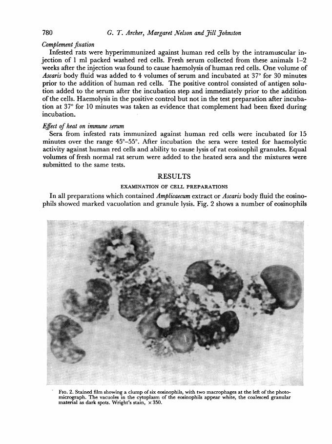
TABLE 1
EFFECT OF SERUM FROM Amplicaecum INFESTED RATS ON EOSINOPHILSAND MACROPHAGES IN THE PRESENCE OF ANTIGEN
Day of Eosinophil Macrophageinfestation granule lysis vacuolation
0 - -
2 _ _4 _ _8 - _
12 + +16 ++ ++20 +++ +++24 +++ +++28 ++ ++32 + +
in which many of the granules have disappeared, leaving clear rounded areas in the cyto-plasm. Other areas have taken up eosin stain and represent concentrations of coalescedeosinophil granule protein. Marked vacuolation was also observed in the mononuclearcells. The most pronounced changes were in macrophage-type cells, with indented nucleiand pale blue cytoplasm. The vacuoles usually appeared white and clear but in some pre-parations the ingested material seemed to have formed a precipitate in the cytoplasm.
Sera from rats infested with Amplicaecum varied in their ability to cause eosinophil andmacrophage changes, as shown in Table 1.
It will be noted that sera collected up to 8 days after infestation were negative in the testsystem and that those collected between 12 and 32 days were positive, the most pronouncedchanges being observed with sera collected 3 weeks after infestation.Table 2 shows the effect of adrenalectomy and of hydrocortisone on the ability of serum
from Amplicaecum infested rats to cause changes in the eosinophils. Adrenalectomy in-creased the activity of the serum, whereas second daily injections of 1 mg hydrocortisonecompletely suppressed activity. Sera collected from sham-operated controls infested withAmplicaecum and from non-operated infested animals gave similar results. The mast cellspresent in the test system showed degranulation and swelling in those preparations whereeosinophil and macrophage vacuolation was most marked. Mast cell degranulation wasmost conspicuous when the serum used was obtained from adrenalectomized animals.
TABLE 2
EFFECT OF ADRENALECTOMY AND HYDROCORTISONE TREATMENT ON THEPRODUCTION OF EOSINOPHIL LYTIC ANTIBODY
Rat serum used collected at the 24th day Eosinophilof infestation with Amplicaecum granule lysis
(a) Non-operated controls +
(b) Sham-operated controls +
(c) Adrenalectomized animals + +
(d) Hydrocortisone-treated animals(1 mg every 2 days)

G. R. Archer, Margaret Nelson and Jill Johnston
EFFECT OF HEAT ON IMMUNE SERUM
Heating immune serum at temperature up to 500 for 15 minutes did not prevent hae-molysis of human red cells or lysis of rat eosinophil granules. Heating at 510 lowered therate of both reactions. Heating at 520 destroyed activity. The addition of an equal volumeof fresh normal serum to serum heated at 520 restored its ability to haemolyse human redcells, though the rate of haemolysis was less than with unheated serum. Lytic activityagainst rat eosinophil granules was not restored.
FIXATION OF COMPLEMENT
The haemolytic activity of immune serum disappeared when the serum was incubatedat 370 for 30 minutes with Ascaris body fluid.
PHASE-CONTRAST STUDIES OF VIABLE CELL PREPARATIONS
Eosinophil granule lysis and vacuolation were seen to occur more rapidly than macro-phage vacuolation. Eosinophil granules burst suddenly at one point in the cell, leaving avacuole in the cytoplasm. In the macrophage, vacuoles appeared to be secondary to pino-cytosis. Small pinocytic vesicles formed at the periphery of the cytoplasm, gradually in-creased in size, and frequently moved towards the perinuclear area.Mast cell disruption and bursting of mast cell granules was frequently observed at the
-same time as eosinophil granule lysis, but it was difficult to interpret the results becausenon-specific mast cell disruption was observed when cover slips were applied firmly tocell preparations on microscope slides.
FRACTIONATION OF Ascaris BODY FLUID
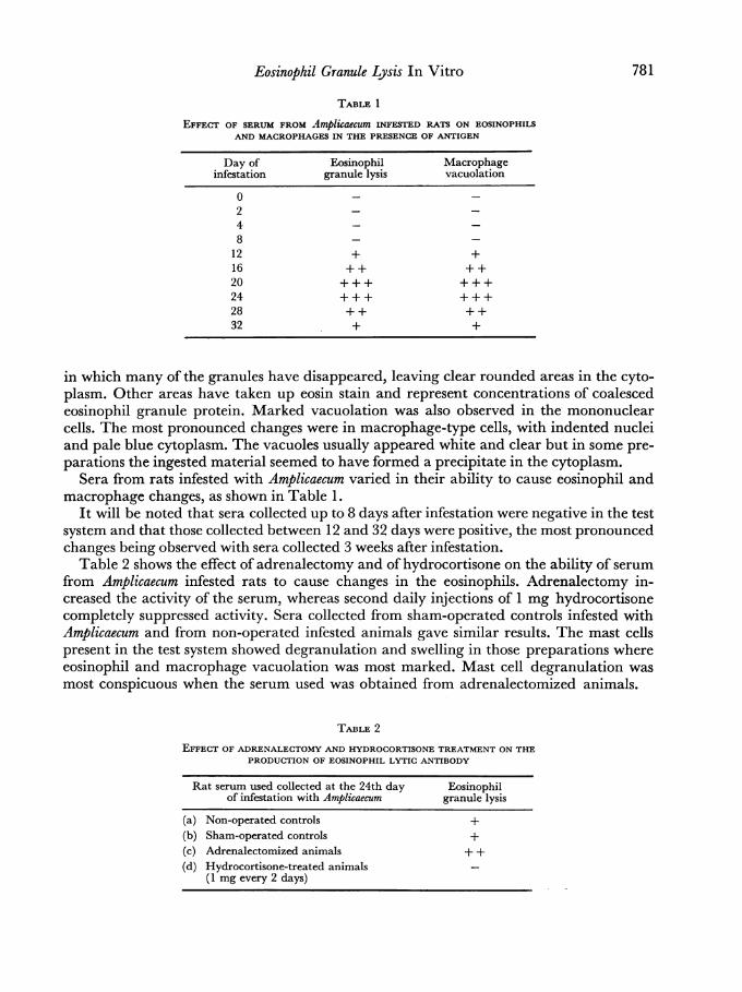
Protein profiles of fractions of Ascaris body fluid separated by DEAE-cellulose columnchromatography are shown in Fig. 3. The pooled Fraction 4-8 was found to be inactive,
0*4_
LO0-3- 4C:0
Z20-30)
CL0
0
Tube No.
FIG. 3. Protein profiles of Ascaris body fluid after fractionation on DEAE-cellulose. Changes of buffer inthe reservoir are indicated by the arrows. Fractions between the dotted lines (20-40, 41-50, 57-62)were submitted to further fractionation (Figs. 4 and 5).
782
Eosinophil Granule Lysis In Vitro
cd
4-
o0
c 1(
c0f.
w o
00
783
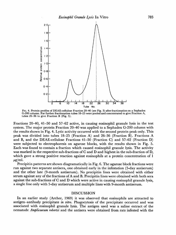
Tube No.FIG. 4. Protein profiles of DEAE-cellulose Fraction 20-40 (see Fig. 3) after fractionation on a SephadexG-200 column. For further fractionation tubes 18-25 were pooled and concentrated to give Fraction A,tubes 26-36 to give Fraction B (Fig. 5).
Fractions 20-40, 41-50 and 57-62 active, in causing eosinophil granule lysis in the testsystem. The major protein Fraction 20-40 was applied to a Sephadex G-200 column withthe results shown in Fig. 4. Lytic activity occurred with the second protein peak only. Thispeak was divided into tubes 18-25 (Fraction A) and 26-36 (Fraction B). Fractions Aand B, and the DEAE-cellulose Fractions 41-50 (Fraction C) and 57-62 (Fraction D)were subjected to electrophoresis on agarose blocks, with the results shown in Fig. 5.Each was found to contain a fraction which caused eosinophil granule lysis. The activitywas marked in the respective sub-fractions ofC and D and highest in the sub-fraction ofD,which gave a strong positive reaction against eosinophils at a protein concentration of 1ug/ml.
Precipitin patterns are shown diagramatically in Fig. 6. The agarose block fractions wererun against two separate antisera, one obtained early in the infestation (5-day antiserum)and the other late (9-month antiserum). No precipitin lines were obtained with eitherserum against any ofthe fractions ofA and B. Precipitin lines were obtained with both seraagainst the sub-fractions of C and D which were active in causing eosinophil granule lysis,a single line only with 5-day antiserum and multiple lines with 9-month antiserum.
DISCUSSIONIn an earlier study (Archer, 1969) it was observed that eosinophils are attracted to
antigen-antibody precipitate in vitro. Phagocytosis of the precipitate occurred and wasassociated with eosinophil granule lysis. The antigen used was a saline extract of thenematode Amplicaecum robertsi and the antisera were obtained from rats infested with the

I Fraction A
08_
06-_
se Anode
- - - ww\AfwW
Fraction C
-4 -2 0 2 4 6 8 10 12 14+ + + -'Aw
Fraction D
-4 -2 0 2 4 6 8 10 12 14
Fraction No.
FIG. 5. Protein profiles after agarose block electrophoresis of Fractions A and B (see Fig. 4) and DEAEFractions 41-50 (C) and 57-62 (D) (see Fig. 3). The ability of the various sub-fractionslto'causeeosinophil granule lysis is indicated as positive (+), weak positive (w) or negative (-).
0O
| - dayantisera
-2
4 x
60-0
8
0.
-20
* I1
.10
.13
9-month 44antisera
6. .10
8
Fraction C
-2
0.
011
6. .10
8
-20
0. .13
6. 100
Fraction D
FIG. 6. Diagram of gel diffusion patterns obtained with sub-fractions of C and D (see Fig. 5). Antiserawere placed in the centre well and the various electrophoresis sub-fractions, numbered according todistance (cm) of migration from the origin towards the anode, in the peripheral wells.
0 4
0 2
Cathod
IC0
c0
0
L.
00
0
0
a:
0 6
0-4
0-2
.A-

Eosinophil Granule Lysis In Vitro 785
parasite. It does not seem likely that insoluble antigen-antibody precipitate is the cause ofthe massive pulmonary eosinophilia which occurs during the 3rd week ofinfestation, whenthe parasites have localized in the liver (Archer, 1968). The experiments now reportedoffer an alternative explanation for the pulmonary eosinophilia, namely, the passagethrough the circulation to the lungs of soluble antigen-antibody complexes formed at sitesremote from the lungs. Eosinophilia is most marked in the loose connective tissue surround-ing the pulmonary vessels and the bronchioles, and this would suggest that the solubleantigen-antibody complexes pass through the walls of the blood vessels in the lungs intothe surrounding loose connective tissue.The ability of preformed soluble antigen-antibody complexes to cause eosinophilia has
been well demonstrated by Litt (1964), who has also shown that guinea-pig eosinophilsare capable ofingesting fluorescent labelled antigen-antibody complexes. The results ofthework reported here support Litt's finding.Another mechanism which may be responsible for pulmonary eosinophilia has been
suggested by Samter, Kofoed and Pieper (1953), who discovered a factor in sensitizedguinea-pig lung which was capable of inducing peritoneal eosinophilia. They proposedthat the active factor was produced following a reaction between the challenging antigenand cell-bound antibody in the lungs. In the present experiments attempts were made toattach antibody to macrophages, eosinophils and mast cells, without success. On theother hand, the addition of antigen to antisera followed by the addition of cells was uni-formly successful in inducing eosinophil and macrophage vacuolation. Thus a cell-boundantibody per se appears to be excluded in so far as these in vitro experiments are concerned,but an interaction between antigen and antibody to produce a complex which binds tocells remains an attractive possibility.
Protein fractions of Ascaris body fluid and saline extracts of Amplicaecum were equallyeffective in the in vitro system. There is known to be a high degree of cross-reactivitybetween Ascaris and Amplicaecum antigens (Archer, 1969), and as Ascaris swum was muchmore readily available than Amplicaecum robertsi, Ascaris body fluid was used in attempts toisolate the antigens responsible for the changes in the eosinophils. Further purification isnecessary, but it is apparent that at least two components of Ascaris body fluid are capableof causing marked eosinophil granule lysis in the presence ofserum from rats infested withAmplicaecum. The fractions containing these two components gave precipitin lines in agarosegel against serum collected from rats five days after infestation with Amplicaecum, suggestinga close relationship between precipitin and eosinophil lytic antibodies. It is of interest thatKent (1963) found a close relationship between precipitin and skin sensitizing antibodies.The early appearance of precipitating antibody and the cross-reactivity between Ampli-caecum and Ascaris (Archer, 1969) suggest that the antigens responsible may be commonlyoccurring antigens and that the animals were naturally immune prior to infestation withAmplicaecum. Infestation would then provide a secondary stimulus, with rapid appearanceofsignificant amounts of antibody in the circulation.An unexpected finding in the present work was the necessity to centrifuge the cells in the
preparations prior to incubation. Eosinophil granule lysis occurred only when this step wasincluded. No adequate explanation can be offered for this phenomenon. It was thoughtthat the ingestion of soluble complexes might be most active when eosinophils and macro-phages were in apposition. However, eosinophil-rich preparations with a few macrophagesand no mast cells and normal peritoneal cavity cell suspensions alike showed vacuolationand granule lysis. The effect ofcentrifugation seems therefore to depend on the production
786 G. T. Archer, Margaret Nelson and Jill Johnstonofa cell-serum interface rather than to an interaction between different cell types. Cochraneand Muller-Eberhard (1968) made the interesting observation that complement complexesare generated more readily on cell surfaces than in free solution. Complement was fixedwhen antigen was added to the antiserum in the present experiments. The observationsof Cochrane and Muller-Eberhard (1968) may explain the necessity to centrifuge thecells in the test system. The cell layer formed during centrifugation may enhance theproduction of antigen-antibody-complement complexes at the cell surface, and thesecomplexes may be responsible for eosinophil granule lysis. This necessity for centrifugationin the in vitro phagocytosis system is at variance with the results of direct observationusing phase contrast, when no centrifugation was involved and eosinophil granule lysisoccurred. A possible explanation, for which no proof is available, is that the cell surfaceis greatly increased in these thin cover slip slide preparations leading to increased pro-duction of antigen-antibody-complement complexes at the cell surface and in closeproximity to the eosinophil granules.The antibody responsible for the induction of the eosinophil and macrophage changes
was shown to be heat labile, to appear in the serum early in infestation, to reach maximumlevels several weeks after the commencement of infestation, and then to decrease in con-centration. The characteristics of heat lability and appearance early in the infestationsuggest that the antibody may be reaginic in type. Ishizaka and Ishizaka (1968) have shownreaginic antibodies to belong to a special class of immunoglobulins termed 'IgE'. Reaginicantibodies are known to be produced in parasite infestation (Jones and Ogilvie, 1967)and Johansson, Mellbin and Vahlquist (1968) have reported high levels of IgE antibodiesin children infested with Ascaris lumbricoides. It is an attractive hypothesis, that reaginic(IgE) antibodies are responsible for eosinophil granule lysis. This would explain the heat-lability of the reaction. An alternative possibility would be the participation of a heat-stable antibody and complement. However, the preliminary experiments using heatedserum suggest that normal serum cannot provide the heat-labile factor necessary foreosinophil granule lysis, and this may be evidence in favour of labile antibody ratherthan complement being involved. Further investigation is required.Serum from infested rats treated with cortisone did not contain lytic activity against
eosinophils. The absence of lytic activity is probably associated with the well-known effectof cortisone in suppressing antibody formation. It does not seem likely that the drug actsdirectly on the parasite, because the rate ofgrowth and migration of the larvae to the liverwas not-impaired.
Vacuole formation in macrophages occurred under the same conditions as eosinophilvacuolation but more slowly. Another difference between eosinophils and macrophageswas the appearance of precipitate in the vacuoles and over the surface of the macrophagesin those preparations which showed the most marked cellular changes. It is not knownwhether this tendency for soluble antigen-antibody to precipitate out on the macrophagesurface bears any relation to in vivo events, but it could be responsible at least in somesituations for the binding of antibody to cell surfaces. A number of workers have suggestedthat reaginic antibodies bind to cell surfaces in the absence of antigen. On the other hand,Ishizaka and Ishizaka (1968) have shown that preformed antigen-IgE complexes are
capable of inducing erythema weal skin reactions when injected into the skin of normalhuman volunteers.
Generalized mast cell disruption occurs in rats infested with Amplicaecum between the1st and 2nd week of infestation (Archer and McGovern, 1968). Mota (1964, 1967) sug-

Eosinophil Granule Lysis In Vitro 787
gested that special antibodies termed mast cell lytic antibodies may be responsible formast cell disruption in vivo, and Becker and Austen (1966) coined the expression 'homocy-totropic antibodies' to describe antibodies which become attached to mast cells early in animmune response. The present experiments suggest that the combination of antigen andantibody rather than antibody alone may attach to mast cells.
ACKNOWLEDGMENTS
The technical assistance of Mrs Angela Trube and Mr Nikolai Kalcenaus is gratefullyacknowledged. Mr Terry Rothwell of the McMaster Laboratories, C.S.I.R.O., kindlyarranged the supply of Ascaris suum.
This work was supported by the National Health and Medical Research Council andthe Asthma Foundation of New South Wales.
REFERENCES
ARCHER, G. T. (1968). 'The function of the eosinophil.'Biblio. haemat., No. 29, Part 1, p. 71 (Proc. 11thCongr. int. Soc. Blood Transf., Sydney 1966).
ARCHER, G. T. (1969). 'Mechanisms ofeosinophilia andmast cell disruption in rats infested with the parasiteAmplicaemum robertsi.' Pathology, 1, 133.
ARCHER, G. T. and HIRsCH, J. G. (1963). 'Motion pic-ture studies on degranulation of horse eosinophilsduring phagocytosis.' J. exp. Med., 118, 287.
ARCHER, G. T. and McGOVERN, V. J. (1968). 'Mastcell changes in rats with eosinophilia.'_J. Path. Bact.,95, 217.
BECKER, E. L. and AUSTEN, K. F. (1966). 'Mechanismsof immunological injury of rat peritoneal mast cells.I. The effect of phosphonate inhibitors on the homo-cytotropic antibody-mediated histamine releaseand the first component of rat complement.' J. exp.Med., 124, 379.
BOSWORTH, N. and ARCHER, G. T. (1961). 'The eosino-phil content of the peritoneal cavity of the rat.'Aust. J. exp. Biol. med. Sci., 39, 165.
COCHRANE, C. G. and MOLLER-EBERHARD, H. J.(1968). 'The derivation of two distinct anaphyla-toxin activities from the third and fifth components ofhuman complement.'J. exp Med., 127, 371.
ISHIZAKA, K. and ISHIZAKA, T. (1968). 'Induction oferythema-weal reactions by soluble antigen-yEantibody complexes in humans.' J. Immunol., 101,68.
JOHANSSON, S. G., MELLBIN, T. and VAHLQUIST, B.(1968). 'Immunoglobulin levels in Ethiopian pre-school children, with special reference to high con-centrations of immunoglobulin E (IgND).' Lancet, i,1118.
JONES V. E. and OGILVIE, B. M. (1967). 'Reaginic anti-bodies and immunity to Nippostrongylus brasiliensis inthe rat. II. Some properties of the antibodies andantigens.' Immunology, 12, 583.
KENT, N. H. (1963). 'Seminar on immunity to parasitichelminths. V. The antigens.' Exp. Parasitol., 13, 45.
LITT, M. (1964). 'Eosinophils and antigen-antibodyreactions.' Ann. N. r. Acad. Sci., 116, 964.
LOWRY, 0. H., ROSEBROUGH, N. J., FARR, A. L. andRANDALL, R. J. (1951). 'Protein measurement withthe folin phenol reagent.' J. biol. Chem., 193, 265.
MOTA, I. (1964). 'The mechanism of anaphylaxis. I.Production and biological properties of 'mast cellsensitizing' antibody.' Immunology, 7, 681.
MOTA, I. (1967). 'Biological characterisation of mouseearly antibodies.' Immunology, 12, 343.
OUCHTERLONY, 0. (1962). 'Diffusion-in-gel methodsfor immunological analysis. II.' Progr. Allergy, 6,30.
SAMTER, M., KOFOED, M. A. and PIEPER W. (1953).'A factor in lungs ofanaphylactically shocked guineapigs which can induce eosinophilia in normalanimals.' Blood, 8, 1078.
SPRENT, J. F. A. (1963). 'The life history and develop-ment of Amplicaecum robertsi, an ascariboid nema-tode of the carpet python (Morelia spilotes variegates).I. Morphology and functional significance of larvalstages.' Parasitology, 53, 7.